HAL Id: hal-00497916
https://hal.archives-ouvertes.fr/hal-00497916
Submitted on 6 Jul 2010HAL is a multi-disciplinary open access
archive for the deposit and dissemination of sci-entific research documents, whether they are pub-lished or not. The documents may come from teaching and research institutions in France or abroad, or from public or private research centers.
L’archive ouverte pluridisciplinaire HAL, est destinée au dépôt et à la diffusion de documents scientifiques de niveau recherche, publiés ou non, émanant des établissements d’enseignement et de recherche français ou étrangers, des laboratoires publics ou privés.
Involvement of mitochondrial ferredoxin and
para-aminobenzoic acid in yeast coenzyme Q
biosynthesis.
Fabien Pierrel, Olivier Hamelin, Thierry Douki, Sylvie Kieffer-Jaquinod,
Ulrich Mühlenhoff, Mohammad Ozeir, Roland Lill, Marc Fontecave
To cite this version:
Fabien Pierrel, Olivier Hamelin, Thierry Douki, Sylvie Kieffer-Jaquinod, Ulrich Mühlenhoff, et al.. Involvement of mitochondrial ferredoxin and para-aminobenzoic acid in yeast coenzyme Q biosynthesis.: pABA is a precursor of yeast coenzyme Q. Chem Biol, 2010, 17 (5), pp.449-59. �10.1016/j.chembiol.2010.03.014�. �hal-00497916�
Involvement of mitochondrial ferredoxin and para-aminobenzoic
acid in yeast coenzyme Q biosynthesis
Fabien Pierrel
1*, Olivier Hamelin
1, Thierry Douki
2, Sylvie
Kieffer-Jaquinod
3, Ulrich Mühlenhoff
4,
Mohammad Ozeir
1, Roland Lill
4and Marc
Fontecave
1,5.
1: Laboratoire de Chimie et Biologie des Métaux; UMR5249 CNRS-CEA-UJF, CEA Grenoble, 17 rue des Martyrs, 38054 Grenoble Cedex 9, France
2 : CEA, INAC, SCIB, UJF & CNRS, LCIB (UMR_E 3 CEA-UJF et FRE 3200), Laboratoire « Lésions des Acides Nucléiques », 17 Rue des Martyrs, F-38054 Grenoble Cedex 9, France. 3 : CEA, DSV, iRTSV, Laboratoire d’Etude de la Dynamique des Protéomes, INSERM, U880, Université Joseph Fourier, Grenoble, France.
4: Institut für Zytobiologie und Zytopathologie, Philipps-Universität Marburg, Robert-Koch-Strasse 6, 35032 Marburg, Germany
5: Collège de France, 11 place Marcellin-Berthelot, 75005 Paris, France
* Contact
Laboratoire de Chimie et Biologie des Métaux iRTSV, Bat K’, P272
CEA Grenoble 17 rue des Martyrs 38054 Grenoble Cedex 9 France
Phone: +33-4-38-78-91-10 Fax: +33-4-38-78-91-24 E-mail:fabien.pierrel@cea.fr
Running title:pABA is a precursor of yeast coenzyme Q
*Manuscript
Summary
Yeast ubiquinone or coenzyme Q6 (Q6) is a redox active lipid with a crucial role in the mitochondrial electron transport chain. At least nine proteins (Coq1p-9p) participate in Q6 biosynthesis from 4-hydroxybenzoate (4-HB). We now show that the mitochondrial ferredoxin Yah1p and the ferredoxin reductase Arh1p are required for Q6 biosynthesis, probably for the first hydroxylation of the pathway. Conditional Gal-YAH1 and Gal-ARH1 mutants accumulate 3-hexaprenyl-4-hydroxyphenol and 3-hexaprenyl-4-aminophenol. Para-aminobenzoic acid (pABA) is shown to be the precursor of 3-hexaprenyl-4-aminophenol and to compete with 4-HB for the prenylation reaction catalyzed by Coq2p. Yeast cells convert U-(13C)-pABA into 13C ring-labeled Q6, a result which identifiespABA as a new precursor of Q6 and implies an additional NH2-to-OH conversion in Q6 biosynthesis. Our study identifies pABA, Yah1p and Arh1p as three new actors in Q6 biosynthesis.
Introduction
Coenzyme Q (ubiquinone or Q) is a lipophilic organic molecule composed of a substituted benzoquinone and a polyprenyl chain containing 6 units in Saccharomyces cerevisiae (Q6) and 10 in humans (Q10). Q has a well known role as an electron carrier in the mitochondrial respiratory chain and also functions as a membrane soluble antioxidant (Bentinger et al., 2007). Moreover, Q has been implicated in modulating the permeability of transition pores (Fontaine and Bernardi, 1999) and is an obligatory cofactor for the proton transport function of uncoupling proteins (Echtay et al., 2001).
4-hydroxybenzoate (4-HB) is the long-known aromatic precursor of the benzoquinone ring of Q (Olson et al., 1963; Rudney and Parson, 1963). Cells synthesize Q from 4-HB and from dimethylallyl diphosphate and isoprenyl diphosphate, the building blocks for the polyprenyl chain. After covalent linkage of the polyprenyl tail to 4-HB to form 3-polyprenyl-4-hydroxybenzoate, a total of 1 decarboxylation, 3 hydroxylation and 3 methylation reactions at the aromatic ring are necessary to yield Q (Figure 1). Current knowledge of the genes involved in Q biosynthesis in eukaryotes is mostly derived from the work carried out on yeast by C. Clarke (Tran and Clarke, 2007). So far, 9 genes (COQ1-9) have been characterized as essential for Q biosynthesis in yeast but only 6 gene products have been assigned a catalytic role (Figure 1)(Kawamukai, 2009). All S. cerevisiae COQ1-9 genes have human relatives. Primary Q10 deficiency, a rare recessive disorder, has now been linked to mutations in five genes: PDSS1 (Mollet et al., 2007) and PDSS2 (Lopez et al., 2006; Mollet et al., 2007) ,which are the relatives of COQ1 ; COQ2 (Mollet et al., 2007); COQ9 (Duncan et al., 2009) and ADCK3/CABC1 the relative of COQ8 (Lagier-Tourenne et al., 2008; Mollet et al., 2008).
Genetic and biochemical data have shown in yeast that a multi-protein Q biosynthetic complex forms and associates with the mitochondrial inner membrane on the matrix side (Tran and Clarke, 2007). The proteins Coq3, Coq4, Coq6, Coq7 and Coq9 are part of the Q
biosynthetic complex (Tran and Clarke, 2007) and steady state levels of these proteins are strongly decreased in any coq null mutants that lack one of the COQ genes (Tran and Clarke, 2007). This explains why yeast coq mutants rarely accumulate biosynthetic Q intermediates that may be diagnostic of the altered reaction in a particular mutant. Indeed, yeast null coq3 to coq9 mutants accumulate the same early intermediate 3-hexaprenyl-4-hydroxybenzoate (HHB) (Figure 1). Only selective mutations altering the activity of a Coq protein without drastically affecting assembly of the Q biosynthetic complex may lead to accumulation of intermediates different from HHB. Only 2 such intermediates have been unambiguously identified and linked to a mutation in a COQ gene: 5-demethoxyubiquinone (DMQ6) accumulates in two coq7 mutants, L237Stop and E233K (Padilla et al., 2004), and 3-hexaprenyl-4,5-dihydroxybenzoate accumulates in an uncharacterized coq3 mutant strain (Clarke et al., 1991). The rare occurrence of intermediates diagnostic of the defective step in Q biosynthesis largely explains why the order of reactions modifying the aromatic ring of 4-polyprenyl-3 hydroxybenzoate is still speculative (Tran and Clarke, 2007).
Coq6p and Coq7p are two mono-oxygenases that function of Q biosynthesis. Coq7p hydroxylates DMQ6 (Padilla et al., 2004; Tran et al., 2006) and Coq6p may catalyse one or both of the two remaining hydroxylation steps (Gin et al., 2003) (Figure 1). Mono-oxygenases are widely distributed proteins that catalyze incorporation of one oxygen atom into substrates. Dioxygen is the source of the oxygen atom and mono-oxygenases usually employ a transition metal (iron, copper) or an organic cofactor (flavin, pterin) to mediate dioxygen activation at their active site. This activation requires two electrons which are provided by a reductant. NAD(P)H can directly reduce the flavin cofactor of flavoprotein mono-oxygenases whereas reduction of the heme iron in cytochrome P450 type oxygenases is accomplished by an associated reductase (Ballou et al., 2005; Munro et al., 2007). The mammalian electron transport machinery of mitochondrial cytochrome P450 consists of two proteins, an
NADPH-ferredoxin reductase called adrenodoxin reductase (AdxR) and a [2Fe-2S] NADPH-ferredoxin called adrenodoxin (Adx). To date, proteins resembling mitochondrial cytochrome P450 mono-oxygenase have not been found in yeast, yet yeast expresses a mitochondrial ferredoxin (Yah1p) and a mitochondrial ferredoxin reductase (Arh1p) which are homologues of Adx and AdxR.
The Yah1p/Arh1p system is located in the mitochondrial matrix and is required for viability (Barros and Nobrega, 1999; Manzella et al., 1998). Yah1p and Arh1p play an essential role in the biogenesis of Fe-S clusters (Lange et al., 2000; Li et al., 2001) and of heme A (Barros et al., 2002), an indispensable cofactor of cytochrome c oxidase. Cox15p which catalyzes the conversion of heme O to heme A is the likely recipient of electrons transferred by Yah1p (Barros et al., 2002) whereas the recipient is still unknown for the biogenesis of Fe-S clusters. Conserved motifs in the primary sequence of Coq6p and Coq7p predict that these two mono-oxygenases employ a flavin and a di-iron cofactor respectively (Gin et al., 2003; Stenmark et al., 2001). The oxidized flavin of Coq6p is a two-electron acceptor whereas the oxidized di-iron (FeIII-FeIII) center of Coq7p will also accept two electrons but one at a time. Therefore the putative direct two-electron reduction of the flavin of Coq6p by hydride transfer from NAD(P)H is not applicable to the di-iron center of Coq7p. The in vivo source of electrons used by Coq7p is still unknown and we hypothesized that the Yah1p/Arh1p system may fulfill this role.
In this work, we show that the Yah1p/Arh1p system is essential for yeast Q6 synthesis. Cells depleted for Yah1p or Arh1p accumulate specific intermediates of Q6 upstream of DMQ6, the substrate of Coq7p. The accumulation of these early intermediates pointed to the involvement of Yah1p/Arh1p in the first hydroxylation reaction of Q6 biosynthesis. Surprisingly, one of the intermediates accumulated in Yah1p-depleted cells contains an amine group. This observation led us to show that para-aminobenzoic acid (pABA) is the precursor
of this intermediate and that pABA enters the Q biosynthetic pathway at the prenylation reaction catalyzed by Coq2p. Moreover, labeling experiments proved that pABA is an in vivo precursor of the benzoquinone ring of Q6. This new, unexpected role of pABA has been overlooked since the discovery of 4-HB as the precursor of the benzoquinone ring of Q more than 40 years ago.
Results
The mitochondrial ferredoxin Yah1p and its associated reductase Arh1p are essential for Q6 biosynthesis
In order to test the possible role of the mitochondrial ferredoxin Yah1p in Q6 biosynthesis, we used the strain Gal-YAH1 in which the native YAH1 promoter was replaced by the GAL10 promoter (Lange et al., 2000). Therefore, the expression of the essential YAH1 gene in the Gal-YAH1 strain is under the control of a galactose-inducible promoter (Lange et al., 2000). When YAH1 expression was repressed by cultivating the Gal-YAH1 strain in a glucose-containing medium, we noted a strong depletion of the Q6 content of the cells together with that of DMQ6 (Figure 2A). Q6 and DMQ6 were detected electrochemically after separation of cell lipid extracts by HPLC. Surprisingly, we detected an additional electroactive compound eluting at around 610 seconds which was absent from the galactose-grown cells (Figure 2A). The striking decrease of Q6 does not result from dying cells because the cells were viable at the end of the glucose culture as assessed by determining the colony forming units on galactose medium. Introduction of a plasmid carrying the YAH1 gene in the Gal-YAH1 strain restored wild-type Q6 levels in glucose-grown cells and abolished the accumulation of the electroactive compound (Figure 2B). To verify that the [2Fe-2S] cluster of Yah1p was required for Q6 generation, we constructed mutant alleles of Yah1p in which conserved Fe-S
cysteine ligands (C103 and C106) are mutated to alanine (Figure S1) (Xia et al., 1996). The electrochromatograms of extracts from glucose-grown Gal-YAH1 strains containing an empty vector and from C103A or C103-106S mutants of Yah1p were similar, with an accumulation of the newly detected metabolite, showing that these cysteines are essential for Yah1p function (Figure 2B and data not shown). The Fe-S center of Yah1p is reduced by electrons provided by the ferredoxin reductase Arh1p which is also an essential protein (Manzella et al., 1998). In order to corroborate the observation that Yah1p is required for Q6 biosynthesis, we generated a Gal-ARH1 strain in which the expression of ARH1 is repressed by growth in glucose medium. After growth of the Gal-ARH1 strain in glucose medium, Arh1p was undetectable in purified mitochondria but Yah1p steady state level was only mildly decreased (Figure 2C). These mitochondria showed little Q6 and accumulated the electroactive compound found in Yah1p-depleted mitochondria (Figure 2D). Altogether, these results show that the absence of a functional ferredoxin / ferredoxin reductase system strongly depletes cellular and mitochondrial levels of Q6 and promotes the accumulation of a novel compound.
A strain deficient in Yah1p accumulates 3-hexaprenyl-4-aminophenol, a product derived from the Q6 biosynthetic pathway
We attempted to identify the origin and nature of the electroactive compound accumulating in Yah1p-deficient cells and found that it is different from DMQ6, the only intermediate of Q6 biosynthesis observed by electrochemical detection so far. Deletion of COQ5 or COQ7, two genes essential for Q6 biosynthesis, abolishes formation of Q6 and results in the accumulation of HHB because many Coq proteins of the Q biosynthetic complex are unstable in these mutants (Figure 1)(Tran and Clarke, 2007). Deletion of COQ5 or COQ7 in the Gal-YAH1 strain prevented the accumulation of the electroactive compound
upon depletion of Yah1p (Figure 3A and data not shown). This result suggests that the electroactive compound is part of the Q6 pathway and is formed downstream of HHB.
In order to determine whether the compound is produced during the Q6 biosynthetic pathway or is a degradation product of Q6, we exploited a strain which accumulates mostly DMQ6 instead of Q6.This strain contains a chromosomal insertion of a sequence coding for a triple hemagglutinin (3HA) epitope tag on the 3’end of COQ7. The presence of the 3HA epitope on the C terminus of Coq7p likely causes the partial inactivation of the protein as evidenced by the accumulation of large amounts of DMQ6 as compared to Q6 (Figure 3B). Depletion of Yah1p in a Gal-YAH1 COQ7-3HA strain resulted in a strong decrease of DMQ6 and still promoted the accumulation of the electroactive compound eluting at around 610 s (Figure 3B). This result demonstrates that this compound is not a degradation product of Q6 and strongly suggests that it originates from Q6 biosynthesis at a step prior to the hydroxylation of DMQ6 catalyzed by Coq7p.
We purified the compound in its reduced form, analyzed it by high resolution mass spectrometry and obtained a m/z ratio of 518.43610 (M+H+). The molecular mass was submitted to different chemical formula prediction software; C36H55NO was the elemental composition which fitted best (M= C36H55NO: 517.42836; observed mass, 517.42828; ppm, 0.2) when C,H,N,O,P,S atoms were included in the calculation. In order to determine if the C36H55NO molecule contains a hexaprenyl moiety like Q6, we obtained two fragmentation spectra of the 518.4 molecular ion (M+H+) on different spectrometers. The fragments at m/z 122 and 162 are consistent with carboxyaminotropylium and carboxyaminopyrilium ions, respectively (Figure S2A). Tropylium and pyrilium ions are typically observed upon fragmentation of Q6 (m/z 197 and 237) and DMQ6 (m/z 167 and 207) (Padilla et al., 2004). The fragmentation observed on the spectrum in Figure S2B is consistent with the successive loss of methine, methylene or methyl groups of the hexaprenyl chain, the lightest fragment
(162.0915) corresponding to C10H12NO. Both fragmentation spectra are indicative of the presence of a hexaprenyl chain in the C36H55NO molecule.
We next characterized the purified C36H55NO molecule by NMR spectroscopy. The resulting 1H NMR spectrum shows well-defined peaks with some of them easily attributable to the hexaprenyl moiety (methyl, methylene and vinylic protons) (Figure 3C and experimental procedures). The aromatic region shows three massifs (6.67 to 6.61 ppm, 6.57 to 6.54 ppm and 6.52 to 6.46 ppm) of equal intensity all situated at rather high field for aromatic protons (Figure 3D). These results strongly suggest the presence of three electro-donating substituents on the aromatic ring. Moreover, the observed multiplicity associated with the value of the coupling constants unambiguously establishes both meta and para substitutions. Finally, the aromatic region of the spectrum was compared to that of the commercially available 3-methyl-4-aminophenol (Figure 3D). Both spectra were almost super-imposable, with identical pattern and coupling constants and slightly different chemical shifts. The UV-visible absorption spectra of the reduced and oxidized forms of the purified compound and of 3-methyl-4-aminophenol show identical absorbance maxima (Figure S2C and S2D). All these results unambiguously identify 3-hexaprenyl-4-aminophenol as the electroactive compound accumulated in Yah1p-deficient cells (Figure 3E). For the sake of simplicity, the numbering of the aromatic carbon atoms of the different Q6 intermediates throughout this manuscript will be as in Figure 3E.
pABA is the precursor of 3-hexaprenyl-4-aminophenol and competes with 4-HB for prenylation by Coq2
The presence of a nitrogen atom in a product synthesized in vivo by the Q6 biosynthetic pathway was surprising. Indeed, C, H and O atoms are the only constituents of the different intermediates of Q biosynthesis described to date (Figure 1). Para-aminobenzoic
acid (pABA) is structurally close to para-hydroxybenzoic acid (4-HB), the precursor of the aromatic ring of Q (Figure 1). We therefore hypothesized that the 3-hexaprenyl-4-aminophenol synthesized in the Yah1p-deficient strain might originate from pABA. The accumulation of 3-hexaprenyl-4-aminophenol increased in the Gal-YAH1 strain upon addition of pABA to YP-glucose (YPD) medium (Figure 4A) showing that pABA is the precursor of aminophenol. We also consistently detected more 3-hexaprenyl-4-aminophenol when the Gal-YAH1 strain was cultured in glucose synthetic complete (SC-2% glu) medium compared to YPD (compare Figure 4A and 4B). This is likely because pABA is an ingredient of synthetic medium at 0.2mg/L.
pABA being a precursor of 3-hexaprenyl-4-aminophenol suggested that pABA is prenylated by the polyprenyl transferase Coq2p, which is known to prenylate 4-HB. Decreased levels of 3-hexaprenyl-4-aminophenol were observed in the Gal-YAH1 strain when 4-HB was added to the culture medium (Figure 4B) suggesting a competition between pABA and 4-HB for prenylation by Coq2p. Addition of 3,4-dihydroxybenzoic acid was without an effect on the accumulation of 3-hexaprenyl-4-aminophenol (data not shown).
A coq5 strain accumulates HHB, the product of the prenylation of 4-HB catalyzed by Coq2p (Figure 1)(Tran and Clarke, 2007). Esterification of HHB with diazomethane stabilizes HHB and facilitates its detection by mass spectrometry (Poon et al., 1995). 3-Hexaprenyl-4-hydroxybenzoic acid methylester (HHBme) (M= C38H56O3: 560.42291; observed mass, 560.42113; ppm, 3.2) was detected spectroscopically eluting at 620 s in chromatograms of diazomethane-treated extracts of the coq5 strain (Figure 4C), and was found to be absent from extracts of the WT strain or from untreated extracts (data not shown). pABA supplementation increased the accumulation of 3-hexaprenyl-4-aminobenzoic acid methylester (HABme) (M= C38H57NO2: 559.43890; observed mass, 559.43931; ppm, 0.7) eluting at 570 s in diazomethane-treated extracts of the coq5 strain (Figure 4C). HABme was
also absent from extracts of the WT strain or from untreated extracts (data not shown). It is apparent in Figure 4C that pABA supplementation increases HABme and decreases HHBme whereas 4-HB supplementation increases HHBme and decreases HABme. This shows that pABA and 4-HB are competing substrates for the prenylation reaction catalyzed by Coq2p.
Concomitant accumulation of hydroxyphenol and 3-hexaprenyl-4-aminophenol by the Yah1p-deficient strain.
SC medium contains pABA which efficiently competes with 4-HB for the prenylation reaction. In order to check if a compound originating from 4-HB may form within the Yah1p-deficient strain, we cultured the Gal-YAH1 strain in YPD instead of SC medium (Figure 2A). Together with 3-hexaprenyl-4-aminophenol at 610 s, we detected an electroactive compound eluting at 800 s in extracts of the Gal-YAH1 strain cultured in YPD (Figure 5A and Figure 4A). The purified compound yielded a pseudo-molecular ion at m/z 519.41962 (M+H+) upon high resolution mass spectrometry analysis. This value is consistent with a chemical formula C36H54O2 (M= C36H54O2: 518.41235; observed mass, 518.4118; ppm, 1.1). The fragmentation spectrum supports the presence of a hexaprenyl tail in the molecule with fragments corresponding to loss of methine, methylene or methyl groups of the hexaprenyl chain (Figure S3A). UV-Vis spectra of the C36H54O2 molecule are similar to the ones of methyl-1,4-benzoquinone (Figure S3B and S3C respectively). These results in addition to the fact that the C36H54O2 molecule is not formed in either the Gal-YAH1 coq5 or Gal-YAH1 coq7 cells (data not shown) strongly suggest that 3-hexaprenyl-4-hydroxyphenol is the electroactive compound eluting at 800 s (Figure 5B).
This conclusion was further substantiated by the finding that 4-HB is the precursor of hexaprenyl-4-hydroxyphenol. Indeed, addition of 4-HB to YPD medium increased 3-hexaprenyl-4-hydroxyphenol and decreased 3-hexaprenyl-4-aminophenol in the
Yah1p-deficient strain (Figure 5C). In contrast, addition of pABA increased the quantity of 3-hexaprenyl-4-aminophenol as noted earlier (Figure 4A), but also decreased the production of 3-hexaprenyl-4-hydroxyphenol (Figure 5C). This competition between pABA and 4-HB for the formation of 3-hexaprenyl-4-aminophenol and 3-hexaprenyl-4-hydroxyphenol parallels the one observed for the biosynthesis of hexaprenyl-4-aminobenzoic acid and 3-hexaprenyl-4-hydroxybenzoic acid (Figure 4C).
pABA is a precursor of Q6
We have shown that 3-hexaprenyl-4-aminophenol and 3-hexaprenyl-4-aminobenzoic acid originate from pABA, suggesting that pABA enters the Q6 biosynthetic pathway (Figure 4A and 4C). In order to check if pABA may be a precursor of Q6 like 4-HB, we synthesized U-(13C)-pABA from U-(13C)-toluene in three steps (Figure S4 and Supplemental experimental procedures). As expected, culture of the Gal-YAH1 strain in YPD containing U-(13C)-pABA predominantly yielded 13C6-labeled 3-hexaprenyl-4-aminophenol at m/z 524.4 (data not shown). A WT strain was cultured in YPD supplemented with 2mg/L of U-(13C)-pABA and Q6 was purified. The mass spectrum of this purified Q6 showed two peaks of comparable abundance at m/z 591.4 and 597.4 (Figure 6A), corresponding respectively to unlabeled Q6 and 13C ring-labeled Q6. This result clearly shows that pABA is a precursor of Q6 in yeast. In order for pABA to be converted into Q6, the amine group must be replaced by an hydroxyl group during the Q6 biosynthetic pathway after the formation of 3-hexaprenyl-4-aminobenzoate. The COQ7-3HA strain mostly accumulated DMQ6 instead of Q6 (Figure 3B). The mass spectrum of DMQ6 purified from the COQ7-3HA strain grown in YPD supplemented with 2mg/L of U-(13C)-pABA displayed two main ions (M+H+) m/z 561.43036 and 567.45111, which correspond to unlabeled DMQ6 (M= C38H56O3: 560.42294; observed mass, 560.42254; ppm, 0.7) and to 13C ring-labeled DMQ6 (M= 13C612C32H56O3: 566.44307;
observed mass, 566.44329; ppm, 0.4) respectively. This result demonstrates that the NH2 -to-OH substitution occurs prior to the hydroxylation of DMQ6 by Coq7p, the penultimate step of Q6 biosynthesis.
We next cultured the W303 WT strain in synthetic medium lacking pABA. To our surprise, cells grown in pABA-free medium accumulated 8 to 10 fold less Q6 than cells grown in rich (YP) medium or in synthetic medium (Figure 6B). Addition of 0.2 mg/L of pABA to pABA-free medium yields synthetic medium and restored normal levels of Q6 (Figure 6B). pABA and 4-HB were equally efficient to support Q6 synthesis when added to pABA-free medium. These results illustrate our conclusion that pABA is a precursor of Q6 and support the idea that most of the Q6 synthesized by yeast grown in synthetic medium actually originates from the pABA present in the growth medium. In order to determine whether S. cerevisiae uses endogenous pABA for Q6 biosynthesis, we cultured the Gal-YAH1 strain in pABA-free medium containing 2% galactose and then in pABA-free medium containing 2% glucose. 3-Hexaprenyl-4-aminophenol was detected in cell lipid extracts (data not shown), showing that endogenously synthesized pABA enters the Q biosynthetic pathway.
Discussion
In this work, we show that the mitochondrial ferredoxin Yah1p and its reductase Arh1p, the yeast homologues of mammalian Adx and AdxR, are essential for Q biosynthesis in yeast. Strains depleted for any of these two essential proteins have a strongly reduced content of Q6 and accumulate aminophenol and 3-hexaprenyl-4-hydroxyphenol (Figure 5C). 3-hexaprenyl-4-aminophenol results from the prenylation of pABA by Coq2p whereas prenylation of 4-HB, the long known ring precursor of Q, will ultimately produce 3-hexaprenyl-4-hydroxyphenol (Figure 7 paths 1 and 4, respectively). pABA and 4-HB differ only by one substituent of benzoic acid, an amine or an hydroxyl
group, and both molecules originate in E. coli and yeast from chorismate, a product of the shikimate pathway. In agreement with our results, pABA has previously been shown to be prenylated by orthologs of Coq2p in E. coli and rat as evidenced by the observation of 3-polyprenyl-4-aminobenzoic acid (Hamilton and Cox, 1971) (Alam et al., 1975). pABA can therefore enter the Q biosynthetic pathway in many organisms but are these organisms capable of converting 3-polyprenyl-4-aminobenzoic acid into Q? Our work clearly shows that, in yeast, 3-hexaprenyl-4-aminobenzoic acid is converted to Q6 since addition of U-(13 C)-pABA to the growth medium of WT yeast strain promotes the biosynthesis of 13C6 labeled Q6 (Figure 6A and Figure 7 path 2). pABA is likely not a precursor of Q in E. coli because a mutant strain deficient in chorismate synthetase activity synthesized Q upon supplementation of the growth medium with 4-HB but not with pABA, despite the formation of 3-polyprenyl-4-aminobenzoic acid (Hamilton and Cox, 1971). Our results with yeast warrant a thorough investigation to determine whether mammalian cells can convert pABA into Q. In vitro experiments by Rudney and collaborators on rat liver demonstrated that pABA is converted to 3-polyprenyl-4-aminobenzoic acid and suggested that this compound was further modified although not into Q (Alam et al., 1975). Therefore pABA has been considered an inhibitor of Q biosynthesis in mammals but only a limited decrease of Q was observed in cancer cells upon culture in a high concentration of pABA (Brea-Calvo et al., 2006).
In view of our observation that Q6 levels drop dramatically when yeast is cultured in pABA-free medium, it is reasonable to postulate that most of the Q6 produced by yeast cultured in SC medium originates from the pABA present in this growth medium. Our results suggest that in pABA-free medium the rate-limiting step of Q6 biosynthesis is the availability of the ring precursors of Q, 4-HB and pABA. Endogenous levels of these molecules allow the synthesis of only 1.4 pmoles of Q6/mg of wet cells under our non-repressive culture conditions. One can then question the physiological significance of the high levels of Q6
(12-16 pmoles of Q6/mg of wet cells) synthesized by yeast grown in SC medium or rich medium. Actually, several authors have shown that only a small percentage of the Q6 synthesized in these growth media is sufficient to sustain respiratory growth (Mollet et al., 2008; Tran et al., 2006), suggesting that, under normal conditions, Q6 is in vast excess for its function as an electron carrier in the respiratory chain. Our results also provide a possible explanation as to why, contrary to E. coli, mutants affecting 4-HB synthesis may not abrogate yeast Q6 biosynthesis and therefore not result in a respiratory deficient phenotype. Previously, authors have speculated that yeast may produce 4-HB by multiple ways; from chorismate via the chorismate pyruvate-lyase reaction similar to the E. coli pathway and from tyrosine like in higher eukaryotes (Clarke, 2000). However, none of the enzymes responsible for these reactions have been identified in yeast. The fact that pABA can be used in Q6 biosynthesis like 4-HB and that pABA is present in standard yeast culture media imply that pABA may support biosynthesis of Q6 in a yeast mutant blocked for the synthesis of 4-HB when cultured in standard conditions.
The conversion of pABA into Q6 suggests an additional step in Q6 biosynthesis: the replacement of the amine by an hydroxyl group at C4 (Figure 7 path 2). This reaction certainly takes place downstream of the hexaprenylation reaction catalyzed by Coq2p since 3-hexaprenyl-4-aminobenzoic acid was detected in a coq5 strain (Figure 4C). The formation of 13
C6-DMQ6 upon supplementation of the growth medium with U-(13C)-pABA proves that the C4 aromatic NH2-to-OH conversion occurs prior to the formation of DMQ6. The accumulation of 3-hexaprenyl-4-aminophenol in the Yah1p-depleted strain suggests that this reaction occurs after the C1 hydroxylation step. Alternatively, the presence of the amine group at this stage may simply result from the lack of hydroxylation of the adjacent C5 position. Indeed, based on the example of bacterial anthranilate and aniline dioxygenases, which catalyze the conversion of aromatic amines into ortho-catechols (Beharry et al., 2003)
(Ang et al., 2007), the NH2-to-OH substitution on C4 may occur together with the hydroxylation on C5. The NH2-to-OH conversion reaction is chemically challenging and future studies will have to elucidate this newly evidenced process in Q6 biosynthesis.
What is the role of Yah1p/Arh1p in Q6 biosynthesis? Deficiency in any of these two proteins results in accumulation of hexaprenyl-4-amino/hydroxyphenol. In order to form 3-hexaprenyl-4-amino/hydroxyphenol from 3-hexaprenyl-4-amino/hydroxybenzoic acid, the decarboxylation and hydroxylation at position 1 must occur without hydroxylation of C5 (Figure 7, paths 1 and 4). Therefore the most upstream reaction of the Q6 biosynthetic pathway to be inhibited in the absence of Yah1p/Arh1p is the C5 hydroxylation of 3-hexaprenyl-4-amino/hydroxybenzoic acid. The identity of the mono-oxygenase that catalyzes the C5 hydroxylation is currently unknown. Coq6p has been initially proposed to catalyze either the C5 or C1 hydroxylations or both (Gin et al., 2003). Our results strongly argue for the involvement of Coq6p in only one of the two reactions since the C1 hydroxylation still proceeds in the Yah1p/Arh1p-depleted strains whereas the C5 hydroxylation does not. Coq6p sequence contains conserved motifs characteristic for flavin-dependent mono-oxygenases such as 4-hydroxybenzoate hydroxylase (Gin et al., 2003). Reduction of the FAD cofactor at the active site of 4-hydroxybenzoate hydroxylase has been extensively studied and shown to proceed via a direct hydride transfer from NADPH (Ballou et al., 2005). By analogy, NAD(P)H is the likely electron donor to Coq6p, making the hydroxylation catalyzed by Coq6 independent of the Yah1p/Arh1p reducing system. We thus propose that the C1 hydroxylation which is unaffected by depletion of Yah1p/Arh1p is catalyzed by Coq6p (Figure 7). This point is difficult to prove since a coq6 strain, like the other coq3 to coq9 null mutants, accumulates only HHB because of the instability of the Q biosynthetic complex (Gin et al., 2003; Hsieh et al., 2007). The involvement of Coq6p in the C1 hydroxylation implies that the C5 hydroxylation is catalyzed by an unknown oxygenase which may require an electron
transport machinery like Yah1p/Arh1p. This scenario would explain why the C5 hydroxylation is abrogated upon depletion of Yah1p or Arh1p.
How to explain that the reactions after the C1 hydroxylation (C2 methylation by Coq5p, C6 hydroxylation by Coq7p and O6 methylation by Coq3p) do not take place in the Yah1p-depleted strain (Figure 7 path 1 and 4)? Yah1p is likely not directly required for the methylation reactions since: (i) methylations are non redox reactions (ii) they employ S-Adenosyl-Methionine (SAM) as a methyl donor, a cofactor whose synthesis is cytosolic and does not require Fe-S containing enzymes whose maturation is generally dependent on Yah1p (Lange et al., 2000). The two O-methylation reactions can not proceed because of the lack of the two corresponding hydroxylations (C5 and C6). The absence of methylation at C2 may be explained in that the C2 carbon will be insufficiently enriched in electrons for an efficient nucleophilic attack to the methyl group of SAM in the absence of the methoxy group on C5. Alternatively, the absence of this methoxy group may prevent substrate binding and recognition by Coq5p. Finally, we envision at least two reasons that could explain the inactivity of Coq7p in the absence of Yah1p/Arh1p. On the one hand, Coq7p may not bind a substrate that lacks the C5 methoxy group and the C2 methyl group. On the other hand, Yah1p/Arh1p may provide electrons for the reduction of the di-iron center of the mono-oxygenase Coq7p, a scenario which was the founding hypothesis of this study. We could not gain any information on this point since Yah1p/Arh1p is required for the hydroxylation of 3-hexaprenyl-4-amino/hydroxybenzoic acid, a reaction upstream of the hydroxylation of DMQ6 which is catalyzed by Coq7p. Further work is needed to test if Yah1p/Arh1p is the physiological reducing system of Coq7p. Answering of this question will probably require the development of an in vitro assay for Coq7p function, a task highly challenging given that, in vivo, Coq7p is part of the Q biosynthetic complex (Tran and Clarke, 2007).
In conclusion, our study has unraveled that pABA serves as a precursor of Q6 similar to 4-HB whose role in Q biosynthesis was identified more than forty years ago. We also demonstrated that the yeast Adx/AdxR homologs Yah1p/Arh1p are required for the first hydroxylation reaction of the yeast Q6 biosynthetic pathway, which represents a new function for these proteins. In the light of these results, it seems important to evaluate whether these new actors of yeast Q6 biosynthesis play a role in mammalian Q biosynthesis.
Significance
Coenzyme Q is a redox active lipid that functions in electron transport chains in cellular membranes and also has an important antioxidant function. Q is a prenylated benzoquinone which biosynthesis starts with the assembly of a polyprenyl chain and its conjugation to 4-HB, the precursor of the benzoquinone moiety (Turunen et al., 2004). Here we demonstrate that yeast can convert pABA into Q making of pABA a new precursor of Q, like the long-known 4-HB. Our finding thus implies that an aromatic NH2-to-OH conversion must occur to synthesize Q from pABA. The reducing system formed by the mitochondrial ferredoxin Yah1p and its associated reductase Arh1p is essential for Fe-S cluster biosynthesis (Lange et al., 2000; Li et al., 2001). This study identifies a new function for Yah1p and Arh1p in Q biosynthesis in yeast. Studies on yeast have provided the basis for the elucidation of the eukaryotic biosynthesis of Q. Sequence identity to yeast genes and complementation of yeast mutants have allowed the characterization of genes implicated in human Q biosynthesis. Mutations in five human genes have been shown to cause primary Q deficiency, which results in encephalomyopathy and severe infantile multisystemic disease. It will be of interest to define whether pABA and the orthologs of Yah1p and Arh1p are involved in mammalian Q biosynthesis.
Experimental Procedures
Yeast strains and culture conditions: Yeast strains used in this study are listed in Table S1.
Yeast strains were transformed using lithium acetate. A 3HA epitope tag was inserted on the 3’ end of COQ7 ORF by PCR as described previously (Longtine et al., 1998) to create the COQ7-3HA strain. This strain was crossed with the Gal-YAH1 gene to isolate the COQ7-3HA Gal-YAH1 strain by selecting the corresponding markers after tetrad dissection. The coq5∆ Gal-YAH1 and coq7∆ Gal-YAH1 strains were obtained in a similar way. In Gal-ARH1, the upstream region from ARH1 was exchanged for the galactose-inducible GAL-L promoter by PCR mediated DNA replacement (Janke et al., 2004).Yeast media were supplemented with either 2% glucose or 2% galactose. Synthetic medium consisted in 1.7g/L YNB and 5g/L ammonium sulfate and the appropriate nutrients to complement strains auxotrophies. Synthetic complete (SC) medium was obtained by adding 2g/L Drop-out mix Synthetic minus Ade, His, Leu, Trp, Ura (US Biological) to synthetic medium. YNB without pABA and folate was from MP Biomedicals and was supplemented with 5ug/L folate. Rich YP medium was prepared as described (Sherman, 2002). Gal-YAH1 and Gal-ARH1 strains were maintained and precultured on galactose medium. Depletion of Yah1p was accomplished by diluting 300 fold the preculture into glucose containing medium and growing the cells for 18 hours at 30°C. Depletion of Arh1p required two subsequent dilutions in glucose medium.
Plasmids : YAH1 ORF with its own promoter (380bp) and terminator (150bp) was cloned into
pRS416 using XhoI and SacI. This vector served as a template to generate the C103A and C103,106S mutants by site directed mutagenesis. Sequencing was used to confirm cloning products in all created vectors. The E. coli strain DH5 was used for plasmid DNA amplification.
Miscellaneous biochemical analysis: Isolation of yeast mitochondria and immunostaining
were performed as described (Diekert et al., 2001) (Harlow and Lane, 1988).
Lipid extraction and quantification of electroactive compounds by HPLC-ECD (electrochemical detection): Lipidextraction and analyses of quinone content were
essentiallyas described (Tran et al., 2006).100 uL glass beads, 50uL H2O and Q4 (used as an internal standard) were added to mitochondria (100ug mitochondrial proteins) or to cell pellets (40mg wet weight). Tubes were wrapped in foil and lipids were extractedby adding 0.6 mL methanol and 0.4 mL petroleum ether and by vortexingfor 3 minutes. The phases were separated by centrifugation (3 minat 1000 × g, 4 °C). The upper petroleum ether layer was transferredto a fresh tube. 0.4 mL petroleum ether was added to theglass bead-methanol containing tube and the extraction was repeated 2 more times. The petroleum ether layers were combined and dried under nitrogen. The lipids were resuspendedin 100 uL mobile phase (98% methanol, 20mM lithium perchlorate) and aliquots were analyzed by reversed-phasehigh pressure liquid chromatography with a C18 column (Betabasic-18, 5 µm, 4.6 × 150 mm, Thermo Scientific) at a flow rate of 1 ml/min. Quinones were quantified with an ESA Coulochem II electrochemical detector and a 5011A analyticalcell (E1, –500 mV; E2, 500 mV). Hydroquinones presentin samples were oxidized with a pre-column 5020 guard cell set in oxidizingmode (E, +550 mV). The maximum output signal of the analytical electrode E2 was set to +10V and was recorded by a 4 channel signal recorder USB10 (Velleman
instruments). The Q4 external standard was used to correct for sample loss during the organic extraction based on its recovery (always greater than 85%).
NMR analysis of 3-hexaprenyl-4-aminophenol: NMR spectra were recorded on a Bruker
EMX 300 MHz spectrometer. 3-hexaprenyl-4-aminophenol: assignable protons: 1H NMR (CD3OD) 6.64 (d, 3J = 8.4 Hz, H2N-C-CH, 1H) , 6.55 (d, , 4J = 2.8 Hz, H2 N-C-CH-CH-C(OH)-CH, 1H), 6.49 (dd, 3J = 8.4 Hz, 4J = 2.8 Hz, H2N-C-CH-CH, 1H), 5.27 (t, 3J = 7.1 Hz,
Ar-CH2-CH, 1H), 5.12 (m, vinylic H, 5H), 3.21 (d, 3J = 7.2 Hz, benzylic H), 2.25-1.90 (m, vinylic CH2 and CH3)
Esterification and analysis of aminobenzoate and 3-hexaprenyl-4-hydroxybenzoate: Dried lipid extracts from yeast cells (700mg wet weight) were
resuspended in 400uL anhydrous diethyl ether. 150uL of diazomethane (0.2 M in diethyl ether) was added, the mixture was incubated for 8 minutes at room temperature and the reaction was quenched by adding 10uL of glacial acetic acid. Samples were dried under nitrogen and HPLC analysis was carried out as described for quinone content quantification except that the signal recorded was the absorbance at 273nm.
Mass spectrometry
For high resolution mass spectrometry analyses, samples in methanol were diluted in 90% acetonitrile, 0.2% formic acid and were infused into the nanospray source of a discovery ORBITRAP instrument (Thermo Fischer Scientific) at a flow rate of 0.5uL min-1. The MS method used a scan range of 150-1600 m/z and was composed of MS and MS/MS events using both the Orbitrap as the analyzer (at a resolution of 30000) to get high precision on the molecular and on the fragments ions. The CID fragmentation collision energy used was set to 35 eV and the MS/MS scan range to 100-2000 m/z. QualBrowser from XCalibur was used to read the spectra.
Analyses involving coupling of HPLC to mass spectrometry were performed with an series 1100 Agilent chromatographic system associated to SCIEX API 3000 triple quadrupolar mass spectrometer. Separation was performed on an octadecylsilyl ODB Uptisphere column (150×2 mm ID, 5 µm particle size, Montluçon, France), using a gradient of acetonitrile in 2 mM ammonium formate. MS2 spectra were obtained by selecting pseudo-molecular ion in quadrupole 1 and fragmenting it in the collision cell. The fragmentation spectrum was recorded in the 50-520 mass unit range.
References
Alam, S.S., Nambudiri, A.M., and Rudney, H. (1975). J-Hydroxybenzoate: polyprenyl transferase and the prenylation of 4-aminobenzoate in mammalian tissues. Arch. Biochem. Biophys. 171, 183-190.
Ang, E.L., Obbard, J.P., and Zhao, H. (2007). Probing the molecular determinants of aniline dioxygenase substrate specificity by saturation mutagenesis. FEBS J 274, 928-939.
Beharry, Z.M., Eby, D.M., Coulter, E.D., Viswanathan, R., Neidle, E.L., Phillips, R.S., and Kurtz, D.M., Jr. (2003). Histidine ligand protonation and redox potential in the rieske dioxygenases: role of a conserved aspartate in anthranilate 1,2-dioxygenase. Biochemistry 42, 13625-13636.
Bentinger, M., Brismar, K., and Dallner, G. (2007). The antioxidant role of coenzyme Q. Mitochondrion 7, S41-S50.
Brea-Calvo, G., Rodriguez-Hernandez, A., Fernandez-Ayala, D.J., Navas, P., and Sanchez-Alcazar, J.A. (2006). Chemotherapy induces an increase in coenzyme Q10 levels in cancer cell lines. Free Radic Biol Med 40, 1293-1302.
Clarke, C.F., Williams, W., and Teruya, J.H. (1991). Ubiquinone biosynthesis in Saccharomyces cerevisiae. Isolation and sequence of COQ3, the 3,4-dihydroxy-5-hexaprenylbenzoate methyltransferase gene. J. Biol. Chem. 266, 16636-16644.
Clarke, C.F. (2000). New advances in coenzyme Q biosynthesis. Protoplasma 213, 134-147.
Diekert, K., de Kroon, A.I., Kispal, G., and Lill, R. (2001). Isolation and subfractionation of mitochondria from the yeast Saccharomyces cerevisiae. Methods Cell Biol 65, 37-51.
Echtay, K.S., Winkler, E., Frischmuth, K., and Klingenberg, M. (2001). Uncoupling proteins 2 and 3 are highly active H+ transporters and highly nucleotide sensitive when activated by coenzyme Q (ubiquinone). Proc. Natl. Acad. Sci. USA 98, 1416-1421.
Fontaine, E., and Bernardi, P. (1999). Progress on the mitochondrial permeability transition pore: Regulation by complex I and ubiquinone analogs. J. Bioenerg. Biomembr. 31, 335-345.
Gin, P., Hsu, A.Y., Rothman, S.C., Jonassen, T., Lee, P.T., Tzagoloff, A., and Clarke, C.F. (2003). The Saccharomyces cerevisiae COQ6 gene encodes a mitochondrial
flavin-dependent monooxygenase required for coenzyme Q biosynthesis. J. Biol. Chem. 278, 25308-25316.
Hamilton, J.A., and Cox, G.B. (1971). Ubiquinone biosynthesis in Escherichia coli K-12. Accumulation of an octaprenol, farnesylfarnesylgeraniol, by a multiple aromatic auxotroph. Biochem. J. 123, 435-443.
Harlow, E., and Lane, D. (1988). (Cold Spring Harbour, NY: Cold Spring Harbour Laboratory Press).
Kawamukai, M. (2009). Biosynthesis and bioproduction of coenzyme Q10 by yeasts and other organisms. Biotechnol. Appl. Biochem. 53, 217-226.
Lagier-Tourenne, C., Tazir, M., Lopez, L.C., Quinzii, C.M., Assoum, M., Drouot, N., Busso, C., Makri, S., Ali-Pacha, L., Benhassine, T., et al. (2008). ADCK3, an ancestral kinase, is mutated in a form of recessive ataxia associated with coenzyme Q(10) deficiency. Am. J. Hum. Genet. 82, 661-672.
Lange, H., Kaut, A., Kispal, G., and Lill, R. (2000). A mitochondrial ferredoxin is essential for biogenesis of cellular iron-sulfur proteins. Proc. Natl. Acad. Sci. USA 97, 1050-1055.
Longtine, M.S., McKenzie, A., 3rd, Demarini, D.J., Shah, N.G., Wach, A., Brachat, A., Philippsen, P., and Pringle, J.R. (1998). Additional modules for versatile and economical PCR-based gene deletion and modification in Saccharomyces cerevisiae. Yeast 14, 953-961.
Mollet, J., Giurgea, I., Schlemmer, D., Dallner, G., Chretien, D., Delahodde, A., Bacq, D., de Lonlay, P., Munnich, A., and Rotig, A. (2007). Prenyldiphosphate synthase, subunit 1 (PDSS1) and OH-benzoate polyprenyltransferase (COQ2) mutations in ubiquinone deficiency and oxidative phosphorylation disorders. J. Clin. Invest. 117, 765-772.
Mollet, J., Delahodde, A., Serre, V., Chretien, D., Schlemmer, D., Lombes, A., Boddaert, N., Desguerre, I., de Lonlay, P., de Baulny, H.O., et al. (2008). CABC1 gene mutations cause ubiquinone deficiency with cerebellar ataxia and seizures. Am. J. Hum. Genet. 82, 623-630.
Olson, R.E., Springer, C.M., Aiyar, A.S., Gold, P.H., Ramsey, G., Dialameh, G.H., and Bentley, R. (1963). Benzoate derivatives as intermediates in biosynthesis of coenzyme Q in rat. J. Biol. Chem. 238, 3146-3148.
Padilla, S., Jonassen, T., Jimenez-Hidalgo, M.A., Fernandez-Ayala, D.J., Lopez-Lluch, G., Marbois, B., Navas, P., Clarke, C.F., and Santos-Ocana, C. (2004). Demethoxy-Q, an intermediate of coenzyme Q biosynthesis, fails to support respiration in Saccharomyces cerevisiae and lacks antioxidant activity. J. Biol. Chem. 279, 25995-26004.
Poon, W.W., Marbois, B.N., Faull, K.F., and Clarke, C.F. (1995). 3-Hexaprenyl-4-hydroxybenzoic acid forms a predominant intermediate pool in ubiquinone biosynthesis in Saccharomyces cerevisiae. Arch. Biochem. Biophys. 320, 305-314. Rudney, H., and Parson, W.W. (1963). Conversion of p-hydroxybenz-aldehyde to benzoquinone ring of ubiquinone in Rhodospirillum rubrum. J. Biol. Chem. 238, 3137-3138.
Sherman, F. (2002). Getting started with yeast. In Guide to Yeast Genetics and Molecular and Cell Biology, Pt B, Volume 350. (San Diego: Academic Press Inc), pp. 3-41.
Tran, U.C., Marbois, B., Gin, P., Gulmezian, M., Jonassen, T., and Clarke, C.F. (2006). Complementation of Saccharomyces cerevisiae coq7 mutants by mitochondrial targeting of the Escherichia coli UbiF polypeptide: two functions of yeast Coq7 polypeptide in coenzyme Q biosynthesis. J. Biol. Chem. 281, 16401-16409.
Tran, U.C., and Clarke, C.F. (2007). Endogenous synthesis of coenzyme Q in eukaryotes. Mitochondrion 7 Suppl, S62-71.
Turunen, M., Olsson, J., and Dallner, G. (2004). Metabolism and function of coenzyme Q. Biochimica et Biophysica Acta-Biomembranes 1660, 171-199.
Acknowledgments
This work was supported in part by a CNRS grant PEPS 2008 (to F.P.) and by the Région Rhône-Alpes program CIBLE 2009 (to F.P.). We thank Dr Dennis Winge and Dr Paul Cobine for critical reading of the manuscript. Dr Sébastien Carret is acknowledged for providing diazomethane and Peggy Charbonnier for technical assistance.
Figure legend
Figure 1: Current model of the eukaryotic coenzyme Q biosynthetic pathway. The names of proteins and intermediates relevant to this study are for S. cerevisiae. The polyprenyl diphosphate tail is assembled from one dimethylallyl pyrophosphate and 5 isopentenyl pyrophosphate in yeast or 9 in humans. Prenylation of 4-hydroxybenzoate (4-HB) by Coq2
yields 3-hexaprenyl-4-hydroxybenzoate (HHB). 7 modifications of the aromatic ring are then needed to produce coenzyme Q6. 5-demethoxyubiquinone (DMQ6) is the substrate of the mono-oxygenase Coq7p. Reactions catalyzed by unidentified or putative proteins are marked with ?.
Figure 2: The yeast ferredoxin/ferredoxin reductase system is required for Q6
biosynthesis: A) Gal-YAH1 cells were cultivated for 18 hours in SC medium containing either
2% galactose (gal) or 2% glucose (glu). Extracts of 10 mg (gal) and 20 mg (glu) of cells were analyzed by electrochemical detection coupled to HPLC (HPLC-ECD). Elution positions of the Q4 standard, of DMQ6 and Q6 and of the electroactive compound (?) accumulated in Yah1p-depleted cells are indicated on the electrochromatogram. B) WT and Gal-YAH1 cells containing an empty vector (vec) or the YAH1 ORF were grown in SC-2% glucose for 18 hours. Extracts of 8mg of cells were analyzed by HPLC-ECD. C) WT and Gal-ARH1 cells were cultivated for 40 h in SC-2% glucose in order to deplete Arh1p to critical levels. Mitochondria were prepared and analysed for Arh1p and Yah1p by immunostaining. Porin (Por1p) served as a loading control. D) Mitochondrial extracts (50ug of mitochondrial proteins) were analyzed by HPLC-ECD. Mitochondria were purified from WT, Gal-YAH1 and Gal-ARH1 cells grown in SC-2% glucose.
Figure 3: 3-hexaprenyl-4-aminophenol is derived from Q6 biosynthesis A) Gal-YAH1 cells and Gal-YAH1 coq5 cells were inoculated from a culture in SC-2% galactose 0.4% glucose into SC-2% glucose and grown for 18 hours. Extracts of 30 mg of cells were analyzed by HPLC-ECD. B) WT, COQ7-3HA and Gal-YAH1 COQ7-3HA cells were grown in YPD for 12 hours. Extracts of 45 mg (WT), 60 mg (COQ7-3HA) and 120 mg (Gal-YAH1 COQ7-3HA) of cells were analyzed by HPLC-ECD. C) 1H NMR spectrum of the purified C36H55NO molecule
in deuterated methanol. D) Superimposition of the aromatic regions of the 1H NMR spectrum of the purified C36H55NO molecule displayed in panel C and of the 1H NMR spectrum of commercial 3-methyl-4-aminophenol (3M4Aphe). E) Chemical structure of 3-hexaprenyl-4-aminophenol and numbering of the aromatic carbon atoms used in the rest of the study.
Figure 4: pABA and 4-HB compete for prenylation by Coq2p. A) 50 mg of Gal-YAH1 cells grown for 18 hours in YPD supplemented with pABA were extracted and analyzed by HPLC-ECD. B) 30 mg of Gal-YAH1 cells grown for 18 hours in SC-2% glucose supplemented with 4-HB were extracted and analyzed by HPLC-ECD. C) coq5 cells were grown in 30mL YPD with the indicated concentration of pABA or 4-HB. Lipid extracts were treated with diazomethane and analyzed by HPLC. The elution profile at 273nm and the elution position and chemical structure of hexaprenyl-4-aminobenzoic acid methylester (HABme) and 3-hexaprenyl-4-hydroxybenzoic acid methylester (HHBme) are shown. Areas of the peaks at 273nm for HABme and HHBme are shown in the inset (A.U., arbitrary units).
Figure 5: Yah1p-deficient cells also accumulate 3-hexaprenyl-4-hydroxyphenol. A) 20 mg of Gal-YAH1 cells grown for 18 hours in YPD were extracted and analyzed by HPLC-ECD. B) Chemical structure of 3-hexaprenyl-4-hydroxyphenol. C) 30 mg of Gal-YAH1 cells grown for 18 hours in YPD supplemented with 1 or 10mg/L 4-hydroxybenzoic acid (4-HB) or para-aminobenzoic acid (pABA) were extracted and analyzed by HPLC-ECD. 3-hexaprenyl-4-aminophenol elutes at around 610 s and 3-hexaprenyl-4-hydroxyphenol at around 800 s and areas of the peaks are shown in the inset (A.U., arbitrary units).
Figure 6: pABA is a precursor of Q6. A) Mass spectrum of Q6 purified from a W303 WT strain grown in YPD medium supplemented with 2mg/L of U-(13C)-pABA. B) The W303 WT strain was grown in synthetic medium, synthetic medium lacking pABA or rich (YP) medium,
with 2% galactose as a carbon source. Cells were harvested in late exponential growth phase and lipids were extracted. Q6 (pmoles per mg of wet weight cell pellet) was quantified by HPLC-ECD and DMQ6 represented less than 10% of Q6 in all samples. Data are the average of at least 3 independent cultures and error bars represent the standard error of the mean.
Figure 7: Dysfunction of the Q6 biosynthetic pathway in Yah1p/Arh1p-depleted yeast
cells. R stands for the hexaprenyl chain. Reactions blocked in the absence of Yah1p/Arh1p
are shown in red. The different series of reactions are numbered path 1 to 4. Coq2p catalyzes the hexaprenylation of pABA (blue) and 4-HB to form 3-hexaprenyl-4-aminobenzoic acid and hexaprenyl-4-hydroxybenzoic acid, respectively. In a WT strain, the conversion of 3-hexaprenyl-4-aminobenzoic acid into Q6 requires the NH2-to-OH conversion on C4 which occurs prior to DMQ6 (path 2). The C5 hydroxylation of 3-hexaprenyl-4-amino/hydroxybenzoic acid catalyzed by an unidentified oxygenase is blocked in the absence of Yah1p/Arh1p (path 2 and 3). In this case, the normal Q6 biosynthetic pathway (path 2 and 3) branches off via decarboxylation of hexaprenyl-4-aminobenzoic acid (path 1) and hydroxybenzoic acid (path 4). Coq6p then synthesize hexaprenyl-4-aminophenol (path 1, framed) and hexaprenyl-4-hydroxyphenol (path 4, framed). 3-hexaprenyl-4-amino/hydroxyphenol accumulate because Coq5p does not catalyze the C2 methylation and Coq7p does not catalyze the C6 hydroxylation in the absence of Yah1p/Arh1p (see discussion).
Figure1
Figure2
Figure3
Figure4
Figure5
Figure6
Figure7
Inventory of Supplemental Information
Supplemental information includes four figures, one table, supplemental experimental procedures and references:
Figure S1 relates to the mutants of Yah1p in Figure 2B.
Figure S2 corresponds to additional characterization of the compound displayed in Figure 3E. Figure S3 corresponds to additional characterization of the compound displayed in Figure 5B. Figure S4 shows the chemical synthesis of labeled pABA used in Figure 6A.
Table S1 contains the yeast strains used in this study.
Supplemental experimental procedures include the description of the chemical synthesis of labeled pABA (Figure S4) and the purification of 3-hexaprenyl-4-aminophenol (Figure 3E).
Supplemental Figures Figure S1 huFdx ---SSS---EDK 6 Yah1 MLKIVTRAGHTARISNIAAHLLRTSPSLLTRTTTTTRFLPFSTSSFLNHGHLKKPKPGEE 60 *:* :: huFdx ITVHFINRDGETLTTKGKVGDSLLDVVVENNLDIDGFGACEGTLACSTCHLIFEDHIYEK 66 Yah1 LKITFILKDGSQKTYEVCEGETILDIAQGHNLDMEG--ACGGSCACSTCHVIVDPDYYDA 118 :.: ** :**. * : *:::**:. :***::* ** *: ******:*.: . *: huFdx LDAITDEENDMLDLAYGLTDRSRLGCQICLTKSMDNMTVRVPETVADARQSIDVGKTS 124 Yah1 LPEPEDDENDMLDLAYGLTETSRLGCQIKMSKDIDGIRVALPQMTRNVNN-NDFS--- 172 * *:************: ******* ::*.:*.: * :*: . :..: *.. huFdx ---SSS---EDK 6 Yah1 MLKIVTRAGHTARISNIAAHLLRTSPSLLTRTTTTTRFLPFSTSSFLNHGHLKKPKPGEE 60 *:* :: huFdx ITVHFINRDGETLTTKGKVGDSLLDVVVENNLDIDGFGACEGTLACSTCHLIFEDHIYEK 66 Yah1 LKITFILKDGSQKTYEVCEGETILDIAQGHNLDMEG--ACGGSCACSTCHVIVDPDYYDA 118 :.: ** :**. * : *:::**:. :***::* ** *: ******:*.: . *: huFdx LDAITDEENDMLDLAYGLTDRSRLGCQICLTKSMDNMTVRVPETVADARQSIDVGKTS 124 Yah1 LPEPEDDENDMLDLAYGLTETSRLGCQIKMSKDIDGIRVALPQMTRNVNN-NDFS--- 172 * *:************: ******* ::*.:*.: * :*: . :..: *..
Figure S1: Sequence alignment of S. cerevisiae mitochondrial ferredoxin Yah1 and the
human ferredoxin (huFdx). The alignment was generated using Clustalw. The conserved
cysteines 52 and 55 shown to be important to coordinate the 2Fe-2S cluster in huFdx are pointed (Xia et al., 1996). The corresponding cysteines 103 and 106 (in red) have been mutated in Yah1.
Figure S2: Fragmentation spectra and UV-Vis spectra of the purified C36H55NO
molecule. A) Fragmentation spectrum obtained on a SCIEX API 3000 mass spectrometer.
The ions at m/z 122 and 162 correspond respectively to carboxyaminotropylium and carboxyaminopyrilium ions. B) Fragmentation spectrum obtained on a discovery ORBITRAP spectrometer. The fragments whose high resolution mass are indicated correspond to loss of methine, methylene or methyl groups of the hexaprenyl chain. C) A similar quantity of purified 3-hexaprenyl-4-aminophenol (3H4Aphe) was injected on the HPLC column with the precolumn electrode set at +550mV (ox) or -500mV (red). The UV-Vis spectra of the oxidized (ox) and reduced (red) forms of 3H4Aphe were recorded at the apex of the elution peak of the product. The absorption maxima of the spectra are indicated. D) UV-Vis spectra of 1 nmole of 3-methyl-4-aminophenol (3M4Aphe) after chromatography on the HPLC column with the precolumn electrode set at +550mV (ox) or -500mV (red).
Figure S3: Fragmentation spectrum and UV-Vis spectra of the purified C36H54O2
molecule. A) Fragmentation spectrum obtained on a discovery ORBITRAP spectrometer. The
fragments whose high resolution mass are indicated correspond to loss of methine, methylene or methyl groups of the hexaprenyl chain. B) UV-Vis spectra of a similar quantity of purified
3-hexaprenyl-4-hydroxyphenol (3H4Hphe) after chromatography on the HPLC column with the precolumn electrode set at +600mV (ox) or -500mV (red). C) UV-Vis spectra of 1 nmole of methyl-1,4-benzoquinone (M1,4benzoQ) after chromatography on the HPLC column with the precolumn electrode set at +600mV (ox) or -1000mV (red).
Figure S4: Chemical synthesis of U-(13C)-p-aminobenzoic acid. The detailed protocol is
described in the supplemental experimental procedures.
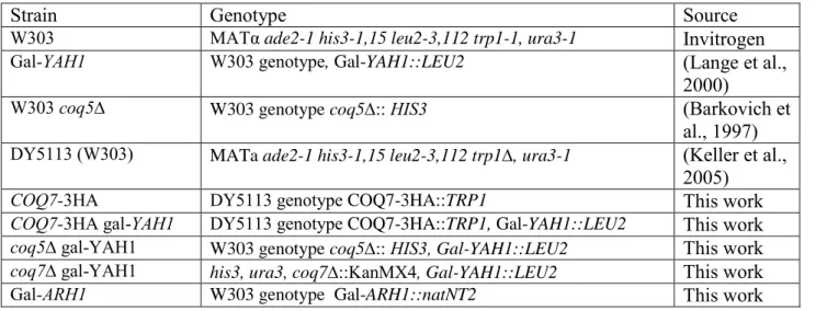
Table S1: S. cerevisiae strains used in this study
Strain Genotype Source
W303 MATα ade2-1 his3-1,15 leu2-3,112 trp1-1, ura3-1 Invitrogen
Gal-YAH1 W303 genotype, Gal-YAH1::LEU2 (Lange et al.,
2000)
W303 coq5∆ W303 genotype coq5:: HIS3 (Barkovich et
al., 1997)
DY5113 (W303) MATa ade2-1 his3-1,15 leu2-3,112 trp1, ura3-1 (Keller et al.,
2005)
COQ7-3HA DY5113 genotype COQ7-3HA::TRP1 This work
COQ7-3HA gal-YAH1 DY5113 genotype COQ7-3HA::TRP1, Gal-YAH1::LEU2 This work
coq5∆ gal-YAH1 W303 genotype coq5:: HIS3, Gal-YAH1::LEU2 This work
coq7∆ gal-YAH1 his3, ura3, coq7::KanMX4, Gal-YAH1::LEU2 This work
Supplemental experimental procedures
Synthesis of U-(13C)-p-aminobenzoic acid: To a solution of 300 mg of U-(13C)-toluene(3.03 mmol) in chloroform (25 mL) were successively added 599 L of 70% HNO3 (2.2 equiv.) and 1.014 g (2.2 equiv.) of phosphomolybdic acid. The resulting solution was refluxed for 5 days then washed with water and dried over Na2SO4. Concentration under vacuum afforded 327 mg (2.27 mmol,75 %) of nitrotoluene as a mixture of two regioisomers (ortho: para ratio, 54:46) unseparable by silica gel chromatography and pure enough to be used in the next step without further purification. 1H NMR (CDCl3) 8.50-8.30 (m, 1H), 7.90-7.50(m, 4H), 7.30-6.75 (m, 3H), 2.60 (dq, 2J 13C-H = 129 Hz, 3J 13C-H = 9.3 Hz, 4J 13C-H = 5.1 Hz, 4J 13C-H = 5.1 Hz, 3H, ortho isomer), 2.47 (dq, 2J 13C-H = 127 Hz, 3J 13C-H = 10.2 Hz, 4J 13C-H = 5.4 Hz, 4J 13C-H = 5.4 Hz, 3H, para isomer).
To a solution of 207 mg (1.5 mmol) of U-(13C)-nitrotoluene (as a 54:46 mixture of ortho and para isomers) in 7 mL of 2% aqueous H2SO4 were successively added 642 mg (2 equiv.) of NaIO4 and 287 mg (2 equiv.) of LiBr. After 30 hours at 95 °C, 2 additional equiv of both NaIO4 and LiBr were added and the resulting solution was stirred for aditionnal 20 hours at 95°C. The aqueous solution was then extracted with EtOAc ( 3 times) and the resulting organic was successively washed with saturated solution of Na2S2O3, twice with water, brine, dried over Na2SO4 then concentrated under vacuum to afford 209 mg (1.24 mmol , 83%) of the mixture of ortho and para U-(13C)-nitrobenzoïc acid as a pale yellow solid. Recristallisation by slow evaporation of diethylether from a solution of the crude product in diethylether resulted in the formation of a mixture of two kinds of crystals. Manual separation
allowed the isolation of 17 mg of pure U-(13C)-para- nitrobenzoic acid. 1H NMR (CD3OD) 8.7-8.5 (m, 2H), 8.12-7.95 (m, 2H).
A suspension of 17 mg (97.7 mol) of U-(13C)-p-nitrobenzoic acid and 5 mg of 10 % Pd-C in 2 mL of methanol was purged with hydrogen then vigorously stirred under H2 atmosphere (1 bar) for two days. After filtration and evaporation of the solvent under vacuum, 14 mg (97.2mmol, 99 %) of U-(13C)-p-aminobenzoic acid were obtained as a pale yellow solid and was pure enough to be used without further purification. . 1H NMR (CD3OD) 7.75 (dm, 2J 13C-H = Hz, 2H), 6.72 (dm, 2J 13C-H = Hz, 2H) , 1H-13C decoupling NMR spectrum: 7.63 (d, JAB= 8.1 Hz, 2H), 6.50 (d, JAB= 8.7 Hz, 2H)
Purification of 3-hexaprenyl-4-aminophenol: A total of 130g of Gal-YAH1 cells (wet
weight) from a culture in SC medium 2%glucose with 10mg/L pABA were used for lipid extraction as described above with volumes scaled up accordingly and without addition of Q4. The petroleum ether extract (400mL) was dried under vacuum. The extract was resuspended in 10mL methanol and lithium perchlorate was added to a final concentration of 40mM. The precipitate was eliminated by centrifugation and the product was purified by multiple
injections on the C18 column using 98% methanol 20mM lithium perchlorate at 0.5mL/min with the precolumn electrode set first at -450mV, then at 700mV and finally at -500mV. The retention time of the product changed from 10min in the reduced form to 16 min in the oxidized form. Finally, the product was injected in its reduced form on the C18 column using 100% methanol as a mobile phase and collected by monitoring the absorbance at 300nm. The pure product was dried under vacuum and resuspended in deuterated methanol for subsequent NMR analysis.
References
Barkovich, R.J., Shtanko, A., Shepherd, J.A., Lee, P.T., Myles, D.C., Tzagoloff, A., and Clarke, C.F. (1997). Characterization of the COQ5 gene from Saccharomyces cerevisiae. Evidence for a C-methyltransferase in ubiquinone biosynthesis. J. Biol. Chem. 272, 9182-9188.
Keller, G., Bird, A., and Winge, D.R. (2005). Independent metalloregulation of Ace1 and Mac1 in Saccharomyces cerevisiae. Eukaryot. Cell 4, 1863-1871.