HAL Id: hal-03236263
https://hal.inrae.fr/hal-03236263
Submitted on 26 May 2021
HAL is a multi-disciplinary open access archive for the deposit and dissemination of sci-entific research documents, whether they are pub-lished or not. The documents may come from teaching and research institutions in France or abroad, or from public or private research centers.
L’archive ouverte pluridisciplinaire HAL, est destinée au dépôt et à la diffusion de documents scientifiques de niveau recherche, publiés ou non, émanant des établissements d’enseignement et de recherche français ou étrangers, des laboratoires publics ou privés.
Distributed under a Creative Commons Attribution| 4.0 International License
production of colibactin in the Escherichia coli
population
Frédéric Auvray, Alexandre Perrat, Yoko Arimizu, Camille Chagneau, Nadège
Bossuet-Greif, Clémence Massip, Hubert Brugère, Jean-Philippe Nougayrède,
Tetsuya Hayashi, Priscilla Branchu, et al.
To cite this version:
Frédéric Auvray, Alexandre Perrat, Yoko Arimizu, Camille Chagneau, Nadège Bossuet-Greif, et al.. Insights into the acquisition of the pks island and production of colibactin in the Escherichia coli population. Microbial Genomics, Society for General Microbiology, 2021, 7 (5), pp.1-16. �10.1099/mgen.0.000579�. �hal-03236263�
OPEN DATA
Insights into the acquisition of the pks island and production of
colibactin in the Escherichia coli population
Frédéric Auvray1,*, Alexandre Perrat1, Yoko Arimizu2, Camille V. Chagneau1, Nadège Bossuet- Greif1, Clémence Massip1,3,
Hubert Brugère1, Jean- Philippe Nougayrède1, Tetsuya Hayashi2, Priscilla Branchu1, Yoshitoshi Ogura4 and
Eric Oswald1,3,*
DOI 10.1099/mgen.0.000579
Received 07 December 2020; Accepted 11 April 2021; Published 07 May 2021
Author affiliations: 1IRSD, INSERM, Université de Toulouse, INRAE, ENVT, UPS, Toulouse, France; 2Department of Bacteriology, Kyushu University,
Fukuoka, Japan; 3CHU Toulouse, Hôpital Purpan, Service de Bactériologie- Hygiène, Toulouse, France; 4Division of Microbiology, Department of Infectious
Medicine, Kurume University School of Medicine, Kurume, Fukuoka, Japan.
*Correspondence: Frédéric Auvray, frederic. auvray@ envt. fr; Eric Oswald, eric. oswald@ inserm. fr
Keywords: colibactin; Escherichia coli; enterobacteria; genotoxin; genetic diversity; pathogenicity island; pks.
Abbreviations: asn, asparagine; CC, clonal complex; CFU, colony forming unit; DR, Direct repeats; ExPEC, extra- intestinal pathogenic E. coli; HGT, horizontal gene transfer; HPI, high pathogenicity island; ICE, integrative and conjugative element; ICL, interstrand cross- link; Int, Integrase; IS, insertion sequence; ML, maximum likelihood; MRCA, most recent common ancestor; NJ, neighbour joining; NRPKS, NRP synthetase; PKS, PK synthase; SNP, Single nucleotide polymorphism; ST, sequence type; tRNA, transfer ribonucleic acid; VNTR, variable number of tandem repeat. Data statement: All supporting data, code and protocols have been provided within the article or through supplementary data files. Six supplementary figures and three supplementary tables are available with the online version of this article.
000579 © 2021 The Authors
Abstract
The pks island codes for the enzymes necessary for synthesis of the genotoxin colibactin, which contributes to the virulence of
Escherichia coli strains and is suspected of promoting colorectal cancer. From a collection of 785 human and bovine E. coli
iso-lates, we identified 109 strains carrying a highly conserved pks island, mostly from phylogroup B2, but also from phylogroups A, B1 and D. Different scenarios of pks acquisition were deduced from whole genome sequence and phylogenetic analysis. In the main scenario, pks was introduced and stabilized into certain sequence types (STs) of the B2 phylogroup, such as ST73 and ST95, at the asnW tRNA locus located in the vicinity of the yersiniabactin- encoding High Pathogenicity Island (HPI). In a few B2 strains, pks inserted at the asnU or asnV tRNA loci close to the HPI and occasionally was located next to the remnant of an integrative and conjugative element. In a last scenario specific to B1/A strains, pks was acquired, independently of the HPI, at a non- tRNA locus. All the pks- positive strains except 18 produced colibactin. Sixteen strains contained mutations in clbB or clbD, or a fusion of clbJ and clbK and were no longer genotoxic but most of them still produced low amounts of potentially active metabolites associated with the pks island. One strain was fully metabolically inactive without pks alteration, but colibactin pro-duction was restored by overexpressing the ClbR regulator. In conclusion, the pks island is not restricted to human pathogenic B2 strains and is more widely distributed in the E. coli population, while preserving its functionality.
DATA SUMMARY
All sequence data of the 785 E. coli used in this study are
freely available from the NCBI BioProject database (https://
www. ncbi. nlm. nih. gov/ bioproject/) under accession number PRJDB5579. This database was updated to include the sequence data obtained using ONT MinION for the E. coli reference strain SP15 and for E. coli strains ECSC054, JML285, KS- NP019, NS- NP030 and SI- NP020. The sequence data of
E. coli strain UPEC129 obtained using the PacBio instrument
were deposited in the NCBI BioProject database and are
avail-able at https://www. ncbi. nlm. nih. gov/ Traces/ study/ under
accession number PRJNA669570. Hybrid MinION- Illumina
and PacBio- Illumina assemblies are available at the NCBI nucleotide database. The genome sequences of 36 other E. coli reference strains and seven non-E. coli strains were retrieved from NCBI.
INTRODUCTION
Escherichia coli is not only a commensal resident of the
human and animal gut, but also a pathogen responsible for intestinal or extra- intestinal infections. E. coli is characterized by a high genetic and phenotypic diversity, with a population distributed into at least eight major phylogenetic groups (A, B1, B2, C, D, E, F and G) [1]. E. coli strains from phylogroup
OPEN ACCESS
B2 are increasingly being found in the faeces of healthy humans in high- income countries and also responsible for extra- intestinal diseases, including urinary tract infections, sepsis, pneumonia and neonatal meningitis [2]. By enabling the exchange of genetic material between bacterial cells, hori-zontal gene transfer (HGT) is a major driving force in the evolution of bacteria, including adaptation to their host and expansion of their ecological niche [3]. HGT- mediated acqui-sition of large genomic islands (GIs) or pathogenicity islands (PAIs) is recognized as a major contributor to the emergence of the various E. coli pathotypes [4]. The E. coli pks patho-genicity island consists of a clbA- clbS gene cluster enabling the biosynthesis of a polyketide (PK) – non- ribosomal peptide (NRP) hybrid genotoxin known as colibactin [5]. This island exhibits typical features of horizontally acquired genomic elements: (i) it is a large (i.e. 54 kb) region with a distinct GC content compared to that of the chromosomal backbone, (ii) it is physically associated with a phage- type integrase gene that probably mediated its insertion into the chromosome and (iii) it is located at a tRNA locus and is flanked by two short (i.e. 17 bp) direct repeats (DRs) reminiscent of those generated upon integrase- mediated insertion of mobile genetic elements [5, 6]. The pks island can be found in other members of the
Enterobacteriaceae such as Klebsiella pneumoniae, Citrobacter koseri and Enterobacter aerogenes [6], and in the honeybee
gut commensal Frischella perrara [7] and the marine sponge commensal Pseudovibrio sp. [8].
Colibactin is a virulence factor for extra- intestinal pathogenic
E. coli (ExPEC) [9–11] and is also a suspected
procarcino-genic factor [12–14]. Colibactin induces DNA interstrand cross- links (ICLs) [15] and double- strand breaks [5] in host eukaryotic cells. Its production involves the sequential action of the Clb proteins, including PK synthases (PKSs), NRP synthetases (NRPSs), hybrid PKS- NRPS, and acces-sory, editing and maturation enzymes [16]. Colibactin was first synthetized as a prodrug called precolibactin, carrying an N- myristoyl- d- asparagine (C14- Asn) side chain that is then cleaved in the periplasm to release the active genotoxin, whose translocation across the bacterial outer membrane remains unknown [17]. The production of colibactin is positively regulated by ClbR [18]. The multi- modular PKS- NRPS assembly line not only produces colibactin but also a set of numerous secondary metabolites with varying modes of action [19, 20]. These include analgesic lipopeptides, such as C12- Asn- GABA, with the ability to diffuse across the epithe-lial barrier and act on sensory neurons to decrease visceral pain in the host [21]. The pks island also contributes to the production of siderophores (enterobactin, salmochelin and yersiniabactin), via its promiscuous phosphopantetheinyl transferase ClbA [10], and siderophore- microcins via its ClbP peptidase [22].
To date, the presence of the pks island has been investigated mostly in E. coli strains isolated from humans with extra- intestinal infections [5, 6, 23, 24]. Here we explored the distribution, conservation and functionality of the pks island in a large collection of non- clinical E. coli strains originating from human and bovine hosts [25]. We found that the pks
island was not only present in phylogroup B2 but also in other genetic phylogroups. We identified different scenarios for its integration into the E. coli genome. The sequence of the pks island is highly conserved and pks- positive strains were over-whelmingly capable of producing colibactin, suggesting that the pks island is under selective pressure for the adaptation of
E. coli to various ecological niches, through the production
of colibactin or other metabolites or pks- encoded enzymatic activities.
METHODS
Bacterial strains used in the study
The E. coli strains were collected in Japan from 418 healthy bovines in 2013 and 2014, 278 healthy humans in 2008, 2009 and 2015, and 89 humans with extra- intestinal infections, either bacteraemia (n=67) in 2002–2008 or urinary tract infection (n=22) in 2006 and 2011. They were described recently [25] and corresponded each to a single isolate; duplicates showing less than five SNPs difference in their whole genomes were excluded from this study. A list of the 109 pks- positive isolates is provided in Table S1 (available in the online version of this article). An additional set of 37 E. coli reference strains (Table S2) and seven non-E. coli strains (Table S3) were included in this study; their genome sequences were downloaded from NCBI, except for E. coli SP15 which was not available and was obtained here (see below).
Impact Statement
Colibactin, a genotoxin associated with the carcino-genicity of certain strains of Escherichia coli, is encoded by a pathogenicity island called pks. We took advantage of a large collection of non- clinical E. coli strains originating from human and bovine hosts to explore the distribution, conservation and functionality of the pks island. We found that the pks island was not only present in phylogroup B2 (and more specifically in certain B2 sublineages), but also in other genetic phylogroups, highlighting its capacity to disseminate though horizontal gene transfer. We identified various genetic pks configurations indica-tive of an introduction of the pks island into E. coli on multiple independent occasions. Despite the existence of various acquisition scenarios, we found that the pks sequences were highly conserved and pks- carrying strains were overwhelmingly capable of producing coli-bactin, suggesting that the pks island is under selective pressure, through the production of colibactin or other secondary metabolites. Future implications include the identification of such metabolites and their biological activities that could be advantageous to E. coli and enable its adaptation to various ecological niches.
Whole genome sequencing
The whole genome sequences of the 785 E. coli isolates were determined by Illumina sequencing [25]. Among these, the genomes of five E. coli strains (ECSC054, JML285, KS- NP019, NS- NP030 and SI- NP020) were further subjected here to long- read sequencing using an Oxford Nanopore Tech-nologies (ONT) MinION device. The DNA libraries were prepared using the rapid barcoding kit (ONT) and sequenced using MinION R9.4.1 flow cells. Long- read sequencing of
E. coli strain UPEC129 was also performed using a Pacific
Biosciences (PacBio) RSII sequencer (Genoscreen). The DNA was extracted using Gentra Puregen Yeast/Bact (Qiagen) and the DNA libraries were prepared using the SMRTbell Template Prep kit (PacBio). Hybrid assembly of Illumina paired- end reads and MinION or PacBio reads was performed using Unicycler (v.0.4.8) [26]. The whole genome sequence of E. coli reference strain SP15 was obtained using Illumina and ONT MinION instruments and assembled as described above.
Sequence and phylogenetic analysis
The core gene- based phylogenetic tree was reconstructed as described previously [25]. Briefly, core genes were determined using Roary [27] and SNP sites were extracted from the core gene alignment using SNP- sites [28]. The maximum- likelihood (ML) tree was reconstructed using RAxML [29] with the GTR- GAMMA model and displayed using iTOL [30].
For the phylogenetic analysis of the entire pks island, the genome sequences of pks- positive strains were aligned with the entire pks island sequence of strain IHE3034 using MUMmer [31] and the SNP sites located therein were identified. After removing SNP sites on the VNTR region, a neighbour- joining (NJ) tree was reconstructed by mega7 [32] using the Tamura–Nei evolutionary model.
Co- phylogenetic analysis of the core- gene- based ML tree and the pks- based NJ tree was performed using the ‘cophylo’ function of the R package Phytools [33].
Sequence type and phylogroup determination was performed as described previously [25].
The pks sequences from four E. coli strains belonging to distinct phylogroups (i.e. SI- NP020, KS- NP019, UPEC129 and ECSC054 from phylogroups A, B1, B2 and D, respec-tively) were extracted from hybrid assemblies and compared at the nucleotide level with that of the reference E. coli strain IHE3034. In addition, the amino acid sequences were obtained for the 19 clb genes of each strain and aligned by muscle with mega7 [32]. The alignment file was analysed with the
sequence identity and similarity online software (http:// imed.
med. ucm. es/ Tools/ sias. html; accessed in July 2020).
The comparison of pks sequences from E. coli and other
bacterial species was performed with blastn (https:// blast.
ncbi. nlm. nih. gov/ Blast. cgi? PAGE_ TYPE= BlastSearch). Each pks region was defined from clbA to clbS and used as the query nucleotide sequence against each pks region as the
subject. Then, the alignment was visualized with the Artemis Comparison Tool (v13.0.0) [34].
The integrase nucleotide and amino acid sequences were aligned using muscle (v3.8.31) and the phylogeny was analysed with PhyML (v3.1/3.0 aLRT) prior to tree
visu-alization with TreeDyn (v198.3) (http://www. phylogeny. fr;
accessed in September 2020).
The CC95 strains were typed for their fimH allele using
FimTyper (v1.0) (https:// cge. cbs. dtu. dk/ services/ FimTyper/;
accessed in November 2020) and were further assigned to subgroups A–E by analysis of the presence of either of the five subgroup- specific genes described previously [35].
PCR analysis of the clbJK fusion gene
The 5651 bp deletion in the clbJ- clbK region resulting in the clbJK fusion gene was tested using a duplex PCR assay, with two primer pairs. The first primer pair (clbK- F, 5′- GACT GCCC AACA TACG CTCCG-3′; clbK- R, 5′- TTGT GTCG TTGT ACTC TCGGC-3′) was used to amplify a 722 bp long DNA fragment that is located within the deleted region and is thus only present in strains with an intact clbJ-
clbK region. The second primer pair consisted of primers
clbJK- F (5′- AGAATTACCCACTGCCACCA-3′) and clbJK- R (5′- GGCGCTAATGGATCAGATGT-3′) flanking the deleted region, and was used to amplify a 1441 bp long DNA fragment only present in strains with a clbJK fusion gene. The strains with an intact clbJ- clbK region or a clbJK fusion gene yielded a 722 or a 1441 bp long amplification product, respectively. The final reaction mixture volume of 50 µl contained 2 µl template DNA, 1× GoTaq Reaction
buffer, 200 µM of each dNTP, 4 mM of MgCl2, 1.25 U of
GoTaq DNA polymerase (Promega) and 0.2 µM of each primer (Eurofins Genomics). Amplification was done in a GeneAmp 9700 thermal cycler (Applied Biosystems), with the following programme: initial denaturation at 95 °C for 2 min; 30 cycles of denaturation at 95 °C for 30 s, annealing at 56 °C for 45 s and extension at 72 °C for 1 min 30 s; and final extension at 72 °C for 5 min. Electrophoresis was carried out in 1 % agarose gel and the PCR products were visualized after Gel Red (Biotium) staining using a Bio- Rad Chemidoc XRS system (Bio- Rad).
In vitro DNA interstrand crosslinking assay
ICL activity was assessed as described previously [15]. Briefly,
3×10⁶ E. coli cells or 6×10⁶ Erwinia oleae cells pre- grown for
3.5 h in Dulbecco's modified Eagle medium (DMEM) with 25 mM HEPES (Invitrogen) were mixed with EDTA (1 mM) and 400 ng of linearized plasmid pUC19 DNA and the mixtures were incubated for 40 min at 37 °C. After pelleting the bacteria, the DNA was purified from the supernatant and analysed by electrophoresis on denaturing (40 mM NaOH – 1 mM EDTA) 1 % agarose gels. ICL activity of Erwinia oleae was also tested in the presence of 400 nM 6- histidine- ClbS, which was purified with HisPur nickel- nitrilotriacetic acid (Ni- NTA) agarose (Thermo Scientific) from a culture of
BL21(DE3) strain hosting the plasmid pET28a- ClbS- His, as described previously [15].
Megalocytosis assay
Non- haemolytic pks- positive strains were tested for mega-locytosis on infected HeLa cells as described previously [5, 36]. Briefly, HeLa cells grown to 50 % confluence in cell culture 96- well plates were inoculated with 5 µl of an overnight culture of bacteria in infection medium (DMEM with 25 mM HEPES) and incubated for 4 h at 37 °C in a 5 %
CO2 atmosphere. Cells were then washed and incubated
for 48–72 h in cell culture medium supplemented with
gentamicin at 200 µg ml−1, and then stained with methylene
blue for microscopy examination.
H2AX phosphorylation assay
HeLa cells were infected as described above and H2AX phos-phorylation was quantified immediately after the 4 h infection step by immunofluorescence as described elsewhere [37].
C14-asn quantification
E. coli strains were grown for 24 h at 37 °C in 10 ml DMEM-
HEPES (Gibco), resuspended in 500 µl Hanks' balanced salt solution (HBSS; Invitrogen) and then crushed with a Precellys instrument (Ozyme). After addition of an internal standard mixture (deuterium- labelled compounds;
400 ng ml−1), cold methanol (MeOH) was added and
samples were solid- phase extracted on HLB plates (OASIS HLB 2 mg, 96- well plate; Waters). Lipids were eluted with
MeOH, evaporated under N2, resuspended in MeOH and
analysed by HPLC/MS- MS analysis (LC- MS/MS) (Meta-ToulLipidomics Facility), as described previously [21].
RESULTS
The pks island was mainly found in specific E. coli lineages from phylogroup B2
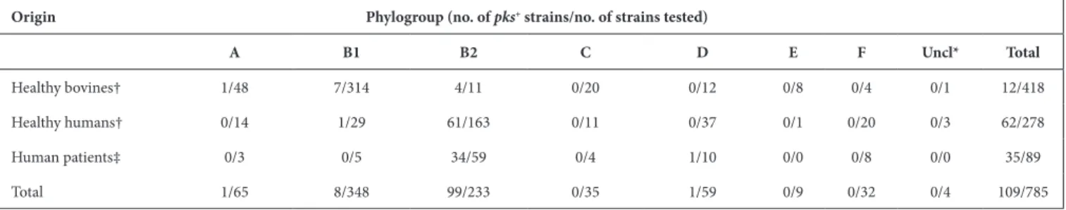
The presence of the pks island was investigated in a collec-tion of 785 E. coli strains [25] belonging to at least 296 different sequence types (STs) and originating mostly from faecal samples of healthy bovines and humans.
Clinical isolates recovered from urine or blood samples of human patients with extra- intestinal infection were also included for comparison. We detected the pks island in 109 E. coli strains, including 62 (22.3 %) out of 278 healthy human faecal isolates and 12 (2.9 %) out of 418 healthy bovine faecal isolates (Table 1, Fig. 1). As expected, a higher proportion of pks- positive strains were found among ExPEC, i.e. 35 (39 %) out of 89 strains, including 14 (63.6 %) out of 22 strains from urinary tract infection and 21 (31.3 %) out of 67 strains from bacteraemia. The vast majority of the 109 pks- positive strains corresponded to B2 isolates (Table 1, Fig. 1) and the pks island was mainly present in specific lineages or STs of the B2 phylogroup (Fig. 1).
Strikingly, the pks island was found in (nearly) 100 % of strains belonging to ST12, ST73, ST95 and ST550, while it was excluded from other STs, such as ST131 and ST357 (Fig. 1, Table 2). Interestingly, these pks- positive and pks- negative STs are found in distinct clusters in the core- genomebased phylogenetic tree (Fig. 1), suggesting that pks acquisition occurred after the divergence of these clusters from a common ancestor. We further characterized the 54 pks- positive strains of ST95 for their fimH allele and affiliation to CC95 subgroups A–E defined previously [35]. We could assign 35 of them to subgroup A (n=22), B (n=12) or E (n=1) (Table S1). The remaining 19 strains, including 15 of serotype O1:H1, did not belong to any of these five subgroups. No pks- positive strain was assigned to CC95 subgroups C or D, in agreement with previous results [35].
Except for four B2 strains originating from healthy bovines, the pks- positive B2 isolates originated from humans, either patients with extra- intestinal infection (n=34) or healthy individuals (n=61) (Fig. 1, Table 1). The low occurrence of pks among bovine isolates probably reflected the low prevalence of B2 strains in cattle [25]. Interestingly, 10 non- B2 pks- positive strains were identified corresponding to one human blood isolate from phylogroup D, one healthy human faecal isolate from group B1, and eight healthy bovine faecal isolates from groups A (n=1) and B1 (n=7) (Fig. 1, Table 1). In contrast to the B2 pks- positive isolates, these strains were scattered throughout the core Table 1. Occurrence of pks in E. coli strains from healthy humans or bovines, and human patients with extra- intestinal infection.
Origin Phylogroup (no. of pks+ strains/no. of strains tested)
A B1 B2 C D E F Uncl* Total Healthy bovines† 1/48 7/314 4/11 0/20 0/12 0/8 0/4 0/1 12/418 Healthy humans† 0/14 1/29 61/163 0/11 0/37 0/1 0/20 0/3 62/278 Human patients‡ 0/3 0/5 34/59 0/4 1/10 0/0 0/8 0/0 35/89 Total 1/65 8/348 99/233 0/35 1/59 0/9 0/32 0/4 109/785 *Unclassified.
†Isolates were collected from faeces of healthy individuals.
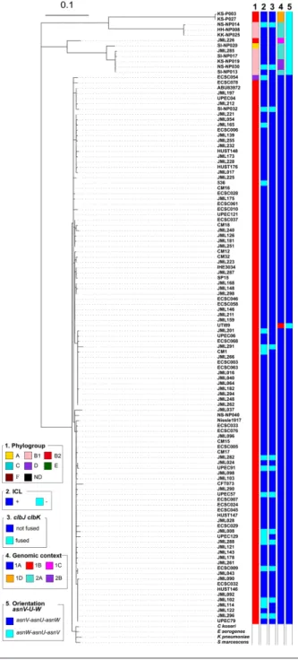
Fig. 1. Phylogenetic relationship and distribution of pks- positive/negative E. coli isolates among 696 human and bovine commensal E.
coli, 89 ExPEC and 37 completely sequenced reference E. coli strains. A core gene- based ML tree was reconstructed based on 271 403
SNPs located on 2000 core genes and rooted on cryptic Escherichia clade 1 strains as outgroups. Origin (column 1), phylogroup (column 2), major sequence type (ST) (i.e. ST identified for at least five strains) (column 3), presence of pks (clbA) (column 4), colibactin activity (ICL) (column 5), presence of the clbJK fusion gene (column 6), genetic pks configuration (see Fig. 3) (column 7), orientation of the asnV-
asnU- asnW region situated downstream of the asnT tRNA gene (column 8) and the number of 5′-ACAGATAC-3′ repeats found in the
clbB- clbR intergenic region (see Fig. 2) (column 9) are shown for each strain. The scale bar shows the number of substitutions per site.
genome phylogenetic tree and were not representative of any particular lineage or ST (Fig. 1, Table 2).
High level of genetic conservation of the pks island among E. coli phylogroups and other enterobacteria
The pks sequence from the B2 reference E. coli strain IHE3034 was compared to that of three non- B2 E. coli isolates, including the single group A isolate (i.e. SI- NP020), the single group D isolate (i.e. ECSC054) and one of the eight B1 isolates (i.e. KS- NP019). An additional B2 isolate (i.e. UPEC129) was also selected for this analysis. To perform this comparison, the whole genomes of these four isolates were assembled from a combination of short and long reads. At the amino acid level, over 99 % identity was observed for each of the 19 clb gene products (Fig. 2). At the nucleotide level, the only variation observed in the pks sequence was the size of the region located between clbB and
clbR which contains a variable number of tandem repeats
(VNTRs) of the motif 5′-ACAGATAC-3′ [6]. This VNTR locus contained between three and 14 repeat units when the whole collection of pks- positive strains was analysed (except for 17 isolates for which the VNTR length could not be calculated), with no apparent correlation with the STs (Fig. 2). Therefore, apart from the size of the VNTR, the pks island was highly conserved among the strains, irrespective of their phylogroup or ST.
Comparison of the pks island nucleotide sequence from B2 reference E. coli strain IHE3034 with that of other pks- positive bacterial species confirmed that it was conserved in other members of the Enterobacteriaceae (Fig. S1) such as K. pneumoniae, Enterobacter aerogenes, C. koseri, Serratia
marcescens and Erwinia oleae. A similar pks island was
present, although less conserved, in F. perrara and
Pseu-dovibrio sp. (Fig. S1).
The pks islands in E. coli from phylogenetic groups B2 and D share a similar genomic environment
To gain insights into the events leading to the acquisition of the pks island into the E. coli population, we analysed Table 2. Distribution of the pks island in the predominant sequence
types (ST) among E. coli strains isolated from healthy bovines, healthy humans and human patients with extra- intestinal infection
Phylogroup ST∗ pks+/no. of strains
A 10 0/22 206 0/5 6126 0/4 B1 20 0/5 29 0/4 56 0/6 58 0/23 101 0/16 109 1/6 154 0/6 155 0/4 156 0/4 164 0/6 205 0/9 223 0/5 278 2/8 295 0/4 297 0/7 300 0/4 327 0/9 332 0/4 446 0/4 677 0/6 679 0/7 718 0/6 765 0/9 795 0/4 906 0/12 1079 0/7 1423 0/5 5487 0/4 B2 12 4/4 73 20/20 95 54/56 131 0/25 357 0/35 Continued
Phylogroup ST∗ pks+/no. of strains
550 12/12 1193 0/4 C 88 0/4 D 32 0/5 38 1/8 69 0/14 349 0/6
*Only STs including at least four strains are listed. Table 2. Continued
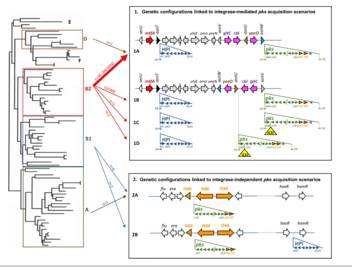
the genomic environment of the pks island in the 109 pks- positive strains and in seven pks- positive E. coli reference strains (i.e. 536, ABU83972, CFT073, Nissle 1917, UTI89, IHE3034 and SP15). Various configurations were found for the pks island genomic environment, suggesting two main scenarios of pks acquisition, depending on the presence or absence of an integrase gene (Fig. 3). The genetic configura-tion typical of B2 strains, named 1A, which is characterized by a pks island carrying an integrase gene and inserted into the asnW tRNA gene in the vicinity of the asnT- located high pathogenicity island (HPI) [5, 6], was found in 96 B2 strains of our collection and in one phylogroup D strain, ECSC054 (Figs 1 and 3).
A similar configuration was found in a few other B2 strains but with variations in the location of the pks island, which was inserted into either the asnV (corresponding to config-urations 1B and 1C found in ST95 reference strain UTI89 and in ST73 strain JML226, respectively) or the asnU tRNA gene (corresponding to configuration 1D found in strains KS- P003 and KS- P027, both belonging to ST95) (Figs 1 and 3) . Besides the difference in the tRNA insertion site, the configuration 1B found in reference strain UTI89 differed from the major configuration 1A by the orientation of the 4309 bp asnW- asnU- asnV tRNA region upstream of the pks island. This region contains three other genes, namely gltC and cbl, encoding two LysR- family transcriptional regula-tors, and yeeO, encoding a flavin mononucleotide (FMN)
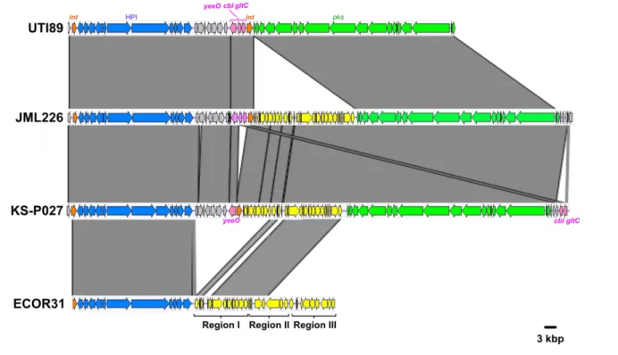
and flavin adenine dinucleotide (FAD) exporter. Configu-rations 1C and 1D possessed the same asnW- asnU- asnV orientation as in UTI89 and carried a 25 kb region between the pks integrase gene and clbS. A 14 kb section from this region exhibited high sequence similarity (>99 %) to inte-grative and conjugative elements (ICEs) identified in E. coli (ICEEc1) and K. pneumoniae (ICEKp1), in particular to the DNA regions I and II from ICEEc1 involved in mating- pair formation (Mpf) and DNA mobilization, respectively (Fig. 4) [6, 38]. This 14 kb section could therefore be consid-ered as an ICE- like element, although it is most probably non- functional given the lack of a complete region II (Fig. 4). The remaining 11 kb section was not homologous to ICEEc1, ICEKp1 or any other ICE, and its role could not be predicted.
The phage- type pks integrase is a tyrosine site- specific recombinase with similarity to the phage P4 integrase C- terminal catalytic domain (INT_P4_C). The integrase genes located at the asnW (configuration 1A) or asnV loci (configurations 1B and 1C) and their gene products were highly conserved and grouped into the integrase family 1 (Fig. S2), whereas the integrase genes located at the asnU locus (configuration 1D) and their gene products shared 94 % nucleotide and 94.6 % amino acid sequence similarity, respectively, with those of family 1 and were thus grouped into the integrase family 2 (Fig. S2).
Fig. 2. Comparison of the pks islands of E. coli strains belonging to phylogroups A, B1, B2 and D. The 19 ORFs of the clbA- clbS gene cluster from the reference E. coli strain IHE3034 sequence (group B2) and MinION- or PacBio- derived sequences of E. coli strains KS- NP019 (group B1), SI- NP020 (group A), UPEC129 (group B2) and ECSC054 (group D) are represented by arrows with the arrowhead representing the direction of transcription. The areas between the corresponding genetic maps shaded in dark and light grey indicate 100 % amino acid identity and ca. 99 % amino acid similarity, respectively. The number of repeated 5′-ACAGATAC-3′ motifs found in the clbB- clbR nucleotide intergenic region of pks- positive E. coli strains and the sequence types (ST) of the corresponding strains are indicated below the clbA- clbS gene cluster, with the number of strains given in parentheses.
Atypical genomic environments of pks islands in
E. coli from phylogenetic groups A and B1
In the nine pks- positive E. coli strains from phylogroups A and B1, two different configurations (named 2A and 2B) were observed that drastically differed from those found in B2/D strains. Their pks islands lacked an integrase gene, were not inserted into a tRNA gene and there were no direct repeats at their chromosomal boundaries (Fig. 3). The pks islands were located in the vicinity of the genes flu (or agn43) and
era encoding the Ag43 autotransporter adhesin and a GTPase
essential for cell growth and viability, respectively. They were flanked on one side by a truncated copy of the IS66 inser-tion sequence (IS) and on the other side by two intact IS66 copies. Two truncated copies of IS3 were also found next to the clbA gene but this was also the case for configurations
1A–1D. Moreover, in these B1/A strains, the HPI was absent (Table 3, Fig. 3, configuration 2A), except for two isolates in which the HPI was present but not in the vicinity of the pks island and not into a tRNA locus (Table 3, Fig. 3, configura-tion 2B). Using PCR assays, it was shown previously that three
E. coli strains from phylogroup B1 (namely U12633, U15156
and U19010) possessed a pks island that co- localized with the HPI and the DNA transfer and mobilization region of an ICEEc1- like element [6], a situation that is reminiscent of that of configuration 1D. However, as the whole genome sequences of these three strains were not available, this could not be confirmed here.
Since the asnW- asnU- asnV tRNA region displayed distinct orientations in pks- positive B2 strains depending on pks Fig. 3. Genetic configurations of the pks island and HPI in E. coli strains and proposed scenarios for their acquisition. Left: schematic phylogenetic tree showing the distribution of E. coli into the main phylogroups. Right: E. coli genetic pks and HPI configurations resulting from proposed acquisition scenarios involving site- specific recombination (configurations 1A, 1B, 1C and 1D) or not (configurations 2A and 2B). The location and orientation of the tRNA genes and the ORFs of the chromosomal regions, including the integrase and genes from pks and the HPI, are indicated by the arrows. Partial and complete insertion sequence (IS) elements are represented by orange arrowheads and arrows, respectively. The ICE- like element (ICEl) found in configurations 1C and 1D is represented as a yellow triangle. DR, direct repeats located at the extremities of the islands (except for the HPI in configurations 1A–1D, one DR lacking at the right border). Middle: arrows connect phylogroups A, B1, B2 and D (left) with the pks and HPI configurations (right). The number of E.
coli isolates belonging to the collection of 785 strains and corresponding to each configuration is indicated (except for configuration
1B which was only found in reference strain UTI89, indicated in parentheses). The thick arrow represents the most frequently found configuration (exemplified here by reference strain IHE3034, indicated in parentheses).
configuration, we further analysed its orientation for the rest of the E. coli collection, i.e. in pks- positive B1/A strains and in pks- negative strains. The ‘asnV- asnU- asnW’ orientation was uniquely found in typical pks- positive B2 strains with configuration 1A, suggesting that, in these strains, pks acquisi-tion at the asnW locus was accompanied by an inversion of the upstream tRNA- encoding region (Figs 1 and 3).
The phylogeny of the pks island globally reflects that of the E. coli core genome
To shed further light on the different pks acquisition scenarios, we constructed a phylogenetic tree of the entire pks sequences (i.e. from clbA to clbS, except for the VNTR- containing region which was excluded from the analysis) from the 109
pks- positive strains. Globally, the pks sequences from the
strains showing distinct pks genomic configurations formed distinct clusters (Fig. 5). The pks sequence of strain UTI89 with a unique configuration (configuration 1B) was clustered with those of strains with configuration 1A (Fig. 5). Remark-ably, the pks sequences of B1/A strains segregated separately from those of B2/D strains. Moreover, the pks sequences with an insertion of an ICE- like element in the B2 (ST73) human strain JML226 (with configuration 1C) or in the pair of B2 (ST95) bovine strains KS- P003 and KS- P027 (with
configuration 1D) also clustered separately and were closer to the pks sequences of B1/A strains than to those of the B2/D strains lacking this ICE- like element (Fig. 5). Finally, the pks sequences of C. koseri, Enterobacter aerogenes, K. pneumoniae and S. marcescens were close to those of E. coli B2/D strains with configuration 1A (Fig. 5) whereas that of Erwinia oleae was more phylogenetically distant and clustered separately (data not shown).
To further assess the evolutionary relationships between the
pks sequences and the genetic background of the strains, a
co- phylogenetic analysis was performed where the phyloge-netic trees based on the pks sequence and the core genome were compared. Globally, congruence was observed between both trees (Fig. 6). It was noticeable that most of the typical
pks- positive B2 strains whose core genomes clustered together
into lineages of clonal complexes (CC) 12, CC14, CC73 and CC95 contained pks sequences that also clustered together in different subgroups of the main pks cluster (Fig. 6). In particular, the CC95 strains that clustered together into subgroups A and B (as defined by fimH typing) or O1:H1 subgroup in the core genome tree also clustered together in the pks tree (Fig. 6). These observations support the hypoth-esis of an introduction of the pks island into CC12, CC14, Fig. 4. Comparison of the chromosomal region covering the HPI and pks island between three atypical E. coli B2 strains (UTI89, JML226 and KS- P027, with genetic configurations 1B, 1C and 1D, respectively) and the integrative conjugative element ICEEc1 from E. coli strain ECOR31. Nucleotide sequence similarity (>99 %) between different DNA regions is indicated by grey areas between the corresponding genetic maps. The pks island and the HPI are represented in green and blue, respectively, and the integrase genes in orange. The yeeO,
cbl and gltC genes located between the asnV and asnW tRNA genes in UTI89 are represented in pink. The region between the HPI and the yeeO gene is represented in grey and the ICE- related region inserted either next to pks (JML226 and KS- P027) or next to the HPI (ECOR31)
is represented in yellow. In strain ECOR31, the ICE is divided into three parts, including region I encoding a mating pair formation system, region II encoding a DNA- processing system, both involved in conjugative transfer, and region III comprising hypothetical genes.
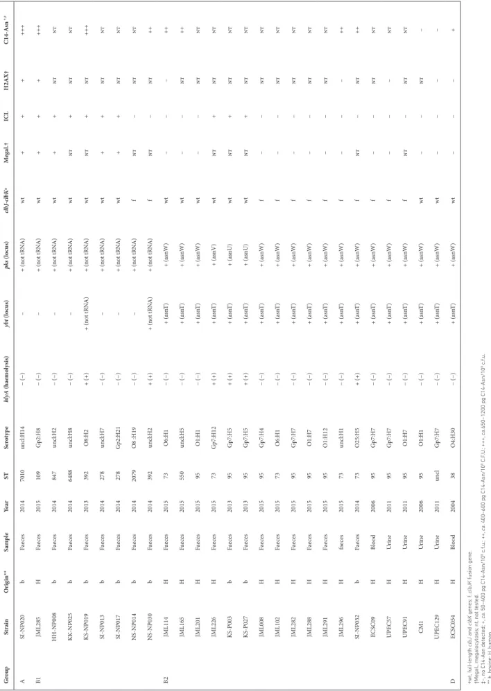
Table 3.
Char
acteristics of B2 and
B2 pks - positiv e E. coli str
ains with atypic
al f eatur es r egar ding pks integrity , functionality or loc ation Grou p Str ai n Orig in** Sa mp le Ye ar ST Se rot yp e hly A (hae mo ly sis) ybt (l oc us) pk s (l oc us) clb clb K∗ Me ga l.† IC L H2A X† A sn †,‡ A NP020 b Fae ces 2014 7010 un cl:H14 − (−) – + (n ot tRN A) wt + + + +++ B1 JML285 H Fae ces 2015 109 G p2:H8 − (−) – + (n ot tRN A) wt + + + +++ NP008 b Fae ces 2014 847 un cl:H2 − (−) – + (n ot tRN A) wt + + nt nt NP025 b Fae ces 2014 6488 un cl:H8 − (−) – + (n ot tRN A) wt nt + nt nt NP019 b Fae ces 2013 392 O8:H2 + (+) + (n ot tRN A) + (n ot tRN A) wt nt + nt +++ NP013 b Fae ces 2014 278 un cl:H7 − (−) – + (n ot tRN A) wt + + nt nt NP017 b Fae ces 2014 278 G p2:H21 − (−) – + (n ot tRN A) wt + + nt nt NP014 b Fae ces 2014 2079 O8 :H19 − (−) – + (n ot tRN A) f nt – nt nt NP030 b Fae ces 2014 392 un cl:H2 + (+) + (n ot tRN A) + (n ot tRN A) f nt – nt ++ B2 JML114 H Fae ces 2015 73 O6:H1 − (−) + (a sn T) + (a snW ) wt – – – ++ JML165 H Fae ces 2015 550 un cl:H5 − (−) + (a sn T) + (a snW ) wt – – nt ++ JML201 H Fae ces 2015 95 O1:H1 − (−) + (a sn T) + (a snW ) wt – – nt nt JML226 H Fae ces 2015 73 G p7:H12 + (+) + (a sn T) + (a snV ) wt nt + nt nt P003 b Fae ces 2013 95 G p7:H5 + (+) + (a sn T) + (a snU) wt nt + nt nt P027 b Fae ces 2013 95 G p7:H5 + (+) + (a sn T) + (a snU) wt nt + nt nt JML008 H Fae ces 2015 95 G p7:H4 − (−) + (a sn T) + (a snW ) f – – nt nt JML102 H Fae ces 2015 73 O6:H1 − (−) + (a sn T) + (a snW ) f – – nt nt JML282 H Fae ces 2015 95 G p7:H7 − (−) + (a sn T) + (a snW ) f – – nt nt JML288 H Fae ces 2015 95 O1:H7 − (−) + (a sn T) + (a snW ) f – – nt nt JML291 H Fae ces 2015 95 O1:H12 − (−) + (a sn T) + (a snW ) f – – nt nt JML296 H fae ces 2015 73 un cl:H1 − (−) + (a sn T) + (a snW ) f – – – ++ NP032 b Fae ces 2014 73 O25:H5 + (+) + (a sn T) + (a snW ) f nt – nt ++ ECSC09 H Bl ood 2006 95 G p7:H7 − (−) + (a sn T) + (a snW ) f – – nt nt UP EC57 H Ur in e 2011 95 G p7:H7 − (−) + (a sn T) + (a snW ) f – – – nt UP EC91 H Ur in e 2011 95 O1:H7 − (−) + (a sn T) + (a snW ) f nt – nt nt CM1 H Ur in e 2006 95 O1:H1 − (−) + (a sn T) + (a snW ) wt – – nt – UP EC129 H Ur in e 2011 un cl G p7:H7 − (−) + (a sn T) + (a snW ) wt – – – – D ECSC054 H Bl ood 2004 38 O4:H30 − (−) + (a sn T) + (a snW ) wt – – – + ∗wt , ful length clbJ and clbK genes; f , clbJK fusion gene. †Megal ., megalocytosis; nt , not tested. ‡−, no Asn detected; +, ca . 50−400 pg Asn/10 8 c.f .u.; ++, ca . 400−600 pg Asn/10 8 C.F .U .; +++, ca .650−1200 pg Asn/10 8 c.f .u. ** b, bo vine; H, human.
CC73 and CC95 through horizontal acquisition by their most recent common ancestor (MRCA) or by the MRCA of each of these lineages, followed by vertical transmission with subtle pks divergence overtime. Since CC95 subgroups C or D contain pks- negative strains [35] (strain ECSC026, subgroup C; this study), we further hypothesize that pks was lost during the evolution of these sublineages.
The fact that a single pks- positive strain from phylogroup D (i.e. ECSC054) possessed a pks island whose sequence clus-tered with that of B2 strains (Fig. 6) suggests that this strain acquired pks from a B2 strain through HGT. The pks- carrying B1/A strains were diverse based on their core genomes and their pks sequences clustered into two separate groups that were distantly related to the major pks cluster of B2 strains (Fig. 6), suggesting the existence of sporadic pks introduc-tion within the B1 and A phylogroups, presumably through HGT from a donor strain different from typical pks- positive B2 strains. Finally, the co- phylogeny also confirmed that the two atypical B2 ST95 strains KS- P03 and KS- P027 clustered with the other B2 strains of ST95 based on the core genome but contained a divergent pks sequence which was closer to those of B1 or A strains (Fig. 6), suggesting that this pair of strains probably acquired their pks islands through HGT, possibly from a donor strain carrying a pks island with an ICE insertion. The same scenario also presumably occurred with the atypical B2 ST73 strain JML226, which clustered with the other B2 strains of ST73 in the core genome tree but carried a pks island characterized by an ICE- like insertion and a sequence closer to those of B1/A strains than to those of B2 ST73 strains.
The functionality of the cluster of genes of the pks island is conserved in the majority of the enterobacterial strains
We next investigated the functionality of the pks islands in E. coli strains belonging to various phylogroups and carrying phylogenetically distinct pks sequences, as well as in the Erwinia oleae strain DAPP- PG531. Production of the genotoxin colibactin was directly investigated through the formation of DNA ICLs (Fig. S3a). The vast majority of the
E. coli strains carrying the pks island (i.e. 83.5 %) produced
ICLs (Fig. 1). DNA- crosslinking was also observed for the
Erwinia oleae strain, and it was abrogated by adding purified
colibactin self- resistance protein ClbS (Fig. S4), confirming the production of a bona fide colibactin by this strain carrying a less conserved sequence of the pks island. The E. coli geno-toxic strains belonged to phylogroups B2, B1 and A (Fig. 1). Eighteen (16.5 %) pks- positive E. coli isolates lacked a detect-able interstrand crosslinking activity, including 15 strains from phylogroup B2, two strains from phylogroup B1 and the single pks- positive strain from phylogroup D (Table 3). These strains did not cluster together in the core genome phylogenetic tree but instead were intertwined among genotoxic strains (Figs 1 and 5). To confirm the absence of genotoxicity, we tested the ability of these ICL- negative strains to trigger megalocytosis in cultured HeLa cells (Fig. S3b) and phosphorylation of histone H2AX (Fig. S3c), a robust Fig. 5. Phylogenetic tree of the entire pks island. SNP analysis was
performed with the pks sequences of the 109 pks- positive E. coli strains and an NJ phylogenetic tree was built. The pks sequences from seven reference E. coli strains (536, ABU83972, CFT073, Nissle 1917, UTI89, IHE3034 and SP15) and other enterobacteria (i.e. C. koseri ATCC BAA-895, Enterobacter aerogenes EA1509E, K. pneumoniae 1084 and
S. marcescens AS012490) were also included in this tree. Phylogroup
(column 1), colibactin activity (column 2), presence of a clbJK fusion gene (column 3), genetic pks configuration (see Fig. 3) (column 4) and orientation of the asnV- asnU- asnW region (see Fig. 3) (column 5) are indicated for each pks- positive strain. The scale bar shows the number of substitutions per site.
marker for DNA damage in eukaryotic cells. To avoid cell lysis during infection, we assessed only non- haemolytic strains. No megalocytosis and no p- H2AX foci were detected in HeLa cells exposed to subsets of ICL- negative strains (Table 3;
n=14 and n=5, respectively), even at a high multiplicity of
infection, confirming the deficiency of these strains in coli-bactin production. These results showed that except for a few
strains, E. coli strains carrying a pks island are overwhelmingly capable of producing the genotoxin colibactin, regardless of their phylogenies and the genomic configurations of their pks islands.
To examine the reasons for the lack of genotoxic activity of the 18 ICL- negative E. coli strains, we further analysed the Fig. 6. Co- phylogeny of pks sequences and E. coli host strains. A comparison generated with Phytools of the E. coli core gene- based ML tree and pks- based NJ tree is shown, including the links between pks and host strains (dashed lines). The phylogenetic groups (A, B1, B2 and D) are indicated. The strains belonging to the major clonal complexes (CCs) are shown with coloured names, including those of CC12 [containing four ST12 strains, one ST961 strain (ECSC078) and one ST5389 strain (ECSC078)], CC14 [containing one ST14 strain (ECSC010) and 12 ST550 strains], CC73 (containing only ST73 strains) and CC95 (containing only ST95 strains). Strains from CC95 that belong to subgroups A and B (as defined by fimH typing) or to serotype O1:H1 are boxed. Seven reference pks- positive E. coli strains (536, ABU83972, CFT073, Nissle 1917, UTI89, IHE3034 and SP15) were included in both trees, whereas the non-E. coli strains carrying a pks island (i.e. C. koseri ATCC BAA-895, Enterobacter aerogenes EA1509E, K. pneumoniae 1084 and S. marcescens AS012490) were included only in the pks- based tree.
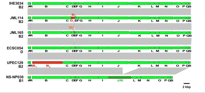
sequence of the pks island from those strains. We identified genetic alterations of the pks island in 16 out of the 18 non- genotoxic isolates. Strain JML114 carried a single nucleotide deletion in clbD at position 172 (A), leading to the segrega-tion of clbD into two ORFs (Fig. 7). Strain JML165 carried an IS1 inserted at the 3′-end of clbD after position 838. In strains UPEC129 and JML201, clbB was segregated into two ORFs due to nucleotide substitutions at positions 452 and 453 (AC to GA) (UPEC129; Fig. 7), and at position 872 (G to A) (JML201; data not shown), respectively. The genetic alterations identified in these four non- genotoxic strains each resulted in premature stop codons in clbB or clbD genes coding enzymes that are essential for the production of colibactin. Twelve ICL- negative strains carried a 5651 bp deletion resulting in a clbJK fusion gene, as shown for strain NS- NP030 in Fig. 7. This deletion presumably resulted from recombina-tion between two copies of a 1480 bp homologous sequence located in clbJ and clbK. A PCR analysis of the corresponding region in the 109 pks- positive strains confirmed the presence of this deletion in the 12 strains, whereas the other 97 pks- positive strains contained full- length clbJ and clbK genes (Fig. 5). The 12 strains carrying the clbJK fusion were detected sporadically in the core genome phylogenetic tree (Fig. 1) and
pks phylogenetic tree (Fig. 5), suggesting that occurrence of
the deletion between clbJ and clbK arose from accidental recombination events. The predicted 2440 aa hybrid ClbJK protein encoded by the clbJK fusion gene lacks the PKS module of ClbK necessary for the formation of stable cross- links [19]. In agreement with this, the strains carrying this fusion were devoid of interstrand crosslinking activity and did not trigger megalocytosis or histone H2AX phosphorylation in infected eukaryotic cells (Table 3). It was reported however
that rat E. coli isolates carrying a clbJK fusion gene caused DNA damage or displayed cytotoxicity to HeLa cells [39, 40]. This discrepancy with our results could be due to the use of distinct experimental conditions. The possibility that these rat isolates might produce additional genotoxins that would mask any colibactin deficiency caused by the clbJK fusion also cannot be excluded. Caution should also be observed during the assembly of sequencing reads as errors including deletions may be caused by the presence of tandem repeats.
For two other non- genotoxic isolates (i.e. CM1 and ECSC054), no mutation disrupting the pks genes was identified (Fig. 7; data not shown), suggesting that mutations located outside the pks island could negatively impact its expression. To test this hypothesis, we used plasmids pASK- clbR and pBAD- clbR, both overexpressing the pks regulator ClbR, and intro-duced either of them into strain CM1 which was susceptible to antibiotic, in contrast to ECSC054. In the resulting CM1 transformants, colibactin activity was restored, as seen by the formation of DNA ICLs (data not shown). Thus, in this strain, the lack of genotoxic activity probably resulted from a negative regulation of the pks island through an unknown mechanism.
The functionality of the pks island was also examined through analysis of the lipid metabolite profiles of selected genotoxic (n=3) and non- genotoxic (n=8) pks- positive strains, and in particular for production of C14- Asn, which was used as an indicator of the activity of the pks biosynthesis machinery. This lipopeptide is synthesized during the initial step of the biosynthesis process involving ClbN and ClbB, prior to elongation and final cleavage through the involvement of ClbC- H- I- J- K and ClbP, respectively [17]. The production of Fig. 7. Comparison of the pks island sequence from the E. coli reference strain IHE3034 with that of a selection of pks- positive but non- genotoxic E. coli isolates. Nucleotide sequence similarity (>99 %) between different DNA regions is indicated by grey areas between the corresponding genetic maps. Fusion of two adjacent ORFs resulting from the deletion of a sequence overlapping the two ORFs is indicated in dark green. Adjacent ORF sequences resulting from the segregation of an original ORF following an insertion or deletion event are indicated in red. The IS1 located in the pks island of strain JML165 is represented in orange.
C14- Asn was detected in all of the ICL- positive strains
exam-ined, SI- NP020, JML285 and KS- NP019 (ca. 650–1200 pg/108
c.f.u.) but not in the ICL- negative strain UPEC129 mutated in clbB (Fig. S5, Table 3). Interestingly, C14- Asn was detected
(ca. 400–600 pg/108 c.f.u.) in three ICL- negative strains
carrying a clbJK fusion gene (SI- NP032, JML296 and NS- NP030) and in two ICL- negative strains carrying a mutated clbD gene (JML114 and JML165). The two non- genotoxic strains carrying intact clb genes (ECSC054 and CM1) produced either a very low level or no detectable C14- Asn, respectively (Fig. S5, Table 3). For strain CM1 transformed with either plasmid pASK- clbR or pBAD- clbR (see above), overexpression of ClbR restored the production of C14- Asn (Fig. S5). These results suggest that even when the pks island does not allow production of active colibactin, enzymes from the pks pathway still produce metabolites with potential biological activities.
DISCUSSION
Acquisition of the pks island in the population of E. coli appears to have involved two distinct mechanisms differing by the presence or absence of a phage- type integrase. The integrase- mediated pks insertion pathway occurred mainly in B2 strains and resulted in pks insertion into either of three
asn tRNA genes (i.e. asnU, asnV or asnW). This potential
for integration into several DNA targets is consistent with the observed conservation and genetic integrity of the pks integrative module, i.e. the integrase gene and the two direct repeats flanking the island. The flexibility of pks insertion is reminiscent of what has been described for the HPI of Yersinia
pseudotuberculosis which is also able to insert into either of the
three Y. pseudotuberculosis asn tRNA genes [41], in contrast to the immobile truncated form of the HPI in E. coli whose right direct repeat is deleted and whose location is fixed at the asnT tRNA gene [42]. A divergent integrase sequence was found for the pks island inserted into the asnU tRNA gene compared to those inserted into the asnV or asnW tRNA gene. As the three asn tRNA sequences are 100 % identical, the use of either of them as an attachment site by slightly different integrases probably reflects distinct histories of pks acquisition. After
pks chromosomal integration, the endogenous pks integrase
promoter is replaced by the promoter of the upstream asn tRNA gene (Fig. S6), a configuration similar to that found for the HPI integrase promoter [43]. Whether the site of integration influences the expression of the pks integrase and hence pks stability at the distinct asn tRNA loci is not known. In contrast to the integrase- mediated pathway, the pks chro-mosomal integration process in the B1 and A E. coli strains remains unclear as no site- specific recombinase- encoding gene was found near pks and chromosomal insertion occurred into a non- tRNA locus. In these strains, pks integration could have involved the participation of IS elements such as the IS66 whose truncated or intact copies were found to flank the pks island.
The co- phylogeny analysis between the core genome- and pks- based phylogenetic trees shed further light on pks acquisition
scenarios. In the case of the typical pks- positive B2 strains belonging to lineages from major CCs (i.e. CC12, CC14, CC73 and CC95), the congruence observed between both trees suggested that the pks island was horizontally acquired by the MRCA of these lineages, or by the MRCA of each of these, and then stably maintained in their descendants through vertical transmission. The fact that strains of certain CC95 subgroups lack the pks island probably suggests that pks was lost during the evolution of these sublineages. Such a loss might be closely linked to the change in the relative fitness of CC95 subgroups underlying the variations observed in their spatial and temporal distribution in several continents [35]. In the case of B1 or A strains, pks acquisition and dissemi-nation probably occurred through sporadic lateral transfer events, as pks- positive B1 or A strains were scarce and not genetically related. The horizontal transferability of the pks island has previously been demonstrated using an in vitro approach where pks could be transferred together with the HPI via F’ plasmid- mediated conjugation from a donor to a recipient E. coli strain [44]. We propose that pks acquisition by the single pks- positive D strain ECSC054 was mediated by HGT, presumably from a B2 donor strain given the pks sequence relatedness observed between the D and B2 strains. HGT was also probably involved in the exchange of the pks island between the three atypical B2 ST73 or ST95 strains and a (yet unknown) phylogenetically distant donor strain, since their pks sequences did not cluster with those from other B2 ST73 or ST95 strains. This hypothesis was further supported by the identification, in these three isolates, of an ICE- like element inserted in their pks island. Similar ICE- like elements have previously been identified in three B1 E. coli isolates and other members of the Enterobacteriacae such as C. koseri,
Enterobacter aerogenes and K. pneumoniae [6]. They could
therefore play a role in pks dissemination in enterobacteria, as proposed for the self- transmissible ICE linked to the HPI identified in the E. coli strain ECOR31 [38]. Due to its lack of a complete DNA mobilization region (region II), we assume that the ICE- linked pks island is no longer self- transferable. It might nevertheless correspond to a remnant of an ancient, complete and self- transmissible ICE- linked pks island that could have behaved as a large complex ICE and spread in enterobacteria before undergoing partial or entire deletion of the ICE region. To date, no bacterial strain carrying a complete ICE linked to pks has been identified and the origin of the pks island therefore remains elusive.
The ecological niche and/or genetic background of the bacterial strains probably had an impact on the acquisition and stable maintenance of pks. The high concentration of pks- positive strains in some CCs of the B2 group such as CC73 and CC95 suggests that pks might have contributed to their ecological and evolutionary success. CC73 and CC95 exhibit a similar phyloge-netic history and are major ExPEC lineages, especially prior to the year 2000 where they were the most commonly detected [1, 45]. They are persistent intestinal colonizers and successful extra- intestinal pathogens with the particularity of exhibiting lower multidrug resistance levels compared to other ExPEC lineages. By contrast, as our collection contained E. coli B2 strains from
STs other than ST73 and ST95, it was interesting to note that pks was absent from STs corresponding to separate B2 lineages in the
E. coli phylogenetic tree, including the ST131 clonal complex
which is associated with multidrug resistance and is now the most predominantly isolated ExPEC lineage worldwide [45]. Consistent with the hypothesis of a pks acquisition by the MRCA(s) of CC73 and CC95 mentioned above, this finding suggests that such acquisition probably occurred after they diverged from the MRCA of CC131, i.e. before going through distinct evolutionary trajectories. The inversion of the upstream
asnW- asnU- asnV tRNA- containing region which probably
accompanied pks insertion into the asnW tRNA gene in the B2 group might have contributed to pks stabilization at this locus. Since the various pks- positive and pks- negative B2 lineages occupy the same ecological niche (i.e. primarily the intestinal tract of humans and animals), horizontal transfer of
pks between them could have been expected, at least to some
extent, but which was not revealed here. Several hypotheses can be proposed to explain this. First, some barriers to HGT might exist between members of distinct CCs, such as restriction- modification systems [46]. Second, pks might have been trans-ferred to recipient strains without providing adaptive value, thus resulting in its rapid loss. Third, as crosstalk between virulence determinants and the chromosome backbone is required for the emergence of virulent clones [1], a specific chromosomal phylogenetic background might be required for appropriate pks expression and production of an adaptive value, thereby consti-tuting a prerequisite for the stable maintenance of the island. The structure of the pks island is very well conserved among the E. coli population, with more than 99 % identity, suggesting that its integrity remains under strong structural and functional evolutionary constraints. We can speculate that transcription and translation of the 19 pks genes of this 54 kb long genomic island would be too high for the bacterial strains if the pks island did not bring a selective advantage to them. This is reinforced by the fact that only 18 out of 109 pks- positive strains lacked genotoxic activity. The importance of the biological role of
pks is highlighted by the numerous activities associated with
this genetic island, including genotoxicity, anti- inflammatory activity, antibiotic and analgesic effects. Given its interplay with siderophores (enterobactin, salmochelin and yersiniabactin) and siderophore- microcins (MccM and MccH47) [10, 22, 47], the pks island contributes to bacterial competition through the acquisition of iron or the production of inhibitory compounds, respectively. Protection of bacterial cells from genomic degrada-tion through the producdegrada-tion of ClbS could also be advantageous to pks- carrrying strains, as this multifunctional protein not only directly inactivates colibactin but also protects bacterial DNA from nucleolytic degradation by nucleases [48]. We also observed that non- genotoxic E. coli strains carrying an altered
pks island still produced the prodrug motif C14- Asn synthesized
at the early stage of the biosynthesis process, suggesting that yet- to- be- discovered bioactive compounds are produced by these strains. Given the high conservation observed for the pks island in E. coli, we can thus speculate that colibactin is a very important genotoxin but that pks- derived synthesis of other secondary metabolites could also be an advantage for E. coli.
Although our collection is characterized by a large diversity of E.
coli strains from various phylogenetic groups and STs, one
limi-tation of this study is that only strains from Japan were included, which may not be representative of the distribution of pks in a global collection of E. coli isolates from worldwide sources. In conclusion, the various genetic configurations of the pks island and its distribution in the E. coli phylogenetic tree imply the existence of various scenarios for the introduction and spread of pks into the E. coli population. The presence of a functional
pks island was demonstrated for the majority of the pks- positive
strains, suggesting that the pks island is under selective pressure for the adaptation of E. coli to various ecological niches, through the production of colibactin or other secondary metabolites. Funding information
This work was supported by funding from the National French Institute of Health and Medical Research (INSERM) to Camille Chagneau and the région Occitanie (grant ALDOCT-000610) and Ministère de l’Agriculture to Alexandre Perrat.
Acknowledgements
We thank Claire Hoede and Sarah Maman (SIGENAE group) and the GENOTOUL bioinformatics platform for providing computational resources. We also thank Pauline Le Faouder and the METATOUL lipi-domic platform for their support in the analysis of the lipid metabolite profiles.
Author contributions
Conceptualization: F.A., T.H., P.B., Y.O., E.O. Methodology: F.A., T.H., P.B., Y.O., E.O. Validation: F.A., T.H., P.B., Y.O., E.O. Formal Analysis: F.A., A.P., C.C., Y.A., T.H., P.B., Y.O., E.O. Investigation: F.A., A.P., Y.A., C.C., N.B.G., C.M., J.P.N., P.B., Y.O., E.O. Resources: T.H., Y.O., E.O. Data Curation: F.A., A.P., Y.A., T.H., P.B., Y.O., E.O. Writing – Original Draft: F.A., T.H., Y.O., E.O. Writing - Review and Editing: F.A., A.P., C.C., H.B., J.P.N., T.H., P.B., Y.O., E.O. Visualization: F.A., Y.A., C.C., J.P.N., P.B., Y.O., E.O. Supervision: F.A., H.B., T.H., Y.O., E.O. Project Administration: F.A., E.O. Funding Acquisi-tion: T.H., Y.O., E.O.
Conflicts of interest
The authors declare that there are no conflicts of interest. References
1. Denamur E, Clermont O, Bonacorsi S, Gordon D. The popula-tion genetics of pathogenic Escherichia coli. Nat Rev Microbiol 2021;19:37-54.
2. Tenaillon O, Skurnik D, Picard B, Denamur E. The population genetics of commensal Escherichia coli. Nat Rev Microbiol 2010;8:207–217. 3. Ochman H, Lawrence JG, Groisman EA. Lateral gene transfer and
the nature of bacterial innovation. Nature 2000;405:299–304. 4. Kaper JB, Nataro JP, Mobley HL. Pathogenic Escherichia coli. Nat
Rev Microbiol 2004;2:123–140.
5. Nougayrède HS, Homburg S, Taieb F, Boury M, Brzuszkiewicz E, Gottschalk G et al. Escherichia coli induces DNA double- strand breaks in eukaryotic cells. Science 2006;313:848–851.
6. Putze J, Hennequin C, Nougayrède JP, Zhang W, Homburg S et al. Genetic structure and distribution of the colibactin genomic island among members of the family Enterobacteriaceae. Infect Immun 2009;77:4696–4703.
7. Engel P, Vizcaino MI, Crawford JM. Gut symbionts from distinct hosts exhibit genotoxic activity via divergent colibactin biosyn-thesis pathways. Appl Environ Microbiol 2015;81:1502–1512. 8. Bondarev V, Richter M, Romano S, Piel J, Schwedt A et al. The
genus Pseudovibrio contains metabolically versatile bacteria adapted for symbiosis. Environ Microbiol 2013;15:2095–2113. 9. Marcq I, Martin P, Payros D, Cuevas- Ramos G, Boury M et al. The