HAL Id: hal-01936999
https://hal.archives-ouvertes.fr/hal-01936999
Submitted on 27 Nov 2018HAL is a multi-disciplinary open access
archive for the deposit and dissemination of sci-entific research documents, whether they are pub-lished or not. The documents may come from teaching and research institutions in France or abroad, or from public or private research centers.
L’archive ouverte pluridisciplinaire HAL, est destinée au dépôt et à la diffusion de documents scientifiques de niveau recherche, publiés ou non, émanant des établissements d’enseignement et de recherche français ou étrangers, des laboratoires publics ou privés.
Bifunctional silica nanoparticles for the exploration of
Pseudomonas aeruginosa biofilm
Léïla Mauline, Marie Gressier, Peter Hammer, Sidney J. L. Ribeiro, José
Maurício A. Caiut, Marie-Joëlle Menu, Christine Roques
To cite this version:
Léïla Mauline, Marie Gressier, Peter Hammer, Sidney J. L. Ribeiro, José Maurício A. Caiut, et al.. Bifunctional silica nanoparticles for the exploration of Pseudomonas aeruginosa biofilm. 2016 IEEE Nanotechnology Materials and Devices Conference (NMDC), Oct 2016, Toulouse, France. pp.1-2, �10.1109/NMDC.2016.7777158�. �hal-01936999�
OATAO is an open access repository that collects the work of Toulouse
researchers and makes it freely available over the web where possible
Any correspondence concerning this service should be sent
to the repository administrator:
tech-oatao@listes-diff.inp-toulouse.fr
This is an author’s version published in:
http://oatao.univ-toulouse.fr/20489
To cite this version:
Mauline, Léïla
and Gressier, Marie
and Hammer, Peter and
Ribeiro, Sidney J. L. and Caiut, José Maurício A. and Menu,
Marie-Joëlle
and Roques, Christine
Bifunctional silica nanoparticles
for the exploration of Pseudomonas aeruginosa biofilm. (2016) In:
2016 IEEE Nanotechnology Materials and Devices Conference
(NMDC), 9 October 2016 - 12 October 2016 (Toulouse, France).
Bifunctional silica nanoparticles for the exploration of
Pseudomonas aeruginosa
biofilm
L. Mauline2, M. Gressier2, P. Hammer3, S.J.L. Ribeiro4, J.M.A. Caiut5 and M.-J. Menu2, C. Roques'* Abstract- Luminescent silica nanoparticles (LSNPs) are
frequently employed for biotechnology applications mainly because of easy functionalization, photo-stability and biocompatibility. Bifunctional silica nanoparticles (BSNPs) are described here as new efficient tools for the understanding of a complex biological system such as biofilms. Photoluminescence is brought by the incorporation of a silylated ruthenium(II) complex, surface properties of the silica particles are designed by reaction with. BSNPs are fully characterized and Zeta potential and contact angle measurements exhibit various surface properties according to the functional groups. Confocal Laser Scanning Microscopy measurements show that the spatial distribution of these nanoparticles inside P AOl biofilm depends more on their hydrophilic/hydrophobic characteristics than on their size.
INTRODUCTION
A biofilm can be described as a community of adhering microorganisms, generally entrapped in a self-produced matrix of extracellular polymeric substances (EPS) [l]. Biofilm formation is a widespread phenomenon with a major economic impact in industrial [2, 3], medical [4, 5] and environmental fields [6, 7].
The particularity of biofilms cornes from its high resistance towards antibiotic and disinfectant agents, greater than planktonic cells and which can be linked to the tridimensional structure and the EPS [8]. Knowledge about transfert limitation through biofilms would provide precious information for the control and/or the eradication ofbiofilm.
Luminescent silica nanoparticles (LSNPs) are frequently employed for biotechnology applications mainly because of easy functionalization, photo-stability and biocompatibility. Bifunctional silica nanoparticles (BSNPs) are described here as new efficient tools for the understanding of a complex biological system such as biofilms.
* Thanks to MESR for a grant and Cofecub for its funding in the project with Brazil.
1Laboratoire de Génie Chimique, UMR-CNRS 5503, Université Paul
Sabatier, 35 chemin des maraîchers, 31062 Toulouse cedex 9, France;
2Centre Interuniversitaire de Recherche et de l'ingénierie des Matériaux,
UMR-CNRS 5085, Université Paul Sabatier, 118 route de Narbonne, 31062 Toulouse cedex 9, France; 3Laborat6rio de Espectroscopia de Fotoelétrons,
LEFE, Institute ofChemistry, Sào Paulo State University, UNESP, CP 355-Araraquara- SP, 14801-970 Brazil; 4lnstitute ofChemistry, Sào Paulo State
University, UNESP, CP 355-Araraquara - SP, 14801-970 Brazil;
5Department of Chemistry, FFLCRP, University of Sào Paulo, Ribeirào
Preto - SP, Brazil, 5
E-mail addresses: mauline@chimie.ups-tlse.fr;
gressier@chimie.ups-tlse.fr; menu@chimie.ups-tlse.fr; ch.roques@wanadoo.fr;
peter@iq.unesp.br; caiut@ffclrp.usp.br; sidney@iq.unesp.br
1. EXPERIMENTAL PART
Silica particles were synthesized by the Sti:iber method in reverse microemulsion. Luminescent characteristics were obtained by incorporation of the [Ru(bpy)2(bpy-Si)]Cb silylated dye [9]. The microemulsions were stirred for 24 h
and then broken by adding acetone (150 cm3). LSNPs were
recuperated by centrifugation and several subsequent washing procedures were performed in order to remove surfactants and excess free dye. The isolated powder 130 mg (95%) was dried under vacuum for 2 h. LSNPs were
activated by mild thermal treatment at 50°C under vacuum
for 30 min to remove physisorbed water. LSNPs (100 mg)
were dispersed in anhydrous acetonitrile (20 cm3) and
different organosilanes bearing functional groups were introduced and the mixtures were allowed to reflux for 24 h. BSNPs were purified by cycles of centrifugation and washings then dried in vacuum for 2h. BSNPs were then characterized: 13C and 29Si Solid-state NMR spectra (Bruker Avance 400WB equipment using dipolar decoupling in combination with Cross-Polarization (CP) and Magic Angle
Spinning (MAS) (100.356 and 79.391 MHz for 13C and 29Si
respectively); Photoluminescence spectra (Jobin Yvon, Fluorolog-3 spectrophotometer model FL3-22); XPS measurements (UNI-SPECS UHV); TEM analyses (JEOL 2010 (200 kV)); Zeta potential (Ç) and dynamic light scattering (DLS) (MAL VERN nanosizer ZS90 equipped with a red laser (633 nm)); Contact angle measurements
using a CDD camera with water drop volume of 4.10-3 cm3•
Bacterial biofilms were grown at 37°C in 24-well or 6-well
microplates containing 2 cm3 or 6 cm3 of MBB, respectively
as previously described [10]. After initial inoculation with a
103 CFU cm-3 bacterial suspension of PAOl, the medium
MBB was renewed at 2, 4, 6, 20, 24 and 48 h (end 72h)
After the period of growth, the wells were rinsed and 2 cm3
or 6 cm3 ofa filtered (0.2 µm) aqueous suspension ofBSNPs
at a concentration of 0.5 or 0.05 mg cm-3 was introduced. Incubation was performed 16 hours before confocal microscopy observations (Leica SP2 confocal laser scanning system). Staining was performed using ConA-Alexa Fluor®488 (Molecular Probe, Invitrogen, France), DNA stain, SYTO45® (Molecular Probe).
Il. RESUL TS AND DISCUSSION
Two adapted procedures have been used for the elaboration of BSNPs, a ternary (cyclohexane/Igepal CO 520/water) [11] and a quaternary (cyclohexane/hexanol/triton X-100/water) microemulsion [12], in order to isolate two sizes of nanoparticles (25±5 nm and 68±2 nm). Different techniques used to characterize LSNPs confirmed the
presence of silica and the ruthenium complex. Surface modification of LSNPs has been performed by grafting reaction of organosilanes on silica particles followed by control of chemical integrity. Figure 1 shows the images of formed drops in contact with the pellets of BSNPs. Ru@Si,
Ru@Si-A, Ru@Si-B and Ru@Si-C are hydrophilic with
contact angles between 31 and 38°, whereas Ru@Si-D
appears very hydrophobie with a contact angle greater than 90°.
,,
---+-�.g �Artgk, d<: oO!'tKt __ ,,.v �6J r"(:
1· ·0,4 10,1a 0,] -O,!i" -39 :1: S 43 S ·32 S Fig. 1. Contact angle measurements (0) formed by the water drop of BSNPs Ru@Si-A, Ru@Si-B, Ru@Si-C and Ru@Si-D.Biofilm penetration.
25 nm BSNPs. Hydrophilic particles Ru@Si, Ru@Si-A,
Ru@Si-B and Ru@Si-C were able to penetrate through the
biofilm regardless of surface charge (Figure 2). The same 2-dimensional snapshots have been performed using hydrophobie Ru@Si-D nanoparticles, results are presented in figure 9 I and Il. The behavior of Ru@Si-D differed from that of the hydrophilic nanoparticles since they aggregated around the biofilm.
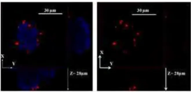
Fig. 2. 3-dimensional snapshot of Ru@Si-B particles in P. aeruginosa biofilm (III). DNA bacteria is labeled with SYT045® (blue; À.exc = 458 nm; À.em = 468 - 488 nm) and Ru@Si-B particles (red; À.exc = 543 nm; À.em = 600 - 700 nm)
68 nm BSNPs. In both cases, hydrophobie and hydrophilic BSNPs, the snapshots and the penetration curves showed a significant decrease of the emission intensity with increasing particle size, indicating deeper penetration of the smaller hydrophilic particles.
Fig. 3. 3-dimensional snapshot of Ru@Si-D particles in P. aeruginosa biofilm (III). DNA bacteria is labeled with SYT045® (blue; À.exc = 458 nm; À.em = 468 - 488 nm) and Ru@Si-D particles (red; À.exc = 543 nm; À.em = 600 - 700 nm)
Ill. CONCLUSION
The present study shows that regardless of size of the hydrophilic particles that they could penetrate throughout the biofilm but a steric effect was observed, the smaller particle diffusing in greater number than the larger ones. For hydrophobie nanoparticles, an accumulation at the periphery of the biofilm was observed regardless of the particle size; this observation suggests that the functionalization of particles could be a key factor in the penetration of particles in the biofilm. These results emphasize that BSNPs are powerful and innovative tools for the study of bacterial biofilms. Contrary to latex particles, silica has the advantage to display a great variety of functional surface groups.
ACKNOWLEDGEMENTS
L.M. thanks MESR for a grant and Cofecub for its funding in the project with Brazil.
REFERENCES
[I] Costerton JW, Lewandowski Z, Caldwell DE, Korber DR, Lappin Scott HM. Ann. Rev. Microbiol. 49:711. 1995.
[2] Lau PCY, Lindhout T, Beveridge TJ, Dutcher JR, Lam JS. J. Bacteriol. 191:6618-6631. 2009.
[3] Little BJ, Lee JS, Ray RI. Electrochim. Acta. 54(1):2-7. 2008. [4] Hall-Stoodley L, Stoodley P. Trends Microbiol. 13(1):7-10. 2005. [5] Huq A, Whitehouse CA, Grim CJ, Alam M, Colwell RR. Curr. Opin.
Biotechnol. 19(3):244-247. 2008.
[6] Singh R, Paul D, Jain RK. Trends in Microbiology.14(9):389-97. 2006.
[7] Wahl M, Goecke F, Labes A, Dobretsov S, Weinberger. Front Microbiol. 3:292. 2012.
[8] Campanac C, Pineau L, Payard A, Baziard-Mouysset G, Roques C. 2002. Antimicrob. Agents Chemother. 46(5):1469-1474. 2002. [9] Bifunctional silica nanoparticles for the exploration of biofilms of
Pseudomonas aeruginosa.
[10] Mauline L, Gressier M, Roques C, Hammer P, Ribeiro SJ, Caiut JM, Menu MJ. Biofouling. 29(7):775-88. 2013.
[11] Khalilzadeh P, Lajoie B, El Rage S, Furiga A, Baziard G, Berge M, Roques C. Can. J. Microbiol. 56(4):317-325. 2010.
[12] Jin Y, Lohstreter S, Pierce DT, Parisien J, Wu M, Hall C, III, Zhao JX. 2008. Chem. Mater. 20:4411-4419. 2008.
[13] Cousinie S, Mauline L, Gressier M, Kandibanda SR, Datas L, Reber C, Menu M-J. NewJ. Chem. 36:1355-1367. 2012.