HAL Id: hal-01314398
https://hal.archives-ouvertes.fr/hal-01314398
Submitted on 11 May 2016
HAL is a multi-disciplinary open access
archive for the deposit and dissemination of
sci-entific research documents, whether they are
pub-lished or not. The documents may come from
teaching and research institutions in France or
abroad, or from public or private research centers.
L’archive ouverte pluridisciplinaire HAL, est
destinée au dépôt et à la diffusion de documents
scientifiques de niveau recherche, publiés ou non,
émanant des établissements d’enseignement et de
recherche français ou étrangers, des laboratoires
publics ou privés.
JOINT DIRECTION AND VOLUME
TOMOGRAPHICAL AB-INITIO RECONSTRUCTION
FOR ELECTRON MICROSCOPY
Bassem Ben Cheikh, Etienne Baudrier, Gabriel Frey
To cite this version:
Bassem Ben Cheikh, Etienne Baudrier, Gabriel Frey. JOINT DIRECTION AND VOLUME
TOMO-GRAPHICAL AB-INITIO RECONSTRUCTION FOR ELECTRON MICROSCOPY. 12th
Interna-tional Symposium on BIOMEDICAL IMAGING: From Nano to Macro, Apr 2015, New-York, United
States. pp.1040-1043, �10.1109/ISBI.2015.7164049�. �hal-01314398�
JOINT DIRECTION AND VOLUME TOMOGRAPHICAL AB-INITIO RECONSTRUCTION
FOR ELECTRON MICROSCOPY
Bassem Ben Cheikh
†Etienne Baudrier
´
?Gabriel Frey
??
University of Strasbourg, CNRS ICube 300 Bd S. Brant - BP 10413 - F-67412 ILLKIRCH, FRANCE
†Sorbonne Universit´es, UPMC Univ Paris 06, CNRS, INSERM, LIB, F-75013, Paris, France
Email: [email protected]
[email protected]
[email protected]
ABSTRACT
This paper deals with the problem of three-dimensional vol-ume reconstruction from two-dimensional projections with unknown orientations. This situation occurs in cryo-electron microscopy. A method determining the orientations and re-constructing the volume jointly from scratch (ab initio) is pre-sented in this work. It is based on a heuristic cost minimiza-tion including a comparison of the input projecminimiza-tions with the projections obtained from the image being reconstructed and the orientations information and a data fitting based on the common-lines. The method is tested on synthetic data and compared to SIMPLE software on real data.
Index Terms— Tomography, Ab-initio Reconstruction, Unknown Directions, 2D, Gray-level Image.
1. INTRODUCTION AND STATE OF THE ART The problem of tomographic reconstruction from projections with unknown directions is encountered in various domains as medical imaging (e.g. when the patient is moving during a X-ray scanner acquisition) and cryo electron microscopy (cryo-EM). In cryo-EM, a thin vitreous ice layer containing multiple specimens is magnified with an electron microscope. Each cryo-EM image is a 2D projection of the 3D scattering density. Because the orientations of the 3D specimens relative to the microscope are not known, it follows that each image is related to an unknown-orientation 2D projection of the 3D specimen. In fact, unlike what is essentially done in medical imaging, cryo-EM don’t allow to rotate the 3D specimen and to take a series of images with known relative orientations be-cause the specimen is rapidly damaged by the electron beam. Therefore, taking multiple images of one specimen in differ-ent oridiffer-entations is replaced by taking one image of many iden-tical specimens where each specimen is in a different random unknown orientation.
The image to reconstruct is a 3D image. Generally, in the first step, the projection directions are estimated. In 3D, it relies on algorithms derived from common line correlation [1]. If the molecule is known to have some preferred ori-entation, then it is possible to find an ab initio 3D structure
using the random conical tilt method [2]. It exists solutions to the ab initio estimation problem of the 3D structure that do not involve tilting [3], [4]. To cope with the high dimen-sional space of the data, the direction-assignment algorithms use dimension reduction [5] or optimization [6, 7]. To re-duce variability, class averages are typically computed from particle images that have already been rotationally and trans-lationally aligned [8]. The choice of reference images for the alignment is, however, arbitrary and can represent a source of bias in the classification process. Ideally one would want to do the 3D reconstruction directly from projections in the form of raw images. This therefore sets the goal for an ab ini-tio reconstrucini-tion algorithm that requires as little averaging as possible.
Once the direction estimation is done, the second step is the reconstruction with known directions. The main families of reconstruction methods are [2]: the algebraic methods, it-erative filtered back projection, and those using the Fourier transform. In the ab-initio case, the sequence of these two steps gives a first reconstructed image whose quality is not always sufficient, and then requires a refinement step. The reconstructed image is used to refine the projection directions and these directions can then be used to improve the data re-construction. This iteration reflects the fact that if the esti-mated directions influence the image reconstruction, the im-age estimation also affects the estimation of directions. But this does not allow the estimated image to play its full role in the estimation of directions, since it was built later. We pro-pose a method that offers this possibility. It is based on a joint estimation of projection directions and image.
The novelty of our method lies in the fact that the image and directions as a single object to reconstruct and that they are jointly estimated. It generalizes our article focused on 2D gray-level image reconstruction [9].
The rest of the paper is structured as follows: in Sec. 2, a model of the acquisition is shown and the problem is speci-fied, the resolution method is detailed and the associated cost function is studied then results are given in Sec. 3. A conclu-sion and perspectives end this paper.
2. PROBLEM AND METHOD
In cryo-EM, 2D images modeled as projections of a 3D single object in different directions are provided. We assume here that the shortcomings of the acquired data (defocusing, con-trast transfer function, aberrations) have been corrected dur-ing a pre-processdur-ing step. These projections come down from the X-ray transform of the object. We first introduce some necessary notations before defining the X-ray transform.
Let B3 = {x ∈ R3| kxk ≤ 1} be the unit ball of R3. We call K the set of the bounded measurable functions f (for the usual measure of Lebesgues) from the unit ball B3in R+such that f is derivable and k∇f k1=R
B3|∇f | < ∞.
Remark2.1. As f is bounded, kf k2=R B3|f |
2 < ∞. Definition2.2 (X-ray transform). Let f a function of K. The X-ray transform of f is given by
Xf(θ, s) = Z
R
f (s + u · θ)du (1)
where θ ∈ S3and s ∈ θ⊥.
The function f is called object and the measurable function s 7→ Xf(θ, s) is called projection of f in the direction θ and denoted π2(θ, f ) or π(θ, f ) if there is no ambiguity.
π(θ, f ) belongs to the set of bounded measurable func-tions with support in B2, denoted B. The projecfunc-tions have the following property that will be exploited for the reconstruc-tion.
Proposition 2.3 (Common line). Let π2(θ1, f ) and π2(θ2, f ) two projections of the functionf . Let d = θ1∧ θ2, γ12 = d⊥ ∩ θ⊥
1 andγ21 = d⊥ ∩ θ⊥2. Then one has the following equality:
π1(γ12, π2(θ1, f )) = π1(γ21, π2(θ2, f ))
whereπ1(α, g) is the 1D projection of the 2D function g in the directionα.
Practically, the planes of projections associated to the two images intersect in a line d called the common line; see Fig. 1 for an illustration.
Fig. 1. Common line principle on two projections Hence, the 1D orthogonal projections of the two images onto the common line are equal. Note that there is a version of the common line property in the Fourier’s space.
Our proposal for reconstructing a 3D function f (standing for a density electron map in the cryo-EM frame) from a set of its 2D projections (P1, . . . , Pn) with unknown directions is to solve the following minimization problem:
( ˜f , ˜Θ) = argmin f,Θ GP (f, Θ) (2) where GP(f, Θ) = βJP(f, Θ) + (1 − β)KP(Θ) (3) β ∈]0, 1] is a weight factor.
The cost function GP is based on three elements which are the current image, current directions and the given projec-tions. GP is formed of a residual norm JP between the given projections and the estimated projections and an attach to the data KP depending only on the given projections and the cur-rent directions. Implementation of (2) relies on the definition of the cost function GP and on an optimization method. In this paper, JP is classically formed of a residual norm that estimates an error between the given projections P and cur-rent projections {π(θ, fcrt), θ ∈ Θcrt} which are two finite subsets of B: JP(f, Θ) = n X i=1 kPi− π2(θi, f )k 2 2
and KP gives a direction consistency based on the common line property KP(Θ) = n X i=1 n X j=i+1 kLi,j− Lj,ik22, where Li,j= π1(γij, Pi), ∀i, j ∈ 1, .., n
To calculate the minimum of the cost function GP we use the metaheuristic optimization algorithm, the Simulated An-nealing, that gives good results to our work in the 2D case [9]. Since this method is an ab initio reconstruction method, the algorithm is initialized with an empty cube and an initial tem-perature T = T0. At each iteration an elementary modifica-tion of the system is applied and evaluated by the variamodifica-tion of cost function. If the variation is negative, the proposed mod-ification is accepted. Otherwise, it is accepted with a proba-bility e−∆GPT . The temperature T decreases at each iteration
toward zero following some annealing schedule. The elemen-tary modification of (Θ, f ) is based on: (1) a modification in the object f by picking randomly a voxel and assigning to it a random value. (2) a modification in the set Θ by selecting randomly a projection and assigning to it a random direction. The process is stopped when the modifications are rejected n0 times. Different types of temperature decay functions have been tested. The exponential decreasing functions give the
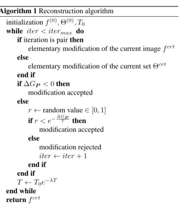
better results with low decay rates λ = 10−3and high initial temperatures T0 = 100. The algorithm of the minimization process is shown in Algo 1.
Algorithm 1 Reconstruction algorithm initialization f(0), Θ(0), T
0 while iter < itermax do if iteration is pair then
elementary modification of the current image fcrt else
elementary modification of the current set Θcrt end if if ∆GP < 0 then modification accepted else r ← random value ∈ [0, 1] if r < e−∆GPT then modification accepted else modification rejected iter ← iter + 1 end if end if T ← T0e−λT end while return fcrt
One of the most important parameters of the algorithm is the weight factor β. The best value is experimentally deter-mined as β = 0.2. Table 1 shows the different reconstruction errors depending on this parameter. Note that the two terms of GP are normalized with the number of projections, the image size and max(Pi) to have a meaningful balance with β.
β 0 0.2 0.4 0.6 0.8 1 MSE f (%) n.a. 0.9 1.9 1.8 2.4 6.7 MSE Θ (%) 3.1 2.2 2.6 2.6 3.2 8.4 Table 1. The reconstruction quality in function of β. The fact that the best value for β is not 0 means that es-timate the directions only from the projections does not give the best results. A consequence is that the choice of recon-struct first the directions and secondly the image (as in the classical methods) does not lead to the best reconstruction. It justifies our proposal of a joint reconstruction of the object and the directions.
3. RESULTS
The proposed method have been applied to 20 gray-level 3D images of different resolutions (m3, m ∈ 16, 32, 64, 128) and different gray-level numbers (2p, p ∈ 1, .., 8), referred to as phantom images (an example is given Fig. 2).
Fig. 2. Example of a phantom from the test database.
The phantoms are generated randomly by a Matlab pro-gram1. For each resolution m, a set of np = 1.7 × m pro-jections uniformly distributed have been computed from the phantoms. A random direction have been assigned to each projections. The importance of the different parameters (ini-tial temperature, temperature decay, acceptance probability, length of each reconstruction phase) has been evaluated ex-tensively, but the study can not be shown here due to the lack of place. The parameters used for the simulation come from a compromise between the reconstruction quality and the pro-cess time. For each of the 20 phantoms, 20 reconstructions have been carried out and only the one with the minimum cost value is retained. The reconstructed volumes have been com-pared to the phantoms and evaluated in term of Mean Square Error (MSE) after rigid registration. The MSE is normalized with the number of gray-scale and the volume of the sphere inscribed in the image support:
M SE = 3 ng4π(N2)3 N X i=1 N X j=1 N X k=1 (I(i, j, k)− ˜I(i, j, k))2 (4)
where ngis the number of gray-levels.
Tab. 2 displays the MSE scores for the different image sizes with 256 gray-level number. For all the tested
resolu-Resolution 163 323 643 1283
MSE (%) 2.8 ± 1.2 2.7 ± 1.2 4.1 ± 1.8 14.2 ± 7.4 Exec. Time 50 sec 20 min 20 hours 9 days
Table 2. Quantitative evaluation of the reconstruction perfor-mance on the phantom base in term of Mean Square Error. tions except 1283, the error remains under 5% of wrong pix-els. The worse result for the resolution 1283is due to the high dimension of the research space. It shows the difficulty of the ab-initio reconstruction and our future work will exploit the lower resolution reconstructions to improve the result for higher resolution by including a multi-resolution process.
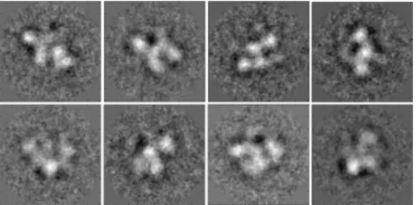
The robustness of the method against white noise has been evaluated. The proposed method has also been applied to real data composed of 225 averaged 1603 projections from the complex TAF7. Fig. 3 shows some examples of these classes. The signal to noise ratio is estimated at 10.02dB. The same protocol as for the phantom reconstruction has been used at
1Images and program are available at http://rhodes.unistra.
Fig. 3. Example of averaged projections of the complex TAF7.
the resolutions 403and 1603but a positive result has been ob-tained only at the resolution 403. A comparison is made with a reconstruction from the averaged projections with SIMPLE software [10]. Then the obtained ab-initio reconstructions are compared to a reference reconstruction obtained with a tilt acquisition. The MSE and the correlation are resp. 1.9784 and 85% for our method and resp 1.5 × 10⁶ and 31% for SIMPLE software. It shows that our method obtains a bet-ter ab-initio reconstruction than the SIMPLE software on the complex TAF7. Fig. 4 shows isosurfaces for the obtained and the tilt volumes after registration.
Fig. 4. Reconstructions of the complex TAF7 with SIMPLE software (left), with our method (center) and with the angular information from a tilt serie (right).
4. CONCLUSION
This paper presents a reconstruction method for the 3D case where the projection orientations are unknown. The method is based on the minimization of a cost depending on the image voxels and the orientations. The minimization uses a simulat-ing annealsimulat-ing process. The proposed method has been tested on 20 synthetic images at 4 resolutions and on real data com-posed of projection averaged classes from the TAF7 complex at resolution 403. The results are promising for the synthetic data and the reconstruction is successful on the real data and shows a good similarity with reference volume.
In the futur, we plan to introduce multiresolution in the method so as to speed up the process and to achieve recon-struction at higher resolutions.
Aknowledgement We thank Gabor Papa¨ı for providing the real data set, the reference volume and the SIMPLE recon-struction of the TAF7 complex.
5. REFERENCES
[1] R. A. Crowther, “Procedures for three-dimensional re-construction of spherical viruses by Fourier synthesis from electron micrographs,” Phils. Trans. R. Soc. Lond. B, vol. 261, no. 837, pp. 221–228, May 1971.
[2] J. Frank, Three-Dimensional Electron Microscopy of Macromolecular Assemblies, Oxford University Press, New York, 2006.
[3] P. A. Penczek, R. A. Grassucci, and J. Frank, “The ribo-some at improved resolution: new techniques for merg-ing and orientation refinement in 3d cryo-electron mi-croscopy of biological particles,” Ultramimi-croscopy, vol. 53, no. 3, pp. 251–270, 1994.
[4] M. Van Heel, “Angular reconstitution: A posteriori as-signment of projection directions for 3d reconstruction,” Ultramicroscopy, vol. 21, no. 2, pp. 111–123, 1987. [5] R. R. Coifman, Y. Shkolnisky, F. J. Sigworth, and
A. Singer, “Graph laplacian tomography from unknown random projections,” IEEE T Image Process, vol. 17, no. 10, pp. 1891–1899, 2008.
[6] C. Yang, E. G. Ng, and P. A. Penczek, “Unified 3-D structure and projection orientation refinement using quasi-Newton algorithm.,” J Struct Biol, vol. 149, no. 1, pp. 53–64, Jan. 2005.
[7] T. Ogura and C. Sato, “A fully automatic 3d reconstruc-tion method using simulated annealing enables accurate posterioric angular assignment of protein projections,” J Struct Biol, vol. 156, no. 3, pp. 371 – 386, 2006. [8] M. van Heel, B. Gowen, R. Matadeen, E. V. Orlova,
R. Finn, T. Pape, D. Cohen, et al., “Single-particle elec-tron cryo-microscopy: towards atomic resolution,” Q Rev biophys, vol. 33, no. 04, pp. 307–369, 2000. [9] B. Ben Cheikh, E. Baudrier, and G. Frey, “A
tomograph-ical reconstruction method from unknown direction pro-jections for 2d gray-level images,” in International Sym-posium on Biomedical Imaging. IEEE, Apr 2014. [10] D. Elmlund and H. Elmlund, “Simple: Software for ab
initio reconstruction of heterogeneous single-particles,” J Struct Biol, vol. 180, no. 3, pp. 420 – 427, 2012.