HAL Id: inserm-00835538
https://www.hal.inserm.fr/inserm-00835538
Submitted on 19 Jun 2013
HAL is a multi-disciplinary open access
archive for the deposit and dissemination of
sci-entific research documents, whether they are
pub-lished or not. The documents may come from
teaching and research institutions in France or
abroad, or from public or private research centers.
L’archive ouverte pluridisciplinaire HAL, est
destinée au dépôt et à la diffusion de documents
scientifiques de niveau recherche, publiés ou non,
émanant des établissements d’enseignement et de
recherche français ou étrangers, des laboratoires
publics ou privés.
echocardiography: the impact of diabetes and age, and
the prognostic value.
Thomas Cognet, Paul-Louis Vervueren, Laurent Dercle, Delphine Bastié,
Rainui Richaud, Matthieu Berry, Pauline Marchal, Matthieu Gautier, Audrey
Fouilloux, Michel Galinier, et al.
To cite this version:
Thomas Cognet, Paul-Louis Vervueren, Laurent Dercle, Delphine Bastié, Rainui Richaud, et al..
New concept of myocardial longitudinal strain reserve assessed by a dipyridamole infusion using
2D-strain echocardiography: the impact of diabetes and age, and the prognostic value.. Cardiovascular
Diabetology, BioMed Central, 2013, 12 (1), pp.84. �10.1186/1475-2840-12-84�. �inserm-00835538�
O R I G I N A L I N V E S T I G A T I O N
Open Access
New concept of myocardial longitudinal strain
reserve assessed by a dipyridamole infusion using
2D-strain echocardiography: the impact of
diabetes and age, and the prognostic value
Thomas Cognet
1,2,3,7*, Paul-Louis Vervueren
2,4, Laurent Dercle
1, Delphine Bastié
1,3, Rainui Richaud
1,3,
Matthieu Berry
2,3, Pauline Marchal
1,2,3, Matthieu Gautier
2,3, Audrey Fouilloux
1,2,3, Michel Galinier
2,5, Didier Carrié
2,3,6,
Pierre Massabuau
2,3, Isabelle Berry
1,5and Olivier Lairez
1,2,3Abstract
Aims: Although dipyridamole is a widely used pharmacological stress agent, the direct effects on myocardium are not entirely known. Diabetic cardiomyopathy can be investigated by 2D-strain echocardiography. The aim of this study was to assess myocardial functional reserve after dipyridamole infusion using speckle-tracking
echocardiography.
Methods: Seventy-five patients referred for dipyridamole stress myocardial perfusion gated SPECT (MPGS) were examined by echocardiography to assess a new concept of longitudinal strain reserve (LSR) and longitudinal strain rate reserve (LSRR) respectively defined by the differences of global longitudinal strain (GLS) and longitudinal strain rate between peak stress after dipyridamole and rest. Twelve patients with myocardial ischemia were excluded on the basis of MPGS as gold standard.
Results: Mean LSR was −2.28±2.19% and was more important in the 28 (44%) diabetic patients (−3.27±1.93%; p = 0.001). After multivariate analyses, only diabetes improved LSR (p = 0.011) after dipyridamole infusion and was not associated with glycaemic control (p = 0.21), insulin therapy (p = 0.46) or duration of the disease (p = 0.80). Conversely, age (p = 0.002) remained associated with a decrease in LSR. LSSR was also correlated to age (p = 0.005). Patients with a LSR < 0% have a better survival after 15 months (log-rank p = 0.0012).
Conclusion: LSR explored by 2D speckle-tracking echocardiography after dipyridamole infusion is a simple and new concept that provides new insights into the impact of diabetes and age on the myocardium with a potential prognostic value.
Keywords: Dipyridamole, Speckle-tracking echocardiography, Diabetes, Strain reserve Introduction
Dipyridamole is a widely used pharmacological agent to test coronary reserve in patients referred for stress myo-cardial perfusion imaging. The mechanism of stress with dipyridamole implies a coronary vasodilatation, which leads to the detection of myocardial ischemia through
coronary steal phenomena. Dipyridamole mainly in-creases the flow supply in the subendocardial layer by decreasing vascular resistance [1], but the resulting ef-fects on myocardium and especially on myocardial strain have not been explored yet. The myocardial functional reserve between peak stress and rest reflects the ability of the myocardium to improve its function during stress testing. Stress testing is recommended in order to detect myocardial ischemia [2], but the role of the myocardial functional reserve during stress remains undervalued even if it provides important information in some
* Correspondence:thomascognet31@gmail.com
1Department of Nuclear Medicine, University Hospital of Rangueil, Toulouse,
France
2Department of Cardiology, University Hospital of Rangueil, Toulouse, France
Full list of author information is available at the end of the article
© 2013 Cognet et al.; licensee BioMed Central Ltd. This is an Open Access article distributed under the terms of the Creative Commons Attribution License (http://creativecommons.org/licenses/by/2.0), which permits unrestricted use, distribution, and reproduction in any medium, provided the original work is properly cited.
situations such as hypertrophic cardiomyopathy or hypo-thyroidism [3,4]. At the same time, the ability of two-dimensional (2D) strain echocardiography to precisely assess myocardial function provides new possibilities for an accurate measurement of the myocardial function re-serve in stress conditions [5,6] and especially in diabetes in which longitudinal strain is altered at baseline [7]. Ac-tually, diabetic patients have early myocardial damage at baseline that requires to be detected by means of mod-ern tools such as biomarkers and imaging [8].
The aim of this study was to assess the effects of di-pyridamole on the myocardium in a population of pa-tients referred for myocardial perfusion imaging by gated single-photon emission computed tomography (MPGS) and to explore the myocardial functional re-serve with dipyridamole by means of a longitudinal strain reserve with speckle-tracking echocardiography.
Methods
Study population
Patients referred for MPGS with previous or non-coronary artery disease (CAD) and without history of myocardial infarction were prospectively included from March to September 2011. All patients underwent a complete phys-ical exam before inclusion. Exclusion criteria were docu-mented allergies to dipyridamole, asthma, systolic blood pressure < 90 mmHg, decompensated heart failure or acute angina, significant valvular disease, hypertrophic car-diomyopathy, atrial fibrillation and the possibility to in-duce stress by effort. All patients were informed of the study design and agreed to the protocol before inclusion. Patients with identified MPGS ischemia were also ex-cluded of the analysis.
Dipyridamole testing and MPGS protocols
All enrolled patients underwent MPGS with intravenous dipyridamole pharmacologic stress using the standard-ized protocols from the European Association of Nuclear Medicine / European Society of Cardiology guidelines [9]. Caffeinated beverages, foods and medications, and medications containing methylxanthine were avoided for at least 12 hours prior to stress testing. Dipyridamole was given as a continuous infusion intravenously at 0.6 mg/kg over the course of 4 minutes. Arterial pressure was recorded before infusion, every 2 minutes during stress and also at the peak of dipyridamole effects (8 mi-nutes). Stress MPGS was performed for all patients with a weight-adjusted dose of 300–400 MBq of 99mTc-tetrofosmin injected 3 minutes after the completion of the dipyridamole infusion. MPGS was acquired 15 to 30 minutes after a radiotracer injection using a Symbia T6 (Siemens Healthcare, Erlangen, Germany) double-headed gamma camera equipped with low-energy, high-resolution collimators. Data was acquired for 180° with 64 frames of
30 and 20 second durations at stress and at rest, respect-ively; a 64 × 64 matrix; 8-frame gating; and a 20% window centred on the 140-keV photo peak of Tc-99m. Rest MPGS was performed only if stress MPGS was considered as pathological, with a 2-fold-higher dose of 99mTc-tetrofosmin injected at least 3 hours after the stress test-ing. All patients underwent low-dose CT using a Symbia T6 system (Siemens Healthcare, Erlangen, Germany) for attenuation correction (130 keV, 30 to 45 mAs).
Echocardiographic protocol
Echocardiography for all patients was performed at rest and 3 minutes after the completion of the dipyridamole infusion, i.e. at peak of stress, with a Imagic KM 60 (Kontron Medical, Saint-Germain en Laye, France) using a 2.5 MHz transducer. A complete two-dimensional grey scale echocardiography including the three standard ap-ical views (four, three and two chambers) with a frame rate > 75 frame/s was performed for each patient. Left ventricle ejection fraction (LVEF) and volumes were assessed before and after the dipyridamole infusion as diastolic parameters. Myocardial strain was measured using speckle-tracking echocardiography.
Data analysis and interpretation
The 17-segments model, as defined by the American Soci-ety of Echocardiography, was used to examine both echo-cardiography and MPGS [10]. The apex segment was then excluded for the analysis.
For the echocardiography, digital data of 3 consecutive heart cycles were recorded and transferred to a personal computer with My Lab Desk workstation (Kontron Med-ical, Saint-Germain en Laye, France) for offline analysis. The endocardial border was defined manually in end sys-tole and automatically tracked frame by frame. Operator assessed optimal evaluation of both quality of tracking and region of interest. Global longitudinal strain (GLS) was obtained by averaging all segmental longitudinal strain curves computed from the conventional apical two-, three- and four-chamber views. Longitudinal strain re-serve (LSR) was defined by the difference between peak systolic global longitudinal strain at the peak of vasodilata-tion with dipyridamole and at rest. The longitudinal strain rate was determined for the left ventricle as the maximal strain rate value (calculated as the temporal derivative of strain) during the ejection phase. The longitudinal strain rate reserve (LSRR) was similarly obtained (Figure 1). Left ventricular ejection fraction (LVEF) was assessed by trans-thoracic echocardiography using the conventional apical two- and four-chamber views and the modified Simpson’s method.
For MPGS studies, off-line analysis was performed on Syngo MI Applications software (Siemens Healthcare, Erlangen, Germany). The images were assessed visually
and by applying automated methods. A single blinded ob-server interpreted echocardiographic and MPGS studies. Myocardial ischemia was defined by at least one reversible myocardial perfusion defect between stress and rest myo-cardial perfusion gated-SPECT and was expressed by the number of segments affected.
Follow up
Data about the occurrence of adverse events were obtained from medical records by direct patients’ inter-views or from the referring physician. The primary end point was defined by all-cause mortality. Patients unable to be interviewed up to 6 months at the date of follow-up were considered as lost to follow-follow-up.
Statistical analysis
Data were expressed as mean +/− SD. Nominal values were expressed as numbers and percentages. Normality was tested by the Kolmogorov-Smirnov test. The associ-ation between the mean values of continuous normally distributed variables were compared using unpaired and paired Student’s t test and the Mann–Whitney rank sum test was used when the samples were not normally dis-tributed or had unequal variances. Comparison between multiple groups was performed with a variance analysis (ANOVA). Nominal variables were investigated by the χ2 test. Linear regression analysis was used to investigate the relation between LSR-LSRR and variables. Conven-tional variables correlated with LSR with a p value < 0.05 at first univariate analyses were used to build the
final multivariate stepwise model. Receiver operating char-acteristic (ROC) curves were computed to determine opti-mal cut-off point for longitudinal strain reserve as well as to calculate area under the curve (AUC) to determine prognostic significance. Multivariate Cox regression model was built to identify echocardiographic parameters as-sociated with all-cause mortality. Survival curve was de-termined according to the Kaplan-Meier method, and cumulative event rates compared by means of the log-rank test. Differences were considered statistically signifi-cant for p-values of < 0.05. All analyses were performed on SPSS software version 20 (SPSS Inc., Chicago, Illinois).
Results
Population
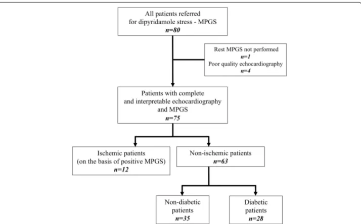
Eighty patients were prospectively included. Four pa-tients (5%) were excluded from the analysis due to a poor resolution of 2D echocardiography as a conse-quence of poor ultrasonic window (obesity or pulmonary disease) that did not allow speckle-tracking imaging and 1 (<1%) due to refusal of gated-SPECT after echocardi-ography. Among the 75 other patients included, 12 were excluded for MPGS ischemia with at least one or more reversible defect segment (Figure 2). Male represented 59% of the 63 patients finally enrolled in the study. The mean age was 70 ± 11 years with a median age of 71 ranging from 46 to 90 years old. Twenty-six patients (41%) had a previous history of coronary artery disease and 28 (44%) suffered from diabetes. Diabetes lasted less than five years in 10/28 patients. Thirty-nine patients
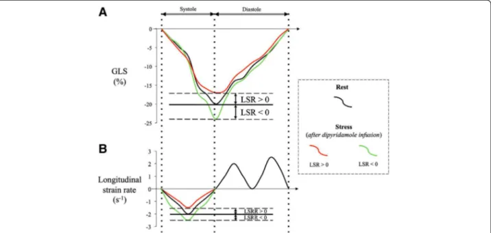
Figure 1 Schematic representation of global longitudinal strain (GLS) curves (Panel A) and longitudinal strain rate curves (Panel B) focused of the systolic phase of the cardiac cycle. Improvement of longitudinal strain reserve (negative LSR) and longitudinal strain rate reserve (negative LSRR) in green curves and decrease of LSR and LSRR (positive LSR and LSRR) in red curves both after dipyridamole infusion as compared to baseline values (black curves).
(62%) reported a NYHA stage 2 and the mean LVEF was 51±14%. Baseline characteristics are presented in Table 1.
Blood pressure and heart rate during stress
Hemodynamic during dipyridamole infusions and echo-cardiographic examinations remained unchanged for all patients. The decrease of systolic blood pressure with di-pyridamole after stress testing was not only insignificant for the whole population (136±22 at rest vs. 130±21 mmHg after dipyridamole infusion, p = 0.14) but the diabetic patients in comparison to the non-diabetics also showed a non-significant variation of systolic blood pres-sure (−3.8±12.4 vs. -2.9±31.6 mmHg; p = 0.88, respect-ively). Results are similar regarding the diastolic blood pressure (p = 0.09). The mean heart rate increased from 70±14 beats/min at rest to 80±18 beats/min after the di-pyridamole infusion (p < 0.001) showing the pharmaco-logical effect of dipyridamole. No examination had to be stopped for safety reasons.
Effects of dipyridamole on strain reserve
The effects of dipyridamole on LSR according to base-line characteristics, coronary risk factors, dyspnea and medications are presented in Table 2. In our general population, the mean GLS before dipyridamole infusion
was −14.5±4.2% and reached −16.8±4.5% at the max-imum effect of vasodilatation. Consequently, the mean LSR was −2.28±2.19%. LSR did not depend on systolic blood pressure (p = 0.99), diastolic blood pressure (p = 0.57) or heart rate (p = 0.85) changes during stress, as LSRR with p-values of 0.89, 0.57 and 0.17, respectively for systolic blood pressure, diastolic blood pressure and heart rate.
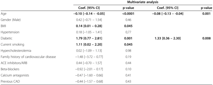
By univariate analysis, only age was associated with a decrease of LSR after dipyridamole infusion whereas patients with diabetes, higher Body Mass Index (BMI) and current smoking showed an improvement of LSR (Table 3). Increasing age was significantly correlated to a decrease of LSR (p < 0.0001) as presented in Figure 3. As shown in Table 4, no difference was observed be-tween diabetic and non-diabetic patients for GLS before stress (−13.9±3.7 vs. -15.0±4.5%; p = 0.30) and after the dipyridamole infusion (−17.2±4.2 vs. -16.5±4.8%; p = 0.55) but LSR was higher in the diabetic population (−3.27±1.93 vs. -1.49±2.08%; p = 0.001). Moreover, GLS of diabetic increased significantly by 24% in stress condi-tions (p = 0.003). Among the 28 patients with diabetes, 22 of them presented also overweight, defined as a BMI > 25 kg/m2 (p < 0.004). GLS before dipyridamole infu-sion was not different between patients with or without overweight (−14.2±3.6 vs. -15.0±4.9%; p = 0.43) but LSR
was significantly higher in patients with overweight (−2.83±1.85 vs. -1.50±2.42%; p = 0.016). After a mul-tivariate analysis, only age (p = 0.001) remained inde-pendently associated with a decrease of LSR after the dipyridamole infusion. Conversely, LSR remained signifi-cantly improved only in diabetic patients (p = 0.008). Among all echocardiographic parameters at baseline and after stress, including systolic, diastolic, hemodynamic and speckle-tracking parameters, only LSR was modified according to the diabetic status (Table 4). LSR was not correlated to the duration of diabetes (p = 0.80) or HbA1c level (p = 0.21) and was not influenced by
dedicated treatments especially insulin therapy (p = 0.46), the presence of retinopathy (p = 0.43) or periph-eral vascular disease (p = 0.34).
LSRR was only associated with aging (p = 0.005) and was not influenced by diabetes (p = 0.57). Moreover, lon-gitudinal strain rate increased by 16% between baseline and peak stress in the diabetic population (p = 0.11).
Prognosis and follow-up
During a mean follow-up of 15±5 months, 6 (10%) pa-tients reached the primary endpoint. Only one patient was lost to follow-up and was excluded for survival
Table 1 Baseline characteristics
Variable All patients (n=63) Diabetic (n=28) Non-diabetic (n=35) p-value Age (yrs) 70 ±11 70±10 72±10 0.04 Male (%) 37 (59) 18 (64) 19 (54) 0.42 BMI (kg/m2) 26.3 ± 3.9 28.1±4.0 24.8±3.2 0.001 SBP (mmHg) 137 ± 21 135±16 138±24 0.55 DBP (mmHg) 76±11 75±12 77±11 0.64 HR (beats/min) 70 ± 14 69±12 71±16 0.59 Dyspnea status (%) NYHA I 12 (19) 7 (25) 5 (14) 0.83 NYHA II 39 (62) 16 (57) 23 (66) 0.65 NYHA III 12 (19) 5 (18) 7 (20) 0.72 Coronary risk factors (%)
Hypertension 45 (71) 19 (48) 26 (71) 0.58 Current smoking 27 (43) 12 (43) 15 (43) 0.99 Hypercholesterolemia 32 (51) 14 (50) 18 (51) 0.91 Family history of cardiovascular 4 (6) 2 (7) 2 (5) 0.82 disease
Previous coronary artery disease (%) 26 (41) 11 (39) 15 (43) 0.78 Medications (%)
ACE inhibitors/ARB 38 (62) 16 (57) 22 (63) 0.44 Beta-blockers 30 (48) 12 (43) 18 (51) 0.50 Calcium antagonists 25 (40) 10 (36) 15 (43) 0.56 Diabetic characteristics
Duration of diabetes (yrs) - 12.0±8.8 -
-HbA1c (%) - 7.0±2.4 -
-Insulin therapy (%) - 20 (71) - -Oral therapy (%) 12 (43)
Metformin therapy (%) - 8 (29) -
-Retinopathy (%) - 11 (39) -
-Peripheral arterial disease (%) - 15 (54) -
-Supra aortic - 6 (21) -
-Lower limb - 11 (39) -
-Values are presented as n (%) or mean +/− SD.
BMI = body mass index; SBP = systolic blood pressure; DBP = diastolic blood pressure; HR = heart rate; ACE = angiotensin-converting enzyme; ARB = angiotensin receptor blocker; LVEF = left ventricle ejection fraction; LVEDV and LVESV = left ventricle end diastolic and end systolic volume.
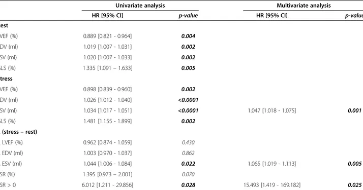
analysis. After multivariate analysis, only the left ven-tricle end systolic volume at stress (HR: 1.047 [95% C.I: 1.018 – 1.075]; p = 0.001; Table 5), the difference of left ventricle end systolic volumes between stress and rest (HR: 1.065 [95% C.I: 1.019 – 1.113]; p = 0.005) and a positive LSR (HR: 15.493 [95% C.I: 1.419 – 169.182]; p = 0.025) remained independently associated with all-cause mortality. ROC curve analysis in Figure 4 identified posi-tive LSR (cut-off value of 0%) as a predictor of all-cause mortality with a sensitivity of 89% and a specificity of 50%, for an area under the curve of AUC = 0.79 (p = 0.021).
The Kaplan-Meier analysis showed better survival in pa-tients with a negative LSR (log-rank p = 0.012). All cause-mortality in the diabetic population was associated with a lower LSR (−0.59±1.17 vs. -3.60±1.77%; p = 0.028) but prognosis was not significantly better in the diabetic popu-lation compared to the non-diabetic since they have a bet-ter LSR (p = 0.102 vs. p = 0.446).
Discussion
LSR assessed with dipyridamole, defined by the differ-ence between GLS after and before a dipyridamole infu-sion, is a new concept of myocardial functional reserve. Our study shows a LSR increase in patients with dia-betes that decreases with aging with a potential interest in prognosis evaluation.
2D-strain echocardiography with speckle-tracking im-aging enables a general analysis of the left ventricle at rest or during stress [11]. Regional analysis during MPGS and perfusion echocardiography with dipyridamole are com-plementary for the assessment of CAD [12] but for the first time, we are reporting the effects of dipyridamole on global longitudinal strain by means of 2D-speckle tracking echocardiography. This study highlights a new concept of LSR during stress with dipyridamole. Palmieri and al. pre-viously experienced a myocardial reserve by means of Doppler tissue imaging. Despite different pharmacological effects, low doses of dobutamine in patients with type 1 diabetes have similar effects, compared to dipyridamole in our study, by improving both global longitudinal strain and longitudinal strain rate of at least 29% [13].
Different deformation modalities such as longitudinal strain [14,15] or torsion [16-18] can be modified by left ventricular load but even if dipyridamole has several sys-temic effects that can lead to hemodynamic changes [19], we show that there are no blood pressure impacts or heart rate variations on LSR.
After a multivariate analysis, only aging is associated with a decrease of LSR during stress with dipyridamole. This result is consistent with the consequences of aging on myocardial deformation at rest: global longitudinal strain declines at rest with aging in a healthy population [20], especially in basal segments [21]. Consistent results were also found with longitudinal strain rate in baseline [22]. These results could be partly explained by a re-duced coronary flow reserve with aging [23] as described with myocardial ischemia [24]. Therefore, LSR may re-flect the physiological age of the myocardium.
Conversely, diabetes is associated with an increase in LSR whereas GLS before the dipyridamole infusion in the diabetic patients is lower but not statistically differ-ent than non-diabetic patidiffer-ents. These results at baseline are different from the studies of Ernande et al. in which GLS was impaired at rest in patients with diabetes [25], even sometimes before diastolic dysfunction [26]. This
Table 2 Effects of dipyridamole on longitudinal strain reserve Coef. 95% for LSR p-value Baseline characteristics Age (yrs) * −0.49 <0.0001 Male † −2.45 0.46 Female −2.04 BMI (kg/m2) * 0.25 0.045 Dyspnea status † NYHA I −2.33 0.98 NYHA II −2.30 NYHA III −2.17 Coronary risk factors †
Hypertension −2.33 0.77 No hypertension −2.15 Diabetic −3.27 0.001 Non-diabetic −1.49 Current smoking −2.92 0.045 No current smoking −1.80 Hypercholesterolemia −2.29 0.98 No hypercholesterolemia −2.27
Family history of cardiovascular disease −0.90 0.19 No family history of cardiovascular disease −2.38
Previous coronary artery disease (CAD) †
CAD −2.02 0.43 No CAD −2.46 Medications † ACE inhibitors/ARB −2.50 0.44 No ACE inhibitors/ARB −2.07 Beta-blockers −1.80 0.10 No beta-blockers −2.72 Calcium antagonists −2.00 0.41 No calcium antagonists −2.47
LSR = longitudinal strain reserve; BMI = body mass index; ACE = angiotensin-converting enzyme; ARB = angiotensin receptor blocker.
*: Spearman correlation. †: ANOVA.
difference for GLS at rest could be explained by the re-cent description of impaired coronary microvascular function in type 2 diabetic patients without CAD [27]. Our smaller population of diabetic patients and a popu-lation partly composed of patients with previous CAD might explain this difference. Several studies confirm the endothelial dysfunction secondary to diabetes [28] but
we show, for the first time, the mechanic consequences of this endothelial dysfunction in diabetic patients, which lead to an exacerbated response to arterial vasodilatation induced by dipyridamole. These results are consistent with the hemodynamic findings of Picchi and al. who previ-ously described an increased basal coronary blood flow at rest in diabetes as a cause of decreased coronary flow
Table 3 Univariate and multivariate linear regression model for longitudinal strain reserve
Multivariate analysis
Coef. [95% CI] p-value Coef. [95% CI] p-value Age −0.10 [−0.14 – -0.05] <0.0001 −0.08 [−0.13 – -0.04] 0.001 Gender (Male) 0.42 [−0.71 – 1.54] 0.46 BMI 0.14 [0.01 – 0.28] 0.045 Hypertension 0.18 [−1.05 – 1.41] 0,77 Diabetic 1.79 [0.77 – 2.81] 0.001 1.33 [0.36 – 2.30] 0.008 Current smoking 1.11 [0.02 – 2.20] 0.045 Hypercholesterolemia 0.02 [−1.09 – 1.13] 0.98 Family history of cardiovascular disease −1.48 [−3.72 – 0.77] 0.19 ACE inhibitors/ARB 0.44 [−0.70 – 1.57] 0.44 Beta-blockers −0.92 [−2.01 – 0.17] 0.10 Calcium antagonists −0.47 [−1.60 – 0.66] 0.41 Previous CAD −0.44 [−1.57 – 0.68] 0.43
BMI = body mass index; ACE = angiotensin-converting enzyme; ARB = angiotensin receptor blocker; MPGS = myocardial perfusion gated SPECT; CAD = coronary artery disease.
Figure 3 Impact of age on LSR and LSRR A. Correlation between increasing age and decreased longitudinal strain rate reserve in Panel B. LSR = longitudinal strain reserve, LSRR = longitudinal strain rate reserve.
reserve with adenosine. Interestingly, as we described with GLS, coronary blood flow is not altered after ad-enosine in the diabetic group compared to the non-diabetic group [29]. The lack of any decrease in systolic or diastolic blood pressure in the diabetic group might also explain these results. Actually, reduced myocardial perfusion with vasodilatator in diabetic patient is asso-ciated with a significant decrease in blood pressure, a consequence of autonomic neuropathy [30]. Coronary metabolic vasodilatation mainly depends on both nitric oxide metabolism, which is impaired in diabetic pa-tients [31], and on adenosine pathways. The predo-minant vasodilatation effects of dipyridamole through A2A-adenosine receptors, which are increased in the
hearts of diabetic rats, [32] could explain the increase
of LSR in the diabetic population. LSR is not influenced by duration of diabetes in contrast to baseline [33]. Gly-caemic control, treatments and vascular disease other than CAD do not influence either LSR. In parallel, left ventricular diastolic function is impaired precociously in patients with insulin resistance and glucose metabol-ism disorders even without overt diabetes [34] but the diastolic functional reserve defined by Jellis et al. is not altered by diabetes during effort conditions [35]. Diabetes seems to alter first the contractile function as described with dobutamine stress echocardiography [36] while myocardial perfusion seems to be main-tained [37]. This hypothesis may explain the improve-ment of the LSR in diabetic patients. As a result, LSR appears to be an interesting and sensitive tool to
Table 4 Echocardiographic parameters according to diabetic status
Echocardiographic parameters All patients (n=63) Diabetic (n=28) Non-diabetic (n=35) p-value Rest LVEF (%) 50.7±14.4 49.8±13.0 51.4±15.5 0.66 EDV (ml) 103.7±46.9 106.7±46.6 101.2±47.6 0.65 ESV (ml) 53.7±39.4 56.6±36.7 51.3±41.8 0.60 TVI LVOT (cm) 22.7±6.0 19.3±5.0 19.3±5.5 0.96 GLS (%) −14.5±4.2 −13.9±3.7 −15.0±4.5 0.30 Longitudinal Strain rate (s-1) −1.19±0.35 −1.15±0.26 −1.22±0.40 0.43 E (cm.s-1) 77±25 78±26 75±25 0.60 A (cm.s-1) 81±29 79±28 82±29 0.77 E/A (cm.s-1) 1.07±0.58 1.14±0.57 1.01±0.59 0.41 DT (s) 205±86 197±88 210±85 0.56 Stress LVEF (%) 54.6±13.9 54.3±14.0 54.9±13.9 0.89 EDV (ml) 98.8±41.6 98.8±36.3 98.9±45.9 0.99 ESV (ml) 48.8±35.6 48.6±31.6 48.9±38.9 0.97 TVI LVOT (cm) 22.7±6.0 23.0±5.0 22.5±6.8 0.76 GLS (%) −16.8±4.5 −17.2±4.2 −16.5±4.8 0.55 Longitudinal Strain rate (s-1) −1.34±0.44 −1.33±0.52 −1.35±0.37 0.86 E (cm.s-1) 84±23 83±23 85±23 0.75 A (cm.s-1) 89±33 88±32 89±35 0.85 E/A (cm.s-1) 1.13±0.77 1.15±0.61 1.10±0.89 0.83 DT (s) 179±73 177±76 180±71 0.87 ∆ (stress – rest) ∆ LVEF (%) 3.9±8.4 4.6±9.8 3.5±7.2 0.60 ∆ EDV (ml) −4.8±25.3 −7.9±30.0 −2.3±20.9 0.39 ∆ ESV (ml) −4.9±20.5 −8.0±24.1 −2.4±17.0 0.28 ∆ TVI LVOT (cm) 3.4±2.5 3.7±2.6 3.2±2.3 0.39 LSR (%) −2.28±2.19 −3.27±1.93 −1.49±2.08 0.001 LSRR (s-1) −0.15±0.34 −0.17±0.43 −0.12±0.26 0.57
LVEF = left ventricle ejection fraction; EDV = end diastolic volume; ESV = end systolic volume, TVI LVOT = time-velocity integral in the left ventricular outflow tract, GLS = global longitudinal strain; DT = mitral deceleration time, LSR = longitudinal strain reserve; LSRR = longitudinal strain rate reserve.
explore the impact of diabetes on myocardium non-invasively, regardless of the characteristics of the diabetes.
Strain rate is load dependent and reflects the regional difference in contractility [38]. In our study, LSRR is not influenced by hemodynamic changes induced by dipyrid-amole. Therefore, LSRR reflects changes in contractility due to myocardial injury only. Moreover, in diabetes, the stability of longitudinal strain rate with stress and the
insignificant increase in LSRR between diabetic and non-diabetic patients results in a homogeneous and stable re-gional contractility. This may be the consequence of an overall and homogeneous myocardial injury. Because subendocardial longitudinal fibres are the most vulnerable in pathological conditions, we deliberately focused our analysis on longitudinal deformation [39]. Longitudinal strain is the most reliable and studied parameter of de-formation modalities and the comparison of radial strain
Table 5 Echocardiographic parameters associated with all-cause mortality in a multivariate Cox regression model
Univariate analysis Multivariate analysis
HR [95% CI] p-value HR [95% CI] p-value Rest LVEF (%) 0.889 [0.821 - 0.964] 0.004 EDV (ml) 1.019 [1.007 - 1.031] 0.002 ESV (ml) 1.020 [1.007 - 1.033] 0.002 GLS (%) 1.335 [1.091 – 1.633] 0.005 Stress LVEF (%) 0.898 [0.839 - 0.960] 0.002 EDV (ml) 1.026 [1.012 - 1.040] <0.0001 ESV (ml) 1.034 [1.017 - 1.051] <0.0001 1.047 [1.018 - 1.075] 0.001 GLS (%) 1.481 [1.155 - 1.899] 0.002 ∆ (stress – rest) ∆ LVEF (%) 0.962 [0.874 - 1.059] 0.430 ∆ EDV (ml) 1.003 [0.970 - 1.037] 0.862 ∆ ESV (ml) 1.044 [1.006 - 1.084] 0.022 1.065 [1.019 - 1.113] 0.005 LSR (%) 1.395 [0.973 – 2.001] 0.070 LSR > 0 6.012 [1.211 - 29.856] 0.028 15.493 [1.419 - 169.182] 0.025
LVEF = left ventricle ejection fraction; EDV = end diastolic volume; ESV = end systolic volume, TVI LVOT = time-velocity integral in the left ventricular outflow tract, GLS = global longitudinal strain; LSR = longitudinal strain reserve; LSRR = longitudinal strain rate reserve.
Figure 4 Prognostic value of LSR A. Kaplan-Meier curve representing the impact of a positive LSR on all-cause mortality in Panel B. LSR = longitudinal strain reserve. AUC = area under the curve, Se = sensitivity and Spe = specitivity.
results in diabetic populations is not reliable among differ-ent studies [25,40].
A positive LSR that reflects lack of myocardial func-tional reserve after dipyridamole infusion appears prom-ising to predict all-cause mortality in a population of patients referred for MPGS even when ischemia is ex-cluded. The cut-off value of 0% of LSR is easy to meas-ure and allows therefore a rapid and reliable evaluation in routine clinical practice.
However, our study has several limitations. First, pres-ence of autonomic neuropathy [41] but also abdominal visceral adipose tissue [42], and the evaluation of aortic stiffness [43] or oxidative stress [44], all associated with myocardial function impairment, could have provided important information. In parallel, our population of diabetic patients is to small to define subset groups according to glycaemic control that could influence evo-lution of LSR and LSRR during follow up. Unfortunately, ischemia cannot be ruled out with certainty and there-fore might interfere with our results. MPGS is an effi-cient and validated exam to assess ischemia and CAD and even if patients with at least only one reversible de-fect segment were excluded, pluritroncular patients could lead to false negative tests. Moreover, coronary angiography should have been interesting to differentiate respective impacts of epicardial coronary artery disease and microvascular dysfunction on strain reserve among patients with diabetes.
Finally, further prospective studies are necessary to de-fine the interaction between diabetes and LSR and a po-tential prognostic value in diabetic patients. The use of the selective adenosine A2A receptor agonist vasodilator
stress agent could be interesting in this context [45]. However, the present results suggest that addition of LSR to dipyridamole stress myocardial contrast perfu-sion echocardiography could improve its prognostic value [46].
Conclusion
LSR assessed after a dipyridamole infusion is a new con-cept of stress examination using speckle-tracking im-aging. LSR increases in the diabetic population and warrants special attention for examinations in the eld-erly population. Myocardial function reserve assessed by LSR after dipyridamole with speckle-tracking echo-cardiography may be of interest for the evaluation of both prognosis and impact of co-morbidities.
Consent
Written informed consent was obtained from the pa-tient for publication of this report and any accompany-ing images.
Abbreviations
2D:Two-dimensional; MPGS: Myocardial perfusion imaging by gated single-photon emission computed tomography; CAD: Coronary artery disease; GLS: Global longitudinal strain; LSR: Longitudinal strain reserve;
LSRR: Longitudinal strain rate reserve; LVEF: Left ventricle ejection fraction; AUC: Area under the curve; BMI: Body mass index.
Competing interests
Doctor Lairez received research support (equipment and software) from Kontron for speckle-tracking echocardiography. However, the current study was not supported by those grants.
Authors’ contributions
TC researched and analyzed data and wrote the manuscript. PLV performed the statistical analysis. LD recorded follow-up information. DB, RR, MB, PM, MG, AF researched data. MG, DC, PM, IB contributed to discussion and reviewing. O.L. led the study, revised the manuscript and gave final approval of the version to be published. All authors read and approved the final manuscript.
Author details 1
Department of Nuclear Medicine, University Hospital of Rangueil, Toulouse, France.2Department of Cardiology, University Hospital of Rangueil, Toulouse,
France.3Cardiac Imaging Centre, University Hospital of Rangueil, Toulouse,
France.4Laboratory of Epidemiology, CJF-INSERM, 94-06, Toulouse, France. 5
Medical School of Rangueil, University Paul Sabatier, Toulouse, France.
6Medical School of Purpan, University Paul Sabatier, Toulouse, France. 7Department of Cardiology, Toulouse University Hospital, 1, avenue Jean
Poulhès, TSA 50032, 31059, Toulouse Cedex 9, France.
Received: 19 March 2013 Accepted: 28 May 2013 Published: 7 June 2013
References
1. Sakanashi M, Noguchi K, Kato T, Matsuzaki T, Kinjo N, Ikema S, Miyagi H, Moromizato H, Nakasone J, Kinjo Y: Investigation on the effect of dipyridamole and papaverine on regional blood flow and cardiac hemodynamics in anesthetized dogs. Arzneimittelforschung 1989, 39:1119–1123.
2. Douglas PS, Garcia MJ, Haines DE, Lai WW, Manning WJ, Patel AR, Picard MH, Polk DM, Ragosta M, Parker Ward R, Weiner RB: ACCF/ASE/AHA/ASNC/ HFSA/HRS/SCAI/SCCM/SCCT/SCMR 2011 Appropriate Use Criteria for Echocardiography. A Report of the American College of Cardiology Foundation Appropriate Use Criteria Task Force, American Society of Echocardiography, American Heart Association, American Society of Nuclear Cardiology, Heart Failure Society of America, Heart Rhythm Society, Society for Cardiovascular Angiography and Interventions, Society of Critical Care Medicine, Society of Cardiovascular Computed Tomography, Society for Cardiovascular Magnetic Resonance American College of Chest Physicians. J Am Soc Echocardiogr 2011, 24:229–267. 3. Ha JW, Ahn JA, Kim JM, Choi EY, Kang SM, Rim SJ, Jang Y, Shim WH, Cho
SY, Oh JK, Chung N: Abnormal longitudinal myocardial functional reserve assessed by exercise tissue Doppler echocardiography in patients with hypertrophic cardiomyopathy. J Am Soc Echocardiogr 2006, 19:1314–1319. 4. Akcakoyun M, Kaya H, Kargin R, Pala S, Emiroglu Y, Esen O, Karapinar H, Kaya
Z, Esen AM: Abnormal left ventricular longitudinal functional reserve assessed by exercise pulsed wave tissue Doppler imaging in patients with subclinical hypothyroidism. J Clin Endocrinol Metab 2009, 94:2979–2983.
5. Cullen MW, Pellikka PA: Recent advances in stress echocardiography. Curr Opin Cardiol 2011, 26:379–384.
6. Moonen M, Lancellotti P, Zacharakis D, Pierard L: The value of 2D strain imaging during stress testing. Echocardiography 2009, 26:307–314. 7. Andersson C, Gislason GH, Weeke P, Hoffmann S, Hansen PR, Torp-Pedersen
C, Sogaard P: Diabetes is associated with impaired myocardial performance in patients without significant coronary artery disease. Cardiovasc Diabetol 2010, 9:3.
8. Zhao CT, Wang M, Siu CW, Hou YL, Wang T, Tse HF, Yiu KH: Myocardial dysfunction in patients with type 2 diabetes mellitus: role of endothelial progenitor cells and oxidative stress. Cardiovasc Diabetol 2012, 11:147.
9. Hesse B, Tagil K, Cuocolo A, Anagnostopoulos C, Bardies M, Bax J, Bengel F, Busemann Sokole E, Davies G, Dondi M, et al: EANM/ESC procedural guidelines for myocardial perfusion imaging in nuclear cardiology. Eur J Nucl Med Mol Imaging 2005, 32:855–897.
10. Cerqueira MD, Weissman NJ, Dilsizian V, Jacobs AK, Kaul S, Laskey WK, Pennell DJ, Rumberger JA, Ryan T, Verani MS: Standardized myocardial segmentation and nomenclature for tomographic imaging of the heart: a statement for healthcare professionals from the Cardiac Imaging Committee of the Council on Clinical Cardiology of the American Heart Association. Circulation 2002, 105:539–542.
11. Ryo K, Tanaka H, Kaneko A, Fukuda Y, Onishi T, Kawai H, Hirata K: Efficacy of longitudinal speckle tracking strain in conjunction with isometric handgrip stress test for detection of ischemic myocardial segments. Echocardiography 2012, 29:411–418.
12. Wei K, Crouse L, Weiss J, Villanueva F, Schiller NB, Naqvi TZ, Siegel R, Monaghan M, Goldman J, Aggarwal P, et al: Comparison of usefulness of dipyridamole stress myocardial contrast echocardiography to technetium-99m sestamibi single-photon emission computed tomography for detection of coronary artery disease (PB127 Multicenter Phase 2 Trial results). Am J Cardiol 2003, 91:1293–1298.
13. Palmieri V, Capaldo B, Russo C, Iaccarino M, Di Minno G, Riccardi G, Celentano A: Left ventricular chamber and myocardial systolic function reserve in patients with type 1 diabetes mellitus: insight from traditional and Doppler tissue imaging echocardiography. J Am Soc Echocardiogr 2006, 19:848–856. 14. Di Bello V, Talini E, Dell'Omo G, Giannini C, Delle Donne MG, Canale ML,
Nardi C, Palagi C, Dini FL, Penno G, et al: Early left ventricular mechanics abnormalities in prehypertension: a two-dimensional strain
echocardiography study. Am J Hypertens 2010, 23:405–412.
15. A'Roch R, Gustafsson U, Johansson G, Poelaert J, Haney M: Left ventricular strain and peak systolic velocity: responses to controlled changes in load and contractility, explored in a porcine model. Cardiovasc Ultrasound 2012, 10:22.
16. A'Roch R, Gustafsson U, Poelaert J, Johansson G, Haney M: Left ventricular twist is load-dependent as shown in a large animal model with controlled cardiac load. Cardiovasc Ultrasound 2012, 10:26. 17. Weiner RB, Weyman AE, Khan AM, Reingold JS, Chen-Tournoux AA,
Scherrer-Crosbie M, Picard MH, Wang TJ, Baggish AL: Preload dependency of left ventricular torsion: the impact of normal saline infusion. Circ Cardiovasc Imaging 2010, 3:672–678.
18. Park HE, Chang SA, Kim HK, Shin DH, Kim JH, Seo MK, Kim YJ, Cho GY, Sohn DW, Oh BH, Park YB: Impact of loading condition on the 2D speckle tracking-derived left ventricular dyssynchrony index in nonischemic dilated cardiomyopathy. Circ Cardiovasc Imaging 2010, 3:272–281. 19. Javadi H, Shariati M, Mogharrabi M, Asli IN, Jallalat S, Hooman A, Seyedabadi
M, Assadi M: The association of dipyridamole side effects with hemodynamic parameters, ECG findings, and scintigraphy outcomes. J Nucl Med Technol 2010, 38:149–152.
20. Sun JP, Lee AP, Wu C, Lam YY, Hung MJ, Chen L, Hu Z, Fang F, Yang XS, Merlino JD, Yu CM: Quantification of left ventricular regional myocardial function using two-dimensional speckle tracking echocardiography in healthy volunteers - A multi-center study. Int J Cardiol 2012.
21. Reckefuss N, Butz T, Horstkotte D, Faber L: Evaluation of longitudinal and radial left ventricular function by two-dimensional speckle-tracking echocardiography in a large cohort of normal probands. Int J Cardiovasc Imaging 2011, 27:515–526.
22. Dalen H, Thorstensen A, Aase SA, Ingul CB, Torp H, Vatten LJ, Stoylen A: Segmental and global longitudinal strain and strain rate based on echocardiography of 1266 healthy individuals: the HUNT study in Norway. Eur J Echocardiogr 2010, 11:176–183.
23. Galderisi M, Rigo F, Gherardi S, Cortigiani L, Santoro C, Sicari R, Picano E: The impact of aging and atherosclerotic risk factors on transthoracic coronary flow reserve in subjects with normal coronary angiography. Cardiovasc Ultrasound 2012, 10:20.
24. Eguchi M, Kim YH, Kang KW, Shim CY, Jang Y, Dorval T, Kim KJ, Sweeney G: Ischemia-reperfusion injury leads to distinct temporal cardiac remodeling in normal versus diabetic mice. PLoS One 2012, 7:e30450. 25. Ernande L, Rietzschel ER, Bergerot C, De Buyzere ML, Schnell F, Groisne L,
Ovize M, Croisille P, Moulin P, Gillebert TC, Derumeaux G: Impaired myocardial radial function in asymptomatic patients with type 2 diabetes mellitus: a speckle-tracking imaging study. J Am Soc Echocardiogr 2010, 23:1266–1272.
26. Ernande L, Bergerot C, Rietzschel ER, De Buyzere ML, Thibault H, Pignonblanc PG, Croisille P, Ovize M, Groisne L, Moulin P, et al: Diastolic dysfunction in patients with type 2 diabetes mellitus: is it really the first marker of diabetic cardiomyopathy? J Am Soc Echocardiogr 2011, 24:1268–1275 e1261.
27. Marciano C, Galderisi M, Gargiulo P, Acampa W, D'Amore C, Esposito R, Capasso E, Savarese G, Casaretti L, Lo Iudice F, et al: Effects of type 2 diabetes mellitus on coronary microvascular function and myocardial perfusion in patients without obstructive coronary artery disease. Eur J Nucl Med Mol Imaging 2012, 39:1199–1206.
28. Hirase T, Node K: Endothelial dysfunction as a cellular mechanism for vascular failure. Am J Physiol Heart Circ Physiol 2012, 302:H499–H505. 29. Picchi A, Limbruno U, Focardi M, Cortese B, Micheli A, Boschi L, Severi S, De
Caterina R: Increased basal coronary blood flow as a cause of reduced coronary flow reserve in diabetic patients. Am J Physiol Heart Circ Physiol 2011, 301:H2279–H2284.
30. Taskiran M, Fritz-Hansen T, Rasmussen V, Larsson HB, Hilsted J: Decreased myocardial perfusion reserve in diabetic autonomic neuropathy. Diabetes 2002, 51:3306–3310.
31. Williams SB, Cusco JA, Roddy MA, Johnstone MT, Creager MA: Impaired nitric oxide-mediated vasodilation in patients with non-insulin-dependent diabetes mellitus. J Am Coll Cardiol 1996, 27:567–574. 32. Grden M, Podgorska M, Szutowicz A, Pawelczyk T: Altered expression of
adenosine receptors in heart of diabetic rat. J Physiol Pharmacol 2005, 56:587–597.
33. Nakai H, Takeuchi M, Nishikage T, Lang RM, Otsuji Y: Subclinical left ventricular dysfunction in asymptomatic diabetic patients assessed by two-dimensional speckle tracking echocardiography: correlation with diabetic duration. Eur J Echocardiogr 2009, 10:926–932.
34. Dinh W, Lankisch M, Nickl W, Scheyer D, Scheffold T, Kramer F, Krahn T, Klein RM, Barroso MC, Futh R: Insulin resistance and glycemic abnormalities are associated with deterioration of left ventricular diastolic function: a cross-sectional study. Cardiovasc Diabetol 2010, 9:63. 35. Jellis CL, Stanton T, Leano R, Martin J, Marwick TH: Usefulness of at rest
and exercise hemodynamics to detect subclinical myocardial disease in type 2 diabetes mellitus. Am J Cardiol 2011, 107:615–621.
36. Cadeddu C, Nocco S, Piano D, Deidda M, Cossu E, Baroni MG, Mercuro G: Early impairment of contractility reserve in patients with insulin resistance in comparison with healthy subjects. Cardiovasc Diabetol 2013, 12:66. 37. Mourmoura E, Vial G, Laillet B, Rigaudiere JP, Hininger-Favier I, Dubouchaud
H, Morio B, Demaison L: Preserved endothelium-dependent dilatation of the coronary microvasculature at the early phase of diabetes mellitus despite the increased oxidative stress and depressed cardiac mechanical function ex vivo. Cardiovasc Diabetol 2013, 12:49.
38. Teske AJ, De Boeck BW, Melman PG, Sieswerda GT, Doevendans PA, Cramer MJ: Echocardiographic quantification of myocardial function using tissue deformation imaging, a guide to image acquisition and analysis using tissue Doppler and speckle tracking. Cardiovasc Ultrasound 2007, 5:27. 39. Mizuno R, Fujimoto S, Saito Y, Nakamura S: Depressed recovery of
subendocardial perfusion in persistent heart failure after complete revascularisation in diabetic patients with hibernating myocardium. Heart 2009, 95:830–834.
40. Ng AC, Delgado V, Bertini M, van der Meer RW, Rijzewijk LJ, Shanks M, Nucifora G, Smit JW, Diamant M, Romijn JA, et al: Findings from left ventricular strain and strain rate imaging in asymptomatic patients with type 2 diabetes mellitus. Am J Cardiol 2009, 104:1398–1401.
41. Piya MK, Shivu GN, Tahrani A, Dubb K, Abozguia K, Phan TT, Narendran P, Pop-Busui R, Frenneaux M, Stevens MJ: Abnormal left ventricular torsion and cardiac autonomic dysfunction in subjects with type 1 diabetes mellitus. Metabolism 2011, 60:1115–1121.
42. Ichikawa R, Daimon M, Miyazaki T, Kawata T, Miyazaki S, Maruyama M, Chiang SJ, Suzuki H, Ito C, Sato F, et al: Influencing factors on cardiac structure and function beyond glycemic control in patients with type 2 diabetes mellitus. Cardiovasc Diabetol 2013, 12:38.
43. van Schinkel LD, Auger D, van Elderen SG, Ajmone Marsan N, Delgado V, Lamb HJ, Ng AC, Smit JW, Bax JJ, Westenberg JJ, de Roos A: Aortic stiffness is related to left ventricular diastolic function in patients with diabetes mellitus type 1: assessment with MRI and speckle tracking strain analysis. Int J Cardiovasc Imaging 2013, 29:633–641.
44. Reinhard H, Hansen PR, Wiinberg N, Kjaer A, Petersen CL, Winther K, Parving HH, Rossing P, Jacobsen PK: NT-proBNP, echocardiographic abnormalities
and subclinical coronary artery disease in high risk type 2 diabetic patients. Cardiovasc Diabetol 2012, 11:19.
45. Murray JJ, Weiler JM, Schwartz LB, Busse WW, Katial RK, Lockey RF, McFadden ER Jr, Pixton GC, Barrett RJ: Safety of binodenoson, a selective adenosine A2A receptor agonist vasodilator pharmacological stress agent, in healthy subjects with mild intermittent asthma. Circ Cardiovasc Imaging 2009, 2:492–498.
46. Gaibazzi N, Reverberi C, Lorenzoni V, Molinaro S, Porter TR: Prognostic value of high-dose dipyridamole stress myocardial contrast perfusion echocardiography. Circulation 2012, 126:1182–1184.
doi:10.1186/1475-2840-12-84
Cite this article as: Cognet et al.: New concept of myocardial longitudinal strain reserve assessed by a dipyridamole infusion using 2D-strain echocardiography: the impact of diabetes and age, and the prognostic value. Cardiovascular Diabetology 2013 12:84.
Submit your next manuscript to BioMed Central and take full advantage of:
• Convenient online submission
• Thorough peer review
• No space constraints or color figure charges
• Immediate publication on acceptance
• Inclusion in PubMed, CAS, Scopus and Google Scholar
• Research which is freely available for redistribution
Submit your manuscript at www.biomedcentral.com/submit