HAL Id: inserm-00796145
https://www.hal.inserm.fr/inserm-00796145
Submitted on 1 Mar 2013
HAL is a multi-disciplinary open access
archive for the deposit and dissemination of
sci-entific research documents, whether they are
pub-lished or not. The documents may come from
teaching and research institutions in France or
abroad, or from public or private research centers.
L’archive ouverte pluridisciplinaire HAL, est
destinée au dépôt et à la diffusion de documents
scientifiques de niveau recherche, publiés ou non,
émanant des établissements d’enseignement et de
recherche français ou étrangers, des laboratoires
publics ou privés.
Description and outcome of a cohort of 8 patients with
WHIM syndrome from the French Severe Chronic
Neutropenia Registry.
Sarah Beaussant Cohen, Odile Fenneteau, Emmanuel Plouvier, Pierre-Simon
Rohrlich, Gerard Daltroff, Isabelle Plantier, Alain Dupuy, Delphine Kerob,
Blandine Beaupain, Pierre Bordigoni, et al.
To cite this version:
Sarah Beaussant Cohen, Odile Fenneteau, Emmanuel Plouvier, Pierre-Simon Rohrlich, Gerard
Dal-troff, et al.. Description and outcome of a cohort of 8 patients with WHIM syndrome from the French
Severe Chronic Neutropenia Registry.. Orphanet Journal of Rare Diseases, BioMed Central, 2012, 7
(1), pp.71. �10.1186/1750-1172-7-71�. �inserm-00796145�
R E S E A R C H
Open Access
Description and outcome of a cohort of 8
patients with WHIM syndrome from the French
Severe Chronic Neutropenia Registry
Sarah Beaussant Cohen
1,2, Odile Fenneteau
3, Emmanuel Plouvier
2, Pierre-Simon Rohrlich
2, Gerard Daltroff
4,
Isabelle Plantier
5, Alain Dupuy
6, Delphine Kerob
7, Blandine Beaupain
1, Pierre Bordigoni
8, Fanny Fouyssac
8,
Anne-Lise Delezoide
9, Gilles Devouassoux
10, Jean François Nicolas
11, Philippe Bensaid
12, Yves Bertrand
13,
Karl Balabanian
14, Christine Bellanne Chantelot
15, Françoise Bachelerie
14*and Jean Donadieu
1*Abstract
Background: WHIM syndrome (WS), a rare congenital neutropenia due to mutations of the CXCR4 chemokine receptor, is associated with Human Papillomavirus (HPV)-induced Warts, Hypogammaglobulinemia, bacterial Infections and Myelokathexis. The long term follow up of eight patients highlights the clinical heterogeneity of this disease as well as the main therapeutic approaches and remaining challenges in the light of the recent development of new CXCR4 inhibitors.
Objective: This study aims to describe the natural history of WS based on a French cohort of 8 patients.
Methods: We have reviewed the clinical, biological and immunological features of patients with WS enrolled into the French Severe Chronic Neutropenia Registry.
Results: We identified four pedigrees with WS comprised of eight patients and one foetus. Estimated incidence for WS was of 0.23 per million births. Median age at the last visit was 29 years. Three pedigrees encompassing seven patients and the fetus displayed autosomal dominant heterozygous mutations of the CXCR4 gene, while one patient presented a wild-type CXCR4 gene. Two subjects exhibited congenital conotruncal heart malformations. In addition to neutropenia and myelokathexis, all patients presented deep monocytopenia and lymphopenia. Seven patients presented repeated bacterial Ears Nose Throat as well as severe bacterial infections that were curable with antibiotics. Four patients with late onset prophylaxis developed chronic obstructive pulmonary disease (COPD). Two patients reported atypical mycobacteria infections which in one case may have been responsible for one patient’s death due to liver failure at the age of 40.6 years. HPV-related disease manifested in five subjects and progressed as invasive vulvar carcinoma with a fatal course in one patient at the age of 39.5 years. In addition, two patients developed T cell lymphoma skin cancer and basal cell carcinoma at the age of 38 and 65 years.
Conclusions: Continuous prophylactic anti-infective measures, when started in early childhood, seem to effectively prevent further bacterial infections and the consequent development of COPD. Long-term follow up is needed to evaluate the effect of early anti-HPV targeted prophylaxis on the development of skin and genital warts.
Keywords: WHIM, CXCR4, Registry, Neutropenia, Myelokathexis, Infections
* Correspondence:francoise.bachelerie@u-psud.fr;jean.donadieu@trs.aphp.fr
14Inserm UMR_S996, Univ. Paris-Sud, Laboratory of Excellence in Research on
Medication and Innovative Therapeutics (LERMIT), 32 rue des Carnets, 92140 Clamart, France
1AP-HP, Registre français des neutropénies chroniques sévères, Centre de
référence des déficits Immunitaires Héréditaires, Service d’Hémato-oncologie Pédiatrique Hôpital Trousseau, 26 avenue du Dr Netter, 75012 Paris, France Full list of author information is available at the end of the article
© 2012 Beaussant Cohen et al.; licensee BioMed Central Ltd. This is an Open Access article distributed under the terms of the Creative Commons Attribution License (http://creativecommons.org/licenses/by/2.0), which permits unrestricted use, distribution, and reproduction in any medium, provided the original work is properly cited.
Beaussant Cohen et al. Orphanet Journal of Rare Diseases 2012, 7:71 http://www.ojrd.com/content/7/1/71
Background
In 1964, Zuelzer reported an exceptional congenital neu-tropenia associated with hyperplasia of mature neutrophils in the bone marrow (BM) – myelokathexis (retention of white blood cells in the BM) [1]. Its acronym (WHIM) derived from the manifestations of Human Papillomavirus (HPV)-induced Warts, Hypogammaglobulinemia, and bacterial Infections together with Myelokathexis [2]. A marked lymphopenia, which affects both T- and B-lymphocytes and NK cells, completes the picture. The clinical onset and complications in WHIM syndrome (WS) are more variable than originally suspected with the notable exceptions of neutropenia and lymphopenia, which are always observed in patients suffering from this disorder [3]. WS is also genetically heterogenous. Most patients present heterozygous autosomal dominant muta-tions of the gene encoding for CXCR4, the receptor of the CXCL12 chemokine (or Stromal cell Derived Factor-1) [4], which notably regulates hematopoiesis and peripheral trafficking of neutrophil and lymphocyte subsets. CXCR4 engagement by CXCL12 induces typical activation of Gαi
protein-dependent pathways. All CXCR4 mutations
described so far result in partial truncations of the recep-tor’s carboxyl terminal tail (C-tail), with the exception of the recently described missense non truncating E343K mutation [5], and impair the desensitization process which precludes further G-protein activation thus leading to enhanced and prolonged responsiveness of CXCR4 mutants to CXCL12 (i.e. gain of function) [6]. Leukocytes from the minority of patients who carry a wild-type (WT) CXCR4 gene presented a similar pattern of aberrant CXCL12/CXCR4 responses [4,7–9] consistent with a role for these dysfunctions in the WS hematological defects [10]. In support of this assumption, a new knock-in mouse strain that harbors a WS-associated heterozygous muta-tion of the CXCR4 gene exhibits striking parallels to the major immunological features of WS (i.e. panleukopenia) and is considered as a valuable model of the human syn-drome [11].
An exhaustive literature review since the first de-scription in 1964 identified 52 cases originating from the United States, Japan or Europe (Additional file 1) [1,2,4,5,9,12–43]. Recurrent infections may be quite se-vere, but other presentations are more indolent while the white blood cell count (WBC) appears to be affected in a large range, from mild lympho-neutropenia to near panleukopenia. Therefore, the therapeutic management of these patients is diverse. Some patients have no prophylactic therapy, while others may receive prophylac-tic antibioprophylac-tics or antiviral therapies such as Immunoglo-bulins (Ig), Granulocyte macrophage colony-stimulating factor (GMCSF), Granulocyte colony-stimulating factor (GCSF) and eventually undergo hematopoietic stem cell transplantation [30]. Recently, plerixafor (or AMD3100),
a small synthetic antagonist of CXCR4 approved for BM hematopoietic progenitor cells transplantation [44], has been tested in WS patients and found to promote the mobilization of neutrophils and lymphocytes to the per-ipheral blood [31,45]. These studies provide the first pharmacological evidence of the causal role of the gain of CXCR4 function in WS-associated panleukopenia and explain the rationale for the use of CXCL12/CXCR4 antagonists in the management of WS. Both the extreme rarity of WS and its clinical heterogeneity prompted us to analyze all cases identified in France through the na-tional Severe Chronic Neutropenia Registry. We collected the patients’ exhaustive clinical, hematological and bio-logical features, as well as their family histories, and performed the genetic and functional analyses of the CXCR4 receptor.
Patients and methods
Organization of the French registry and data monitoring
All patients included in this study were registered in the French Severe Chronic Neutropenia Registry. The regis-try was created in 1993; since then, enrollment has been prospective. We included all types of congenital neutro-penia [46]. The registry received national certification in the year 2008 by the French health authorities. Thirty-five French pediatric hematology-oncology clinical units participate in this registry. Data monitoring was based on the review of medical records collected by a clinical research associate who visited each center yearly, and multiple sources ascertained the completeness of each case. The patient or his legal guardians provided written informed consent before being included in the registry. Several reports of the registry are available elsewhere [47-49].
We used the common definition of WS. Briefly, WS was diagnosed in patients with both neutropenia (defined by at least one absolute neutrophil count (ANC) below 1.5 G/L) and myelokathexis on the BM smear after cen-tral review. In addition to this phenotype, we considered CXCR4 mutations as diagnostic criteria. When available, we screened patients for biological CXCR4 dysfunctions. All these criteria were found in one patient (UPN 5592) who presented no CXCR4 mutation and whose cyto-logical findings have been previously reported [50]. The patient UPN 5446 has been partially reported elsewhere [51] as well as the functional studies from three indivi-duals of the cohort (UPN 5592, UPN 5446 and UPN 5231) [7,8].
Phenotyping
We only analyzed routinely recorded parameters such as auxologic status and major medical events requiring medical management. Ig levels were analyzed according to age and expressed as standard deviations (SD) [52].
Beaussant Cohen et al. Orphanet Journal of Rare Diseases 2012, 7:71 Page 2 of 14
We obtained systematic recording of infectious episodes from the patients’ written medical history. We defined severe infections as those that would be life threatening without appropriate antibiotic or antifungal therapy and that entail medical supervision or hospitalization. These events were exhaustively registered in the medical records. Minor infections were those for which patients did not seek medical surveillance such as stomatological or ear, nose and throat (ENT) infections. These events were often omitted in the medical records. Warts were considered as a WS manifestation when their number was equal or superior to ten or when they presented as genital condylomata acuminita. We performed HPV genotyping for one case of skin warts and one case of ano-genital warts following a Quantitative PCR on fro-zen biopsies with HPV-specific primers.
CXCR4 gene testing
The patients or their parents gave their written informed consent for genetic and functional testing. Genomic DNA was extracted from blood with standard proce-dures. The coding sequence and exon-intron boundaries of the CXCR4 gene (encompassing two exons) were amplified using the primers and PCR conditions described elsewhere. PCR products were sequenced in both directions with the ABI PRISM Big Dye Terminator v1.1 Ready Reaction Cycle Sequencing kit (Applied Biosystems) on an ABI PRISM 3100 Genetic Analyzer (Applied Biosystems). Sequences were analyzed with the Seqscape software v2.2 (Applied Biosystems). We numbered mutations as recommended by the Human Genome Variation Society (http://www.hgvs.org/), using the reference sequence NM_003467.2.
Functional evaluation of the CXCL12/CXCR4 axis
Studies aimed at investigating the functioning of CXCR4 and other chemokine receptors (CCR5, CCR7, CXCR7) were performed as previously described [7,8].
Statistical analysis
Stata software version 10 was used for all statistical ana-lyses. Lower and upper interquartile, and median values depict the distribution of quantitative variables. Consid-ering that we ascertained the completion of the enrol-ment only from 1990 to 2006, we only took into consideration the years 1990–2006 to calculate the epi-demiological parameters. Our research extracted the number of births per year in France (metropolitan areas excluding Reunion Island, French Polynesia or French Antilles) from the records of the Institut National de la Statistique et des Etudes Economiques (www.insee.fr). Incidence at birth was supposed to satisfy a Poisson dis-tribution. For survival analysis the endpoints were death, and the first episode of cancer. The period taken into
account was the time interval from birth to the first date when the event was observed or to the last examination when no event occurred. The Kaplan-Meier method was used to estimate survival rates. The cut-off date was February 1st, 2012. Data from the foetus was not included in the statistical analysis for survival.
Literature review
In order to identify all publications related to WS, we first screened PubMed, with the key words: myelo-kathexis and WHIM. We then checked the bibliography of each article in order to identify additional references and to avoid duplicates.
Results
Demographic data
We analyzed data from eight subjects (including 6 males) presenting WS and one 32 weeks gestational age medic-ally terminated foetus, originating from four pedigrees (Table 1). The family trees of the four pedigrees are shown in the Additional file 2. The median age at the last follow up was 29 years [p25 8 years – p75 40 years]. De-finitive WS diagnosis, as defined by the detection of a CXCR4 mutation or of the characteristic CXCR4 dys-functions in leukocytes, was established at the median age of 20 years (SD 25.9) (min 1 year, max 75.6 years), but for the three cases born in the last ten years this diag-nosis was made before the age of 2 years, facilitated by an informative family history in two cases and by more readily available CXCR4 sequencing. WS-linked symp-toms first occurred at the median age of 1.3 years (SD 2.32) [min birth- max 5.1 years]. The initial clinical mani-festations of WS were bacterial infections for five patients, skin warts for one patient, and isolated neutro-penia for two patients. Incidence at birth was 0.23 per million births and the 95% confidence interval limits were 0.0019 – 0.29 per million births.
Bacterial, fungal and mycobacterial infections
Five patients had a history of repeated ENT infections in childhood, whereas two patients (UPN 55476 and 5780) reported less than one ENT episode per year, and an-other patient treated with prophylactic Ig since the age of 6 months (UPN 5447) reported no ENT episodes. Severe bacterial infections manifested in seven cases as mastoiditis, osteoarthritis, nephritis, angiocholitis, bac-terial adenotidis and armpit, submandibulary or pul-monary abscesses. The first episode of severe bacterial infection was recorded at the median age of 10.05 years [min 1.24 years – max 33.7 years]. However, the fre-quency of these severe bacterial infections was variable; three patients experienced less than one episode every five years. Nevertheless, four patients without a history of overt pulmonary infections developed COPD and
Beaussant Cohen et al. Orphanet Journal of Rare Diseases 2012, 7:71 Page 3 of 14
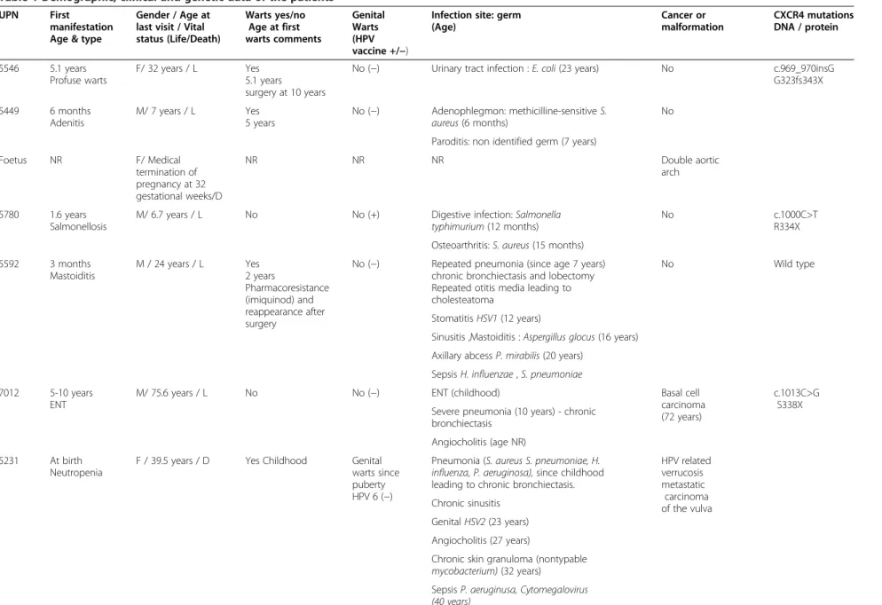
Table 1 Demographic, clinical and genetic data of the patients
UPN First
manifestation Age & type
Gender / Age at last visit / Vital status (Life/Death) Warts yes/no Age at first warts comments Genital Warts (HPV vaccine +/−)
Infection site: germ (Age) Cancer or malformation CXCR4 mutations DNA / protein 5546 5.1 years Profuse warts F/ 32 years / L Yes 5.1 years surgery at 10 years
No (−) Urinary tract infection : E. coli (23 years) No c.969_970insG
G323fs343X 5449 6 months Adenitis M/ 7 years / L Yes 5 years No (−) Adenophlegmon: methicilline-sensitive S. aureus (6 months) No Paroditis: non identified germ (7 years)
Foetus NR F/ Medical termination of pregnancy at 32 gestational weeks/D NR NR NR Double aortic arch 5780 1.6 years Salmonellosis
M/ 6.7 years / L No No (+) Digestive infection: Salmonella
typhimurium (12 months)
No c.1000C>T
R334X Osteoarthritis: S. aureus (15 months)
5592 3 months Mastoiditis M / 24 years / L Yes 2 years Pharmacoresistance (imiquinod) and reappearance after surgery
No (−) Repeated pneumonia (since age 7 years) chronic bronchiectasis and lobectomy Repeated otitis media leading to cholesteatoma
No Wild type
Stomatitis HSV1 (12 years)
Sinusitis ,Mastoiditis : Aspergillus glocus (16 years) Axillary abcess P. mirabilis (20 years)
Sepsis H. influenzae , S. pneumoniae 7012 5-10 years
ENT
M/ 75.6 years / L No No (−) ENT (childhood) Basal cell
carcinoma (72 years)
c.1013C>G S338X Severe pneumonia (10 years) - chronic
bronchiectasis Angiocholitis (age NR) 5231 At birth
Neutropenia
F / 39.5 years / D Yes Childhood Genital warts since puberty HPV 6 (−)
Pneumonia (S. aureus S. pneumoniae, H.
influenza, P. aeruginosa), since childhood
leading to chronic bronchiectasis.
HPV related verrucosis metastatic carcinoma of the vulva Chronic sinusitis Genital HSV2 (23 years) Angiocholitis (27 years)
Chronic skin granuloma (nontypable
mycobacterium) (32 years)
Sepsis P. aeruginusa, Cytomegalovirus
(40 years) Beaussant Cohen et al. Orphanet Journal of Rare Diseases 2012, 7 :71 Page 4 of 14 http://ww w.ojrd.com/ content/7/1 /71
Table 1 Demographic, clinical and genetic data of the patients (Continued)
Sputum Candida albicans , P aeruginosa,
E. Coli (40 years)
Intestinal tract C. jejuni, S. aureus ,
C. albicans (40 years) 5446 5-10 years ENT M /40.6 years / D Yes Childhood HPV 2, 5,23
Yes (−) Stomatitis HSV 1 (24 years) EBV negative
cutaneous T-cell lymphoma (37 years) Repeated pneumonia, since infancy
complicated by lung abcesses and chronic bronchiectasis :
S. aureus, H. influenzae , C. albicans,
M. morganii (33 years), S. pneumoniae
(34 years), B. catarrhalis (35 years),
Proteus mirabilis (36 years)
Purulent pericarditis
Pneumococci (35 years)
Skin lesions Molluscum
contagiosum and Onycomycosis
(NR germ)
Atypical Mycobacteria hepatitis (40.6 years)
5447 2 months
Neutropenia
M / 8.6 years / L No No (+) No severe infection Tetralogy of Fallot
ATB: Antibiotic prophylaxis; CXCR4: Chemokine (C-X-C-motif) receptor type 4; D: Death; DNA: deoxyribonucleic acid; EBV: Epstein Barr virus; ENT: Ears nose throat infection; HPV: Human Papillomavirus; Ig: Immunoglobulin prophylaxis; L: Life, NR: Non reported; PLTs: Platelets; UPN : Unique patient number; + Present; - Absent.
Beaussant Cohen et al. Orphanet Journal of Rare Diseases 2012, 7 :71 Page 5 of 14 http://ww w.ojrd.com/ content/7/1 /71
bronchiectasis. These were recorded at the age of 8 years for one patient and after the age of 30 years for three other patients. These long-term complications were asso-ciated with late anti-bacterial prophylaxis (including Ig and antibiotic rotations). Conversely, the only patient so far free from any infection at the age of 8.6 years was diagnosed with WS at the age of 6 weeks during a pre-operative screening, and has since been maintained on continuous Ig prophylaxis (UPN 5447).
Identified pathogens were Staphylococcus aureus (three cases), Escherichia coli, Streptococcus pneumoniae and Haemophilus influenzae (two cases each), Salmonella typhimurium, Aspergillus glocus, Pseudomonas aeruginosa, Morganella morganii, Moraxella catarrhalis, Proteus mirabilis, and Candida albicans (one case each). Atypical Mycobacterium was observed in two patients. In one case, mycobacteria infection was limited to the skin and although the organism was not identified, the referent physician based the diagnosis on the presence of an epithelioid granuloma on a skin biopsy and on the sensibil-ity to rifampicine, isoniazide and pyrazinamid. In the sec-ond case, the laboratory identified Mycobacterium gordonae in the patient’s sputum. Meanwhile, the patient developed severe liver failure responsible of death. Although the liver biopsy did not show any granuloma, displaying only a non-specific inflammation, we hypothesize that the mycobac-teria infection likely contributed to this liver failure.
HPV-induced warts
Phenotypically, five patients showed varying degrees of HPV-induced lesions. Skin warts generally first appeared on hands or feet, but could also affect the face. They devel-oped for three patients at the ages of 5.1, 5 and 2 years, the date of onset remaining unknown for the other two patients. The three patients exempt from warts were aged 75.6, 8.6 and 6.7 years. There was no association between the age of the skin warts onset and the severity of the wart proliferation. Extensive pharmaco-resistant cutaneous verrucosis that reappeared after surgical ablation severely impaired the quality of life for one patient (UPN 5592).
Besides warts, one male and one female patient reported anogenital condylomata acuminata (vulva, va-gina, cervix or rectum mucosa). Vulvar and perianal lesions were first recorded at the age of 18 years for the female patient (UPN 5231). In addition, she suffered from recurrent sporadic genital infections by herpes viruses since the age of 23 years, despite oral acyclovir prophylaxis, and aggressive treatment with intravenous valaciclovir and foscarnet. Later, she exhibited intract-able multifocal dysplastic HPV-induced lesions despite several years of repeated cervical conizations and exten-sive surgery (i.e., vulvectomy, pelvectomy, colostomy, hysterectomy) and ultimately developed invasive cancer with an early fatal course at the age of 39.5 years. The
male patient (UPN 5446), whose numerous skin warts showed HPV serotypes 2, 5 and 23, also presented severe condylomata acuminata of the ano-rectal area by the age of 31 years that were surgically removed. We per-formed HPV serotyping on the anogenital lesions of the female patient (UPN 5231) for low risk types 6, 11, 44, 53 and 54 and high risk types 45, 31, 18 and 16 and 33, and only HPV type 6 was detected. This finding was unexpected considering the lesions’ progression to car-cinoma. To try to prevent the development of these mucosal HPV lesions, two of the youngest patients (UPN 5780 and 5447) were vaccinated at 3 and 5 years of age with quadrivalent HPV vaccine (designed against HPV serotypes 6, 11, 16, and 18). Of note, neither patient has presented warts until now.
Hematological and Immunological parameters
At the time of WS diagnosis, median WBC was 1.050 G/L (p25: 0.7 - p75: 3.7 G/L), ANC was 0.23 G/L (p25: 0.2 - p75: 0.4 G/L), absolute lymphocyte count (ALC) was 0.63 G/L (p25: 0.42 - p75: 0.7 G/L), and abso-lute monocyte count (AMC) was 0.09 (p25: 0.083 - p75: 0.24 G/L). The median hemoglobin (Hb) level was 11.5 g/dL (p25: 10.2 g/dL - p75 12 g/dL) and platelet count was 201 G/L (p25 172 - p75 223 G/L). Transient anemia (< 7 g/dL) was observed in two patients, and transient mild thrombocytopenia (between 50 and 120 G/L) was observed in three patients, without obvi-ous etiology in both cases. During routine follow-up, a median of 16 baseline complete blood cell count (CBC) values per patient was available. For all cases, WBC and ANC fluctuated with time, without any detectable regu-lar variation or significant change observed over time, with the exception of the transiently normalized blood count values recorded during infectious episodes. The Additional file 3 illustrates the dynamic variation of the WBC of four patients at the occasion of sepsis or of GCSF therapy. Taking into account all the available CBC recorded during the routine follow up of the patients (Table 2), with a median number of 17 per patient, the median WBC was 1.1 G/L (p25: 0.85 - p75: 1.7 G/L), the ANC was 0.19 G/L (p25: 0.15 - p75: 0.37 G/L), the ALC was 0.63 G/L (p25: 0.0.42 - p75: 0.7 G/L), and the AMC was 0.1 (p25: 0.09 - p75: 0.13 G/L). The median Hb level was 11 g/dL (p25: 10.6 g/dL - p75 12.1 g/dL) and the median platelet count was 212 G/L (p25 181 - p75 336 G/L). Seven BM smears were available for central review, all showing rich BM: the granulocytic lineage represented a total of 36% of BM cells, with 18.5% ma-ture granulocytes, of which 2.75% were monocytes and 23% were lymphocytes. Examination confirmed myelo-kathexis in all cases (Figure 1). Four patients were trea-ted with 5 μg/kg/day GCSF resulting in a significant increase in the peripheral ANC from a median of 0.340 ×
Beaussant Cohen et al. Orphanet Journal of Rare Diseases 2012, 7:71 Page 6 of 14
109/L before treatment, to a maximum median of 1.499 × 10⁹/L under GCSF. AMC and ALC were not significantly modified (0.11 versus 0.10 G/L and 0.57 versus 0.35 × 10⁹/L, respectively before and under GCSF treatment). The total duration of the GCSF treatment was extremely short in one patient (13 days). The three patients who received long term GCSF therapy, interrupted treatment in the absence of significant efficiency to prevent recurrent infections. Indeed, for patient UPN 5446, despite three
years of apparent normalization of the ANC consecutive to daily Lenograstim subcutaneous injections, the number of infectious pulmonary episodes was not diminished. The registry recorded similar findings for patients UPN 5231 and 5592.
The total number of lymphocytes was extremely low among patients with median CD3+ T cells determined at 0.465 × 10⁹/L (SD 0.41) (min 0.1, max 1.33), median CD4+ T cells at 0.25 × 10⁹/L (SD 0.33) (min 0.02, max 0.89),
Table 2 Hematological, immunological features and summary of medical management
UPN ANC ALC AMC PLTs Ig G Ig A Ig M CD3 CD4 CD8 CD19 GCSF
×109/L ×109/L ×109/L ×109/L g/L g/L g/L ×109/L ×109/L ×109/L ×109/L ATB Ig Other 5546 0.16 0.38 0.08 223 0.33 0.2 0.1 No GCSF No ATB No Ig 5449 0.16 2.08 0.16 452 11.6 (6 mo) 0.12 0.199 1.33 0.89 0.35 0.16 No GCSF No ATB No Ig Foetus NR NR NR NR NR NR NR NR NR NR NR NR
5780 0.16 1.27 0.1 367 2.83* 0 0.53 1 0.77 0.13 0.07 GCSF short course / efficient
No ATB Early Ig+
5592 1.32 0.3 .021 60 - 0.8 0 0.1 0.02 0.02 0.09 GCSF since age 5 years, on
demand since age 8 years / repeated infections despite GCSF :
ATB+ Ig +
Warts treated by surgery and imiquinod with no efficiency
7012 0.22 0.58 0.09 202 9.69 1.89 <0.25 0.67 0.30 0.42 0.01 No GCSF
No ATB No Ig
5231 0.4 0.17 0.07 212 4.2 0.5 0.4 0.3 0.04 0.04 0 GCSF since age 28 years
/repeated infections despite GCSF
No ATB but Aciclovir prophylaxis Ig +
5446 0.34 0.42 0.12 193 7.2 0 0 0.33 0.15 0.19 0.04 GCSF since age 34 years / no
effect on bronchitis Withdrawn ATB+ Ig + 5447 0.14 1.2 0.1 306 1.8** 1.0 0 0.6 0.54 0.04 0 No GCSF No ATB Ig +
ALC: Absolute lymphocyte count; AMC: Absolute Monocyte count; ANC: Absolute neutrophil count; GCSF: Granulocyte colony stimulating factor;
Ig: Immunoglobulin prophylaxis; NR: Non reported; PLTs: Platelets; UPN : Unique patient number; + Present; - Absent **Antibody against vaccine: normal titers.
Beaussant Cohen et al. Orphanet Journal of Rare Diseases 2012, 7:71 Page 7 of 14
median CD8+ T cells at 0.11 × 10⁹/L (SD 0.14) (min 0.02, max 0.42), and median CD19+ B cells at 0.02 × 10⁹/L (SD 0.05) (min 0, max 0.16). Despite the low total lymphocyte count, the proportion of lymphocyte subsets was apparently preserved (Table 2).
We evaluated vaccine antigen responses in two patients. Patient UPN 5780 showed neither anti-tetanus nor anti-H. influenzae b antibodies (Abs) despite vaccin-ation 6 months prior, whereas the patient UPN 7012, who had been vaccinated more than ten years ago, pre-sented low anti-tetanus Abs, normal anti-pneumococcal Abs, but no anti-poliovirus Abs. Ig levels were evaluated in seven patients. IgG levels were mildly low (between −2 and −3 SD) in two subjects and normal in four sub-jects. IgA and IgM levels were low in five subjects and normal in two subjects. Five patients received monthly Ig replacement therapy including two children since the diagnosis of WS, and three adults who were also treated with GCSF because of severe recurrent bacterial infec-tions. Prophylactic Ig therapy seemed efficient in pre-venting bacterial infections, particularly in the younger patients of the cohort, who did not present any further severe bacterial infection since the introduction of Ig prophylaxis. Interestingly, three patients who received neither Ig nor GCSF were free of any bacterial infection.
Genetics
We screened CXCR4 mutations for all the patients in-cluding the foetus: seven patients among three families
presented mutations of the CXCR4 gene and one patient had a WT CXCR4 gene (Table 1). We found a new mu-tation 969_970insG in two related patients, a C1013→G substitution in three patients from one pedigree, and a C1000→T substitution in one patient (respectively, in the CXCR4 proteins G323fsX343, S338X and R334X). These nonsense or frameshift mutations led to C-tail trunca-tions of the CXCR4 protein.
Long term outcome and associated pathology
Patient UPN 5446 developed cutaneous T-cell lymphoma of the right ankle at the age of 37 years uncorrelated with an Epstein Barr virus (EBV) infection. His father, UPN 7012, presented basal cell carcinoma at the age of 72 years. Two premature deaths were reported in two siblings respectively at 39.5 and 40.6 years. The causes of death were metastatic vulvar carcinoma and possible Mycobacteria-related liver failure. Severe cardiac cono-truncal malformations were present in two subjects from unrelated pedigrees with different CXCR4 mutations (Table 1). The first case was diagnosed with Tetralogy of Fallot (TOF) at birth and was successfully operated at the age of two months. The second was diagnosed in utero with a double aortic arch that led to the medical termin-ation of the pregnancy at 32 gesttermin-ational weeks. Fertility seemed unperturbed in WS patients. One woman gave birth to two children with non-complicated pregnancies, had one miscarriage and one terminated pregnancy. The second woman bore no children. Among affected men, two of the three adult males had children.
A
B
C
D
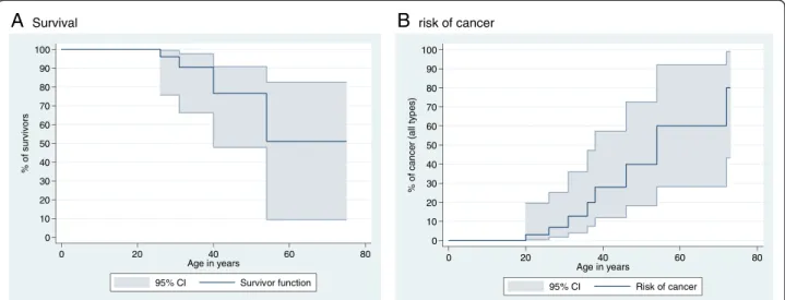
Figure 1 Bone marrow smears from four patients with myelokathexis, revealing over 50% of neutrophils with abnormal nuclei encompassing 3 to 5 lobes connected by long thin chromatin filaments, and less than 10% vacuolated mature neutrophils visible on images A and C. (May-Grunwald-Giemesa stain; original magnification x 1000). A, patient UPN 5780; B, patient UPN 5446; C, patient UPN 5546; D, patient UPN 5449.
Beaussant Cohen et al. Orphanet Journal of Rare Diseases 2012, 7:71 Page 8 of 14
Discussion
In this study we describe the French national cohort of patients with WS, thereby adding 8 patients and 4 pedi-grees to the 52 previously reported distinct cases origin-ating from 32 pedigrees (Additional file 1). The genetic studies performed in 27 pedigrees find that 25 pedigrees (including 3 from this report) bear CXCR4 mutations. R334X, the most frequent mutation, is reported in 15 pedigrees (Table 3). The S338X and S339fs342X muta-tions are respectively detected in 3 and 2 pedigrees, while the other mutations (E343X, G336X, S341fsX365, the previously unreported G323fs343X mutation herein described and the missense mutation E343K) are reported only once. This survey underlines the extreme rarity of the WS disease, as the birth ratio observed in France can be estimated to 0.23 cases per million births. This incidence rate has been calculated for the period 1990–2006. Nevertheless, we cannot formally exclude the possibility that there may exist some individuals, most likely sporadic cases, with milder clinical courses that do not yet require medical attention and who have therefore not been included in the registry. This low in-cidence rate, although never calculated worldwide, is in accordance with the scarcity of reported cases as shown in the Additional file 1.
This cohort offers new information about the
hematological and infectious profiles of WS. Interest-ingly, besides the constant neutropenia and lymphope-nia, all 8 patients present monocytopenia and half of the patients present profound monocytopenia below 0.1 G/ L, contrasting with the monocytosis commonly observed in other congenital neutropenias, such as the elastase neutrophil expressed (ELANE) syndrome. Of particular
note, susceptibility to mycobacterial infections may be added to the infection spectrum of WS. Considering the monocytopenia and the infectious profile, composed by pyogenic infections, warts and mycobacteria, WS pre-sents certain similarities with the Mono-MAC syn-drome, now identified as the consequence of GATA2 mutations [53,54]. The major phenotypic difference be-tween the two syndromes is the BM myelokathexis fea-ture the WS.
We previously reported, in leukocytes derived from two patients (UPN 5231 and 5446) from pedigree 4 car-rying a mutated CXCR4 receptor, that the increased Gαi
protein-dependent signaling (e.g. CXCL12-induced
chemotaxis) was associated with the inability of CXCR4 to be uncoupled from G proteins (i.e. desensitized) and internalized in response to CXCL12 [7], which is in agreement with other studies (reviewed in [55,56]). Functional studies could not be assessed in two patients (UPN 5546 and 5449) from pedigree 1 who did not give their consent. In this genetic form of WS, impaired CXCR4 desensitization and internalization resulted from distal truncations of the receptor’s C-tail thereby remov-ing potential phosphorylation sites involved in the at-tenuation process. Interestingly, we also described a similar pattern of CXCR4 dysfunctions in leukocytes from one subject carrying a WT CXCR4 open reading frame and myelokathexis (UPN 5592) with a full WS phenotype (pedigree 3). This suggests that altered CXCL12/CXCR4-mediated signaling is a common bio-logical trait of WS caused by different genetic mutations, and that for the patients with a WT CXCR4, the genetic cause(s) may involve an effector protein of the CXCL12/ CXCR4 axis [7]. Later, fibroblasts and EBV-immortalized B cells derived from this patient were found to display dysfunctions of G protein coupled receptor kinase 3 (GRK3) pointing to the key role of this kinase in the regulation of the CXCR4 receptor attenuation [8]. How-ever, the genetic anomalie(s) causing the GRK3 dysregu-lation, and the subsequent impairment in CXCR4 inactivation, remain unknown.
Defining WS with certitude is a difficult exercise in the absence of identified CXCR4 mutations since none of the terms of the WS acronym (Warts Hypogammaglo-bulinemia Infections and Myelokathexis) have full sens-ibility or specificity. Warts concern 58% of the cases in literature and in agreement with this frequency, five out of eight cases in our cohort. Hypogammaglobulinemia is usually very mild with reported levels between −1 and −2 SD and rarely below. Accordingly, in this cohort, patients presented variable IgG levels with no cases exhi-biting frank hypogammaglobulinemia below −3 SD. Al-though infection susceptibility is on the contrary almost always present, severe infections are rare. Finally, myelo-kathexis is not pathognomomic of WS, as it is observed
Table 3 Literature summary of CXCR4 gene mutations reported in WHIM Syndrome pedigrees – information from 27 informative pedigrees including the two WT CXCR4 Nucleotide change Amino acid change Number of reported pedigrees (including present study) Reference
c.969_970insG G323fs343X 1 Present study
c.1000C>T R334X 15 [4,43,59,64–66] and present study c.1006G>T G336X 1 [66] c.1013C>G S338X 3 [7,21,62] and present study c.1016-17delCT S339fsX342 2 [4,63] c.1021delT S341fsX365 1 [41] c.1027G>T E343X 1 [4] c.1027G>A E343K 1 [5]
Wild Type No mutation 2 [7,9,32] and
present study,
Beaussant Cohen et al. Orphanet Journal of Rare Diseases 2012, 7:71 Page 9 of 14
in other situations such as in neutropenia linked to the glucose 6 phosphatase, catalytic subunit 3 (G6PC3) [57], CXCR2 loss-of-function mutations [58] or in gastric can-cer [59]. In addition, for three patients from this study, BM smears were initially described as showing only an absence of myeloid blockage, while we identified the typ-ical multi-lobular nuclei of myelokathexis only at the time of the central review. Moreover there is one described WS case with documented CXCR4 mutation but no myelokathexis (P6 [41]). On the contrary, the constant panleukopenia and monocytopenia [3,56,60] are not included in the acronym of the WS. This panleu-kopenia is also reported in the literature. Indeed, taking into account all original publications displayed in the Additional file 1, the median lowest WBC is 1.65 (mean 1.96; p25: 1 - p75: 2.60 G/L), the median lowest ANC is 0.33 (mean of 0.46; p25: 0.13 - p75: 0.75), with constant lymphopenia and deep monocytopenia (< 0.2 G/l). According to these observations we now suggest WILM (Warts, Infections, Leukopenia, and Myelokathexis) as a new acronym of the syndrome.
To make the definitive diagnosis of WS in the ab-sence of any CXCR4 mutation, we propose that the as-sociation of these ‘WILM’ features together with the gain of CXCR4 function are needed. Under such con-ditions, two pedigrees with WT CXCR4 have been described, one originating from Slovenia [9,15], and one from our survey, for which a GATA2 mutation was excluded. In addition to these findings, we have studied two patients with GATA2 mutations who pre-sented WS-like features (Mycobacterium avium infection, Warts and Neutropenia) together with an impaired CXCR4 internalization [7,8,61]. This suggests some eventual inter-play between CXCR4-dependant signalling and GATA2 that could account for the manifestations of neutropenia and warts of the GATA2 syndrome [62] and beyond, of the more recently described Serine threonine kinase 4 (STK4)-linked syndrome [63].
Of particular note was the apparent paradox between the profoundly altered WBC and the relatively indolent clinical presentations of WS both in our cohort and in the literature. Indeed, ANC, ALC (including both T- and B-cell subpopulations) and AMC are close to those observed in Severe Combined Immunodeficiency Dis-ease patients. However, the spectrum of infections pre-sented by WS patients is rather limited: they only exhibit a higher susceptibility to bacterial infections from encapsulated Gram-positive and -negative bacteria, staphylococcus and mycobacteria. Among the eleven published cases, for which the bacterial pathogens re-sponsible for infection were identified (Additional file 1), Streptococcus pneumonia was reported in seven cases, Haemophilus influenza in six cases, Staphylococcus aureus in three cases, and Proteus mirabilis, Clostridium
perfringens and Pseudomonas aeruginosa in one case each. Finally, apart from two reported cases of herpes infections, one case of severe chicken pox [13], one case of rubella and two cases of influenza [5], WS patients mainly present a particular susceptibility to HPV infec-tion that manifests as cutaneous (hand/feet/face) and ano-genital mucosal lesions which abnormally often pro-gress to cancerous lesions. Although we cannot dismiss the hypothesis that the WS immunodeficiency eventually affects anti-HPV immune responses, we rather favor the possibility that the CXCL12/CXCR4 axis represents a host susceptibility factor for HPV-infection and asso-ciated carcinogenic evolution [64]. Low risk HPVs that infect the mucosa (such as HPV-6 and −11) do not usu-ally cause cancer in the general population, yet develop as severe dysplasia and carcinoma in WS patients. It must further be pointed out that certain types of infec-tion are not reported in WS such as pulmonar pneumo-cystosis, zona or severe flu. Thus, the apparent paradox of WS patients is to exhibit a profoundly altered im-mune function and yet a limited susceptibility to viral and bacterial pathogens, with the notable exception of HPV. This paradox may be explained if we consider that the disease is mainly related to the neutropenia, which, in contrast to other congenital neutropenias, is likely due to an impaired release from the BM that can be transiently overcome during infection, thus resulting in normalization of BM cytology and peripheral neutrophil counts [15,42,43].
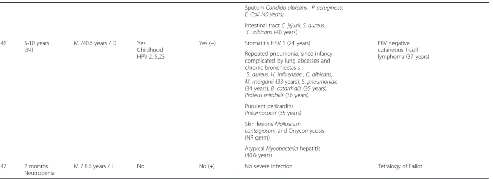
The long-term outcome of WS patients from both our survey and the 52 previously reported patients indicated five deaths (Figure 2A). No death was reported before the age of 20 years with the excep-tion of the medically terminated foetus described in this study. In addition to the two patients herein reported, whose deaths were caused by HPV-induced genital cancer and liver failure, the causes of deaths were lymphoma in two cases (at the ages of 26 [28] and 54 years [41]) and bacterial meningitis in one case (at the age of 31 years [2]). Three other malig-nancies were reported including one patient who declared a non-lethal B lymphoma at the age of 31 years followed by a maxillary carcinoma [19], and another patient who developed a genital cancer of un-specified etiology at age of 20 years [5]. The overall cancer risk in this population is estimated to be about 30% by the age of 40 years, with an age of onset be-yond the third decade (Figure 2B). Genital warts, which are reported for 23% of the literature cases and for 28.6% of cases herein described (2 cases), mark a turning point in the natural course of the disease due to their likelihood to progress to intractable multi-focal dysplasia and invasive cancer with an early fatal course despite repeated aggressive surgical removal.
Beaussant Cohen et al. Orphanet Journal of Rare Diseases 2012, 7:71 Page 10 of 14
Lastly, the association between WS and TOF has been re-cently reported [65], which included the patient UPN 5447, reported as P3. This association is strengthened by the herein reported fetus, aborted at 32 gestational weeks due to a double aortic arch, a cardiac abnormality belonging to the same group of conotruncal malformations as TOF.
Even though WS is quite rare, the amount of data collected until now can help to determine the medical management of afflicted patients. The current standard therapeutic approach often associates oral antibiotic prophylaxis, Ig and GCSF. The benefit of Ig prophylaxis is obvious when proposed early in life, as was the case for the two youngest patients of this cohort, but less evident when started later in life, notably after the de-velopment of permanent lung damage due to previous infections. The use of GCSF is questionable. Undoubt-edly, neutropenia is corrected by GCSF, yet infections do not seem correlated directly to the ANC, and no chronic stomatitis is reported. In addition, patients in our cohort withdrew from GCSF due to the lack of effi-ciency of the treatment against infections and this pat-tern was equally frequent in the literature [40]. Long
term prophylactic rotational antibiotic therapy is
recorded in two patients with COPD (Table 1) in addition to their Ig prophylaxis which seemed to lower the incidence of subsequent pulmonary infection epi-sodes. However, although prophylactic measures can be beneficial on bacterial infections they have no impact on HPV-related disease.
Moreover, for the management of HPV-induced cuta-neous and genital warts, standard methods such as cauterization and laser therapy seem relatively inefficient [34]. Aggressive surgical removal, Interferon or Cidofovir
administration, as well as topical Imiquimod treatments all proved equally unsatisfying, as lesions tended to re-appear. This stresses the need for a better understanding of the physiopathologic mechanisms accounting for the susceptibility to HPV. Prophylactic quadrivalent HPV vaccine administration to a WS patient was recently tested and was associated with protective immunity [25] suggesting that such a vaccine, if its long-term activity can be demonstrated, could be used as early prophylactic treatment for young WS patients and for newly diag-nosed WS cases. Hematopoietic stem cell transplant-ation has been proposed once, but with regards to the natural history of the disease, it seems difficult to propose a pre-emptive program for all patients, as the benefit expected by a transplant is to prevent infections in the third or fourth decade.
We should stress that although the actual management of WS is prophylactic, treatments that specifically target the CXCL12/CXCR4 axis are currently being evaluated. Indeed, two independent phase I clinical trials based on the acute administration of plerixafor have provided the pharmacological evidence of the causal role of CXCR4 dysfunctions in the WS-associated panleukopenia. How-ever, whether such treatment may help to treat or to prevent HPV lesions, EBV lymphoma or Mycobacteria infection, remains to be investigated. Moreover, consid-ering that the management of WS will need efficient and safe in the long run chronic treatments aimed at nor-malizing, but not abolishing the CXCR4 signaling, and because the long-term safety of AMD3100 may be ques-tioned [66,67], alternative new inhibitory compounds need to be characterized. The pre-clinical mouse model of the WS [66,67] will be useful for such analyses.
r e c n a c f o k s i r
B
l a v i v r u SA
0 10 20 30 40 50 60 70 80 90 100 % of s u rv iv o rs 0 20 40 60 80 Age in years 95% CI Survivor function 0 10 20 30 40 50 60 70 80 90 100 % of c a nc e r ( a ll t y pe s ) 0 20 40 60 80 Age in years 95% CI Risk of cancerFigure 2 Graphical representation of survival (A) and of the risk of cancer (B) after pooling the 52 published cases of patients with WHIM Syndrome and the 8 cases reported in this survey.
Beaussant Cohen et al. Orphanet Journal of Rare Diseases 2012, 7:71 Page 11 of 14
Additional files
Additional file 1: Literature review of 52 published cases of WHIM syndrome. ** Antibody against vaccine: Normal titers; ALC: Absolute lymphocyte count; AMC: Absolute Monocyte count; ANC: Absolute neutrophil count; ATB: Antibiotic prophylaxis; CXCR4: Chemokine (C-X-C-motif) receptor type 4; EBV: Epstein Barr virus; F: Female, ENT: Ears nose throat infection; GCSF: Granulocyte colony stimulating factor; GMCSF: Granulocyte macrophage colony-stimulating factor; HPV: Human Papillomavirus; Ig: Immunoglobulin prophylaxis; M: Male; NA: Non applicable; NR: Non reported. UPN: Unique patient number; SP: Streptococcus pneumoniae; HI: Haemophilus influenzae; SA: Staphylococcus aureus; PM: Proteus mirabilis; PA: Pseudomonas aeruginosa; NR: Not reported. + Present; - Absent.
Additional file 2: Family Trees of the four WHIM syndrome pedigrees described in this study, consistent with autosomal-dominant inheritance and sporadic cases.
Additional file 3: Sequential variation of blood counts in four patients illustrating the dynamic of variations of ANC (absolute neutrophil count), ALC (absolute lymphocyte count) and AMC (absolute monocyte count), at the occasion of sepsis and 5μg/kg/day Granulocyte colony-stimulating factor (GCSF) therapy.
Abbreviations
Abs: Antibodies; ALC: Absolute lymphocyte count; AMC: Absolute monocyte count; ANC: Absolute neutrophil count; BM: Bone Marrow; CBC: Complete blood count; COPD: Chronic obstructive pulmonary disease; C-tail: Carboxyl terminal tail; CXCR4: Chemokine (C-X-C motif) receptor type 4;
CXCL12: Chemokine (C-X-C motif) ligand 12; ELANE: Elastase neutrophil expressed; EBV: Epstein Barr virus; GCSF: Granulocyte colony-stimulating factor; GMCSF: Granulocyte macrophage colony-stimulating factor; GRK3: G-protein coupled receptor kinase 3; G6PC3: Glucose 6 phosphatase catalytic subunit 3 gene; g.w.: Gestational weeks; Hb: Hemoglobin; HPV: Human Papillomavirus; Ig: Immunoglobulins; PCR: Polymerase chain reaction; SD: Standard deviation; STK4: Serine threonine kinase 4 syndrome; TOF: Tetralogy of Fallot; UPN: Unique patient number; WBC: White blood cell count; WS: WHIM (Warts Hypogammaglobulinemia Infections Myelokathexis) syndrome.
Competing interests
The authors declare no competing interests.
Authors’ contributions
OF carried out the bone marrow central review, CBC performed the molecular genetic studies, KB and FB were responsible for the CXCR4 functional studies and analyses. EP, PSR, GD, IP, AD, DK, PB, FF, ALD, GD, JFN, PB, and YB who are referent physicians for the patients, revised the manuscript and contributed to the acquisition and analysis of data with SBC, JD and KB. BB visited each centre yearly to update the register. FB, SBC and JD wrote the manuscript. All authors read and approved the final manuscript.
Acknowledgements
The authors thank Dr Nathalie Aladjidi M.D., Dr Martine Gardembas M.D., Dr Denis Martin M.D., Dr Jean René Nelson M.D. and Dr Mireille Plasse M.D. who are following the patients and contributed to data collection, as well as the patients and their families for their participation in this study and Dr. F. Arenzana-Seisdedos (Inserm U819 & Laboratoire de Pathogénie Virale, Institut Pasteur, Paris, France) for invaluable support. The French registry is supported by grants from Amgen SAS, Chugai SA, GIS Maladies Rares, Institut de Veille Sanitaire and Inserm. This research is a study from the Centre de Référence des Déficits Immunitaires Héréditaires (CEREDIH: the French National Reference Center for Primary Immune Deficiencies, www.ceredih.fr) and has been supported by the Société Française d’Hématologie et d’Immunologie Pédiatrique. The K.B. laboratory is supported by the Agence Nationale de la Recherche (ANR, grant number 2010 JCJC 1104 01), the Université Paris-Sud and Inserm; FB was supported by the ANR (ANR-07-MRAR-029), AP-HP and E-rare (07 E-RARE 013–01), and K.B. and F.B. are members of the Laboratory
of Excellence LERMIT supported by a grant from ANR (Investissements d´Avenir).
Author details
1AP-HP, Registre français des neutropénies chroniques sévères, Centre de
référence des déficits Immunitaires Héréditaires, Service d’Hémato-oncologie Pédiatrique Hôpital Trousseau, 26 avenue du Dr Netter, 75012 Paris, France.
2
Service d’Onco-Hématologie Pédiatrique, Centre Hospitalo-Universitaire de Besançon, Paris, France.3AP-HP, Hôpital Robert Debré Laboratoire
d’Hématologie, 75019 Paris, France.4Service de Pédiatrie, Centre Hospitalier
Belfort-Montbéliard, Paris, France.5
Service d’hématologie, Centre Hospitalier de Roubaix, Paris, France.6Service de Dermatologie, CHU
Rennes-Pontchaillou, 35033 Rennes, France.7AP-HP Service de Dermatologie
Hôpital Saint Louis, 75010 Paris, France.8Service de Médecine Infantile II,
Hopitaux de Brabois, Vandoeuvre les Nancy Cedex, Paris, France.9AP-HP,
Service de Biologie du Développement, Hôpital Robert Debré, 75019 Paris, France.10Service de Pneumologie, Centre Hospitalier Lyon-Sud, Paris, France. 11
Service d’immunologie clinique et allergologie, Centre Hospitalier Lyon-Sud, Paris, France.12
Service d’Hématologie Pédiatrique, Hôpital Robert Debré, 75019 Paris, France.13Institut d’Oncologie et Hématologie Pédiatrique, Centre Hospitalo-Universitaire Lyon, Paris, France.14Inserm UMR_S996, Univ.
Paris-Sud, Laboratory of Excellence in Research on Medication and Innovative Therapeutics (LERMIT), 32 rue des Carnets, 92140 Clamart, France.1515
AP-HP, Hôpital Pitié-Salpêtrière, Département de Génétique, Univ. Pierre et Marie Curie, 75013 Paris, France.
Received: 28 May 2012 Accepted: 14 September 2012 Published: 25 September 2012
References
1. ZUELZER WW: "Myelokathexis"–A New Form of Chronic
Granulocytopenia. Report of a case. N Engl J Med 1964, 270:699–704.
2. Wetzler M, Talpaz M, Kleinerman ES, King A, Huh YO, Gutterman JU,
Kurzrock R: A new familial immunodeficiency disorder characterized by severe neutropenia, a defective marrow release mechanism, and hypogammaglobulinemia. Am J Med 1990, 89:663–672.
3. Dotta L, Tassone L, Badolato R: Clinical and genetic features of Warts,
Hypogammaglobulinemia, Infections and Myelokathexis (WHIM) syndrome. Curr Mol Med 2011, 11:317–325.
4. Hernandez PA, Gorlin RJ, Lukens JN, Taniuchi S, Bohinjec J, Francois F,
Klotman ME, Diaz GA: Mutations in the chemokine receptor gene CXCR4 are associated with WHIM syndrome, a combined immunodeficiency disease. Nat Genet 2003, 34:70–74.
5. Liu Q, Chen H, Ojode T, Gao X, Naya-O'Brien S, Turner NA, Ulrick J, Decastro
R, Kelly C, Cardones AR, et al: WHIM syndrome caused by a single amino acid substitution in the carboxy-tail of chemokine receptor CXCR4 LIU2012. Blood 2012, 120:181–189.
6. Bachelerie F: CXCL12/CXCR4-axis dysfunctions: Markers of the rare
immunodeficiency disorder WHIM syndrome. Dis Markers 2010, 29:189–198.
7. Balabanian K, Lagane B, Pablos JL, Laurent L, Planchenault T, Verola O,
Lebbe C, Kerob D, Dupuy A, Hermine O, et al: WHIM syndromes with different genetic anomalies are accounted for by impaired CXCR4 desensitization to CXCL12. Blood 2005, 105:2449–2457.
8. Balabanian K, Levoye A, Klemm L, Lagane B, Hermine O, Harriague J, Baleux
F, Arenzana-Seisdedos F, Bachelerie F: Leukocyte analysis from WHIM syndrome patients reveals a pivotal role for GRK3 in CXCR4 signaling. J Clin Invest 2008, 118:1074–1084.
9. Bohinjec J: Myelokathexis: chronic neutropenia with hyperplastic bone
marrow and hypersegmented neutrophils in two siblings. Blut 1981, 42:191–196.
10. Kawai T, Choi U, Cardwell L, DeRavin SS, Naumann N, Whiting-Theobald NL, Linton GF, Moon J, Murphy PM, Malech HL: WHIM syndrome
myelokathexis reproduced in the NOD/SCID mouse xenotransplant model engrafted with healthy human stem cells transduced with C-terminus-truncated CXCR4. Blood 2007, 109:78–84.
11. Balabanian K, Brotin E, Biajoux V, Bouchet-Delbos L, Lainey E, Fenneteau O, Bonnet D, Fiette L, Emilie D, Bachelerie F: Proper desensitization of CXCR4 is required for lymphocyte development and peripheral
compartmentalization in mice. Blood 2012, 119:5722–5730.
Beaussant Cohen et al. Orphanet Journal of Rare Diseases 2012, 7:71 Page 12 of 14
12. Alapi K, Erdos M, Kovacs G, Marodi L: Recurrent CXCR4 sequence variation in a girl with WHIM syndrome. Eur J Haematol 2007, 78:86–88.
13. Aprikyan AA, Liles WC, Park JR, Jonas M, Chi EY, Dale DC: Myelokathexis, a congenital disorder of severe neutropenia characterized by accelerated apoptosis and defective expression of bcl-x in neutrophil precursors. Blood 2000, 95:320–327.
14. Bassan R, Viero P, Minetti B, Comotti B, Barbui T: Myelokathexis: a rare form of chronic benign granulocytopenia. Br J Haematol 1984, 58:115–117. 15. Bohinjec J, Andoljsek D: Neutrophil-releasing activity of recombinant
human granulocyte-macrophage colony stimulating factor in myelokathexis. Br J Haematol 1992, 82:169–170.
16. Cernelc P, Andoljsek D, Mlakar U, Pretnar J, Modic M, Zupan IP, Zver S: Effects of molgramostim, filgrastim and lenograstim in the treatment of myelokathexis. Pflugers Arch 2000, 440:R81–R82.
17. Chae KM, Ertle JO, Tharp MD: B-cell lymphoma in a patient with WHIM syndrome. J Am Acad Dermatol 2001, 44:124–128.
18. Christ MJ, Dillon CA: Myelokathexis in a mother and infant: a second case suggesting dominant inheritance. Mil Med 1997, 162:827–828.
19. Cipriani NA, Blair E, Taxy JB: WHIM syndrome and oral squamous cell carcinoma. Oral Surg Oral Med Oral Pathol Oral Radiol Endod 2010, 109:105–108. 20. Ganser A, Ottmann OG, Erdmann H, Schulz G, Hoelzer D: The effect of
recombinant human granulocyte-macrophage colony-stimulating factor on neutropenia and related morbidity in chronic severe neutropenia. Ann Intern Med 1989, 111:887–892.
21. Goddard EA, Hughes EJ, Beatty DW: A case of immunodeficiency characterized by neutropenia, hypogammaglobulinaemia, recurrent infections and warts. Clin Lab Haematol 1994, 16:297–302.
22. Gorlin RJ, Gelb B, Diaz GA, Lofsness KG, Pittelkow MR, Fenyk JR Jr: WHIM syndrome, an autosomal dominant disorder: clinical, hematological, and molecular studies. Am J Med Genet 2000, 91:368–376.
23. Gulino AV, Moratto D, Sozzani S, Cavadini P, Otero K, Tassone L, Imberti L, Pirovano S, Notarangelo LD, Soresina R, et al: Altered leukocyte response to CXCL12 in patients with warts hypogammaglobulinemia, infections, myelokathexis (WHIM) syndrome. Blood 2004, 104:444–452.
24. Hagan JB, Nguyen PL: WHIM syndrome. Mayo Clin Proc 2007, 82:1031. 25. Handisurya A, Schellenbacher C, Reininger B, Koszik F, Vyhnanek P, Heitger
A, Kirnbauer R, Forster-Waldl E: A quadrivalent HPV vaccine induces humoral and cellular immune responses in WHIM immunodeficiency syndrome. Vaccine 2010, 28:4837–4841.
26. Hess U, Ganser A, Schnurch HG, Seipelt G, Ottmann OG, Falk S, Schulz G, Hoelzer D: Myelokathexis treated with recombinant human granulocyte-macrophage colony-stimulating factor (rhGM-CSF). Br J Haematol 1992, 80:254–256.
27. Hord JD, Whitlock JA, Gay JC, Lukens JN: Clinical features of myelokathexis and treatment with hematopoietic cytokines: a case report of two patients and review of the literature. J Pediatr Hematol Oncol 1997, 19:443–448. 28. Imashuku S, Miyagawa A, Chiyonobu T, Ishida H, Yoshihara T, Teramura T,
Kuriyama K, Imamura T, Hibi S, Morimoto A, et al: Epstein-Barr virus-associated T-lymphoproliferative disease with hemophagocytic syndrome, followed by fatal intestinal B lymphoma in a young adult female with WHIM syndrome. Warts, hypogammaglobulinemia, infections, and myelokathexis. Ann Hematol 2002, 81:470–473. 29. Krill CE Jr, Smith HD, Mauer AM: Chronic idiopathic granulocytopenia.
N Engl J Med 1964, 270:973–979.
30. Krivan G, Erdos M, Kallay K, Benyo G, Toth A, Sinko J, Goda V, Toth B, Marodi L: Successful umbilical cord blood stem cell transplantation in a child with WHIM syndrome. Eur J Haematol 2010, 84:274–275.
31. McDermott DH, Liu Q, Ulrick J, Kwatemaa N, Naya-O'Brien S, Penzak SR, Filho JO, Priel DA, Kelly C, Garofalo M, et al: The CXCR4 antagonist plerixafor corrects panleukopenia in patients with WHIM syndrome. Blood 2011, 118:4957–4962.
32. Mentzer WC Jr, Johnston RB Jr, Baehner RL, Nathan DG: An unusual form of chronic neutropenia in a father and daughter with
hypogammaglobulinaemia. Br J Haematol 1977, 36:313–322. 33. O'Regan S, Newman AJ: Graham RC: 'Myelokathexis'. Neutropenia with
marrow hyperplasia. Am J Dis Child 1977, 131:655–658.
34. Palm MD, Tyring SK, Rady PL, Tharp MD: Human papillomavirus typing of verrucae in a patient with WHIM syndrome. Arch Dermatol 2010, 146:931–932.
35. Plebani A, Cantu-Rajnoldi A, Collo G, Allavena P, Biolchini A, Pirelli A, Clerici SM, Masarone M: Myelokathexis associated with multiple congenital
malformations: immunological study on phagocytic cells and lymphocytes. Eur J Haematol 1988, 40:12–17.
36. Sanmun D, Garwicz D, Smith CI, Palmblad J, Fadeel B: Stromal-derived factor-1 abolishes constitutive apoptosis of WHIM syndrome neutrophils harbouring a truncating CXCR4 mutation. Br J Haematol 2006,
134:640–644.
37. Siedlar M, Rudzki Z, Strach M, Trzyna E, Pituch-Noworolska A, Blaut-Szlosarczyk A, Bukowska-Strakova K, Lenart M, Grodzicki T, Zembala M: Familial occurrence of warts, hypogammaglobulinemia, infections, and myelokathexis (WHIM) syndrome. Arch Immunol Ther Exp (Warsz ) 2008, 56:419–425.
38. Taniuchi S, Yamamoto A, Fujiwara T, Hasui M, Tsuji S, Kobayashi Y: Dizygotic twin sisters with myelokathexis: mechanism of its neutropenia. Am J Hematol 1999, 62:106–111.
39. Taniuchi S, Masuda M, Fujii Y, Izawa K, Kanegane H, Kobayashi Y: The role of a mutation of the CXCR4 gene in WHIM syndrome. Haematologica 2005, 90:1271–1272.
40. Tarzi MD, Jenner M, Hattotuwa K, Faruqi AZ, Diaz GA, Longhurst HJ: Sporadic case of warts, hypogammaglobulinemia,
immunodeficiency, and myelokathexis syndrome. J Allergy Clin Immunol 2005, 116:1101–1105.
41. Tassone L, Notarangelo LD, Bonomi V, Savoldi G, Sensi A, Soresina A, Smith CI, Porta F, Plebani A, Notarangelo LD, et al: Clinical and genetic diagnosis of warts, hypogammaglobulinemia, infections, and myelokathexis syndrome in 10 patients. J Allergy Clin Immunol 2009, 123:1170–1173. 42. Weston B, Axtell RA, Todd RF III, Vincent M, Balazovich KJ, Suchard SJ, Boxer
LA: Clinical and biologic effects of granulocyte colony stimulating factor in the treatment of myelokathexis. J Pediatr 1991, 118:229–234. 43. Wetzler M, Talpaz M, Kellagher MJ, Gutterman JU, Kurzrock R:
Myelokathexis: normalization of neutrophil counts and morphology by GM-CSF. JAMA 1992, 267:2179–2180.
44. Calandra G, Bridger G, Fricker S: CXCR4 in clinical hematology. Curr Top Microbiol Immunol 2010, 341:173–191.
45. Dale DC, Bolyard AA, Kelley ML, Westrup EC, Makaryan V, Aprikyan A, Wood B, Hsu FJ: The CXCR4 antagonist plerixafor is a potential therapy for myelokathexis, WHIM syndrome. Blood 2011, 118:4963–4966.
46. Donadieu J, Fenneteau O, Beaupain B, Mahlaoui N, Bellanne CC: Congenital neutropenia: diagnosis, molecular bases and patient management. Orphanet J Rare Dis 2011, 6:26.
47. Beaupain B, Leblanc T, Reman O, Hermine O, Vannier JP, Suarez F, Lutz P, Bordigoni P, Jourdain A, Schoenvald M, et al: Is pegfilgrastim safe and effective in congenital neutropenia? An analysis of the French Severe Chronic Neutropenia registry. Pediatr Blood Cancer 2009,
53:1068–1073.
48. Bellanne-Chantelot C, Clauin S, Leblanc T, Cassinat B, Rodrigues-Lima F, Beaufils S, Vaury C, Barkaoui M, Fenneteau O, Maier-Redelsperger M, et al: Mutations in the ELA2 gene correlate with more severe expression of neutropenia: a study of 81 patients from the French Neutropenia Register. Blood 2004, 103:4119–4125.
49. Donadieu J, Leblanc T, Bader-Meunier B, Barkaoui M, Fenneteau O, Bertrand Y, Maier-Redelsperger M, Micheau M, Stephan JL, Phillipe N, et al: Analysis of risk factors for myelodysplasias, leukemias and death from infection among patients with congenital neutropenia. Experience of the French Severe Chronic Neutropenia Study Group. Haematologica 2005, 90:45–53.
50. Latger-Cannard V, Bensoussan D, Bordigoni P: The WHIM syndrome shows a peculiar dysgranulopoiesis: myelokathexis. Br J Haematol 2006, 132:669. 51. Vinurel H, Freymond N, Pacheco Y, Devouassoux G: [The Whim syndrome:
a rare cause of diffuse bronchiectasis. Immune defect of CXCR4 and chronic bronchial suppuration]. Rev Mal Respir 2008, 25:614–618. 52. Buckley RH, Dees SC, O'Fallon WM: Serum immunoglobulins. I. Levels in normal
children and in uncomplicated childhood allergy. Pediatrics 1968, 41:600–611. 53. Hsu AP, Sampaio EP, Khan J, Calvo KR, Lemieux JE, Patel SY, Frucht DM, Vinh
DC, Auth RD, Freeman AF, et al: Mutations in GATA2 are associated with the autosomal dominant and sporadic monocytopenia and mycobacterial infection (MonoMAC) syndrome. Blood 2011, 118:2653–2655.
54. Kazenwadel J, Secker GA, Liu YJ, Rosenfeld JA, Wildin RS, Cuellar-Rodriguez J, Hsu AP, Dyack S, Fernandez CV, Chong CE, et al: Loss-of-function germline GATA2 mutations in patients with MDS/AML or MonoMAC syndrome and primary lymphedema reveal a key role for GATA2 in the lymphatic vasculature. Blood 2012, 119:1283–1291.
Beaussant Cohen et al. Orphanet Journal of Rare Diseases 2012, 7:71 Page 13 of 14
55. Diaz GA, Gulino AV: WHIM syndrome: a defect in CXCR4 signaling. Curr Allergy Asthma Rep 2005, 5:350–355.
56. Kawai T, Malech HL: WHIM syndrome: congenital immune deficiency disease. Curr Opin Hematol 2009, 16:20–26.
57. McDermott DH, De Ravin SS, Jun HS, Liu Q, Priel DA, Noel P, Takemoto CM, Ojode T, Paul SM, Dunsmore KP, et al: Severe congenital neutropenia resulting from G6PC3 deficiency with increased neutrophil CXCR4 expression and myelokathexis. Blood 2010, 116:2793–2802. 58. Eash KJ, Greenbaum AM, Gopalan PK, Link DC: CXCR2 and CXCR4
antagonistically regulate neutrophil trafficking from murine bone marrow. J Clin Invest 2010, 120:2423–2431.
59. Maran R, Mittelman M, Cohen AM, Djaldetti M: Myelokathexis and monocytosis in a patient with gastric cancer. Acta Haematol 1992, 87:210–212.
60. Diaz GA: CXCR4 mutations in WHIM syndrome: a misguided immune system? Immunol Rev 2005, 203:235–243.
61. Doncker AV, Balabanian K, Bellanne-Chantelot C, de GS, Revest M, Bachelerie F, Lamy T: Two cases of disseminated Mycobacterium avium infection associated with a new immunodeficiency syndrome related to CXCR4 dysfunctions. Clin Microbiol Infect 2011, 17:135–139.
62. Ostergaard P, Simpson MA, Connell FC, Steward CG, Brice G, Woollard WJ, Dafou D, Kilo T, Smithson S, Lunt P, et al: Mutations in GATA2 cause primary lymphedema associated with a predisposition to acute myeloid leukemia (Emberger syndrome). Nat Genet 2011, 43:929–931.
63. Abdollahpour H, Appaswamy G, Kotlarz D, Diestelhorst J, Beier R, Schaffer AA, Gertz EM, Schambach A, Kreipe HH, Pfeifer D, et al: The phenotype of human STK4 deficiency. Blood 2012, 119:3450–3457.
64. Chow KY, Brotin E, Ben KY, Carthagena L, Teissier S, Danckaert A, Galzi JL, Renzana-Seisdedos F, Thierry F, Bachelerie F: A pivotal role for CXCL12 signaling in HPV-mediated transformation of keratinocytes: clues to understanding HPV-pathogenesis in WHIM syndrome. Cell Host Microbe 2010, 8:523–533.
65. Badolato R, Dotta L, Tassone L, Amendola G, Porta F, Locatelli F, Notarangelo LD, Bertrand Y, Bachelerie F, Donadieu J: Tetralogy of Fallot is an Uncommon Manifestation of Warts, Hypogammaglobulinemia, Infections, and Myelokathexis Syndrome. J Pediatr 2012, Epub ahead of print. 66. Dugan MJ, Maziarz RT, Bensinger WI, Nademanee A, Liesveld J, Badel K,
Dehner C, Gibney C, Bridger G, Calandra G: Safety and preliminary efficacy of plerixafor (Mozobil) in combination with chemotherapy and G-CSF: an open-label, multicenter, exploratory trial in patients with multiple myeloma and non-Hodgkin's lymphoma undergoing stem cell mobilization. Bone Marrow Transplant 2010, 45:39–47.
67. Hendrix CW, Collier AC, Lederman MM, Schols D, Pollard RB, Brown S, Jackson JB, Coombs RW, Glesby MJ, Flexner CW, et al: Safety, pharmacokinetics, and antiviral activity of AMD3100, a selective CXCR4 receptor inhibitor, in HIV-1 infection. J Acquir Immune Defic Syndr 2004, 37:1253–1262.
doi:10.1186/1750-1172-7-71
Cite this article as: Beaussant Cohen et al.: Description and outcome of a cohort of 8 patients with WHIM syndrome from the French Severe Chronic Neutropenia Registry. Orphanet Journal of Rare Diseases 2012 7:71.
Submit your next manuscript to BioMed Central and take full advantage of:
• Convenient online submission
• Thorough peer review
• No space constraints or color figure charges
• Immediate publication on acceptance
• Inclusion in PubMed, CAS, Scopus and Google Scholar
• Research which is freely available for redistribution
Submit your manuscript at www.biomedcentral.com/submit
Beaussant Cohen et al. Orphanet Journal of Rare Diseases 2012, 7:71 Page 14 of 14