HAL Id: hal-01204331
https://hal.archives-ouvertes.fr/hal-01204331
Submitted on 31 May 2020
HAL is a multi-disciplinary open access
archive for the deposit and dissemination of
sci-entific research documents, whether they are
pub-lished or not. The documents may come from
teaching and research institutions in France or
abroad, or from public or private research centers.
L’archive ouverte pluridisciplinaire HAL, est
destinée au dépôt et à la diffusion de documents
scientifiques de niveau recherche, publiés ou non,
émanant des établissements d’enseignement et de
recherche français ou étrangers, des laboratoires
publics ou privés.
Extent of horizontal gene transfer in evolution of
Streptococci of the salivarius group
Christine Delorme, Claire Poyart, Stanislav Ehrlich, Pierre Renault
To cite this version:
Christine Delorme, Claire Poyart, Stanislav Ehrlich, Pierre Renault. Extent of horizontal gene transfer
in evolution of Streptococci of the salivarius group. Journal of Bacteriology, American Society for
Microbiology, 2007, 189 (4), pp.1330-1341. �10.1128/JB.01058-06�. �hal-01204331�
0021-9193/07/$08.00⫹0 doi:10.1128/JB.01058-06
Copyright © 2007, American Society for Microbiology. All Rights Reserved.
Extent of Horizontal Gene Transfer in Evolution of Streptococci of
the Salivarius Group
䌤
†
Christine Delorme,
1* Claire Poyart,
2S. Dusko Ehrlich,
1and Pierre Renault
1Laboratoire de Ge´ne´tique Microbienne, Institut National de Recherche Agronomique, 78352 Jouy-en-Josas Cedex, France,1and
INSERM E44, Institut Cochin, Centre National de Re´fe´rence des Streptocoques, Service de Bacte´riologie Hoˆpital Cochin, Faculte´ de Me´decine Paris 5, 27 rue du faubourg Saint Jacques, 75679 Paris Cedex 14, France2
Received 19 July 2006/Accepted 19 October 2006
The phylogenetically closely related species Streptococcus salivarius and Streptococcus vestibularis are oral bacteria that are considered commensals, although they can also be found in human infections. The relation-ship between these two species and the relationrelation-ship between strains isolated from carriers and strains responsible for invasive infections were investigated by multilocus sequence typing and additional sequence analysis. The clustering of several S. vestibularis alleles and the extent of genomic divergence at certain loci support the conclusion that S. salivarius and S. vestibularis are separate species. The level of sequence diversity in S. salivarius alleles is generally high, whereas that in S. vestibularis alleles is low at certain loci, indicating that the latter species might have evolved recently. Cluster analysis indicated that there has been genetic exchange between S. salivarius and S. vestibularis at three of the nine loci investigated. Horizontal gene transfer between streptococci belonging to the S. salivarius group and other oral streptococci was also detected at several loci. A high level of recombination in S. salivarius was revealed by allele index association and split decom-position sequence analyses. Commensal and infection-associated S. salivarius strains could not be distin-guished by cluster analysis, suggesting that the pathogen isolates are opportunistic. Taken together, our results indicate that there is a high level of gene exchange that contributes to the evolution of two streptococcal species from the human oral cavity.
Streptococcus salivarius and Streptococcus vestibularis belong
to the salivarius group of viridans streptococci (18). Many of the viridans streptococcal species are part of the normal mi-crobial flora of humans. These commensal species are most prevalent in the oral cavity but also reside in the gastrointes-tinal and urogenital tracts. Viridans streptococci have emerged as important nosocomial pathogens, and they typically cause significant septicemia when mucosal lesions are present and host defense mechanisms are compromised (36, 53, 57). S.
salivarius and S. vestibularis have been associated with severe
human infections, such as, meningitis, endocarditis, and bac-teremia (9, 11, 14, 31, 42, 47). Molecular taxonomy based on sequencing and comparison of the 16S rRNA or sodA genes demonstrated that these two species are closely related (34, 46). The population structure of several oral streptococci has been studied previously (7, 19, 26, 38). Genetic typing of com-mensal or opportunistic pathogens has revealed considerable diversity, sometimes even within a single host. Data obtained in a Streptococcus mitis population analysis support the concept that the strains are transient, and this species appears to be maintained by clonal replacement of evolving strains rather than by stable strains (19, 26). Almost all isolates of
Strepto-coccus mutans obtained either from 30 individuals or from a
small group of families displayed distinctive restriction
frag-ment length polymorphism or restriction endonuclease analy-sis patterns (7, 38). Fitzsimmons et al. suggested that the high degree of diversity observed in several mucosal bacteria may be a mechanism for avoiding immune elimination (19). S. mitis and Streptococcus oralis strains isolated from the blood of neu-tropenic cancer patients were also highly diverse, as they all had distinct fingerprint patterns (57). The population structure and genetic diversity of S. salivarius and S. vestibularis have been not extensively investigated, in spite of the fact that S. salivarius is the predominant oral species, especially during mouth coloniza-tion, and can be associated with caries (2, 35, 43).
Multilocus sequence typing (MLST), a method based on the nucleotide sequences of⬃500-bp internal fragments of multi-ple (usually about seven) housekeeping genes, has been widely used to study global epidemiology and bacterial population structure (15). MLST can reveal highly clonal populations, as well as freely recombining populations, and the latter are ex-emplified by Streptococcus uberis or Streptococcus pneumoniae populations (8, 16). Several reports have indicated that most streptococcal species have a highly recombinational population structure (33, 37, 58). The genetic relationships of group B, C, G, and A streptococcal isolates from asymptomatic carriers and from human infections were investigated by phylogenetic analysis of MLST data (3, 32, 33). Here we used the MLST method to investigate the genetic relationship between strains of S. salivarius and strains of S. vestibularis. Below we describe an analysis of data obtained from 27 S. salivarius strains and 9
S. vestibularis strains recovered from patients with septicemia
or from the oral cavities of healthy individuals. Our results document the population structure of S. salivarius, the rela-tionship between the two species, and the extent of gene ex-change in the evolution of these oral streptococci.
* Corresponding author. Mailing address: Laboratoire de Ge´ne ´t-ique Microbienne, Institut National de Recherche Agronom´t-ique, 78352 Jouy-en-Josas Cedex, France. Phone: (33) 1 34 65 25 26. Fax: (33) 1 34 65 25 21. E-mail: christine.delorme@jouy.inra.fr.
† Supplemental material for this article may be found at http://jb .asm.org/.
䌤Published ahead of print on 3 November 2006.
(This work was presented in part at the 7th ASM Confer-ence on Streptococcal Genetics, Saint Malo, France, June 2006.)
MATERIALS AND METHODS
Bacterial strains.S. salivarius (n⫽ 27) and S. vestibularis (n ⫽ 9) strains are listed in Table 1. These strains were isolated in different countries and from different sources over a 50-year period (1954 to 2004) and were obtained from the Institut Pasteur Collection (CIP), the Belgium Coordinated Collections of Microorganisms (LMG), Paris Cochin Hospital (CCH), and our laboratory col-lection (JIM). Strains were obtained from the oral cavity (n⫽ 18), human blood (n⫽ 10), and breast milk (n ⫽ 1). Commensal strains were isolated from different oral cavities; oral cavities 1 and 2 were the oral cavities of 3-year-old twin girls.
Strains were grown overnight at 37°C on M17 broth medium containing glu-cose or lactose at final concentration of 1% in an anaerobic atmosphere. Samples were collected from oral cavities by scraping the tongue surface with sterile wooden tongue depressors that were used directly for isolation on M17 medium plates containing glucose. Both genetic identification and phenotypic identifica-tion were performed for all the strains. Phenotypic identificaidentifica-tion was performed
with the rapid ID 32 Strep system (bioMe´rieux) or using a focused scheme for assessing hydrolysis of urea and acid production in lactose-, raffinose-, sorbitol-, or inulin-containing media. Hydrolysis of urea and a failure to ferment raffinose and inulin are characteristics of all S. vestibularis strains tested (10). Genetic identification at the species level was carried out by sequencing 16S rRNA and the sodA gene (46).
Multilocus sequence typing.We searched for candidate MLST loci among housekeeping genes which were used previously for MLST of other gram-posi-tive bacteria and which could be amplified with degenerate primers (ddlA, tkt,
glcK, pyrE, and sodA) or whose sequences were available in a database for closely
related species (ilvC, pepO, thrS, and dnaE). Furthermore, we checked to make sure that the genes were usually found in distant locations in previously described streptococcal genomes, especially the genome of the closely related species
Streptococcus thermophilus. The nucleotide sequences of internal fragments of
the following nine genes that were selected were determined: ilvC (encoding ketol-acid reductoisomerase), ddlA (encodingD-alanineD-alanine ligase), glcK (encoding glucose kinase), pepO (encoding endopeptidase), thrS (encoding threonyl-tRNA synthetase), tkt (encoding transketolase), pyrE (encoding orotate phosphoribosyltransferase), dnaE (encoding DNA polymerase III), and sodA (superoxide dismutase). For the sodA gene, we used primers and an amplifica-tion procedure described previously (46). Primers used in this study are listed in
TABLE 1. Strains and MLST data
Strain ST
Allele
Sourceb
Origin Yr
glck ddlA pepO ilvC thrS pyrE dnaE sodA tkta S. salivarius strains
CIP53.158 1 2 10 7 13 4 15 14 13 NA NV Ithaca, NY 1954
CIP55.126 2 2 11 7 13 4 15 14 13 NA NV France 1955
CIP102505 3 7 6 1 4 13 12 20 2 NA NV New York, NY 1971
JIM8222 4 16 26 13 16 17 22 13 8 NA Oral cavity Germany 2000
JIM8223 5 9 7 18 10 4 16 18 20 NA Oral cavity Germany 2000
JIM8224 6 14 30 16 10 15 19 12 5 NA Oral cavity Germany 2000
JIM8221 7 4 3 3 4 14 12 17 14 NA Oral cavity Germany 2000
CIP102503T 8 3 7 1 8 1 13 17 12 NA Human blood New York, NY 1950
CIP104994 9 6 6 4 5 5 11 20 14 NA Human blood Tourcoing, France 1996
LMG13104 10 1 15 17 14 2 14 22 10 NA NV Munchen, Germany 1983
LMG13106 11 1 17 22 15 4 3 24 2 NA NV Hospital, London, United
Kingdom
1983
LMG13108 12 15 25 7 11 8 24 16 6 NA NV Hospital, London, United
Kingdom
1983
LMG13109 13 15 27 20 9 12 21 7 2 NA NV Hospital, London, United
Kingdom K
1983
LMG14652 14 5 14 3 5 5 15 21 14 NA Human blood Ostersund, Sweden 1993
JIM8421 15 15 23 11 14 11 2 23 11 NA Breast milk Finland 2001
JIM8775 16 2 16 6 12 4 15 17 7 NA Oral cavity 1 France 2004
JIM8777 18 1 9 17 6 7 4 19 10 NA Oral cavity 1 France 2004
JIM8776 17 2 12 12 7 6 10 15 9 NA Oral cavity 1 France 2004
JIM8774 17 2 12 12 7 6 10 15 9 NA Oral cavity 2 France 2004
JIM8773 21 17 4 21 9 1 20 9 4 NA Oral cavity 2 France 2004
JIM8771 19 16 8 6 14 9 23 8 1 NA Oral cavity 2 France 2004
JIM8772 20 8 28 2 8 10 25 17 7 NA Oral cavity 2 France 2004
CCHSS1 22 16 29 14 17 20 18 11 3 NA Human blood Hospital Cochin, France 2003
CCHSS2 23 14 1 8 10 16 19 10 2 NA Human blood Cochin Hospital, France 2001
CCHSS3 24 5 13 19 5 4 1 6 14 NA Human blood Cochin Hospital, France 2002
CCHSS4 25 4 24 5 5 5 11 20 2 NA human blood Cochin Hospital, France 2001
CCHSS7 26 15 2 16 12 6 17 17 1 NA human blood Cochin Hospital, France 2004
S. vestibularis strains
CIP103363T 1 12 19 10 3 3 9 2 16 5 Oral cavity Hospital, London, United
Kingdom
1987
LMG17854 2 11 20 9 1 18 7 4 17 4 Vestibular mucosa Malmo¨, Sweden 1966
LMG14646 2 11 20 9 1 18 7 4 17 4 Vestibular mucosa Malmo¨, Sweden 1966
LMG14645 3 13 21 9 1 19 7 4 15 4 Vestibular mucosa Malmo¨, Sweden 1966
LMG14647 4 10 18 9 2 18 5 1 15 3 Vestibular mucosa Malmo¨, Sweden 1966
LMG17855 4 10 18 9 2 18 5 1 15 3 Vestibular mucosa Malmo¨, Sweden 1966
LMG17856 4 10 18 9 2 18 5 1 15 3 Human dental plaque Malmo¨, Sweden 1966
CCHSV5 5 10 22 15 1 21 8 5 18 1 Human blood Cochin Hospital, France 2002
CCHSV6 6 10 5 10 1 3 6 3 19 2 Human blood Cochin Hospital, France 2004
aNA, not amplified. bNV, not available.
Table 2. One bacterial colony, freshly grown on an agar plate, was suspended in 50l of TES (10 mM Tris-HCl, 1 mM EDTA, 25% sucrose), and DNA was extracted by lysis in a thermocycler at 95°C for 10 min and at 4°C for 15 min. The DNA obtained was used immediately for PCR amplification performed with the GeneAmp 9700 PCR system (Perkin-Elmer) by using the following cycling parameters: 5 min at 94°C, followed by 30 cycles of 30 s at 94°C, 30 s at 50°C, and 1 min at 72°C. Amplified products (459 to 527 bp of internal gene fragments) were examined on a 1.5% agarose gel. They were used for direct sequencing by a fluorescent sequencing procedure with the same PCR primers that were used for the initial PCR amplification. The sequence of each fragment on both strands was determined with an Applied Biosystems 370A DNA sequencer by using a Bigdye Terminator sequencing kit according the manufacturer’s protocol (Per-kin-Elmer). To ensure the accuracy of the sequence, amplicons obtained from at least two bacterial colonies were used for sequence determination.
PCR amplification of ilvC, pepO, pyrE, and tkt genes for sequencing.Primer pairs for the tkt locus did not amplify the DNA fragments of all S. salivarius isolates. Therefore, alternative sets of primers were designed using sequence information available in public databases. The primers listed in Table 2 (tkt-up2,
tkt-dn2, tkt-up3, tkt-dn3, tkt-up4, tkt-dn4, tkt-dn5, tkt-up6, and tkt-dn6) were used
in all possible up and dn combinations. The large region around the ilvC, pepO, and tkt genes was sequenced by primer walking, using primers designed on the basis of the regions sequenced. For amplification of the region containing the
ilvB, ilvN, ilvC, and tyrS genes, we used ilvB-up3, ilvC-dn3, ilvC-up5, and tyrS-do4
(Table 2). For amplification of the region containing the dexS and pepO genes, we used up6, up4, dn6, dn4, dn5, dn7, pepO-up5, and pepO-up7 (Table 2). For amplification of the region containing the tkt gene, we used tkt-dn7 and tkt-up8 for both species, tkt-salup, tkt-saldn, tkt-saldn8, and tkt-saldo9 for S. salivarius, and tkt-vesup, tkt-vesdo, tkt-vesup9, and tkt-vesdo9 for S. vestibularis (Table 2).
For pyrE sequence alignment, the nucleotide sequence of the mobile element found in allele 11 (S. salivarius) was not taken into account.
Data treatment and statistical analysis.For each locus, all the sequences were compared, and arbitrary allele numbers were assigned to the different sequences. The combination of alleles at each locus defined an allelic profile or sequence type (ST) for a strain. Strains with the same allelic profile were assigned to the same ST. The STs were identified by arbitrary numbers.
The number of polymorphic nucleotide sites and the maximal and average levels of nucleotide divergence of alleles (expressed as percentages) at a given
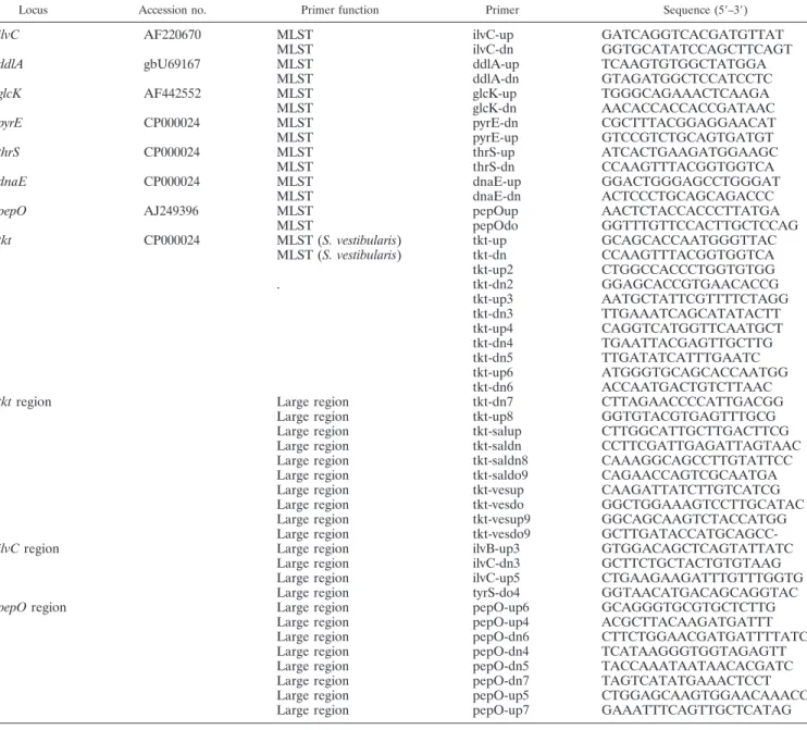
TABLE 2. Oligonucleotide primers for S. salivarius and S. vestibularis
Locus Accession no. Primer function Primer Sequence (5⬘–3⬘)
ilvC AF220670 MLST ilvC-up GATCAGGTCACGATGTTAT
MLST ilvC-dn GGTGCATATCCAGCTTCAGT
ddlA gbU69167 MLST ddlA-up TCAAGTGTGGCTATGGA
MLST ddlA-dn GTAGATGGCTCCATCCTC
glcK AF442552 MLST glcK-up TGGGCAGAAACTCAAGA
MLST glcK-dn AACACCACCACCGATAAC
pyrE CP000024 MLST pyrE-dn CGCTTTACGGAGGAACAT
MLST pyrE-up GTCCGTCTGCAGTGATGT
thrS CP000024 MLST thrS-up ATCACTGAAGATGGAAGC
MLST thrS-dn CCAAGTTTACGGTGGTCA
dnaE CP000024 MLST dnaE-up GGACTGGGAGCCTGGGAT
MLST dnaE-dn ACTCCCTGCAGCAGACCC
pepO AJ249396 MLST pepOup AACTCTACCACCCTTATGA
MLST pepOdo GGTTTGTTCCACTTGCTCCAG
tkt CP000024 MLST (S. vestibularis) tkt-up GCAGCACCAATGGGTTAC
MLST (S. vestibularis) tkt-dn CCAAGTTTACGGTGGTCA tkt-up2 CTGGCCACCCTGGTGTGG . tkt-dn2 GGAGCACCGTGAACACCG tkt-up3 AATGCTATTCGTTTTCTAGG tkt-dn3 TTGAAATCAGCATATACTT tkt-up4 CAGGTCATGGTTCAATGCT tkt-dn4 TGAATTACGAGTTGCTTG tkt-dn5 TTGATATCATTTGAATC tkt-up6 ATGGGTGCAGCACCAATGG tkt-dn6 ACCAATGACTGTCTTAAC
tkt region Large region tkt-dn7 CTTAGAACCCCATTGACGG
Large region tkt-up8 GGTGTACGTGAGTTTGCG
Large region tkt-salup CTTGGCATTGCTTGACTTCG
Large region tkt-saldn CCTTCGATTGAGATTAGTAAC
Large region tkt-saldn8 CAAAGGCAGCCTTGTATTCC
Large region tkt-saldo9 CAGAACCAGTCGCAATGA
Large region tkt-vesup CAAGATTATCTTGTCATCG
Large region tkt-vesdo GGCTGGAAAGTCCTTGCATAC
Large region tkt-vesup9 GGCAGCAAGTCTACCATGG
Large region tkt-vesdo9
GCTTGATACCATGCAGCC-ilvC region Large region ilvB-up3 GTGGACAGCTCAGTATTATC
Large region ilvC-dn3 GCTTCTGCTACTGTGTAAG
Large region ilvC-up5 CTGAAGAAGATTTGTTTGGTG
Large region tyrS-do4 GGTAACATGACAGCAGGTAC
pepO region Large region pepO-up6 GCAGGGTGCGTGCTCTTG
Large region pepO-up4 ACGCTTACAAGATGATTT
Large region pepO-dn6 CTTCTGGAACGATGATTTTATC
Large region pepO-dn4 TCATAAGGGTGGTAGAGTT
Large region pepO-dn5 TACCAAATAATAACACGATC
Large region pepO-dn7 TAGTCATATGAAACTCCT
Large region pepO-up5 CTGGAGCAAGTGGAACAAACC
locus were calculated using the MEGA software (version 3; http://www .megasoftware.net) (39). Phylogenetic analyses of the nucleotide sequences of each housekeeping gene separately and the concatenated sequence containing
ddlA, thrS, pyrE, dnaE, and sodA were performed using the neighbor-joining (NJ)
method and the same software. Because of their extensive nucleotide diversity, the glcK, ilvC, and pepO sequences could not be used in the MLST scheme, so we removed them from the concatenated sequence. A Kimura two-parameter dis-tance model was used to estimate disdis-tances for nucleotide sequences. To deter-mine the significance of the groups observed in trees constructed by the NJ method, a bootstrap analysis with 1,000 replicates was performed.
The split decomposition method was used to assess the degree of tree-like structure for alleles found for each locus (30). The sequence alignments were converted to NEXUS files, and the split decomposition analysis was performed with the SPLITSTREE 3.1 program (http://bibiserv.techfak.uni-bielefeld.de /splits/).
A statistical analysis based on the dN/dSratio (41), which was the ratio of the
number of substitutions that changed the amino acid sequence (dNis number of
nonsynonymous substitutions per nonsynonymous site) to the number of silent substitutions (dSis the number of synonymous substitutions per synonymous
site), was performed by using methods described with the START program (K. Jolley; http://mlst.zoo.ox.ac.uk/links/START). The standardized index of associ-ation (IS
A) was calculated as described by Haubold and Hudson (24). In our
study, the set of S. salivarius isolates analyzed for linkage equilibrium did not contain CIP55.126 and JIM8774, which are considered to be very closely related to CIP53.128 and JIM8776, respectively.
Nucleotide sequence accession numbers.The sequences of all alleles have been deposited in the GenBank database under accession numbers DQ460524 to DQ460540 (ilvC fragment), DQ460609 to DQ460638 (ddlA fragment), DQ460507 to DQ460523 (glcK fragment), DQ460541 to DQ460562 (pepO fragment), DQ460588 to DQ460608 (thrS fragment), DQ460478 to DQ460482 (tkt frag-ment), DQ460563 to DQ460587 (pyrE fragfrag-ment), DQ460483 to DQ460506 (dnaE fragment), and EF054776 to EF054795 (sodA fragment). The sequences of large regions have been deposited in the GenBank database under accession numbers DQ46995 (dexS and pepO of S. salivarius LMG13109), DQ464996 (dexS and pepO of S. vestibularis LMG14645), DQ464998 (ilvB, ilvN, ilvC, and tyrS of
S. salivarius LMG13109), DQ464994 (ilvB, ilvN, ilvC, and tyrS of S. vestibularis
LMG14645), DQ464997 (tkt and tRNAThr
of S. salivarius LMG13109), and DQ464993 (orf1, tkt, and trkA2 of S. vestibularis LMG14645).
RESULTS
Development of an MLST scheme for S. salivarius and S.
vestibularis.Twenty-seven S. salivarius strains and nine S.
ves-tibularis strains isolated from different sources (oral cavity,
breast milk, and human blood) and in different countries over a 50-year period were collected (Table 1). Misclassification due to the natural intraspecies phenotypic variability and the high level of 16S rRNA sequence identity (99%) between S.
saliva-rius and S. vestibularis was corrected by characterization of
the sodA gene sequence. The type strains of S. salivarius (CIP102503 [⫽ NCTC8618]) and S. vestibularis (CIP103363 [⫽ NCTC 12166]) were used as standards. The nucleotide se-quences of 404- to 527-bp internal fragments were determined for eight and nine loci in S. salivarius and S. vestibularis, re-spectively, as tkt could not be amplified for S. salivarius with the first set of primers. The polymorphic sites for the ddlA alleles are shown in Fig. 1 and for the eight other loci in Fig. S1 in the supplemental material. The average levels of sequence diver-sity for the S. salivarius and S. vestibularis alleles were 5.8% and 2%, respectively.
In the S. salivarius population, a high number of unique alleles was found for each gene, and there was no particularly predominant allele. The number of alleles per locus for the 27
S. salivarius strains ranged from 13 (glcK) to 24 (ddlA). The
highest level of nucleotide sequence divergence for a locus ranged from 4.9% (sodA) to 45.3% (pepO) (Table 3). The
FIG. 1. Polymorphic nucleotide sites of ddlA alleles of S. salivarius and S. vestibularis: variable sites in each allele of the ddlA locus from the 27 S. salivarius strains and 9 S. vestibularis strains. The site numbers at the top are in vertical format, and S. vestibularis alleles are shaded.
dN/dSratio was calculated as a measure of the degree of se-lection in the population. For S. salivarius, the dN/dSratios for
glcK, pyrE, ilvC, sodA, and dnaE were⬍0.25. In contrast, the dN/dSratio for the thrS, ddlA, and pepO loci were⬎1, indicat-ing that there was a lower degree of selection against amino acid change (32).
Table 1 summarizes the allelic profiles of the 27 S. salivarius strains used in this study. Each unique combination of allele numbers represents one allelic profile or ST. For S. salivarius, 26 different allelic profiles were found (ST1 to ST26), which allowed discrimination of all but two strains in our collection (ST17) (Table 1). Most STs differed at at least three of seven loci; the exceptions were ST1 and ST2, which differed at only one locus (Table 1). ST1 and ST2 were represented by two strains from the Institut Pasteur Collection (CIP53.158 and CIP55.126) and differed by only one nucleotide in the ddlA allele (Fig. 1 and Table 1). This is in sharp contrast to the high levels of variability observed for most other isolates and sug-gests that CIP53.158 and CIP55.126 originated from the same strain. The two ST17 strains, JIM8776 and JIM8774, are prob-ably independent isolates of the same strain originating from the oral cavities of twin girls (Table 1). However, analysis of multiple isolates from the same two oral cavities revealed three different STs in one oral cavity and four different STs in the other, showing that S. salivarius isolates from the same indi-vidual are genetically heterogeneous. For the commensal and disease-associated S. salivarius isolates, the concatenated ddlA,
thrS, pyrE, dnaE, and sodA sequences (total length, 2,359 bp)
did not allow us to clearly distinguish the two classes, as judged from the tree shown in Fig. 2.
For the set of nine S. vestibularis strains, the maximal level of nucleotide sequence divergence ranged from 0.6% (pyrE) to 38.2% (pepO) (Table 3), and six different allelic profiles were found, (ST1 to ST6) (Table 1). Two and three strains belonged to ST2 and ST4, respectively. All of these strains were isolated from vestibular mucosa at the same time and place (1966 in Malmo¨, Sweden). The strains with the same ST might corre-spond to multiple isolates of the same strains (Table 1). Be-cause of the low number of independent isolates in our set, we could not use S. vestibularis in the MLST scheme.
Evidence of intraspecific recombination in S. salivarius ob-tained by statistical analysis.Two types of statistical analysis
were used to characterize intragenic recombination at S.
sali-varius loci analyzed by MLST.
(i) Standardized index of association.The index of associ-ation has been widely used to analyze the degree of linkage disequilibrium between alleles in MLST and multilocus en-zyme electrophoresis data (49). Haubold and Hudson de-scribed the standardized index of association, which does not
TABLE 3. Genetic diversity at S. salivarius and S. vestibularis loci
Locus
Length of sequence
(bp)
No. of alleles No. of variable sites Maximal % divergence
S. salivarius (27 isolates) S. vestibularis (9 isolates) S. salivarius (27 isolates) S. vestibularis (9 isolates) Within S. salivarius Within S. vestibularis Between S. salivarius and S. vestibularis glcK 470 13 4 65 4 11.9 0.9 11.7 pepO 460 19 3 193 138 45.3 38.2 41.2 ilvC 492 14 3 82 3 15.5 0.6 16 sodA 404 15 5 29 3 4.9 0.7 11.6 thrS 497 17 4 60 27 6.1 4 6.8 pyrE 519 20 5 69 3 7.3 0.6 7.3 dnaE 480 19 5 76 34 6.7 6.3 11.1 ddlA 459 24 6 75 50 8.1 8.8 9.3 tkta 527 5 4 0.6
aThe tkt locus was amplified only in S. vestibularis isolates.
FIG. 2. Phylogenetic tree based on concatenated sequences of five housekeeping genes (ddlA, thrS, pyrE, dnaE, and sodA). The tree was constructed using the neighbor-joining method. Bootstrap values that areⱖ80% are indicated at nodes. Scale bar ⫽ 0.005 nucleotide sub-stitution per site. Disease-related isolates are indicated by an asterisk.
depend on the number of loci analyzed (24). An IS
Avalue of zero is expected for alleles in linkage equilibrium, indicating that alleles are distributed independent of each other because of free recombination (24). The IS
Avalue calculated for our S.
salivarius data was close to zero (0.057), suggesting that there
was free recombination and thus a high level of gene exchange.
(ii) Split decomposition analysis. Evidence of recombina-tion was obtained by split decomposirecombina-tion analysis using the SPLITSTREE 3.1 program. The algorithm used in this soft-ware is able to display conflicting results in the phylogenetic descent of sequences. A tree-like structure is created when the descent is clonal, but an interconnecting network-like structure appears when recombination plays a role in the evolutionary history of the population analyzed (30, 52). The results ob-tained with unique S. salivarius alleles are shown in Fig. 3 and in Fig. S2 in the supplemental material. All but one split graph have network-like structures consistent with a recombinational population structure. The sole exception is the ddlA split graph, which is closer to a bush-like structure.
Clustering S. salivarius and S. vestibularis allele trees. The relationship between S. salivarius and S. vestibularis strains was analyzed further by using the nucleotide sequences of the al-leles at each MLST locus. Allele trees that included all S.
salivarius and S. vestibularis alleles were constructed for each of
the seven loci by using the NJ method (48) (Fig. 4). On the one
hand, as expected for a separate species, S. vestibularis alleles are clustered together in the glcK, pyrE, dnaE, sodA, and ilvC trees, and this clustering is supported by bootstrap values of 100%. On the other hand, the ddlA, pepO, and thrS allele trees do not allow discrimination between S. vestibularis and S.
sali-varius alleles (Fig. 4). For example, in the case of pepO, the S. vestibularis pepO15 allele (see Fig. S1 in the supplemental
material) clearly clusters with S. salivarius alleles such as
pepO14 (5.2% divergence), while it exhibits more than 38%
divergence with other S. vestibularis alleles. In the same way, the ddlA and thrS alleles of both species are mixed in different branches (Fig. 1; see Fig. S1 in the supplemental material).
Origin of S. salivarius and S. vestibularis genes.In S.
saliva-rius, the maximal level of divergence of nucleotide sequences
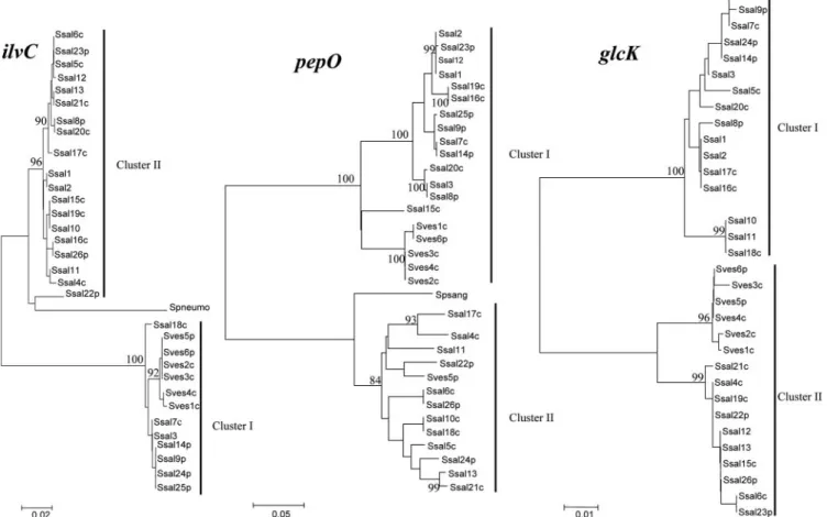
ranged from less than 10% (4.9 to 8.1%) for five loci to more than 10% for three loci (11.9 to 43.5%; pepO, ilvC, and glcK) (Table 3). In the same way, in S. vestibularis the maximal level of divergence of nucleotide sequences was 38.2% for the pepO locus (Table 3). Interestingly, the pepO, ilvC, and glcK trees were each divided into two clusters strongly supported by a bootstrap value of 100% (Fig. 4). None of the clusters corre-sponded to known features, such as species or origin (com-mensal versus pathogen or country). For each locus, the aver-age levels of divergence were similar within the cluster, whereas they were three- to fourfold higher between the
clus-FIG. 3. Split decomposition analysis of alleles present in 27 S. salivarius strains: split graphs for thrS, pyrE, and ddlA alleles. In some graphs several alleles are connected to each other by multiple pathways, forming an interconnected network and suggesting that there were recombination events. The numbers are allele numbers. Split graphs for ilvC, pepO, glcK, dnaE, and sodA alleles are shown in Fig. S2 in the supplemental material.
ters (Table 4). Interestingly, the pepO cluster II sequences were more closely related to the Streptococcus parasanguinis pepO se-quence (GenBank accession no. AF116532) than to cluster I sequences (Fig. 4). In the same way, ilvC cluster II sequences were more closely related to S. thermophilus (GenBank accession no. AE008420 and AE007356) and S. pneumoniae (GenBank acces-sion no. AF220670) ilvC sequences than to cluster I sequences (Fig. 4).
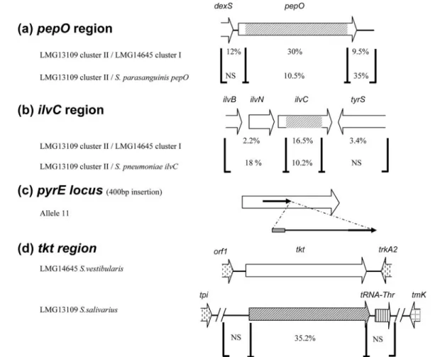
The levels of divergence for pepO and ilvC regions in strains having alleles that belong to different clusters are summarized in Fig. 5. The 1,783-bp cluster II pepO sequence exhibited 10.5% divergence from S. parasanguinis pepO, compared with 30% divergence from the cluster I pepO allele (Fig. 5a).
Sim-ilarly, the 725-bp cluster II ilvC sequence exhibited 5.1% and 10.2% divergence from S. thermophilus and S. pneumoniae ilvC sequences, respectively, compared with 16.5% divergence from cluster I ilvC alleles (Fig. 5b). These results suggest that there was horizontal gene transfer (HGT) of the pepO and ilvC alleles from different streptococcal species. A similar process seems to take place at the glcK locus, as the alleles are split in two well-supported clusters. However, neither of the clusters appears to be closely related to known sequences (Table 4 and Fig. 4), suggesting that there was HGT from an uncharacter-ized streptococcal species.
Genomic features that distinguish S. salivarius and S.
ves-tibularis.The maximal level of nucleotide divergence was high (4.9 to 45%) at all loci in S. salivarius. In contrast, several S.
vestibularis loci (glcK, ilvC, tkt, sodA, and pyrE) diverged very
little (maximal divergence, ⬍0.9% [Table 3]). These differ-ences in the levels of allele divergence indicate that these loci evolved differently and might be used to develop rapid test to differentiate the two species.
Strikingly, the tkt loci were significantly different in S.
sali-varius and S. vestibularis as the S. salisali-varius gene could not be
amplified with several oligonucleotide pairs. Successful ampli-fication of an internal fragment was achieved only with oligo-nucleotides tkt-up6 and tkt-dn6, and the fragment was
se-FIG. 4. Phylogenetic relationships among 27 S. salivarius strains and 9 S. vestibularis strains. Trees are shown for glcK, ilvC, and pepO loci. Ssal, S. salivarius; Sves, S. vestibularis; p, pathogen; c, commensal; Spneumo, S. pneumoniae; Spsang, S. parasanguinis. The strain numbers correspond to ST numbers shown in Table 1. The trees were constructed by using the neighbor-joining method. Clusters and bootstrap values that areⱖ80% are indicated. The scale bars indicate the number of nucleotide substitutions per site.
TABLE 4. Genetic diversity of clusters at the glcK, ilvC, and pepO loci
Locus
No. of alleles Maximal % divergence Cluster I Cluster II Within
cluster I Within cluster II Between cluster I and cluster II glcK 7 9 3.9 2.6 11.9 ilvC 11 6 2.1 4.2 16 pepO 11 11 13.2 12.9 45.3
quenced. The level of divergence of the S. salivarius and S.
vestibularis tkt gene sequences was 35.2% (Fig. 5d).
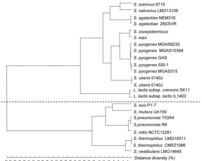
Phyloge-netic analysis showed that two types of tkt genes are present in streptococci (Fig. 6). The tkt gene of S. salivarius belongs to the first group (highest level of identity with Streptococcus pyogenes and Streptococcus agalactiae genes, 78%), whereas the tkt gene of S. vestibularis belongs to the second group (highest level of identity with S. thermophilus gene, 98%). Further amplification by inverse PCR and sequencing of the flanking regions showed that the genetic organization in the tkt region was different in the two species (Fig. 5d). The S. vestibularis tkt gene is flanked by an ABC transporter gene (highest level of homology to str0310 from S. thermophilus) and trkA2 (encoding potassium uptake protein); a similar organization occurs in S.
thermophi-lus. By contrast, the S. salivarius tkt gene is followed by a
tRNAThrgene and is situated in a region flanked by tpi
(en-coding triose phosphate isomerase) and tmk (thymidylate ki-nase), which are adjacent in the S. thermophilus genome (data not shown). PCR experiments showed that the genetic organi-zation of the tkt region is typical for each species (S. salivarius
and S. vestibularis) (data not shown). The trkA2 and tpi loci are located 170 kb apart in the S. thermophilus genome, indicating that a major rearrangement occurred during differentiation of the S. vestibularis and S. salivarius loci.
DISCUSSION
In the present study, we carried out a comparative sequence analysis of several loci to explore the population structure of
S. salivarius and S. vestibularis. Due to the small number of S. vestibularis isolates, an MLST scheme was obtained only for S. salivarius. The analysis allowed us (i) to investigate the genetic
diversity within S. salivarius and S. vestibularis isolates and the relationships between them, (ii) to evaluate the relationships of commensal and pathogen strains, and (iii) to characterize extensive HGT in the two populations.
MLST of S. salivarius.As a first step in developing an MLST method, the internal fragments of eight loci (dnaE, thrS, pyrE,
ilvC, pepO, ddlA, sodA, and glcK) were amplified from all
strains tested. One locus, tkt, could not be amplified from S.
FIG. 5. HGT events detected in S. salivarius pepO, ilvC, pyrE, and tkt genes. Regions where there are high and low levels of diversity are indicated by brackets, and the level of diversity is shown for each region. NS, no significant homology. (a) In the pepO region, 2,095-bp nucleotide sequences of LMG13109 cluster II and LMG14645 cluster I were compared. The regions were compared with the S. parasanguinis pepO region. (b) In the ilvC region, 2,765-bp nucleotide sequences of LMG13109 cluster II and LMG14645 cluster I were compared. The regions were compared with the S. pneumoniae ilvC region. (c) In allele 11 of the pyrE locus, a 234-bp DNA insertion containing 30 bp of S. salivarius IS1139 (gray box) and 204 bp of DNA whose function and origin are unknown is present and is flanked by a 157-bp duplication (arrows). (d) In the tkt region, 2,506-bp nucleotide sequences of S. salivarius LMG13109 and S. vestibularis LMG14645 were compared. orf1 exhibits the highest level of homology to str0310 from S. thermophilus.
salivarius strains. The eight loci were polymorphic, and the
maximal levels of divergence of three genes, ilvC, pepO, and
glcK, were more than 12%. These high levels of divergence,
which could have resulted from interspecific HGT events (see below), prevented use of these three loci in an MLST scheme for S. salivarius.
An analysis of alleles and STs showed that the diversity in S.
salivarius is very high. The variability is comparable to the
maximal sequence divergence (5 to 32%) and the mean nucle-otide diversity (16.6%) reported for group C and G strepto-cocci and for S. oralis, respectively (33, 55). Like S. salivarius, these streptococci are considered to be commensals, even though they are sometimes associated with human disease. The level of S. salivarius diversity is significantly higher than the levels of diversity of group A streptococci (maximal diver-gence, 1.4 to 6.1% [17]) and group B streptococci (1.2 to 2.5% variable nucleotide sites [32]). We found that S. salivarius iso-lates from the same individual exhibit a high degree of genetic diversity, as observed previously for several other mucosal streptococci (S. mitis, S. mutans, and S. oralis) (19, 38, 57). The phylogenetic tree of concatenated sequences of S. salivarius did not allow us to cluster strains isolated from human blood specifically. We concluded that commensal and potentially
pathogenic strains do not belong to distinct S. salivarius pop-ulations. The lack of clear separation of commensal and clin-ical S. salivarius isolates resembles the findings reported for several other commensal streptococci, such as group C and G streptococci (S. mitis and S. oralis), which also exhibit oppor-tunistic pathogenicity (33, 55).
Genetic relationship of S. salivarius and S. vestibularis isolates.
S. salivarius and S. vestibularis are included in the salivarius
group of viridans streptococci (18). In 1988, S. vestibularis was described as a new oral species (56), while further analysis showed that it is very closely related to S. salivarius (34, 46). In this work, we found that the levels of relatedness between S.
vestibularis and S. salivarius are very different at different
ge-netic loci.
Two findings support the notion that S. salivarius and S.
vestibularis should be considered separate species: (i) the
strongly supported clusters of S. vestibularis alleles at the glcK,
pyrE, dnaE, sodA, and ilvC loci and (ii) the extent of genomic
divergence in particular loci, such as the divergence ob-served for tkt. Incidentally, the great differences in the se-quence and genetic organization of the tkt locus could prob-ably be used to develop a rapid test for discriminating these two bacteria.
FIG. 6. Additive distance tree for tkt genes from streptococci and lactococci. Alignment was performed with ClustalW, and the tree was constructed by the flexible method. The levels of identity between the two clusters range from 54% to 58%, whereas the levels of identity are more than 73% for the genes within each cluster.
By contrast, the lack of resolution of S. vestibularis and S.
salivarius at three loci (ddlA, pepO, and thrS) suggests that
there is frequent gene exchange between these two species. Considering the mosaic structure of these three S. vestibularis genes and the low levels of divergence found at five other loci (glcK, ilvC, pyrE, sodA, and tkt), we suggest that S. vestibularis may be a recently emerging population which evolves by inter-specific gene exchange. However, analysis of a larger sample of
S. vestibularis strains is necessary to test this hypothesis more
stringently and to better define the evolutionary scheme of S.
vestibularis.
Intraspecific recombination in S. salivarius and S.
vestibu-laris populations.Analysis of different loci of S. salivarius and
S. vestibularis indicated that the loci have different evolutionary
histories. Extensive intraspecific recombinational exchanges should have occurred to explain the features observed.
The noncongruence between allele phylogenetic trees and the low index of association of MLST alleles can be explained by frequent recombination events. The extent of the conflicting phylogenetic results is illustrated by the fact that a strain which has a certain position in one allele tree can occupy a very different position in another allele tree, with both positions strongly supported by bootstrap analysis. The conflicting phy-logenetic signals found for seven S. salivarius loci in split de-composition analysis, coupled with a low association index value (IS
A, 0.057), indicate that intraspecific recombination occurs frequently in S. salivarius and plays a major role in generating sequence diversity between strains. A specific gene transfer event is revealed by the presence of pyrE allele 11 in two strains isolated from human blood 5 years apart (CIP104994 isolated in 1996 and CCHSS4 isolated in 2001). This allele contains a specific DNA insertion consisting of 30 bp of S. salivarius insertion sequence IS1139 (40), followed by 204 bp of DNA whose origin is unknown (Fig. 5c). The events required to generate this allele were unlikely to occur inde-pendently twice, suggesting that the allele was exchanged hor-izontally. In S. vestibularis, split decomposition analysis sug-gested that intragenic recombination occurred at four of the nine loci (not shown). Taken together, these results support the conclusion that intraspecific recombination occurs exten-sively in the S. salivarius population and probably in S.
vestibu-laris.
HGT between oral streptococci. The presence of longer branches in ilvC, glcK, and pepO S. salivarius split graphs (see Fig. S2 in the supplemental material) are consistent with the importation of divergent genes from other species (27). This hypothesis is supported by the higher levels of homology of cluster II alleles of ilvC and pepO loci with S.
pneumoniae and S. parasanguinis alleles, respectively, than
with the other S. salivarius allele clusters. S. pneumoniae and
S. parasanguinis are oral streptococci which might come in
contact with S. salivarius and S. vestibularis in the buccal cavity. Evaluation of the extent of HGT in ilvC and pepO genes by sequence analysis indicated that exchange of 725-and 1,783-bp internal gene fragments, respectively, took place (Fig. 5a and b). Differences in chromosomal localiza-tion and high levels of nucleotide divergence between tkt genes from S. salivarius and S. vestibularis are also consistent with an HGT event. Detailed cluster analysis of these se-quences and the presence of multiple variants in clusters
suggest that the transfers are ancient. For example, the transfer of the ilvC divergent allele, possibly from the mitis group, likely preceded the emergence of the food species S.
thermophilus in the salivarius group (4, 28). Notably, alleles
of each cluster are still maintained in the population. The extent of recombination and HGT in S. salivarius and S.
vestibularis populations suggests that these bacteria have
effi-cient mechanisms for gene acquisition and recombination, al-though they are not known to be naturally competent (6). Natural transformation has been described for S. pneumoniae and several viridans streptococci, such as S. mutans and
Strep-tococcus sanguinis (1, 20, 44), and plays an important role in
generating the high levels of genotypic diversity in these spe-cies. Recombinational events are responsible for the dissemi-nation of genes encoding virulence factors, such as antibiotic resistance markers, quinolone resistance, immunoglobulin A1 protease, and competence-stimulating peptide (5, 21, 25, 45, 54). Altered penicillin binding protein genes are directly in-volved in penicillin resistance, and DNA sequences closely related to them appear to have been distributed horizontally between S. pneumoniae and viridans streptococci (12, 13, 22). Gene flow from human pathogens to commensal streptococci, described previously for groups A, C, and G (33, 50) and for the oral mitis group (55) and also observed here, may be involved in the evolutionary model of the commensal strepto-cocci, which can be associated with bacterial infections. Re-cently, phylogenetic analyses of more than 20 streptococcal species revealed several examples of interspecific homologous recombination in housekeeping genes of viridans streptococci (29). Evidence for HGT and recombination events in this study associated S. salivarius and S. vestibularis with this group of oral streptococci, which have great potential to be receptors of foreign genes.
The absence of clustering of S. salivarius strains isolated from blood suggests that these strains are opportunistic patho-gens. However, the high potential of S. salivarius and S.
ves-tibularis for acquisition of new alleles at loci involved in general
functioning of the cell and the possible acquisition of antibiotic resistance determinants (23, 51) raise the possibility that strains isolated from human infections acquired DNA that promoted their pathogenic behavior. Further studies are re-quired to examine this possibility.
ACKNOWLEDGMENTS
We thank Per S. J. Saris and Christina Jerusalem for providing S. salivarius and S. vestibularis strains.
REFERENCES
1. Avery, O. T., C. M. MacLeod, and M. McCarty. 1995. Studies on the chem-ical nature of the substance inducing transformation of pneumococcal types. Induction of transformation by a desoxyribonucleic acid fraction isolated from Pneumococcus type III. 1944. Mol. Med. 1:344–365.
2. Becker, M. R., B. J. Paster, E. J. Leys, M. L. Moeschberger, S. G. Kenyon,
J. L. Galvin, S. K. Boches, F. E. Dewhirst, and A. L. Griffen.2002. Molecular analysis of bacterial species associated with childhood caries. J. Clin. Micro-biol. 40:1001–1009.
3. Bisharat, N., D. W. Crook, J. Leigh, R. M. Harding, P. N. Ward, T. J. Coffey,
M. C. Maiden, T. Peto, and N. Jones.2004. Hyperinvasive neonatal group B
Streptococcus has arisen from a bovine ancestor. J. Clin. Microbiol. 42:2161–
2167.
4. Bolotin, A., B. Quinquis, P. Renault, A. Sorokin, S. D. Ehrlich, S. Kulakauskas,
A. Lapidus, E. Goltsman, M. Mazur, G. D. Pusch, M. Fonstein, R. Overbeek, N. Kyprides, B. Purnelle, D. Prozzi, K. Ngui, D. Masuy, F. Hancy, S. Burteau, M. Boutry, J. Delcour, A. Goffeau, and P. Hols.2004. Complete sequence
and comparative genome analysis of the dairy bacterium Streptococcus
ther-mophilus. Nat. Biotechnol. 22:1554–1558.
5. Bracco, R. M., M. R. Krauss, A. S. Roe, and C. M. Macleod. 1957. Trans-formation reactions between Pneumococcus and three strains of Strepto-cocci. J. Exp. Med. 106:247–259.
6. Buckley, N. D., C. Vadeboncoeur, D. J. LeBlanc, L. N. Lee, and M. Frenette. 1999. An effective strategy, applicable to Streptococcus salivarius and related bacteria, to enhance or confer electroporation competence. Appl. Environ. Microbiol. 65:3800–3804.
7. Caufield, P. W., and T. M. Walker. 1989. Genetic diversity within
Strepto-coccus mutans evident from chromosomal DNA restriction fragment
poly-morphisms. J. Clin. Microbiol. 27:274–278.
8. Coffey, T. J., G. D. Pullinger, R. Urwin, K. A. Jolley, S. M. Wilson, M. C.
Maiden, and J. A. Leigh.2006. First insights into the evolution of
Strepto-coccus uberis: a multilocus sequence typing scheme that enables investigation
of its population biology. Appl. Environ. Microbiol. 72:1420–1428. 9. Conte, A., P. Chinello, R. Civljak, A. Bellussi, P. Noto, and N. Petrosillo.
2005. Streptococcus salivarius meningitis and sphenoid sinus mucocele. Case report and literature review. J. Infect. 52:27–30.
10. Coykendall, A. L. 1989. Classification and identification of the viridans strep-tococci. Clin. Microbiol. Rev. 2:315–328.
11. Cunliffe, N. A., and A. J. Jacob. 1997. Streptococcus vestibularis bacteraemia. J. Infect. 34:85.
12. Dowson, C. G., T. J. Coffey, C. Kell, and R. A. Whiley. 1993. Evolution of penicillin resistance in Streptococcus pneumoniae; the role of Streptococcus
mitis in the formation of a low affinity PBP2B in S. pneumoniae. Mol.
Microbiol. 9:635–643.
13. Dowson, C. G., A. Hutchison, N. Woodford, A. P. Johnson, R. C. George, and
B. G. Spratt.1990. Penicillin-resistant viridans streptococci have obtained altered penicillin-binding protein genes from penicillin-resistant strains of
Streptococcus pneumoniae. Proc. Natl. Acad. Sci. USA 87:5858–5862.
14. Doyuk, E., O. J. Ormerod, and I. C. Bowler. 2002. Native valve endocarditis due to Streptococcus vestibularis and Streptococcus oralis. J. Infect. 45:39–41. 15. Enright, M. C., and B. G. Spratt. 1999. Multilocus sequence typing. Trends
Microbiol. 7:482–487.
16. Enright, M. C., and B. G. Spratt. 1998. A multilocus sequence typing scheme for Streptococcus pneumoniae: identification of clones associated with serious invasive disease. Microbiology 144:3049–3060.
17. Enright, M. C., B. G. Spratt, A. Kalia, J. H. Cross, and D. E. Bessen. 2001. Multilocus sequence typing of Streptococcus pyogenes and the relationships between emm type and clone. Infect. Immun. 69:2416–2427.
18. Facklam, R. 2002. What happened to the streptococci: overview of taxo-nomic and nomenclature changes. Clin. Microbiol. Rev. 15:613–630. 19. Fitzsimmons, S., M. Evans, C. Pearce, M. J. Sheridan, R. Wientzen, G.
Bowden, and M. F. Cole.1996. Clonal diversity of Streptococcus mitis biovar 1 isolates from the oral cavity of human neonates. Clin. Diagn. Lab. Immu-nol. 3:517–522.
20. Gaustad, P. 1979. Genetic transformation in Streptococcus sanguis. Distri-bution of competence and competence factors in a collection of strains. Acta Pathol. Microbiol. Scand. Sect. B 87:123–128.
21. Gonzalez, I., M. Georgiou, F. Alcaide, D. Balas, J. Linares, and A. G. de la
Campa.1998. Fluoroquinolone resistance mutations in the parC, parE, and
gyrA genes of clinical isolates of viridans group streptococci. Antimicrob.
Agents Chemother. 42:2792–2798.
22. Hakenbeck, R., A. Konig, I. Kern, M. van der Linden, W. Keck, D.
Billot-Klein, R. Legrand, B. Schoot, and L. Gutmann.1998. Acquisition of five high-Mrpenicillin-binding protein variants during transfer of high-level
beta-lactam resistance from Streptococcus mitis to Streptococcus pneumoniae. J. Bacteriol. 180:1831–1840.
23. Hartley, D. L., K. R. Jones, J. A. Tobian, D. J. LeBlanc, and F. L. Macrina. 1984. Disseminated tetracycline resistance in oral streptococci: implication of a conjugative transposon. Infect. Immun. 45:13–17.
24. Haubold, B., and R. R. Hudson. 2000. LIAN 3.0: detecting linkage disequi-librium in multilocus data. Linkage analysis. Bioinformatics 16:847–848. 25. Havarstein, L. S., R. Hakenbeck, and P. Gaustad. 1997. Natural
compe-tence in the genus Streptococcus: evidence that streptococci can change pherotype by interspecies recombinational exchanges. J. Bacteriol. 179: 6589–6594.
26. Hohwy, J., J. Reinholdt, and M. Kilian. 2001. Population dynamics of
Strep-tococcus mitis in its natural habitat. Infect. Immun. 69:6055–6063.
27. Holmes, E. C., R. Urwin, and M. C. Maiden. 1999. The influence of recom-bination on the population structure and evolution of the human pathogen
Neisseria meningitidis. Mol. Biol. Evol. 16:741–749.
28. Hols, P., F. Hancy, L. Fontaine, B. Grossiord, D. Prozzi, N.
Leblond-Bour-get, B. Decaris, A. Bolotin, C. Delorme, S. Dusko Ehrlich, E. Guedon, V. Monnet, P. Renault, and M. Kleerebezem.2005. New insights in the molec-ular biology and physiology of Streptococcus thermophilus revealed by com-parative genomics. FEMS Microbiol. Rev. 29:435–463.
29. Hoshino, T., T. Fujiwara, and M. Kilian. 2005. Use of phylogenetic and phenotypic analyses to identify nonhemolytic streptococci isolated from bac-teremic patients. J. Clin. Microbiol. 43:6073–6085.
30. Huson, D. H. 1998. SplitsTree: analyzing and visualizing evolutionary data. Bioinformatics 14:68–73.
31. Idigoras, P., A. Valiente, L. Iglesias, P. Trieu-Cout, and C. Poyart. 2001. Meningitis due to Streptococcus salivarius. J. Clin. Microbiol. 39:3017. 32. Jones, N., J. F. Bohnsack, S. Takahashi, K. A. Oliver, M. S. Chan, F. Kunst,
P. Glaser, C. Rusniok, D. W. Crook, R. M. Harding, N. Bisharat, and B. G. Spratt.2003. Multilocus sequence typing system for group B streptococcus. J. Clin. Microbiol. 41:2530–2536.
33. Kalia, A., M. C. Enright, B. G. Spratt, and D. E. Bessen. 2001. Directional gene movement from human-pathogenic to commensal-like streptococci. Infect. Immun. 69:4858–4869.
34. Kawamura, Y., X. G. Hou, F. Sultana, H. Miura, and T. Ezaki. 1995. De-termination of 16S rRNA sequences of Streptococcus mitis and Streptococcus
gordonii and phylogenetic relationships among members of the genus Strep-tococcus. Int. J. Syst. Bacteriol. 45:406–408.
35. Kazor, C. E., P. M. Mitchell, A. M. Lee, L. N. Stokes, W. J. Loesche, F. E.
Dewhirst, and B. J. Paster.2003. Diversity of bacterial populations on the tongue dorsa of patients with halitosis and healthy patients. J. Clin. Micro-biol. 41:558–563.
36. Kennedy, H. F., D. Morrison, D. Tomlinson, B. E. Gibson, J. Bagg, and C. G.
Gemmell.2003. Gingivitis and toothbrushes: potential roles in viridans strep-tococcal bacteraemia. J. Infect. 46:67–70.
37. King, S. J., J. A. Leigh, P. J. Heath, I. Luque, C. Tarradas, C. G. Dowson,
and A. M. Whatmore.2002. Development of a multilocus sequence typing scheme for the pig pathogen Streptococcus suis: identification of virulent clones and potential capsular serotype exchange. J. Clin. Microbiol. 40:3671– 3680.
38. Kulkarni, G. V., K. H. Chan, and H. J. Sandham. 1989. An investigation into the use of restriction endonuclease analysis for the study of transmission of mutans streptococci. J. Dent. Res. 68:1155–1161.
39. Kumar, S., K. Tamura, I. B. Jakobsen, and M. Nei. 2001. MEGA2: molec-ular evolutionary genetics analysis software. Bioinformatics 17:1244–1245. 40. Lortie, L. A., G. Gagnon, and M. Frenette. 1994. IS1139 from Streptococcus
salivarius: identification and characterization of an insertion sequence-like
element related to mobile DNA elements from gram-negative bacteria. Plas-mid 32:1–9.
41. Nei, M., and T. Gojobori. 1986. Simple methods for estimating the numbers of synonymous and nonsynonymous nucleotide substitutions. Mol. Biol. Evol. 3:418–426.
42. Partridge, S. M. 2000. Prosthetic valve endocarditis due to Streptococcus
vestibularis. J. Infect. 41:284–285.
43. Pearce, C., G. H. Bowden, M. Evans, S. P. Fitzsimmons, J. Johnson, M. J.
Sheridan, R. Wientzen, and M. F. Cole.1995. Identification of pioneer viridans streptococci in the oral cavity of human neonates. J. Med. Microbiol.
42:67–72.
44. Perry, D., and H. K. Kuramitsu. 1981. Genetic transformation of
Strepto-coccus mutans. Infect. Immun. 32:1295–1297.
45. Poulsen, K., J. Reinholdt, C. Jespersgaard, K. Boye, T. A. Brown, M. Hauge,
and M. Kilian.1998. A comprehensive genetic study of streptococcal immu-noglobulin A1 proteases: evidence for recombination within and between species. Infect. Immun. 66:181–190.
46. Poyart, C., G. Quesne, S. Coulon, P. Berche, and P. Trieu-Cuot. 1998. Identification of streptococci to species level by sequencing the gene encod-ing the manganese-dependent superoxide dismutase. J. Clin. Microbiol. 36: 41–47.
47. Ruoff, K. L., S. I. Miller, C. V. Garner, M. J. Ferraro, and S. B. Calderwood. 1989. Bacteremia with Streptococcus bovis and Streptococcus salivarius: clin-ical correlates of more accurate identification of isolates. J. Clin. Microbiol.
27:305–308.
48. Saitou, N., and M. Nei. 1987. The neighbor-joining method: a new method for reconstructing phylogenetic trees. Mol. Biol. Evol. 4:406–425. 49. Smith, J. M., N. H. Smith, M. O’Rourke, and B. G. Spratt. 1993. How clonal
are bacteria? Proc. Natl. Acad. Sci. USA 90:4384–4388.
50. Sriprakash, K. S., and J. Hartas. 1996. Lateral genetic transfers between group A and G streptococci for M-like genes are ongoing. Microb. Pathog.
20:275–285.
51. Stadler, C., and M. Teuber. 2002. The macrolide efflux genetic assembly of
Streptococcus pneumoniae is present in erythromycin-resistant Streptococcus salivarius. Antimicrob. Agents Chemother. 46:3690–3691.
52. Suerbaum, S., M. Lohrengel, A. Sonnevend, F. Ruberg, and M. Kist. 2001. Allelic diversity and recombination in Campylobacter jejuni. J. Bacteriol.
183:2553–2559.
53. Tunkel, A. R., and K. A. Sepkowitz. 2002. Infections caused by viridans streptococci in patients with neutropenia. Clin. Infect. Dis. 34:1524–1529. 54. Whatmore, A. M., V. A. Barcus, and C. G. Dowson. 1999. Genetic diversity
of the streptococcal competence (com) gene locus. J. Bacteriol. 181:3144– 3154.
55. Whatmore, A. M., A. Efstratiou, A. P. Pickerill, K. Broughton, G. Woodard,
D. Sturgeon, R. George, and C. G. Dowson.2000. Genetic relationships between clinical isolates of Streptococcus pneumoniae, Streptococcus oralis, and Streptococcus mitis: characterization of “atypical” pneumococci and
or-ganisms allied to S. mitis harboring S. pneumoniae virulence factor-encoding genes. Infect. Immun. 68:1374–1382.
56. Whiley, R. A., and J. M. Hardie. 1988. Streptococcus vestibularis sp. nov. from the human oral cavity. Int. J. Syst. Bacteriol. 38:335–339.
57. Wisplinghoff, H., R. R. Reinert, O. Cornely, and H. Seifert. 1999. Molecular relationships and antimicrobial susceptibilities of viridans group streptococci
isolated from blood of neutropenic cancer patients. J. Clin. Microbiol. 37: 1876–1880.
58. Zadoks, R. N., Y. H. Schukken, and M. Wiedmann. 2005. Multilocus se-quence typing of Streptococcus uberis provides sensitive and epidemiologi-cally relevant subtype information and reveals positive selection in the vir-ulence gene pauA. J. Clin. Microbiol. 43:2407–2417.