HAL Id: hal-01480854
https://hal-univ-rennes1.archives-ouvertes.fr/hal-01480854
Submitted on 1 Mar 2017HAL is a multi-disciplinary open access archive for the deposit and dissemination of sci-entific research documents, whether they are pub-lished or not. The documents may come from teaching and research institutions in France or abroad, or from public or private research centers.
L’archive ouverte pluridisciplinaire HAL, est destinée au dépôt et à la diffusion de documents scientifiques de niveau recherche, publiés ou non, émanant des établissements d’enseignement et de recherche français ou étrangers, des laboratoires publics ou privés.
Early expansion of circulating granulocytic
myeloid-derived suppressor cells predicts development of
nosocomial infections in septic patients
Fabrice Uhel, Imane Azzaoui, Murielle Grégoire, Céline Pangault, Joelle
Dulong, Jean-Marc Tadié, Arnaud Gacouin, Christophe Camus, Luc Cynober,
Thierry Fest, et al.
To cite this version:
Fabrice Uhel, Imane Azzaoui, Murielle Grégoire, Céline Pangault, Joelle Dulong, et al.. Early expan-sion of circulating granulocytic myeloid-derived suppressor cells predicts development of nosocomial infections in septic patients. American Journal of Respiratory and Critical Care Medicine, American Thoracic Society, 2017, 196 (3), pp.315-327. �10.1164/rccm.201606-1143OC�. �hal-01480854�
Early expansion of circulating granulocytic
myeloid-derived suppressor cells predicts development of
nosocomial infections in septic patients.
Fabrice Uhel, MD, PhD1,2,3, Imane Azzaoui, PhD3,4,5,6, Murielle Grégoire, PhD3,4,5,6, Céline Pangault, PharmD, PhD3,4,5,6, Joelle Dulong, MBiol3,4,5,6, Jean-Marc Tadié, MD, PhD1,2,3,5, Arnaud Gacouin, MD1,2, Christophe Camus, MD, PhD1,2, Luc Cynober, PharmD, PhD7, Thierry Fest, MD, PhD3,4,5,6, Yves Le Tulzo, MD, PhD1,2,3,5, Mikael Roussel, MD, PhD3,4,5,6 and Karin Tarte, PharmD, PhD3,4,5,6
1 CHU Rennes, Maladies Infectieuses et Réanimation Médicale, F-35033 Rennes,
France
2 Inserm, CIC-1414, Faculté de Médecine, Université Rennes 1, F-35043 Rennes,
France.
3
Inserm, U917, Faculté de Médecine, Université Rennes 1, F-35043 Rennes, France
4 CHU Rennes, Pôle Biologie, F-35033 Rennes, France 5
Université Rennes 1, UMR917, F-35043 Rennes, France
6 EFS Bretagne, F-35016 Rennes, France 7
AP-HP Hôpital Cochin, Service de Biochimie, and Dept Nutrition EA 4466, Faculté de Pharmacie Paris Descartes F-75014 Paris, France
Corresponding author
Fabrice Uhel, Service des Maladies Infectieuses et Réanimation Médicale, CHU Pontchaillou, 2 rue Henri Le Guilloux, F-35033 Rennes Cedex 9, France; e-mail:
fabrice.uhel@chu-rennes.fr; Phone: +33(0) 299 284 248; Fax: +33(0) 299 284 164. Karin Tarte, INSERM U917, Faculté de Médecine, 2 Avenue du Pr Léon Bernard, F-35043 Rennes France, E-mail: karin.tarte@univ-rennes1.fr, Phone: +33 (0) 223 234 512, fax: +33 (0) 223 234 958.
Authors' contribution
Raised funds: FU, KT, TF. Designed and supervised research: KT, MR, TF, YLT. Designed experiments: FU, IA, JD, CP. Performed experiments: FU, IA, MG, LC. Provided samples: FU, JD, JMT, AG, CC, YLT. Analyzed data: FU, IA, JD, CP, JMT, AG, CC, MR, KT. Wrote the paper: FU, KT. Critically revised the manuscript: all authors.
Grant support
This work was supported by research grants from the French Intensive Care Society (SRLF) and the National Institute of Cancer (INCa Recherche Translationnelle 2010).
Descriptor numbers
4.12 – 7.08 – 7.19 – 10.07
Running title
At a Glance Commentary
Scientific Knowledge on the Subject: Sepsis is characterized by a persistent immune
dysfunction responsible for nosocomial infections and poor outcome. The role of myeloid-derived suppressor cells (MDSCs), described as potent inhibitors of immune responses in cancer and inflammatory conditions, still needs to be clarified in septic patients.
What This Study Adds to the Field: We demonstrate that monocytic (M)-MDSCs are
expanded in ICU septic and non-septic patients and that granulocytic (G)-MDSCs are more specifically expanded in septic patients. Both subsets independently inhibit T-cell proliferation. G-MDSCs are made of immature and mature granulocytes expressing high levels of degranulation markers and produce arginase 1. Importantly, early expansion of G-MDSCs, unlike M-MDSCs, is associated with subsequent occurrence of nosocomial infections, suggesting a major role for those cells in sepsis-induced immune suppression. MDSCs may thus become interesting therapeutic targets to restore immune capacities of septic patients.
(130 words)
This article has an online data supplement, which is accessible from this issue's table of content online at www.atsjournals.org
ABSTRACT
Rationale: Sepsis induces a sustained immune dysfunction responsible for poor
outcome and nosocomial infections. Myeloid-derived suppressor cells (MDSCs) described in cancer and inflammatory processes may be involved in sepsis-induced immune suppression but their clinical impact remains poorly defined.
Objectives: To clarify phenotype, suppressive activity, origin, and clinical impact of
MDSCs in septic patients.
Methods: Peripheral blood transcriptomic analysis was performed on 29 septic
patients and 15 healthy donors. A second cohort of 94 consecutive septic patients, 11 severity-matched intensive care patients and 67 healthy donors was prospectively enrolled for flow cytometry and functional experiments.
Measurements and Main Results: Genes involved in MDSC suppressive functions,
including S100A12, S100A9, MMP8 and ARG1, were upregulated in the peripheral blood of septic patients. CD14posHLA-DRlow/neg monocytic (M)-MDSCs were expanded in intensive care septic and non-septic patients and CD14negCD15pos low-density granulocytes/ granulocytic (G)-MDSCs were more specifically expanded in septic patients (p<.001). Plasma levels of MDSC mediators S100A8/A9, S100A12, and Arginase 1 were significantly increased. In vitro, CD14pos- and CD15pos-cell depletion increased T-cell proliferation in septic patients. G-MDSCs, made of immature and mature granulocytes expressing high levels of degranulation markers, were specifically responsible for arginase 1 activity. High initial levels of G-MDSCs, arginase 1, and S100A12 but not M-MDSCs were associated with subsequent occurrence of nosocomial infections.
Conclusions: M-MDSCs and G-MDSCs strongly contribute to T-cell dysfunction in
septic patients. More specifically, G-MDSCs producing arginase 1 are associated with a higher incidence of nosocomial infections and appear to be major actors of sepsis-induced immune suppression.
Abstract word count: 247
INTRODUCTION
Sepsis is one of the leading causes of admission in intensive care units (ICUs). Despite a decreased overall mortality rate during the past three decades, the prognosis remains hampered by a high long term mortality secondary to nosocomial infections with opportunistic pathogens or viral reactivations, an increased risk of cardiovascular events and cancers, and frequent hospital readmissions causing increased healthcare costs (1-4). This unfavorable outcome has been largely attributed to a persistent immunological impairment affecting both innate and adaptive immunity (5). To date, the most described features are i) monocyte dysfunction illustrated by reduced expression of HLA-DR and impaired production of inflammatory cytokines after bacterial challenge in vitro (6, 7), ii) granulocyte functional impairment (8), iii) lymphopenia and lymphocyte dysfunction associated with an increased proportion of regulatory T-cells (Tregs) (9, 10) and iv) enhanced systemic indoleamine 2,3-dioxygenase (IDO) activity (11).
During the past decade, much attention has focused on a heterogeneous population of cells from the myeloid lineage called myeloid-derived suppressor cells (MDSCs) which have been demonstrated to play potent immunosuppressive functions in cancers and inflammatory diseases (12, 13). In cancer, they are induced by many soluble factors (such as S100A8/A9, GM-CSF, G-CSF, IL-6, IL-10, VEGF or TGF-ß) as a consequence of a maturational block and/or dysregulated myelopoiesis. Various mechanisms have been proposed to explain their suppressive activity, including depletion of arginine and tryptophan by arginase 1 and IDO, respectively; production of reactive oxygen species (ROS) by NADPH oxidase; release of IL-10 or
TGF-ß; and induction of Tregs (14-17). Two main subsets, monocytic (M)-MDSCs and granulocytic (G)-MDSCs, have been identified, but their definition and role in humans remain elusive due to the lack of specific phenotypic markers (18).
CD14posHLA-DRlow/neg cells described more than 15 years ago as TLR-unresponsive cells in septic patients were recently reconsidered as M-MDSCs (19) but were not evaluated for their suppressive functions. Conversely, G-MDSCs with various phenotypes, including CD15pos cells with decreased CD10 and CD16 expression, LinnegCD33posCD15posHLA-DRneg cells, or CD15pos low-density granulocytes (LDGs) that co-purify with peripheral blood mononuclear cells (PBMCs) after density gradient centrifugation, were shown to be expanded in septic and in ICU non-septic patients, and to display T-cell inhibitory activity (20-22). Especially, CD15pos LDGs were recently proposed to variably display either arginase or ROS-dependent inhibitory effects with a potential impact of causative pathogens (19, 20). However, the low number of patients studied and the controversial phenotype and origin of putative suppressor cells preclude any definitive conclusion on the clinical impact, mechanisms of action, and role of MDSCs in septic patients.
Consequently, our objectives were i) to highlight the various putative MDSC subsets within the circulating myeloid compartment, ii) to characterize their suppressive activity, iii) to clarify their origins, and iv) to study their clinical impact in septic patients.
MATERIALS AND METHODS
Patients and healthy donor participants
This study was performed in the ICU at Rennes University Hospital. 94 consecutive adult septic patients and 11 severity-matched ICU non-septic patients were prospectively enrolled, and compared with 67 healthy donors. The study design was approved by the local institutional review board and written informed consents were obtained. Pregnant women, patients younger than 18 years old, patients with malignancy, HIV infection, or receiving immunosuppressive agents were excluded. The standard criteria were used for diagnosis of severe sepsis and septic shock (23). The Simplified Acute Physiology Score (SAPS II), the Sepsis-related Organ Failure Assessment (SOFA) score and the Logistic Organ Dysfunction (LOD) system at admission in ICU were used to assess sepsis severity (24-26). Patients received standard enteral or parenteral nutrition. The occurrence of nosocomial infections during hospitalization was recorded prospectively and defined as previously described (27).
Samples
Blood heparinized samples were collected within 3 days following sepsis diagnosis. The delay between sampling and beginning of laboratory procedures was <1h. PBMCs were isolated by Ficoll density gradient, and plasma samples were stored at -80°C until use. Whole blood RNA samples collected on PAXGene Blood RNA tubes (PreAnalytiX) at the same time-point came from a previously published cohort of septic patients with similar inclusion/exclusion criteria (11) and were stored at -80°C before RNA extraction.
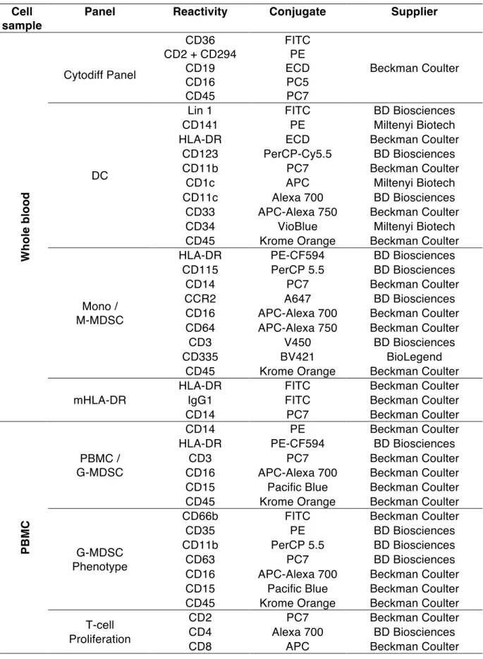
Flow cytometry
White blood cell differentials were obtained using the CytodiffTM panel (Beckman Coulter) (28). Myeloid subpopulations including MDSCs were then quantified on whole blood or PBMCs using multicolor antibody panels and Flow-count fluorospheres (Beckman Coulter) (Table E1). Samples were run on a Navios flow cytometer and data were analyzed using Kaluza software (Beckman Coulter). Gating strategy was defined as previously described (Figure E1) (29).
Quantitative real-time PCR
Quantitative RT-PCR were performed on whole blood RNA using Taqman array custom microfluidic cards (Applied Biosystems) and on purified granulocyte subsets using Fluidigm system. Methods are detailed in the online data supplement.
Cytokines and enzymes activity assessment
Plasma levels of Myeloperoxidase (MPO), neutrophil gelatinase-associated lipocalin (NGAL) (R&D Systems), S100A8/9, S100A12, and Arginase I (Hycult Biotech) were determined by ELISA. Plasma levels of G-CSF, IL-10, and IL-12 were measured using a Milliplex map magnetic bead kit (EMD Millipore). IDO and arginase activities were respectively evaluated by measuring kynurenine and tryptophan levels by high-performance liquid chromatography, and ornithine and arginine concentrations by ion-exchange chromatography (11, 30).
Cell isolation and culture
Proliferation of T cells from whole or CD14/CD15-depleted PBMCs was assessed by CFSE dilution. Details are provided in the online data supplement. For cytological analyses, cytospin slides were stained with May-Grünwald-Giemsa.
Statistical analysis
Statistical analyses were performed with GraphPad Prism 6.0 software. Comparisons between groups were performed using Mann-Whitney U test, Wilcoxon matched-pairs signed test for matched samples. Kruskall-Wallis or Friedman (matched samples) with Dunn's correction tests were performed if multiple comparisons were requested. Correlations between continuous variables were investigated using nonparametric Spearman rank correlation test. Unsupervised hierarchical clustering with Spearman’s rank distance and average linkage was performed using Cluster 3.0 (31). Results were displayed using Treeview (http://jtreeview.sourceforge.net). A receiver operating characteristic (ROC) plot was performed to determine the best G-MDSC threshold to discriminate between patients with high versus low risk of nosocomial infections. Kaplan-Meier curves were compared with the Mantel-Cox log-rank test. Gene set enrichment analysis (GSEA) was performed using the BROAD Institute GSEA software (http://www.broad.mit.edu/gsea/) on a previously published dataset of septic patients (GSE65682) using the panel of genes defined for the TLDA analyses.
RESULTS
Peripheral blood transcriptomic analysis reveals a myeloid suppressive signature in septic patients.
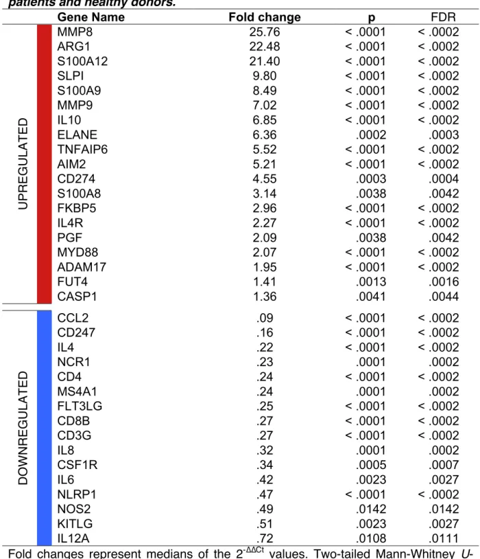
A transcriptomic analysis targeting 44 immune-related genes, including cell subset specific markers and functional pathways, was conducted on a previously published cohort of 29 septic patients and 15 healthy donors. Unsupervised hierarchical clustering analysis discriminated septic patients from healthy donors (Figure 1A). More importantly, genes associated with MDSC recruitment, phenotype, and suppressive functions, including MMP8, MMP9, ARG1, S100A8, S100A9,
S100A12, CD274 (PD-L1), IL4R, and IL10 were upregulated whereas genes
associated with adaptive immunity and inflammation, including CD4, MS4A1 (CD20),
CD8B, CD3G, IL8, and IL6 were downregulated in septic patients (Table 1). A gene
set enrichment analysis (GSEA) confirmed that upregulated genes were enriched in a large external cohort of septic patients (32) (Figure 1B). Of note, a hierarchical clustering analysis performed among septic patients could not find any segregation according to the severity of illness, i.e. septic shock versus severe sepsis (Figure
E2). ARG1 and S100A9 expression correlated with granulocyte count and inversely
correlated with lymphocyte count (Figure 1C). Noteworthy, even if both CD247 (CD3ζ) and CD3G (CD3γ) were downregulated, the CD247/CD3G ratio was further decreased in septic patients and inversely correlated with granulocyte count (Figure
1D). Those data raise the hypothesis that a suppressive activity burden by myeloid
Peripheral blood myeloid cells display a suppressive phenotype in septic patients.
To further study the potential role of myeloid cells in sepsis-induced immune dysfunction, we set up an independent prospective cohort of 94 septic patients and 67 healthy donors. The population characteristics are summarized in Table 2 and sepsis etiology is detailed in Table 3. As human MDSCs are heterogeneous and poorly defined, we started by performing an extensive description of the circulating myeloid compartment in a cohort of 36 septic patients and 26 healthy donors (Figure
2A). As previously described (11), the CD14pos monocyte count was increased in septic patients compared to healthy donors. Interestingly, this difference was essentially related to the CD14posCD16pos subtype (p<.001). Moreover, consistently with previous reports, the inflammatory CD14dimCD16pos subset was strongly decreased (p<.001) (33). As expected, HLA-DR expression in septic patients was decreased on all monocyte subtypes (p<.001) (Figures E3A & E3B). In agreement, the number of CD14posHLA-DRlow/neg monocytes, previously described as M-MDSCs, was markedly increased in septic patients (143.2 x10⁶/L [Interquartile range (IQR), 35.7-340.9 x10⁶/L]) compared to healthy donors (4.0 x10⁶/L [IQR, 2.0-17.5 x10⁶/L], p<.001). Besides an increase in circulating mature and immature granulocytes (p<.001), we observed in septic patients a high proportion of CD14negCD15pos LDGs (28.7% of PBMCs [IQR, 18.2-52.9%] in septic patients, vs. .8% [IQR, .31-2.1%] in healthy donors, p<.001), previously described as G-MDSCs (34). The absolute LDG count was also increased in septic patients. Interestingly, both proportion (rho=.35, p=.005) and absolute count (rho=.44, p<.001) of LDGs correlated with immature unlike mature granulocyte counts. The CD14posHLA-DRlow/neg count and the proportion of LDGs did not correlate. These subsets were not statistically different
between severe sepsis and septic shock patients (data not shown). Importantly, whereas M-MDSC were significantly increased in ICU non-septic patients (p<.001), raising a similar level than in septic patients, G-MDSCs increase was more specific to septic patients (Figure 2B) and persisted for at least 14 days (Figure E3C). Of note, whereas the proportion of LDGs did not differ according to the Gram staining of the causative microorganism, the number of circulating CD14posHLA-DRlow/neg monocytes was higher in patients with Gram-negative sepsis (Figure E3D). Lastly, the three dendritic cell (DC) subtype counts (plasmacytoid DC, and both subsets of myeloid DC expressing CD1c or CD141) were dramatically decreased in septic patients (Figure E4A).
To confirm and extend this work, we studied the characteristic features of MDSC activity at the protein level (Figure 2C). In agreement with their increased RNA levels in peripheral blood, concentrations of calcium-binding proteins S100A8/A9 and S100A12 involved in MDSC recruitment (35) were increased in the peripheral blood of septic patients (p<.001). Plasma level of G-CSF, which has been reported to take part in the recruitment of G-MDSCs (36), was also increased in septic patients (p=.001). The plasma kynurenine/tryptophan ratio reflecting IDO activity was markedly increased in patients with sepsis (.213 [IQR, .092-.658], +719%, p<.001) compared with healthy donors (.026 [IQR, .023-.035]). As previously described, this increase was not associated with an upregulation of IDO1 expression (data not shown)(11). Conversely, plasma levels of arginase 1 were increased in septic patients (119.3 ng/mL [IQR, 68.2-242.5 ng/mL], vs. 18.3 ng/mL [IQR, 9.6-23.4 ng/mL], p<.001), correlated with immature granulocyte count (rho=.28, p=.03) and
were associated with a decreased arginine concentration (35.3 µmol/L [IQR, 23.9-59.7 µmol/L] vs. 66.5 µmol/L [IQR, 52.1-95.1 µmol/L], p<.001). Finally, the increased IL10/IL12 ratio at protein and transcriptomic levels was consistent with an overall suppressive environment (Figure E4B), as highlighted by the significant decrease in the various lymphocyte subsets (Figure E4C).
Altogether, these data indicate a similar expansion of CD14posHLA-DRlow/neg monocytes in ICU septic and non-septic patients, and a more intense and specific increase of CD15posCD14neg LDGs in septic patients. Those subsets are consistent with M-MDSC and G-MDSC phenotypes respectively, and occur in a systemic suppressive environment.
CD14pos monocytes and CD15pos LDGs from septic patients display a MDSC activity
in vitro.
As MDSC definition in humans lacks phenotypic specificity and relies on their suppressive properties, we next assessed the influence of CD14pos and CD15pos cells on T-cell proliferation. The proliferation of both CD4pos and CD8pos T-cells was strongly reduced in septic patients (Figure 3A). Depletion of CD14pos cells resulted in a significant increase in the proportion of proliferating CD4pos (+144%, p=.002) and CD8pos (+124%, p=.004) T-cells in septic patients, whereas it was associated with a decreased CD4pos and CD8pos T-cell proliferation in healthy donors (-15%, p=.02 and -17%, p=.008; respectively) (Figures 3B & E5). Similarly, depletion of CD15pos cells in septic patients increased CD4pos and CD8pos T-cell proliferation by +60% (p=.014) and +65% (p=.037) respectively, whereas it had no effect on healthy donor T-cell proliferation, in agreement with the lack of LDGs in normal context. These results
indicate that both CD14pos monocytes, essentially made up of HLA-DRlow/neg cells, and CD15pos LDGs should be considered as MDSCs in sepsis. We then assessed if sepsis-related MDSCs displayed Arginase 1 activity by measuring arginine and ornithine concentrations in culture supernatants. PBMCs from septic patients but not those from healthy donors exerted an Arginase 1 activity in vitro, as demonstrated by an increased ornithine/arginine ratio (p=.009). This activity was completely abrogated after CD15pos LDG depletion but was not affected by CD14pos monocyte depletion (p<.001) (Figure 3C). In addition, arginase activity strongly correlated with the number of LDGs in culture (rho=.90, p<.001) further reinforcing the demonstration that LDGs, unlike monocytes, are key arginase 1 producers in septic patients.
High initial levels of G-MDSC are associated with a higher risk of secondary nosocomial infections
Among septic patients, 21 (22%) developed one or more nosocomial infection during hospital stay (Table E2). The first episode occurred 6 days after ICU admission (median 20 days [IQR, 12-28 days]). The initial proportion of LDGs correlated with SOFA score (rho=.28, p=.019) and was significantly higher in patients who further developed nosocomial infections (45.5% of PBMCs [IQR, 24.5-69.4%] compared to patients who did not (26.5% [IQR, 16.3-45.7%, p=.008) (Figure 4A). Similarly, the initial peripheral immature granulocyte count was 2.5 fold higher (2.1 x10⁶/L [IQR, .6-4.3 x10⁶/L] in patients with nosocomial infections vs. .9 x106/L [IQR, .3-2.3 x10⁶/L] in patients without nosocomial infections, p=.046) whereas the mature granulocyte count was not significantly different. A ROC plot determined that LDGs>36% was the best threshold to identify patients with the highest risk of
developing nosocomial infections (area under the curve (AUC) .70 (95% confidence interval [CI] .57-.83). The cumulative incidence of nosocomial infections was thus significantly higher in patients with LDGs>36% (Hazard ratio 2.83; 95%CI 1.18–8.11; Log rank p=.023). Conversely, the peripheral CD14posHLA-DRlow/neg monocyte count was not associated with nosocomial infection occurrence (Figure 4B).
In line with those results, development of nosocomial infections was also associated with higher initial plasma concentrations of S100A12 (p=.027) and G-CSF (p=.013) (Figure 4C). More importantly, early levels of arginase 1, specifically produced by G-MDSCs, correlated with initial SOFA (rho=.49, p=.011, Spearman) and SAPS II (rho=.41, p=.035, Spearman) scores, and were significantly higher in patients who developed nosocomial infections (p=.043).
Low-density granulocytes in septic patients are composed of immature and mature granulocytes expressing high levels of degranulation markers.
Given the importance of LDGs in the outcome of septic patients, we finally sought to better characterize them. Detailed phenotypic phenotyping of LDGs revealed high expression of CD11b (αM-integrin, expressed by mice MDSCs) and CD64 (FcγRI, upregulated on myeloid cells from septic patients (41)), and intermediate levels of CD33 and CD115 (colony-stimulating factor 1 receptor). We also observed two levels of CD16 expression, consistent with mature and immature stages (Figure E6A).
As granulocytes that co-purify in the PBMC-fraction were called LDGs, we referred to granulocytes which sediment with erythrocytes as high-density granulocytes (HDGs). To determine if LDGs and HDGs could be morphologically and
phenotypically distinguished, we compared CD15pos cells from density gradient interface (LDGs) and pellet (HDGs). Based on their expression of CD16 and CD11b maturation markers, we noticed that LDGs were significantly enriched for immature granulocytes (Figure 5A). Indeed, LDGs contained a higher proportion of CD15posCD16negCD11bpos/low promyelocytes and myelocytes (4.4±2.3% vs. .4±.2%, p=.004), a higher proportion of CD15posCD16low metamyelocytes (51.5±12.3% vs. 38.9±12.1%, p=.008) and a lower proportion of mature neutrophils (44.0±13.5% vs. 60.7±12.2%, p=.004) compared to HDGs. Morphological analysis confirmed that HDGs could be considered as a homogeneous population of mature neutrophils, whereas LDGs were composed of neutrophils at different stages of maturation, including mature segmented neutrophils as well as more immature banded neutrophils and myelocytes (Figure 5B). Consistently, qPCR performed on paired HDGs/LDGs from 5 septic patients showed a higher expression of genes involved in early granulocytic maturation in LDGs (CEBPE, CEBPA, and RUNX1) (Figure 5C).
Since arginase 1 has been previously shown to be stocked inside azurophilic granules of human granulocytes (37), we hypothesized that LDGs might result from neutrophil activation and degranulation. As compared to HDGs, LDGs expressed higher surface levels of CD35 (p<.05), CD63 (p<.01) and CD66b (p<.01) on both mature and immature subsets, indicating degranulation of secretory vesicles, azurophilic, and specific granules; respectively (38) (Figure 5D). Furthermore, LDGs had lower side and forward scatter (SSC and FSC) than HDGs, suggesting that they were smaller and had lower granularity (Figures 5E, E6B). Consistently, plasma levels of neutrophil granule proteins MPO and NGAL were significantly increased in septic patients compared to healthy donors (p<.001) (Figure 5F).
DISCUSSION
A vast majority of patients surviving the initial phase of sepsis display signs of immune suppression directly related to persistent lymphopenia and T-cell exhaustion (39, 40). In this study, we demonstrated that CD14posHLA-DRlow/neg M-MDSCs were expanded in ICU septic and non-septic patients and that CD14negCD15pos LDGs/G-MDSCs were more specifically expanded in septic patients. Both subsets independently inhibited T-cell proliferation in vitro. Especially, G-MDSCs responsible for an arginase activity were composed of immature and mature granulocytes displaying high levels of degranulation markers. More importantly, we demonstrated that the early expansion of G-MDSC predicted the development of nosocomial infections in septic patients.
Of note, our conclusions were based on a single center exploratory study. Despite the relatively large number of patients included, the design and the number of events did not allow to perform a reliable multivariate analysis of risk factors associated with ICU-acquired infections. A multicentric study will allow confirming the specific role of G-MDSCs in the promotion of nosocomial infections in septic patients.
The decreased expression of HLA-DR on monocytes has been associated with poor prognosis and increased risk of nosocomial infections, and is currently considered an essential surrogate marker of monocyte unresponsiveness (41). "Endotoxin tolerance", defined by the reduced capacity of monocytes to release pro-inflammatory cytokines associated with a preserved or enhanced ability to release
anti-inflammatory mediators in response to bacterial compounds, has thus been one of the most described features of sepsis-induced immunosuppression (42). For the first time, we described CD14posHLA-DRlow/neg monocytes not only as hypo-responsive cells, but also as actively suppressive M-MDSCs. Indeed, as previously described in cancer patients, we demonstrated that CD14posHLA-DRlow/neg monocytes were responsible for a strong inhibition of T-cell proliferation in vitro. However, we only found a trend toward higher initial number of peripheral M-MDSCs in patients who further developed nosocomial infections. This result could be explained by the fact that only persisting but not initial low monocyte HLA-DR expression predicts mortality in septic shock (7, 43). Moreover, we demonstrated that expansion of M-MDSC was not specific to septic patients and could reflect a more global response to injury in ICU patients. As described in mice, stress-induced endogenous production of cortisol, as well as corticosteroid medications, could be involved in this process (44). Consequently, the precise role of those cells during immune response as well as their mechanisms of action deserves to be further described.
Besides M-MDSCs, granulocyte subsets also appear to display potent suppressive functions during sepsis. To date, neutrophils had been mainly shown to be affected by a severe dysfunction, including impaired capacities of bacterial clearance, reduced production of ROS and decreased recruitment to infected tissues (45). Patients with the most severe dysfunction were more vulnerable to nosocomial infections (8). As recently reported, we found a high proportion of LDGs/G-MDSCs in septic patients compared to healthy donors (19, 20, 46). Moreover, we revealed for the first time that this increased in G-MDSC was much stronger in septic patients
than in severity-matched non-septic ICU patients and should thus be considered as a more specific feature of the septic immune microenvironment. G-MDSCs were specifically responsible for an arginase activity. They were associated with a decreased CD3ζ chain expression, and were directly able to inhibit T-cell proliferation. Unlike previously described, we didn't find any association with the Gram staining of the causative organism (19). This discrepancy may be explained by the larger size of our cohort, reflecting a higher diversity of infections. Arginase is one of the most described mechanisms of MDSC-mediated suppression (16). However, we cannot exclude that other mechanisms such as ROS production or PD-L1 expression could also be involved in septic patients. In agreement, arginase 1 inhibition failed to restore T-cell proliferation in vitro (data not shown) and CD274/PD-L1 expression was increased in the peripheral blood of septic patients.
Although LDGs have been described as a unique population of granulocytes that co-purify with PBMCs after density centrifugation, their origins in septic patients remained unclear. We demonstrated that G-MDSC consisted of both immature and mature cells displaying high levels of degranulation markers. As previously described, the immature subset may originate from the bone marrow as a result of emergency myelopoiesis (47). Consistently, we found high levels of G-CSF in the plasma from septic patients. Thus, simultaneously to the reconstitution and expansion of the primary granulocytic compartment, development of G-MDSCs may represent an important mechanism of regulation of the immune response. Accordingly, a recent prospective immunomonitoring study revealed that circulating immature granulocytes predicted early sepsis deterioration and could be responsible
for immunosuppression through the induction of T-cell lymphopenia (21). Besides this process of increased myelopoiesis, animal studies strongly suggest the existence of an early disruption of myeloid maturation and differentiation (47, 48). Especially, persistent inflammatory environment like cancer or sepsis may prevent MDSC from further differentiating into mature myeloid cells. Contrary to the initial statement that defined MDSCs as immature cells, we demonstrated that G-MDSCs were also composed of mature cells, expressing high levels of degranulation markers and displaying lower size and granularity than HDGs. This phenotype may reflect an alternate activation state resulting in the release of suppressive molecules such as arginase 1 from intracellular granules to the circulation. Those results are consistent with previous description of LDGs in patients with HIV infection or renal cell carcinomas (49, 50). Conversely, in a mouse tumor model, Sagiv et al. demonstrated that mature LDGs may also derive from HDGs upon TGF-ß stimulation but not by a degranulation process (51), suggesting a high diversity and plasticity of granulocytes, depending on various pathological processes.
Importantly, we demonstrated that a high level of G-MDSCs at the initial phase of sepsis predicted the development of subsequent nosocomial infections in medical-ICU septic patients. Arginase 1 produced by G-MDSCs plays a major role in the plasma arginine depletion (the so-called arginine-deficiency syndrome (52)) and the subsequent poor prognosis, as previously described in ICU non-septic patients (22). Granulocytes that have been considered for a long while as primary short-lived effectors of the host defense appear to display an important plasticity during sepsis. The massive recruitment of neutrophils from the bone marrow combined with a
delayed apoptosis results in a markedly increased number of circulating neutrophils of various degrees of maturation (53, 54). Pathogen-derived factors as well as endogenous alarmins such as S100 proteins may subsequently promote polarization of specific subpopulations towards suppressive phenotypes. Of note, MDSC counts were not significantly different between survivors and non-survivors (data not shown). This result might be explained by the important decrease in sepsis mortality during the past decades (1). Moreover, a large prospective observational study recently demonstrated that ICU-acquired infections contributed only modestly to overall mortality (55). Consequently, occurrence of nosocomial infections might be a more relevant endpoint than overall mortality regarding consequences of sepsis-induced immunosuppression.
CONCLUSIONS
Altogether, our results show that MDSCs are major actors of the sepsis-induced immune suppression. CD14posHLA-DRlow/neg M-MDSCs and CD15pos G-MDSCs strongly contribute to T-cell dysfunction in septic patients and the early expansion of arginase-1-producing G-MDSCs specifically promotes the development of nosocomial infections. Controlling the expansion of MDSCs or blocking their suppressive functions may represent promising novel therapeutic approaches in sepsis, as currently developed for cancer (56-58).
ACKNOWLEDGEMENTS
We are thankful for the clinicians of the Polyvalent ICU of Saint Brieuc, of the Surgical and ICU of Rennes and for the French Blood Bank (EFS) of Rennes for providing samples. The authors acknowledge the Centre de Ressources Biologiques (CRB-santé) of Rennes (BB-0033-00056, http://www.crbsante-rennes.com) for managing samples, and all patients who participated in this study.
AUTHOR DISCLOSURES
REFERENCES
1. Kaukonen K-M, Bailey M, Suzuki S, Pilcher D, Bellomo R. Mortality Related to Severe Sepsis and Septic Shock Among Critically Ill Patients in Australia and New Zealand, 2000-2012. JAMA 2014;311:1308.
2. Luyt C-E, Combes A, Deback C, Aubriot-Lorton M-H, Nieszkowska A, Trouillet J-L, Capron F, Agut H, Gibert C, Chastre J. Herpes Simplex Virus Lung Infection in Patients Undergoing Prolonged Mechanical Ventilation. Am J
Respir Crit Care Med 2007;175:935–942.
3. Yende S, D'Angelo G, Kellum JA, Weissfeld L, Fine J, Welch RD, Kong L, Carter M, Angus DC. Inflammatory Markers at Hospital Discharge Predict Subsequent Mortality after Pneumonia and Sepsis. Am J Respir Crit Care Med 2008;177:1242–1247.
4. Ou S-M, Chu H, Chao P-W, Lee Y-J, Kuo S-C, Chen T-J, Tseng M, Shih C-J, Chen Y-T. Long-term Mortality and Major Adverse Cardiovascular Events in Sepsis Survivors: A Nationwide Population-based Study. Am J Respir Crit Care
Med 2016;rccm.201510–2023OC–80.doi:10.1164/rccm.201510-2023OC.
5. Hotchkiss RS, Monneret G, Payen D. Sepsis-induced immunosuppression: from cellular dysfunctions to immunotherapy. Nat Rev Immunol 2013;13:862– 874.
6. Rigato O, Salomao R. Impaired production of interferon-gamma and tumor necrosis factor-alpha but not of interleukin 10 in whole blood of patients with sepsis. Shock 2003;19:113–116.
7. Le Tulzo Y, Pangault C, Amiot L, Guilloux V, Tribut O, Arvieux C, Camus C, Fauchet R, Thomas R, Drénou B. Monocyte Human Leukocyte Antigen–DR Transcriptional Downregulation by Cortisol during Septic Shock. Am J Respir
Crit Care Med 2004;169:1144–1151.
8. Stephan F, Yang K, Tankovic J, Soussy C-J, Dhonneur G, Duvaldestin P, Brochard L, Brun-Buisson C, Harf A, Delclaux C. Impairment of polymorphonuclear neutrophil functions precedes nosocomial infections in critically ill patients. Crit Care Med 2002;30:315–322.
9. Le Tulzo Y, Pangault C, Gacouin A, Guilloux V, Tribut O, Amiot L, Tattevin P, Thomas R, Fauchet R, Drénou B. Early circulating lymphocyte apoptosis in human septic shock is associated with poor outcome. Shock 2002;18:487–494. 10. Venet F, Chung C-S, Kherouf H, Geeraert A, Malcus C, Poitevin F, Bohé J, Lepape A, Ayala A, Monneret G. Increased circulating regulatory T cells (CD4(+)CD25 (+)CD127 (-)) contribute to lymphocyte anergy in septic shock patients. Intensive Care Med 2009;35:678–686.
11. Tattevin P, Monnier D, Tribut O, Dulong J, Bescher N, Mourcin F, Uhel F, Le Tulzo Y, Tarte K. Enhanced indoleamine 2,3-dioxygenase activity in patients with severe sepsis and septic shock. J Infect Dis 2010;201:956–966.
12. Gabrilovich DI, Nagaraj S. Myeloid-derived suppressor cells as regulators of the immune system. Nat Rev Immunol 2009;9:162–174.
13. Solito S, Marigo I, Pinton L, Damuzzo V, Mandruzzato S, Bronte V. Myeloid-derived suppressor cell heterogeneity in human cancers. In: Rose NR, editor.
Ann N Y Acad Sci 2014;1319:47–65.
14. Huang B. Gr-1+CD115+ Immature Myeloid Suppressor Cells Mediate the Development of Tumor-Induced T Regulatory Cells and T-Cell Anergy in Tumor-Bearing Host. Cancer Research 2006;66:1123–1131.
15. Mougiakakos D, Jitschin R, Bahr von L, Poschke I, Gary R, Sundberg B, Gerbitz A, Ljungman P, Le Blanc K. Immunosuppressive CD14+HLA-DRlow/neg IDO+ myeloid cells in patients following allogeneic hematopoietic stem cell transplantation. Leukemia 2013;27:377–388.
16. Rodríguez PC, Quiceno DG, Zabaleta J, Ortiz B, Zea AH, Piazuelo MB, Delgado A, Correa P, Brayer J, Sotomayor EM, Antonia S, Ochoa JB, Ochoa AC. Arginase I production in the tumor microenvironment by mature myeloid cells inhibits T-cell receptor expression and antigen-specific T-cell responses.
Cancer Research 2004;64:5839–5849.
17. Schmielau J, Finn OJ. Activated granulocytes and granulocyte-derived hydrogen peroxide are the underlying mechanism of suppression of t-cell function in advanced cancer patients. Cancer Research 2001;61:4756–4760. 18. Damuzzo V, Pinton L, Desantis G, Solito S, Marigo I, Bronte V, Mandruzzato S.
Complexity and challenges in defining myeloid-derived suppressor cells.
Cytometry 2014;88:77–91.
19. Janols H, Bergenfelz C, Allaoui R, Larsson A-M, Rydén L, Björnsson S, Janciauskiene S, Wullt M, Bredberg A, Leandersson K. A high frequency of MDSCs in sepsis patients, with the granulocytic subtype dominating in gram-positive cases. J Leukoc Biol 2014;96:685–693.
20. Darcy CJ, Minigo G, Piera KA, Davis JS, McNeil YR, Chen Y, Volkheimer AD, Weinberg JB, Anstey NM, Woodberry T. Neutrophils with myeloid derived suppressor function deplete arginine and constrain T cell function in septic shock patients. Crit Care 2014;18:R163.
21. Guérin E, Orabona M, Raquil M-A, Giraudeau B, Bellier R, Gibot S, Béné M-C, Lacombe F, Droin N, Solary E, Vignon P, Feuillard J, Francois B. Circulating Immature Granulocytes With T-Cell Killing Functions Predict Sepsis Deterioration*. Crit Care Med 2014;42:2007–2018.
22. Gey A, Tadie M, Caumont-Prim A, Hauw-Berlemont C, Cynober L, Fagon J-Y, Terme M, Diehl J-L, Delclaux C, Tartour E. Granulocytic myeloid-derived suppressor cells inversely correlate with plasma arginine and overall survival in critically ill patients. Clin Exp Immunol 2015;180:280–288.
23. Levy MM, Fink MP, Marshall JC, Abraham E, Angus D, Cook D, Cohen J, Opal SM, Vincent J-L, Ramsay G. 2001 SCCM/ESICM/ACCP/ATS/SIS International Sepsis Definitions Conference. Crit Care Med 2003;31:1250–1256.
24. Le Gall JR, Lemeshow S, Saulnier F. A new Simplified Acute Physiology Score (SAPS II) based on a European/North American multicenter study. JAMA 1993;270:2957–2963.
25. Le Gall JR, Klar J, Lemeshow S, Saulnier F, Alberti C, Artigas A, Teres D. The Logistic Organ Dysfunction system. A new way to assess organ dysfunction in the intensive care unit. ICU Scoring Group. JAMA 1996;276:802–810.
26. Vincent JL, Moreno R, Takala J, Willatts S, De Mendonça A, Bruining H, Reinhart CK, Suter PM, Thijs LG. The SOFA (Sepsis-related Organ Failure Assessment) score to describe organ dysfunction/failure. On behalf of the Working Group on Sepsis-Related Problems of the European Society of Intensive Care Medicine. Intensive Care Med 1996. pp. 707–710.
27. Camus C, Salomon S, Bouchigny C, Gacouin A, Lavoué S, Donnio P-Y, Javaudin L, Chapplain J-M, Le Tulzo Y, Bellissant E. Short-Term Decline in All-Cause Acquired Infections With the Routine Use of a Decontamination Regimen Combining Topical Polymyxin, Tobramycin, and Amphotericin B With Mupirocin and Chlorhexidine in the ICU. Crit Care Med 2014;42:1121–1130. 28. Roussel M, Benard C, Ly-Sunnaram B, Fest T. Refining the white blood cell
differential: The first flow cytometry routine application. Cytometry 2010;77A:552–563.
29. Ziegler-Heitbrock L, Ancuta P, Crowe S, Dalod M, Grau V, Hart DN, Leenen PJM, Liu YJ, MacPherson G, Randolph GJ, Scherberich J, Schmitz J, Shortman K, Sozzani S, Strobl H, Zembala M, Austyn JM, Lutz MB. Nomenclature of monocytes and dendritic cells in blood. Blood 2010;116:e74– e80.
30. Loï C, Zazzo J-F, Delpierre E, Niddam C, Neveux N, Curis E, Arnaud-Battandier F, Cynober L. Increasing plasma glutamine in postoperative patients fed an arginine-rich immune-enhancing diet—A pharmacokinetic randomized controlled study*. Crit Care Med 2009;37:501–509.
31. Eisen MB, Spellman PT, Brown PO, Botstein D. Cluster analysis and display of genome-wide expression patterns. Proc Natl Acad Sci USA 1998;95:14863– 14868.
32. Scicluna BP, Klein Klouwenberg PMC, van Vught LA, Wiewel MA, Ong DSY, Zwinderman AH, Franitza M, Toliat MR, Nürnberg P, Hoogendijk AJ, Horn J, Cremer OL, Schultz MJ, Bonten MJ, van der Poll T. A Molecular Biomarker to Diagnose Community-acquired Pneumonia on Intensive Care Unit Admission.
Am J Respir Crit Care Med 2015;192:826–835.
33. Poehlmann H, Schefold JC, Zuckermann-Becker H, Volk H-D, Meisel C. Phenotype changes and impaired function of dendritic cell subsets in patients with sepsis: a prospective observational analysis. Crit Care 2009;13:R119. 34. Brandau S, Moses K, Lang S. The kinship of neutrophils and granulocytic
myeloid-derived suppressor cells in cancer: cousins, siblings or twins?
Seminars in Cancer Biology 2013;23:171–182.
35. Sinha P, Okoro C, Foell D, Freeze HH, Ostrand-Rosenberg S, Srikrishna G. Proinflammatory S100 Proteins Regulate the Accumulation of Myeloid-Derived Suppressor Cells. J Immunol 2008;181:4666–4675.
Dependent Mechanism. In: Blagosklonny MV, editor. PLoS ONE 2011;6:e27690–15.
37. Munder M. Arginase I is constitutively expressed in human granulocytes and participates in fungicidal activity. Blood 2005;105:2549–2556.
38. Faurschou M, Borregaard N. Neutrophil granules and secretory vesicles in inflammation. Microbes and Infection 2003;5:1317–1327.
39. Boomer JS, To K, Chang KC, Takasu O, Osborne DF, Walton AH, Bricker TL, Jarman SD, Kreisel D, Krupnick AS, Srivastava A, Swanson PE, Green JM, Hotchkiss RS. Immunosuppression in patients who die of sepsis and multiple organ failure. JAMA 2011;306:2594–2605.
40. Drewry AM, Samra N, Skrupky LP, Fuller BM, Compton SM, Hotchkiss RS. Persistent Lymphopenia After Diagnosis of Sepsis Predicts Mortality. Shock 2014;42:383–391.
41. Monneret G, Finck M-E, Venet F, Debard A-L, Bohé J, Bienvenu J, Lepape A. The anti-inflammatory response dominates after septic shock: association of low monocyte HLA-DR expression and high interleukin-10 concentration.
Immunology Letters 2004;95:193–198.
42. Cavaillon J-M, Adib-Conquy M. Bench-to-bedside review: endotoxin tolerance as a model of leukocyte reprogramming in sepsis. Crit Care 2006;10:233. 43. Monneret G, Lepape A, Voirin N, Bohé J, Venet F, Debard A-L, Thizy H,
Bienvenu J, Gueyffier F, Vanhems P. Persisting low monocyte human leukocyte antigen-DR expression predicts mortality in septic shock. Intensive
Care Med 2006;32:1175–1183.
44. Zhang K, Bai X, Li R, Xiao Z, Chen J, Yang F, Li Z. Endogenous glucocorticoids promote the expansion of myeloid-derived suppressor cells in a murine model of trauma. Int J Mol Med 2012;30:277–282.
45. Kovach MA, Standiford TJ. The function of neutrophils in sepsis. Current
Opinion in Infectious Diseases 2012;25:321–327.
46. Mathias B, Delmas AL, Ozrazgat-Baslanti T, Vanzant EL, Szpila BE, Mohr AM, Moore FA, Brakenridge SC, Brumback BA, Moldawer LL, Efron PA. Human Myeloid-derived Suppressor Cells are Associated With Chronic Immune Suppression After Severe Sepsis/Septic Shock. Ann Surg 2016;[Epub ahead of print].doi:10.1097/SLA.0000000000001783.
47. Cuenca AG, Delano MJ, Kelly-Scumpia KM, Moreno C, Scumpia PO, Laface DM, Heyworth PG, Efron PA, Moldawer LL. A paradoxical role for myeloid-derived suppressor cells in sepsis and trauma. Mol Med 2011;17:281–292. 48. Brudecki L, Ferguson DA, McCall CE, Gazzar El M. Myeloid-Derived
Suppressor Cells Evolve during Sepsis and Can Enhance or Attenuate the Systemic Inflammatory Response. In: Bäumler AJ, editor. Infect Immun 2012;80:2026–2034.
49. Rodriguez PC, Ernstoff MS, Hernandez C, Atkins M, Zabaleta J, Sierra R, Ochoa AC. Arginase I-Producing Myeloid-Derived Suppressor Cells in Renal
Research 2009;69:1553–1560.
50. Cloke T, Munder M, Taylor G, Müller I, Kropf P. Characterization of a Novel Population of Low-Density Granulocytes Associated with Disease Severity in HIV-1 Infection. In: Boasso A, editor. PLoS ONE 2012;7:e48939.
51. Sagiv JY, Michaeli J, Assi S, Mishalian I, Kisos H, Levy L, Damti P, Lumbroso D, Polyansky L, Sionov RV, Ariel A, Hovav A-H, Henke E, Fridlender ZG, Granot Z. Phenotypic Diversity and Plasticity in Circulating Neutrophil Subpopulations in Cancer. CellReports 2015;10:562–573.
52. Popovic PJ, Zeh HJ, Ochoa JB. Arginine and immunity. The Journal of nutrition 2007;
53. Taneja R, Parodo J, Jia SH, Kapus A, Rotstein OD, Marshall JC. Delayed neutrophil apoptosis in sepsis is associated with maintenance of mitochondrial transmembrane potential and reduced caspase-9 activity*. Crit Care Med 2004;32:1460–1469.
54. Drifte G, Dunn-Siegrist I, Tissières P, Pugin J. Innate Immune Functions of Immature Neutrophils in Patients With Sepsis and Severe Systemic Inflammatory Response Syndrome*. Crit Care Med 2013;41:820–832.
55. van Vught LA, Klein Klouwenberg PMC, Spitoni C, Scicluna BP, Wiewel MA, Horn J, Schultz MJ, Nürnberg P, Bonten MJM, Cremer OL, van der Poll T, MARS Consortium. Incidence, Risk Factors, and Attributable Mortality of Secondary Infections in the Intensive Care Unit After Admission for Sepsis.
JAMA 2016;315:1469–1479.
56. Xu J, Escamilla J, Mok S, David J, Priceman S, West B, Bollag G, McBride W, Wu L. CSF1R Signaling Blockade Stanches Tumor-Infiltrating Myeloid Cells and Improves the Efficacy of Radiotherapy in Prostate Cancer. Cancer
Research 2013;73:2782–2794.
57. Qin H, Lerman B, Sakamaki I, Wei G, Cha SC, Rao SS, Qian J, Hailemichael Y, Nurieva R, Dwyer KC, Roth J, Yi Q, Overwijk WW, Kwak LW. Generation of a new therapeutic peptide that depletes myeloid-derived suppressor cells in tumor-bearing mice. Nature Medicine 2014;1–8.doi:10.1038/nm.3560.
58. Lai D, Qin C, Shu Q. Myeloid-derived suppressor cells in sepsis. Biomed Res
FIGURE LEGENDS
Figure 1: Peripheral blood transcriptomic analysis reveals a myeloid suppressive signature in septic patients.
(A) Hierarchical clustering of 44 immune-related genes, in septic patients and healthy donors (HD). The level of expression of each gene was determined on whole blood RNA by qRT-PCR (TLDA) in 15 HD and 29 septic patients, and analyzed by unsupervised hierarchical clustering with Spearman’s rank distance and average linkage. Color intensity is related to the expression fold change; red = increased expression, green = decreased expression, black = no significant variation.
(B) Gene set enrichment analysis (GSEA) plot for upregulated genes involved in MDSC signature (Table 1) on a published cohort of septic patients (GSE65682). (C) Correlation between gene expression and lymphocyte or granulocyte counts. (D) Ratio of CD247 (CD3ζ) to CD3G (CD3γ) peripheral blood expression and correlation with granulocyte count. Box = interquartile range and median, whiskers = range, points = outlying values; comparisons between groups were performed using Mann-Whitney U test. Spearman's rank correlation coefficients (rho) and p values are indicated for each correlation. ** p<.01.
Figure 2: Peripheral blood myeloid cells display a suppressive phenotype in septic patients.
(A) Myeloid cell subsets in septic patients (sepsis) and healthy donors (HD). Whole blood monocyte subset counts were determined by flow cytometry in 36 septic patients and 26 healthy donors. Mature and immature granulocytic subset counts
patients and 44 healthy donors. The proportion of low-density granulocytes (LDGs) among PBMCs is shown for 88 septic patients and 16 healthy donors. The absolute peripheral LDG count is inferred from the ratio of LDGs to CD3pos lymphocytes among PBMCs and the absolute CD3pos lymphocyte count. Spearman's rank correlation coefficients (rho) and p values are indicated for each correlation.
(B) CD14posHLA-DRlo/neg monocyte and LDG counts were compared to values obtained in 11 intensive care unit non-septic patients (ICU). Kruskall Wallis with Dunn's multiple comparisons test.
(C) Inducers and mediators of MDSC activity in septic patients and healthy donors (HD). Plasma levels of S100A8/A9, S100A12 proteins and arginase 1 enzyme were determined by ELISA in 16 healthy donors and 73 septic patients. Plasma levels of G-CSF were determined by ELISA in 10 healthy donors and 25 septic patients. Plasma concentration of Arginine (19 healthy donors, 73 sepsis) and kynurenine / tryptophan ratio (19 healthy donors, 21 sepsis) were measured by high-performance liquid chromatography. Box = interquartile range and median, whiskers = range, points = outlying values; comparisons between groups were performed using Mann-Whitney U test; * p<.05, ** p<.01, *** p<.001, **** p<.0001; ns, non significant.
Figure 3: CD14pos monocytes and CD15pos low-density granulocytes display a myeloid-derived suppressor cell activity in septic patients.
(A) T-cell proliferation is deeply impaired in septic patients. Fresh PBMCs obtained from septic patients (n=10) or healthy donors (HD, n=8) were stimulated with anti-CD3/anti-CD28 antibodies after CFSE labeling. The proportions of CD4pos and CD8pos proliferated T-cells (≥ G2 generation) were determined at day 4 by flow
cytometry. Box = interquartile range and median, whiskers = range, points = outlying values; comparisons between groups were performed using Mann-Whitney U test. ** p<.01, **** p<.0001.
(B) CD14pos monocytes and CD15pos low-density granulocytes (LDGs) suppress in
vitro T-cell proliferation in septic patients. Fresh PBMCs obtained from septic patients
or healthy donors (HD) were depleted of CD14pos monocytes (PBMC–CD14pos cells), of CD15pos LDGs (PBMC–CD15pos cells), or not depleted (PBMC), and stimulated with anti-CD3/anti-CD28 antibodies after CFSE labeling. The proportion of CD4pos and CD8pos proliferated T-cells (≥ G2 generation) was determined at day 4 by flow cytometry. Dashed lines represent the median of healthy donor values (n=8), points and lines represent paired values for septic patients. Proliferation values with or without depletion were compared using the Wilcoxon matched-pairs signed rank test. * p<.05, ** p<.01.
(C) LDGs are responsible for an arginase activity in vitro. The concentrations of arginine and ornithine were determined by high-performance liquid chromatography in the supernatants of PBMCs depleted or not of CD14pos or CD15pos cells. Friedman with Dunn's multiple comparisons test between depleted and not-depleted PBMCs; ** p<.01. Spearman's rank correlation coefficient (rho) and p value is indicated for each correlation.
Figure 4: High initial levels of granulocytic myeloid-derived suppressor cells predict occurrence of nosocomial infections.
(A) High initial proportion of low-density granulocytes (LDGs) and elevated immature neutrophil count predict the development of nosocomial infections. The mature and
immature granulocytic subset counts on a white blood cells differential and the proportion of low-density granulocytes (LDGs) among PBMCs were determined by flow cytometry in 88 septic patients, of which 21 (22%) developed nosocomial infection(s) (NI). The Kaplan-Meier curve represents the cumulative incidence of nosocomial infections according to initial G-MDSC proportion. Grey line, MDSC < 36% of PBMCs; Dark line, G-MDSCs > 36% of PBMCs. Censored subject (vertical hash marks) represent patients who were either discharged from the hospital or who died without events.
(B) Whole blood CD14posHLA-DRlow/neg (M-MDSC) count was determined by flow cytometry in 36 patients. Among them, 9 (25%) developed nosocomial infection(s). (C) Plasma levels of S100A12 protein were determined by ELISA in 73 patients, 18 (24,7%) of which developed nosocomial infection(s). Plasma levels of G-CSF and arginase 1 enzyme were respectively determined by multiplex Luminex assay and ELISA in 26 patients. Among them, 6 (23,1%) developed nosocomial infection(s). Box = interquartile range and median, whiskers = range, points = outlying values; comparisons between groups were performed using Mann-Whitney U test. The Mantel-Cox log-rank test was used to compare the Kaplan-Meier curves. * p<.05; ** p<.01; ns, non-significant.
Figure 5: Characterization of low-density granulocytes in septic patients.
(A) Low-density granulocytes (LDGs) are enriched for immature cells compared to high-density granulocytes (HDGs). The proportion of mature CD11bposCD16high polymorphonuclear cells (PMN), immature CD11bposCD16low metamyelocytes (MM), CD11bposCD16neg myelocytes (M) and CD11bnegCD16neg promyelocytes (PM) in the
LDGs versus HDGs fractions in septic patients were determined by flow cytometry after Ficoll density centrifugation. Error bars represent ±SEM. **p <.01. The proportions of granulocyte subsets between LDGs and HDGs were compared with Wilcoxon matched-pairs signed rank test; ** p<.01.
(B) HDGs are a homogeneous population of mature neutrophils, whereas LDGs are composed of neutrophils at different stages of maturation, including mature segmented neutrophils as well as more immature banded neutrophils and myelocytes. May-Grünwald-Giemsa-stained cytospin slides of purified CD15pos cells from density gradient interface (LDGs) and pellet (HDGs).
(C) LDGs express higher levels of genes involved in the granulocytic maturation. Gene expression was evaluated by qPCR on paired LDGs and HDGs purified from 5 septic patients. For each gene, the relative expression (mRNA 2-∆∆Ct) in paired LDGs and HDGs was compared (ratio of the mean expression in LDGs to the mean expression in HDGs).
(D, E) LDGs express high levels of degranulation markers. Mean fluorescence intensity (MFI) of degranulation surface markers (CD35, CD63, CD66b), size (forward scatter, FS) and granularity (SS, side scatter) were compared by flow cytometry between LDGs and HDGs purified from 9 septic patients. Box = interquartile range and median, whiskers = range, points = outlying values; Wilcoxon matched-pairs signed rank test; * p<.05, ** p<.01.
(F) Plasma levels of neutrophil granule proteins are increased in septic patients. Plasma levels of myeloperoxidase (MPO) and neutrophil gelatinase-associated lipocalin (NGAL) were determined by ELISA in 16 healthy donors (HD) and 74 septic
patients. Box = interquartile range and median, whiskers = range, points = outlying values; Mann-Whitney U test; **** p<.0001.
TABLES
Table 1. Differentially expressed genes in the peripheral blood from septic patients and healthy donors.
Gene Name Fold change p FDR
U PR EG U L AT ED MMP8 25.76 < .0001 < .0002 ARG1 22.48 < .0001 < .0002 S100A12 21.40 < .0001 < .0002 SLPI 9.80 < .0001 < .0002 S100A9 8.49 < .0001 < .0002 MMP9 7.02 < .0001 < .0002 IL10 6.85 < .0001 < .0002 ELANE 6.36 .0002 .0003 TNFAIP6 5.52 < .0001 < .0002 AIM2 5.21 < .0001 < .0002 CD274 4.55 .0003 .0004 S100A8 3.14 .0038 .0042 FKBP5 2.96 < .0001 < .0002 IL4R 2.27 < .0001 < .0002 PGF 2.09 .0038 .0042 MYD88 2.07 < .0001 < .0002 ADAM17 1.95 < .0001 < .0002 FUT4 1.41 .0013 .0016 CASP1 1.36 .0041 .0044 D O W N R EG U L AT ED CCL2 .09 < .0001 < .0002 CD247 .16 < .0001 < .0002 IL4 .22 < .0001 < .0002 NCR1 .23 .0001 .0002 CD4 .24 < .0001 < .0002 MS4A1 .24 .0001 .0002 FLT3LG .25 < .0001 < .0002 CD8B .27 < .0001 < .0002 CD3G .27 < .0001 < .0002 IL8 .32 .0001 .0002 CSF1R .34 .0005 .0007 IL6 .42 .0023 .0027 NLRP1 .47 < .0001 < .0002 NOS2 .49 .0142 .0142 KITLG .51 .0023 .0027 IL12A .72 .0108 .0111
Fold changes represent medians of the 2-∆∆Ct values. Two-tailed Mann-Whitney U-test. FDR represent false discovery rate adjusted p-values.
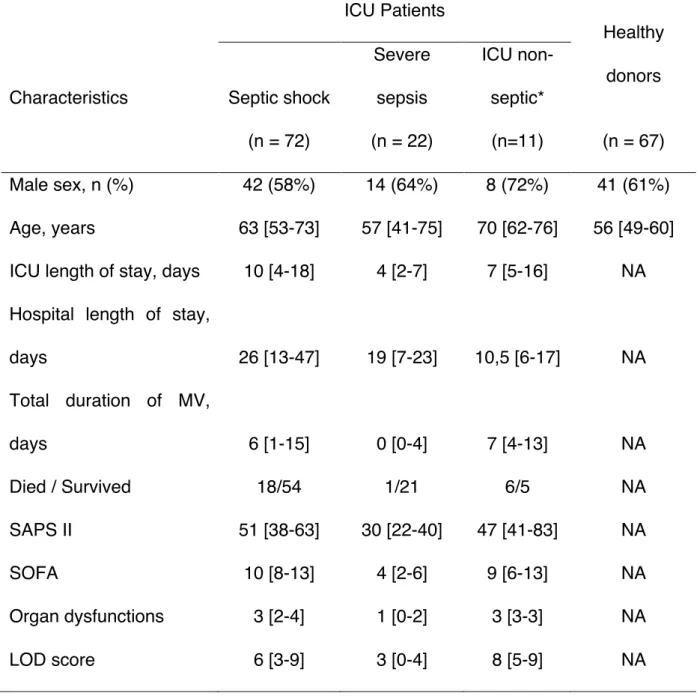
Table 2. Characteristics of patients and healthy donors.
ICU Patients
Healthy donors Characteristics Septic shock
Severe sepsis ICU non-septic* (n = 72) (n = 22) (n=11) (n = 67) Male sex, n (%) 42 (58%) 14 (64%) 8 (72%) 41 (61%) Age, years 63 [53-73] 57 [41-75] 70 [62-76] 56 [49-60] ICU length of stay, days 10 [4-18] 4 [2-7] 7 [5-16] NA Hospital length of stay,
days 26 [13-47] 19 [7-23] 10,5 [6-17] NA Total duration of MV, days 6 [1-15] 0 [0-4] 7 [4-13] NA Died / Survived 18/54 1/21 6/5 NA SAPS II 51 [38-63] 30 [22-40] 47 [41-83] NA SOFA 10 [8-13] 4 [2-6] 9 [6-13] NA Organ dysfunctions 3 [2-4] 1 [0-2] 3 [3-3] NA LOD score 6 [3-9] 3 [0-4] 8 [5-9] NA
Data are expressed as median [interquartile range] unless otherwise indicated. ICU, intensive care unit; LOD, Logistic Organ Dysfunction; MV, Mechanical ventilation; NA, Not applicable; SAPS, Simplified Acute Physiology Score; SOFA, Sepsis-related Organ Failure Assessment.
Table 3. Sites of infection and microorganisms isolated in septic patients.
Characteristics Septic shock
(n = 72)
Severe sepsis (n = 22) Infection site, no. of patients
Respiratory tract 23 8
Intra-abdominal/pelvis 11 1
Urinary tract 8 5
Vascular 7 2
Central nervous system 3 3
Other sources 19 3
Unknown 1 0
Bacteremia 39 11
Isolates, no. of patients
Gram positive 37 9
Gram negative 18 9
Miscellaneous 5 0
Candida 1 0
A.
D.
CD247 / CD3GC.
LGALS9 HMOX1 VEGFA AGER FKBP5 CD163 SLPI IL10 MMP8 S100A12 S100A9 ARG1 MMP9 TNFAIP6 AIM2 MYD88 IL4R ADAM17 S100A8 CD274 CASP1 FUT4 ELANE PGF FLT1 NOS2 IL8 CCL2 KITLG PTGS2 CSF1R IL12A MS4A1 IL6 CD4 CD247 FLT3LG CD3G CD8B NLRP1 NCR1 IL4 LGALS3 TGFB1 HD 3.00 2.00 1.00 0.00 -1.00 -2.00 -3.00 Sepsis 0.8 0.7 0.6 0.5 0.4 0.3 0.2 0.1 0.0 30 20 10 0 -10 -20 -30sepsis (positively correlated)
HD (negatively correlated)
Enrichment score (ES)
Ranked list metric (PreRanked)
Zero cross at 5457
B.
2 500 5 000 7 500 10 000 Rank in Ordered Dataset
Upregulated genes
CD14posHLA-DRlo/neg
monocytes LDGs LDGs
Mature Gran Imm Gran LDGs
C.
A
. S100A8/A9 S100A12 Arginase 1 Arginine IDO G-CSF LDGs CD14posCD16neg monocytes CD14 posCD16pos monocytes CD14 dimCD16pos monocytes CD14posHLA-DRlo/neg
monocytes CD14pos
monocytes
A.
CD8pos CD8pos Sepsis HDC.
B.
CD4pos CD4pos CD8pos CD4posA
. LDGs Imm Gran Mature NeutroC
.B
. CD14posHLA-DRlo/negmonocytes
A.
D.
Distribution of mature / immature granulocytes
MFI MFI MFI
CD35
CD63
CD66b
FS
SS
MFI MFIB.
LDG
HDG
E.
F.
MPO
NGAL
C.
Online data supplement
Early expansion of circulating myeloid-derived suppressor
cells predicts development of nosocomial infections in
septic patients.
Fabrice Uhel, MD, PhD1,2,3, Imane Azzaoui, PhD3,4,5,6, Murielle Grégoire, PhD3,4,5,6, Céline Pangault, PharmD, PhD3,4,5,6, Joelle Dulong, MBiol3,4,5,6, Jean-Marc Tadié, MD, PhD1,2,3,5, Arnaud Gacouin, MD1,2, Christophe Camus, MD, PhD1,2, Luc Cynober, PharmD, PhD7, Thierry Fest, MD, PhD3,4,5,6, Yves Le Tulzo, MD, PhD1,2,3,5, Mikael Roussel, MD, PhD3,4,5,6 and Karin Tarte, Pharm D, PhD3,4,5,6
SUPPLEMENTAL METHODS
Quantitative real-time PCR
Total RNA was extracted using PAXgene blood RNA kit (Qiagen). cDNA was then generated using Multiscribe Reverse Transcriptase and High-Capacity cDNA reverse Transcription kit (Invitrogen). For quantitative RT-PCR on whole blood, we used Taqman array microfluidic cards and Taqman Universal Master Mix from Applied Biosystems (Invitrogen). A panel of genes related to myeloid suppressor cells was selected based on literature knowledge. Gene expression was measured by the ABI Prism 7900HT Sequence Detection System. 18S, CDKN1B and ELF1 were determined as appropriate internal standards using TaqMan endogenous control assays (Invitrogen) and geNorm algorithm analysis (https://genorm.cmgg.be). For each sample, the gene CT values were determined, normalized to the geometric mean value of the 3 housekeeping genes, and compared to the median value obtained from healthy donors using the 2-∆∆CT method.
Quantitative RT-PCR were also performed on purified low- (LDG) and high-density granulocytes (HDG) from 5 septic patients. cDNA was prepared using Fluidigm Reverse Transcription Master Mix (Fluidigm, Sunnyvale, CA). The qPCR was performed in triplicate using 96.96 Dynamic Array integrated fluidics circuit and the BioMark HD System from Fluidigm. CDKN1B and PUM1 were determined as appropriate internal standards using TaqMan endogenous control assays (Invitrogen) and geNorm algorithm analysis (https://genorm.cmgg.be). For each LDG sample, the mean Ct value for the gene of interest was calculated, normalized to the geometric mean value of housekeeping genes, and compared with value obtained from the paired HDG samples using the 2-∆∆Ct method. For each gene, results were expressed as the ratio of paired LDG to HDG 2-∆∆Ct values.
Cell isolation and culture
Fresh PBMCs were obtained from healthy donors or septic patients after Ficoll density centrifugation. Cells were magnetically depleted or not for monocytes or LDGs using CD14 or CD15 microbeads (Miltenyi Biotech). Purity and viability after depletion evaluated by flow cytometry were always >97%. Purified cells were labeled with carboxyfluorescein succinimidyl ester (CFSE; Interchim) and cultured in 96-well round-bottom plates with RPMI 1640-10% human AB serum (Biowest) in the presence of anti-CD3 and anti-CD28 monoclonal antibodies (0.6 μg/mL, Sanquin). After 4 days, cells were labeled with anti-CD2,
identify viable cells. CFSE dilution was assessed by flow cytometry. Results were analyzed with ModFit LT software (Verity Software House) and expressed as the cumulated proportion of proliferated T-cells (≥ G2 generation).