Long-range silencing and position effects at telomeres
and centromeres: parallels and differences
S. Perrod and S. M. Gasser *
University of Geneva, Department of Molecular Biology, Quai Ernest-Ansermet 30, 1211 Geneva 4 (Switzerland), Fax: +41 22 379 6868, e-mail [email protected]
Abstract. Most of the human genome is compacted into
heterochromatin, a form that encompasses multiple forms of inactive chromatin structure. Transcriptional si-lencing mechanisms in budding and fission yeasts have provided genetically tractable models for understanding heritably repressed chromatin. These silent domains are typically found in regions of repetitive DNA, that is, ei-ther adjacent to centromeres or telomeres or within the tandemly repeated ribosomal DNA array. Here we ad-DOI 10.1007/s00018-003-3246-x
© Birkhäuser Verlag, Basel, 2003
dress the mechanisms of centromeric, telomeric and lo-cus-specific gene silencing, comparing simple and com-plex animals with yeast. Some aspects are universally shared, such as histone-tail modifications, while others are unique to either centromeres or telomeres. These may reflect roles for heterochromatin in other chromosomal functions, like kinetochore attachment and DNA ends protection.
Key words. Silencing; PEV; yeast; SIR protein; epigenetics; Drosophila; telomeres; rDNA.
Comparison of long-range silencing with heterochromatin
Long-range silencing generates a heritable, transcription-ally inactive chromatin structure that is associated with stable posttranslational modifications of histones, such as methylation or hypoacetylation (for reviews [1 – 3]). Such repression is not promoter specific, but rather region spe-cific, and can act over large stretches of DNA. A re-pressed state can persist through meiotic and mitotic cell divisions and usually leads to late replication in S phase. Whereas gene silencing shares the key properties of inac-cessibility and epigenetic inheritance with heterochro-matin, transcriptionally silent domains are not always cy-tologically visible, even though this trait was traditionally used to distinguish heterochromatin from euchromatin [4]. The absence of a cytologically distinct hetero-chro-matin is particularly pronounced in budding yeasts, which nonetheless has provided a powerful model for mechanisms of chromatin-mediated repression.
It is helpful to distinguish different types of heterochro-matin; the most common are called facultative (as in tis-sue-specific domain inactivation) and constitutive, which
*Corresponding author.
generally refers to noncoding satellite repeats. Faculta-tive heterochromatin can be specific to one of two ho-mologous chromosomes, or to a certain cell type or de-velopmental stage, and leads to the repression of unique sequences rich in genes. Constitutive heterochromatin re-mains condensed in almost all somatic cells of a given organism and usually involves repetitive, noncoding DNA sequences. Such gene-poor regions are typically, al-though not exclusively, located near centromeres or telomeres and can induce repression of nearby genes in an epigenetic manner. This phenomenon is called posi-tion effect variegaposi-tion. The precise funcposi-tion of simple re-peat DNA is poorly understood, but it is likely to serve a structural role in chromosome pairing and segregation (reviewed in [5, 6]), which only indirectly results in transcriptional silencing. Here we compare the silencing that occurs in Saccharomyces cerevisiae (budding yeast) with the constitutive heterochromatin found at the cen-tromeres of Schizosaccharomyces pombe (fission yeast) and higher eukaryotes, like Drosophila and human beings (see fig. 1). By using ‘heterochromatin’ as a collective term to describe a range of compact interphase chromatin structures that are distinct from transcriptionally active and structurally accessible ‘euchromatin,’ we can indeed include silent loci in yeast in this category.
Budding yeast silencing
Silent domains within S. cerevisiae chromosomes gener-ally cover only a few kilobases, a distance that is not sur-prising given the compact organization of the budding yeast genome. Except for the irregular TG repeat at telomeres, budding yeast lacks simple repeat DNA, and genes have few introns and lie within 1 – 2 kb of each other. Perhaps because of the complexity and sheer size of higher eukaryotic genomes, many more proteins are implicated directly or indirectly in the formation of hete-rochromatin in complex organisms than in yeast. Indeed, constitutive heterochromatin constitutes > 30 % of the genome in Drosophila and > 55 % in humans and mice, whereas heritably silent chromatin represents < 1 % of the total budding yeast genome.
Nonetheless, three distinct chromosomal regions of S. cerevisiae confer a heritable state of transcriptional re-pression (i. e., epigenetic silencing) on otherwise func-tional promoters: (i) the mating-type loci HML and HMR
(homothallic mating-type locus left or right), (ii) telo-meres and (iii) the rDNA array (reviewed in [7, 8]). Genes located near or within these domains are either com-pletely inert for transcription or exhibit a variegated state of repression that is relatively stable within a single cell. Importantly, repression is position, and not promoter, specific. Maintenance of silencing is essential for pre-serving mating competence in yeast, because derepres-sion of the HM loci leads to expresderepres-sion of both mating-type programs, allowing haploid cells to acquire the prop-erties of a nonmating diploid cell. On the other hand, the relaxation of silencing at the rDNA repeats leads to re-combination and excision of rDNA repeats [9] which cor-relates with a shortened life span [10]. In S. pombe (fis-sion yeast), centromeres are composed of large repetitive sequence elements that also confer position-dependent repression of Pol II-transcribed genes. This centromere-induced repression, although absent in budding yeast, is the most widely conserved type of heterochromatin and thus will be examined first.
Figure 1. Trans-acting factors required for repression. Top: Propagation of silencing at budding yeast telomeres and HM loci involves mul-tiple steps. Bottom: Key elements for targeting repression in diverse organisms. The cis-acting sequences necessary for the binding of trans-acting factors have not yet been determined for the budding yeast rDNA or for ribosomal repression or PEV in Drosophila. In fission yeast, siRNA are required at least for the initiation of repression and in addition for the maintenance of repression at centromeres. Only the se-quence of siRNA necessary for centromeric silencing is known. Localization of Swi6 at the silent mating-type locus and at centromeres requires Clr4/Rik1, Clr3/Clr6 and Chp1, whereas HP1 independently initiates methylation in a small domain of the chromocenter.
Centromeres
Repetitive centromeric sequences and centromeric identity
The centromere is an essential chromosomal landmark that provides a site for the attachment of mitotic and mei-otic spindle microtubules, which in turn mediate mitmei-otic chromosome segregation. Centromeric activity in bud-ding yeast is conferred by a specific sequence of roughly 125 bp, whereas centromeres of fission yeast and higher eukaryotes are composed of large repetitive DNA do-mains packaged into a heterochromatic structure (for de-tails see table 1; for centromere reviews [11, 12]). Be-cause these ‘regional’ centromeres cannot be easily de-lineated on the basis of their sequence, it has been difficult to determine the minimal size of a functional centromere in most organisms.
In fission yeast, Drosophila and mammals, heterochro-matin is an integral part of the centromere, and the estab-lishment and maintenance of heterochromatin correlates with centromere function [5]. Centric heterochromatin may not only provide a site for microtubule attachment, but may also contribute to the nucleation of sister chro-matid cohesion [13]. The occasional creation of new cen-tromeres (i. e., neocencen-tromeres) at novel chromosomal sites that lack sequence homology with natural cen-tromeres [14, 15] suggests that the structural organization of centromeric domains, rather than the DNA sequence per se, is the primary relevant parameter for their func-tion. Centromere choice may also reflect complex para-meters such as timing of replication, subnuclear position-ing and/or other heritable features of chromatin structure [11, 12, 16].
One conserved heritable feature of centromeres is the presence of a special histone H3 variant, which is found exclusively within the core centromeric region. This spe-cial histone is called CENP-A in mammals (centromeric protein A), Cid (centromere identifier) in Drosophila, Cse4 in S. cerevisiae and Cnp1 in S. pombe, and it re-places histone H3 in specialized centromeric nucleo-somes (reviewed in [11, 16]). The presence of this H3 variant appears to be crucial for kinetochore assembly and distinguishes the inner plate of the centromere from pericentric heterochromatin, which contains normal his-tone H3. In an attempt to explain this distribution, a re-cent study found that core regions of Drosophila cen-tromeres replicate as isolated domains early in S phase, prior to the replication of flanking heterochromatin. Moreover, histone H3-containing nucleosome assembly was shown to be inhibited during the replication of these core centromeric sequences [17]. One suggestion for the perpetuation of this special centromeric chromatin is therefore that pericentromeric heterochromatin se-questers centromeres from an H3-specific assembly ma-chinery, promoting incorporation of the
centromere-spe-cific variant [17]. Alternatively, a histone exchange factor may replace H3 with a H3 variant in a localized and repli-cation-independent manner.
Pericentromeric heterochromatin mediates PEV
Pericentromeric heterochromatin and its associated phe-nomenon of position-effect variegation (PEV) are only observed in organisms having large regional centromeres. A mere 125 bp is necessary and sufficient for the assem-bly of a functional S. cerevisiae centromere, and this se-quence is not flanked by heterochromatin, nor does it si-lence genes. In contrast, now-classic studies by H. J. Müller (reviewed in [18, 19]) showed that Drosophila genes that were transposed by natural or induced genetic rearrangements to sites near pericentric heterochromatin frequently assume a variegated pattern of expression. This chromosomal position effect can spread over dis-tances of 1 Mbp or more, generally reflecting a gradient of gene inactivation that is inversely correlated with dis-tance [18]. Recent data show that the local context of a gene also influences the degree of PEV, and suggest that the repression process can also be discontinuous and modulated by promoter strength [20]. Indeed, some genes are specifically adapted to be expressed exclusively in a heterochromatic context [19]. Genes that encode proteins structurally important for heterochromatin have been identified in screens for dominant mutations that enhance or suppress PEV, called E(var)s and Su (var)s, respec-tively [18]. Approximately 120 modifiers of PEV have been identified to date, and more can be expected. Initiation of PEV at the molecular level is poorly under-stood, although the products of three Su(var) genes, HP1 [heterochromatin protein 1 encoded by Su(var)2-5], Su(var)3-7, and Su(var)3-9 are strong candidates for structural components of pericentric heterochromatin in flies. All three colocalize to pericentric regions on poly-tene chromosomes, coimmunoprecipitate as members of a protein complex and interact in two-hybrid assays [21 – 22]. Only the Su(var)3-7 protein has affinity for DNA, it binds through a cluster of N-terminal zinc fin-gers. Deletion studies however show that the Su(var)3-7 C-terminal domain mediates targeting to pericentric het-erochromatin and that HP1 localization occurs indepen-dent of the Su(var)3-7 N-terminus [23]. The cluster of zinc fingers may nonetheless mediate RNA binding (see below). Although transgene inactivation by PEV is corre-lated with HP1 deposition, this deposition alone is not sufficient to confer gene repression. Indeed, HP1 also lo-calizes to over 200 euchromatic sites on polytene chro-mosomes, indicating that additional conditions must be met for promotion of a transcriptionally inactive chro-matin state [24, 25].
In the case of fly PEV, the spread of centric heterochro-matin can be assessed cytologically on the basis of a
vis-ible change in the banding pattern of polytene chromo-somes. Nonetheless, this cis-spreading model of centric heterochromatin does not adequately explain the ability of PEV to repress genes located several megabases away [18]. Such phenomena led to the suggestion that trans-interactions between different heterochromatic regions and the three-dimensional organization of chromosomes in the interphase nucleus may be important for PEV
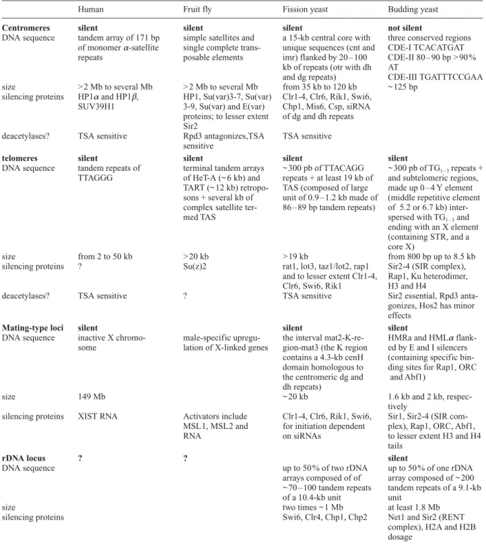
[18, 26]. Indeed, in many cells Drosophila centromeric heterochromatin is clustered at the ‘chromocenter,’ a zone of ~ 25 % of the nuclear volume that is readily visu-alized by the detection of the HP1 protein [27]. In some cell types, Drosophila interphase chromosomes also ex-hibit a Rabl orientation, in which the centromeres and the bulk of heterochromatin are positioned at one end of the nucleus and telomeres at the opposite end [18]. In sum-Table 1. The different heterochromatic loci found in human, fruit flies, fission yeast and budding yeast.
Human Fruit fly Fission yeast Budding yeast
Centromeres silent silent silent not silent
DNA sequence tandem array of 171 bp simple satellites and a 15-kb central core with three conserved regions of monomer a-satellite single complete trans- unique sequences (cnt and CDE-I TCACATGAT repeats posable elements imr) flanked by 20 – 100 CDE-II 80 – 90 bp > 90 %
kb of repeats (otr with dh AT
and dg repeats) CDE-III TGATTTCCGAA size > 2 Mb to several Mb > 2 Mb to several Mb from 35 kb to 120 kb ~ 125 bp
silencing proteins HP1aand HP1b, HP1, Su(var)3-7, Su(var) Clr1-4, Clr6, Rik1, Swi6, SUV39H1 3-9, Su(var) and E(var) Chp1, Mis6, Csp, siRNA
proteins; to lesser extent of dg and dh repeats Sir2
deacetylases? TSA sensitive Rpd3 antagonizes,TSA TSA sensitive sensitive
telomeres silent silent silent silent
DNA sequence tandem repeats of terminal tandem arrays ~ 300 pb of TTACAGG ~ 300 pb of TG1 – 3repeats + TTAGGG of HeT-A (~ 6 kb) and repeats + at least 19 kb of and subtelomeric regions,
TART (~ 12 kb) retropo- TAS (composed of large made up 0 – 4 Y element sons + several kb of unit of 0.9 – 1.2 kb made of (middle repetitive element complex satellite ter- 86 – 89 bp tandem repeats) of 5.2 or 6.7 kb)
inter-med TAS spersed with TG1 – 3and
ending with an X element (containing STR, and a core X)
size from 2 to 50 kb > 20 kb > 19 kb from 800 bp up to 8.5 kb
silencing proteins ? Su(z)2 rat1, lot3, taz1/lot2, rap1 Sir2-4 (SIR complex), and to lesser extent Clr1-4, Rap1, Ku heterodimer, Clr6, Swi6, Rik1 H3 and H4
deacetylases? TSA sensitive ? TSA sensitive Sir2 essential, Rpd3 anta-gonizes, Hos2 has minor effects
Mating-type loci silent silent silent
DNA sequence inactive X chromo- male-specific upregu- the interval mat2-K-re- HMRa and HMLa flank-some lation of X-linked genes gion-mat3 (the K region ed by E and I silencers
contains a 4.3-kb cenH (containing specific bin-domain homologous to ding sites for Rap1, ORC the centromeric dg and and Abf1)
dh repeats)
size 149 Mb ~ 20 kb 1.6 kb and 2 kb,
respec-tively
silencing proteins XIST RNA Activators include Clr1-4, Clr6, Rik1, Swi6, Sir1, Sir2-4 (SIR com-MSL1, MSL2 and for initiation dependent plex), Rap1, ORC, Abf1,
RNA on siRNAs to lesser extent H3 and H4
tails
rDNA locus ? ? silent
DNA sequence up to 50 % of two rDNA up to 50 % of one rDNA
arrays composed of of array composed of ~ 200 ~ 70 – 100 tandem repeats tandem repeats of a 9.1-kb of a 10.4-kb unit unit
size two times ~ 1 Mb at least 1.8 Mb
silencing proteins Swi6, Clr4, Chp1, Chp2 Net1 and Sir2 (RENT
complex), H2A and H2B dosage
mary, two epigenetic mechanisms appear to contribute to the cell-to-cell phenotypic variation that is typical for PEV: the cis-spreading effect of adjacent heterochro-matin and a trans-effect due to chromosomal interactions that themselves may involve by heterochromatin binding factors.
HP1 and its target, methylated lysine 9 of histone H3, are conserved
What targets the complex of HP1/Su(var)3-7/Su(var)3-9 to pericentric heterochromatin? Answers have been pro-vided in part by recent studies showing that the hy-poacetylation and subsequent methylation of a particular lysine (K9) in histone H3 is a conserved marker for
hete-rochromatin that provides a specific target for the binding of HP1 and related proteins [28]. In this aspect, fission yeast, flies and mammalian cells have all contributed to a coherent model of heterochromatin assembly.
The S. pombe homologue of HP1, Swi6, is found at peri-centric heterochromatic regions, the imr and otr repeats (see table 1). Similarly, of the three human HP1 homo-logues, two, HP1aand HP1b, localize to heterochromatin,
whereas a third one, HP1g, is primarily found at
euchro-matic sites (reviewed in [28]). All HP1-like proteins have a characteristic N-terminal chromodomain (CD), a short variable hinge region and a C-terminal chromoshadow domain (CSD), which mediates self-dimerization and protein-protein interaction. Although HP1 appears to bind specifically to > 20 proteins, its most crucial binding partner may well be the methylated lysine 9 (K9me) of
his-tone H3, which it binds through its chromodomain [29, 30]. Importantly, recent studies have shown that Su(var)3-9 is a conserved histone methyltransferase (HMTase) specific for H3 K9[31 – 33]. Consistently, both
a mutation in the CD domain of HP1 and a deletion of Su(var)3-9 lead to the delocalization of HP1 from hete-rochromatin.
Although, H3 K9meis recognised by HP1, the two markers
do not always colocalize [34 – 36]. For example, the inac-tive X chromosome contains extensive H3 K9
methyla-tion, yet no HP1 is bound to this chromosome [35, 36]. Moreover, as mentioned above, HP1 localizes to certain euchromatic bands in polytene chromosomes that show no staining for H3 K9me[36]. Although conclusions from
these immunoanalyses must take into account whether the antibodies recognize di- and trimethyl forms of H3 K9
equally, they clearly show that H3 K9meis not a sufficient
condition for HP1 recruitment. Previous studies had shown that HP1 becomes delocalized from the pericentric foci in RNAse-treated cells [34, 35, 37]. This observation was extended by demonstration that the HP1 hinge do-main binds single- and possibly double-stranded RNAs and that this hinge, together with the CD domain, is suf-ficient to ensure the proper localization of HP1 [38]. In
conclusion, a combination of a specific histone-tail mod-ification and an as yet unidentified RNA apparently tar-gets HP1 and presumably triggers the establishment of a repressed heterochromatin structure. This minimal model certainly does not exclude the participation of other com-ponents, for example, the recently characterized HP2 pro-tein [39].
Methylation at histone H3 K9and the formation
of heterochromatin
The three centromeres of S. pombe chromosomes are composed of large inverted-repeat structures that sur-round the central core domain (cnt), called the imr (in-nermost repeat) and otr (outer repeat). Reporter genes inserted at any of these domains are subject to position-effect variegation (reviewed in [40]), although those in-serted in cnt are only weakly repressed compared with those inserted in imr and otr repeats. The initial trans-acting factors identified as necessary for the mainte-nance of heterochromatin were Clr1-4, Clr6, Swi6 and Rik1 [40].
Genetic studies in fission yeast have been crucial for identifying the key molecular events that lead to the for-mation of pericentric heterochromatin. Consistent with data discussed above, the S. pombe mechanism starts with covalent modifications of histone tails, that is, deacetyla-tion and methyladeacetyla-tion, which appear to act in concert to es-tablish a “histone code” that signals heterochromatin as-sembly [1]. Notably, the fission yeast homologue of Su(var)3-9, Clr4, has been shown to possess an intrinsic H3 K9methylation activity that can propagate this
modi-fication throughout the imr and otr repeats [32]. The deacetylation of H3 K9 and K14 must occur prior to
methylation, however, in a reaction mediated by Clr6 and Clr3, homologues of the histone deacetylases Rpd3 and Hda1. The final player recruited is Swi6, the closest ho-mologue to HP1 in fission yeast. Because deletion of the Clr4 methyltransferase abolishes Swi6 localization at centromeres, one can infer that as in flies, the binding of Swi6 to centromeres depends on H3 K9methylation. The
converse is not true, consistent with the idea that Swi6 (HP1) acts downstream of Clr4 (the H3 K9
methyltrans-ferase). Finally, deletion of either Rik1 or Chp1, a second chromodomain protein mainly confined to the otr re-gions, has been shown to abolish both H3 K9methylation
and centromeric Swi6 localization [32, 41].
These observations suggest a progressive model for hete-rochromatin assembly based on an ordered series of his-tone modifications. Deacetylation of hishis-tone H3 by Clr3 and Clr6 creates conditions favoring the deposition of H3 K9 methylation by the recruitment of Chp1, Rik1 and
Clr4. H3 K9meis a specific binding site for Swi6, which in
turn assembles nucleosomes into heterochromatin. HP1-like molecules HP1-like Swi6 may act in concert with the
pas-sage of the replication fork, because mutations in DNA Pola [32] also affect the centromeric localization of Swi6, and vertebrate HP1 associates with the replication-associated nucleosome assembly factor CAF1 [42]. In addition to replication factors, other trans-acting factors were identified in genetic screens for mutations that re-sult in the disruption of centromeric silencing in a subdo-main-specific manner. For example, silencing in the otr repeats is affected by mutations in 11 csp loci (cen-tromere suppressor of position effect; [43]), whereas Mis6 affects silencing only at the imr and cnt domains to which it binds [41].
The observations that HP1 localizes to a restricted inter-nal domain of the chromocenter in Su(var)3-9-deficient flies and that a chimeric HP1 containing the CD of Poly-comb (Pc) relocalizes with Su(var)3-9 to Pc binding sites [21] suggest that the competence to nucleate pericentric heterochromatin is gained through HP1 binding. The spreading of pericentric heterochromatin, on the other hand, requires both HP1 and Su(var)3-9. In addition, the distribution of H3 K9meis diffuse throughout the nucleus
when HP1 is absent [21], indicating that methylation and HP1 promote their localization mutually and spread in a self-perpetuating manner.
A role for RNAi in the establishment of heterochromatin
Three recent studies have solved the enigma concerning the role of RNA in the targeting HP1 for heterochromatin nucleation, by showing that small interfering RNAs (siRNA) are necessary for heterochromatin formation at fission yeast centromeres [44 – 46]. The siRNAs are end products of RNA interference (RNAi), a mechanism that processes double-stranded RNA into short sense and an-tisense RNA oligonucleotides of 21 – 25 nts. These siR-NAs in turn inhibit the accumulation of homologous mes-senger RNA (mRNA) transcripts of cognate genes (re-viewed in [47]). Intriguingly, deletion of the RNAi machinery genes ago1 (Argonaute), dcr1 (Dicer) and rdp1 (RdRP) has been found to be correlated with the loss of pericentric heterochromatin, induction of transcription of pericentric reporter genes, loss of H3 K9meand
delocali-zation of Swi6 [44]. The work proposes that in wild-type cells, the reverse strand of the dh repeat is transcribed, leading to a rapid cleavage of the ensuing dsRNA by the RNAi machinery. As a result, in RNAi mutants dh reverse transcripts accumulate. Naturally, the dh forward tran-script also accumulates in these mutants, reflecting a loss of silencing. Consistently, in swi6 mutants the forward dh transcript accumulates but the reverse one does not [44]. Finally dh and also dg siRNA are essential for the appro-priate targeting of Clr4, Rik1 and Chp1 to centromeres and for the maintenance of pericentromeric heterochro-matin [44, 45].
Establishment and maintenance of pericentric hete-rochromatin in human and Drosophila cells will probably similarly require the RNAi machinery, because the genes required for RNAi are conserved and the localization of HP1 and H3 K9meto pericentric heterochromatin is
sensi-tive to RNAse [35]. Some aspects of this mechanism may also be conserved in the phenomenon of female-specific X-chromosome inactivation, a complex developmentally regulated event that requires transcription of the X-chro-mosome-specific Xist RNA and its antisense strand. X inactivation and PEV differ in many ways, however, be-cause Xist RNA is not required for maintenance once re-pression has been established, and methylation of the in-active X is both Su(var)3-9 and RNAse insensitive. Fi-nally, HP1 does not bind on the inactive X chromosome [35]. Nonetheless, X inactivation may present a paradigm for other instances of facultative or cell-type-specific het-erochromatin.
Telomeres
Telomeres and telomeric position effect
The ends of linear chromosomes are stabilized by a struc-ture called the telomere. If telomeres were to rely exclu-sively on conventional DNA polymerases to complete their replication, they would undergo terminal attrition and loss of genetic information. This problem has been solved in most eukaryotes by the ribonucleoprotein en-zyme called telomerase, which extends the TG-rich strand of telomeric DNA by means of a self-templating mechanism (for review [48]). Thus, telomeres typically end in tandem arrays of G-rich telomeric repeats. In bud-ding and fission yeast the terminal TG-rich repeats are ir-regular and extend only 300 bp, although they abut other middle-repetitive subtelomeric motifs (see table 1). Drosophila telomeres are exceptions to this general struc-ture, as chromosomes end with retrotransposon-like ele-ments and length is apparently maintained by recombina-tion and retrotransposirecombina-tion [49].
Telomeres do not simply function as buffer zones to pre-vent the loss of essential sequences but have specialized functions that protect DNA ends. These capping func-tions impede chromosomal fusion (end-to-end joining) and degradation, which would otherwise lead to genomic instability. Paradoxically, telomeres exploit some of the same cellular machinery that mediates nonhomologous end-joining events at internal double-strand breaks, such as the Ku heterodimer, the Mre11/Xrs2/Rad50 complex and the flap-processing complex of Rad27 and Dna2 (viewed in [50]). At telomeres these are thought to help re-cruit or facilitate telomerase action rather than promote recombination. When they are not being replicated, telomeres may have increased local folding, which could also facilitate the silencing of nearby promoters.
The SIR complex mediates silencing at budding yeast telomeres
As mentioned above, the extreme ends of S. cerevisiae chromosomes contain ~ 300 bp of an irregular TG1 – 3
re-peat lying terminal to subtelomeric sequences. These in-clude up to four tandem copies of Y¢ elements (i. e., a middle repetitive sequence of either 5.2 or 6.7 kb), short internal TG1 – 3 repeats, and X element composed of
subtelomeric repeats (STRs) and a conserved 437-bp core (fig. 2; [51]). Whereas the TG-rich portion of the telomeres (called the telosome) is free of nucleosomes, the subtelomeric repeats are nucleosomal [52]. Of the se-quence-specific proteins that recognize the TG repeats (i. e., Cdc13, Tel2, Rap1), only Rap1 is absolutely re-quired for the budding yeast telomeric-position effect (TPE), but the Ku heterodimer (Ku70 and Ku80), which binds telomeres because of its affinity both for free DNA ends and Sir4, contributes to silencing in at least two ways (see details below).
Rap1 has been suggested to recruit the two silent infor-mation proteins, Sir3 and Sir4, to telomeres, catalyzing the first step of a pathway that would lead to propagation of the silencing-proficient SIR complex (i. e., Sir2, Sir3 and Sir4, [53]). Recent data show, however, that Rap1 ini-tially recruits Sir4 alone, because Sir4 can be detected at
telomere ends in the absence of Sir2 or Sir3 [54, 55]. Given that the converse is not true, Sir4 appears to have an essential initiator function for the recruitment or stabi-lization of Sir2 and Sir3 at telomeres. The spreading of the SIR complex along subtelomeric nucleosomes is fully dependent on an intact SIR complex, on the nicotinamide adenine dinucleotide (NAD)-dependent deacetylase ac-tivity of Sir2 [54, 55], and on the deacetylated N-terminal tails of histones H3 and H4 [53]. This multiple-step path-way could be envisioned thus: First, Rap1 binds specifi-cally to the TG repeats. Second, Rap1 recruits Sir4, which in turn recruits Sir2 and stabilizes the Rap1-Sir3 interac-tion. Finally, the full SIR complex would be able to spread along the TG-rich sequences because of affinity of Sir3 and Sir4 for the hypoacetylated tails of H3 and H4. This spread may require the active deacetylation of histone tails adjacent to the SIR complex by Sir2, because such spreading appears to be counteracted by a histone acety-lase called Sas2 [56, 57]. The action of Sir2 deacetyacety-lase activity on adjacent nucleosomes may in fact be neces-sary for the cooperative, self-propagating nature of the linear and nucleosome-mediated spreading of SIR com-plexes along the chromosome.
To date only H3 K9ac, H3 K14acand H4 K16ac have been
demonstrated to be substrates of Sir2 in vitro [58].
Be-Figure 2. Protosilencers, STARs, silencers and profile of repression at budding yeast telomeres and HM loci. In budding yeast, native telomeres are composed of X and Y¢ elements (i. e., X1Y¢0 – 4). The X and Y¢ elements each contain a protosilencer that can cooperate with telomeres to induce repression, as well as a STAR (subtelomeric anti-silencing region, see [107]). Therefore, if Y¢ elements are present
at telomeres, they are flanked by STARs and insulated from repression. Repression is only observed at the X core element and ~ 2 kb centromere proximal. The HM loci are flanked by two silencers and are thus efficiently repressed, as even partial silencers cooperate to promote repression in this context [108].
cause Sir2 deacetylates a large variety of acetyl-lysine-containing proteins with equal efficiency, both the sub-nuclear environment and proteins that bind Sir2 are likely to help determine substrate specificity in vivo. Lysines at positions 9, 14, 18, 23 and 27 on H3 and positions 5, 8, 12 and 16 on H4 have been shown to be underacetylated in silent chromatin, as have lysine 7 on H2A, and lysines 11 and 16 on H2B [59]. This result suggests either that other histone deacetylases are required to maintain telomeres in their hypoacetylated state or that the his-tones have been assembled into silent chromatin in an un-deracetylated state and have been shielded from the action of acetylases. Since few deacetylase mutants other than SIR2 derepress TPE, the latter is more likely true.
Finally, as mentioned above, a further catalyst for Sir4 re-cruitment to telomeres is the yKu heterodimer, which is associated with termini both through its end-binding function and its affinity for Sir4 [60, 61]. Strains defi-cient in ku70 or ku80 lose telomeric repression, which can be restored through extreme telomere lengthening (i. e., an increase in Rap1 binding sites), by an increase in the dosage of Sir4 or by relief of the competition imposed by Rif proteins (Rap1-interacting factors) for the access of SIR proteins to Rap1 [62]. In brief, the Ku heterodimer aids but does not replace Rap1 in the recruitment of SIR proteins to telomeres. In addition, Ku plays an important role is anchoring telomeres to the nuclear periphery [63, 64], a localization that helps create subnuclear compart-ments enriched for Rap1 and SIR factors [65]. The im-portance of this nuclear organization for TPE is discussed below.
In summary, the establishment of silent chromatin at yeast telomeres requires the initial recruitment of Sir4 by Rap1 and yKu. Sir4 may be sufficient to recruit the intact Sir2-3-4 complex to TG1 – 3repeats. The spreading of this
SIR complex along nucleosomes, however, depends on a careful balance of the three SIR proteins and the deacety-lase activity of Sir2, reminiscent of the means used by HP1 and Su(var)3-9 to initiate the formation of hete-rochromatin in S. pombe, Drosophila and mammalian cells. To date, however, no methylated forms of H3 K9
have been detected in budding yeast, nor have homo-logues to the RNAi enzymes been reported in budding yeast. Thus, although conceptually similar, the mecha-nisms for repressed chromatin assembly at centromeres and telomeres are genetically distinct.
Barriers, insulators and the propagation of the repressed state
The most widely accepted model of telomeric silencing suggests that repressive chromatin propagates continu-ously inward from the telomere and diminishes with in-creasing distance from the terminus [53, 66]. Although
this is true at artificial telomeres (i. e., terminal trunca-tions lacking subtelomeric repeats), the extent of repres-sion at native telomeres seems more limited [67]. For ex-ample, at native ends the subtelomeric Y elements are re-sistant to silencing along their entire length, although repression can be reestablished at the proximal X element by elements known as protosilencers (fig. 2; [67, 68]). In-deed, the Y elements appear to be flanked by insulators or barrier elements that protect sequences within the domain from repression. Thus, silencing at native telomeres seem in fact discontinuous, with maximal repression being found adjacent to the protosilencers element (an ARS consensus) in the subtelomeric core X element and ex-tending inward with decreasing strength for ~ 3 kb. Al-though SIR proteins can be coimmunoprecipitated with the Y repeats, it is not clear whether they propagate con-tinuously along its length or bind to flanking clusters of Rap1or Ku molecules [69]. Recent studies suggest that the extent of Sir3 spreading is determined by the balance of two antagonistic histone-modifying enzymes: the Sir2 deacetylase and the Sas2 histone acetyltransferase [56, 57]. Surprisingly, the deletion of SAS2 leads to a loss, rather than improvement, of TPE at truncated telomeres possibly due to the uncontrolled spreading of Sir proteins inwards [56].
Yeast telomeres are anchored at the nuclear periphery and have a fold-back structure
In budding yeast, telomeric components such as the yKu complex mediate the tethering of telomeres with struc-tural components of the nuclear envelope, leading to the clustering of telomeres into a limited number of discrete foci at the nuclear periphery (see references in [63 – 65, 70, 71]). Telomere clustering is correlated with an accu-mulation of SIR proteins and Rap1 in perinuclear foci, and the ensuing high concentration of SIR proteins has been shown to be a critical factor for efficient subtelom-eric repression (reviewed in [70]). Despite the increased local concentration of SIR proteins at telomeres, their overexpression singly or in pairs (e. g., Sir3 and Sir4) still improves repression efficiency at telomeres, indicating that the SIR complex is limiting under normal growth conditions [65, 66].
Live imaging of GFP-lac repressor-tagged telomeres de-signed to determine whether the peripheral clustering of telomeres facilitates silencing, or whether silent chro-matin itself is responsible for the creation of telomeric foci, has been performed under a range of mutant and wild-type conditions [64]. The deletion of SIR4 leads to a slight but reproducible drop in telomere anchoring, al-though the extent of delocalization varies from end to end [64, 71]. Telomere displacement is even more striking in strains lacking the yKu70 or yKu80 protein [63, 64], but because SIR proteins are themselves displaced from
telomeres in ku-deficient strains [63], and because a sub-population of the Ku heterodimer is displaced from telomeres in sir-deficient strains [60], determining whether yKu and SIR complexes define two independent anchoring pathways required restoration of telomeric si-lencing in a strain lacking yKu. This restoration was achieved by deletion of the RIF1 gene, which encodes a Rap1-binding factor that competes with Sir3 and Sir4 for access to Rap1 [62]. In a rif1 ku70 double mutant, the per-inuclear anchoring of the lacop-tagged Tel VI-R was
re-stored, coincident with the restoration of TPE, in a SIR-dependent manner. This SIR-mediated telomere anchor-ing is cell-cycle dependent, beanchor-ing more pronounced in S-phase than in G1-phase nuclei [64], a dependence that is strikingly correlated with an ill-defined S-phase re-quirement for the establishment of silent chromatin (see below). In summary, telomere anchoring studies indicate that SIR-mediated tethering functions in parallel to the Ku-mediated pathway, which itself anchors telomeres in a repression-independent manner. Thus the clustering of telomeres into foci can both precede and result from si-lencing.
In contrast to initial suggestions [72, 73], the myosin-like proteins 1 and 2 play no significant role in the Ku-medi-ated anchorage pathway, as monitored by either fluores-cence in situ hybridization (FISH) or live telomere-imag-ing techniques [64, 74]. Indeed, telomeres lengthen and TPE improves in mlp1 mlp2 cells [74]. Although the membrane-associated partner for Ku remains obscure, a candidate perinuclear anchor for Sir4 has been identified as Esc1 ([75]; A. Taddei and S. M. Gasser, unpublished). Recently, de Bruin and colleagues have provided data suggesting that budding yeast telomeres not only cluster but fold back upon themselves [76]. Initial support for this idea was based on the observation that Rap1, as well as the Ku heterodimer, are not only recovered associated to the terminal TG-rich sequences but also bind repressed subtelomeric nucleosomes extending several kilobases from the TG repeats [60, 77, 78]. Although trans-interac-tions between telomeres could account for this result, a recent study based on a Gal4-mediated gene-activation assay argues that chromosomal termini fold inward to al-low long-range protein-protein interactions in cis, in a Sir3-dependent manner [76].
Parallels between mammalian and yeast telomeres
Homologues of Sir3 and Sir4 have been found to date only in Trypanosomes and other Ascomycetes. Nonetheless, certain parallels can be drawn between budding yeast, mammalian and fission yeast telomeres. Indeed, among these organisms both the TG-rich repeats and some of the ligands for these repeats (i.e., homologues of Rap1, Cdc13 and the Ku heterodimer) are conserved, although Drosophila telomeres again are a special case, for which
no sequence-specific proteins have been identified to date.
Mammalian TG-repeat tracts are conserved in sequence (TTAGGG) but are variable in length, showing organism-and cell-type-specific diversity. Lengths range from 5 – 30 kb in humans and up to > 150 kb in mice. As in budd-ing yeast, the fission yeast TG repeat covers < 500 bp, although subtelomeric repeats can extend up to 19 kb. A Rap1-like factor is conserved in both human and fission yeast, yet in these organisms it lacks the conserved Myb-related DNA binding domain [79], and thus it does not bind TG repeats. Other related Myb-box-containing fac-tors, such as TRF1 and TRF2 in humans [80, 81] and Taz1 and Teb1 in S. pombe [82, 83], bind the repeated sequence and mediate the association of human or fission yeast Rap1 to telomeres. Throughout evolution the structural DNA binding motif of telomeric proteins is conserved: yeast Rap1 binds to an iterated consensus by means of two internal Myb-like DNA binding domains [79], whereas the human TRF1 and TRF2 and the S. pombe Taz1 ho-modimerize, allowing the Myb domain of each subunit to contact one repeat unit [81, 82]. TRF2 is thought to play an additional role in protecting the telomere end from dou-ble-stranded-break repair factors, by aiding the formation of t loops [84, 85]. In t loops the single-stranded G-rich tail of the repeat folds back to anneal with internal repeats, forming a stable terminal loop [84]. Although t loops have been observed in Trypanosoma and mouse cells, as well as in humans, the frequency of their occurrence remains un-clear, and it is likely that telomeres are stabilized by more than one mechanism [85].
Given their high degree of structural conservation, it was reasonable to speculate that telomeric silencing should also be conserved. Indeed, TPE exists in fission yeast [86], Drosophila [87], and in human somatic cells [88, 89]. To date little is known about the proteins that func-tion in human and Drosophila TPE, but a luciferase re-porter has been shown to be expressed 10-fold less effi-ciently when integrated near a truncated telomere in HeLa cells than when at internal sites. This telomere-proximal repression was sensitive to a general inhibitor of histone deacetylases, called trichostatin A, which does not inhibit the activity of the Sir2 family of NAD-depen-dent deacetylases [88]. Therefore, although Sir2 is con-served and may affect Drosophila PEV [90], there is no evidence that it affects TPE in organisms other than bud-ding yeast.
In Drosophila, TPE and PEV appear to be qualitatively different, because pericentric insertions are generally much more repressed than telomeric insertions. Consis-tently, none of the 25 genes that influence the severity of centromeric variegation in trans have been found to affect TPE (reviewed in [91]). Moreover, all telomere-associ-ated repressed transgenes analyzed to date have been shown to be embedded in or adjacent to
telomere-associ-ated sequences (TAS) and not within the retrotransposons HeT-A and TART [87, 92, 93]. This result may indicate that only the TAS repeats are packaged into a repressive chromatin structure [87].
Interestingly, HP1 was shown to associate with fly telo-meres, even those lacking TAS, HeT-A or TART retro-transposons [24]. Because HP1 mutants show increased levels of telomere-telomere fusion [24], it was suggested that HP1 might be involved in telomere stabilization, rather than in telomeric silencing (fly telomeric silencing is reviewed in [93]). Like those of budding yeast, Drosophila telomeres are found clustered at the nuclear periphery [94], although their mechanism of anchoring and the importance of this clustering for telomeric re-pression are unknown.
The telomeres of fission yeast are also subject to tran-scriptional silencing, which results in the variegated ex-pression of integrated reporter genes [86, 95]. Whereas TPE is only slightly affected by mutations affecting cen-tromeric trans-acting factors (i. e., Clr1-4, Clr6, Swi6 and Rik1; [40]), another group of factors, rat1, lot2, lot3/taz1 and rap1, specifically affect repression in telomeric sites [95, 96]. In lot2 and taz1 mutants, telomeres no longer as-sociated with the spindle pole body at the premeiotic horsetail stage, although it is not known whether these mutations significantly impair telomere clustering in in-terphase of vegetatively growing cells [95, 97].
Silent mating-type loci
Both budding and fission yeasts are able to switch mating type because they maintain transcriptionally silent copies of both mating-type determinants in their haploid genomes. The repression at these silent loci is very simi-lar to that found at either the telomeres or the centromeres of the species concerned. That is, repression of the S. cerevisiae mating-type loci requires Rap1 and the SIR proteins, whereas Clr1-4, Clr6, Rik1 and Swi6 serve this role in S. pombe. It is unclear whether the silencing mech-anisms initially evolved for mating-type maintenance or for centromeric and/or telomeric functions.
Repression of silent mating-type loci in budding yeast
In budding yeast, haploid cells are ‘sexed’ with either a-type or a-type mating preferences, and this is determined by the MAT locus (mating-type locus) at which either a1 and a2 or a1 and a2 mating-type genes are expressed. Two related loci found near the left and right telomeres of chromosome III, called HMLa and HMRa, contain iden-tical copies of the mating-type genes that are transcrip-tionally repressed. These silent copies allow mating-type switching to occur, a genetic event that changes the
infor-mation present at the MAT locus through gene-conversion with either the HMLa or HMRa silent loci serving as donors (HMLa donating its sequences to MATa, and HMRa to MATa). In haploid cells the HM loci must re-main transcriptionally silent, because simultaneous coex-pression of a and a mating-type gene products results in a nonmating a/a diploid cell.
The HM loci are flanked by two specific cis-acting se-quences, the E and I silencers (< 250 bp) on which the es-tablishment and maintenance of their repression depends [98, 99]. Each silencer is composed of a specific combi-nation of at least two of the three possible cis-acting sites that bind the trans-acting factors Rap1, ORC and Abf1. These trans-acting factors act as ‘docking surfaces’ for the Sir2-4 proteins, either directly or through Sir1, as for Orc1. Like telomeric silencing, HM repression requires an intact complex of Sir2, 3 and 4, whereas Sir1 facili-tates the establishment of repression by helping recruit Sir4 to the silencer (reviewed in [7, 8]. Because the dele-tion of SIR1 leads to semistable silencing and mimics cer-tain alleles of Rap1 or mutations in the ARS sequence, it is thought primarily to aid the establishment of repression and not long-term maintenance like other SIR proteins [100].
Repression at telomeres in budding yeast can be consid-ered a rudimentary or less robust form of its HM silenc-ing. For example, whereas telomeric silencing is totally dependent on both histone H3 and H4 tails, HMLa si-lencing is affected only by mutations in H4, and HMRa repression does not require either the histone H3 or H4 N-termini [101 – 104]. If HM repression is weakened, e. g. by silencer mutation it also becomes fully dependent on histone tail integrity [105]. In addition, whereas telom-eric silencing is exquisitely sensitive to SIR protein dosage [66] or to expression of subdomains of these pro-teins, HM repression is only mildly affected. Again, it is not true in silencer mutated strains [106].
The key to the robustness of HM silencing is redundancy. Redundancy is apparent in the organization of the HM loci, in that each domain is flanked by two silencers, even though a single silencer is sufficient to repress an adja-cent promoter [98, 99]. Moreover, telomeric TG tracts which have a similar function, are found only on one side of the silenced gene. Intriguingly, natural protosilencers are found in subtelomeric repeats of native telomeres and cooperate with TG-bound Rap1 to promote repression [67, 68, 107, 108], but these are often counteracted by other subtelomeric sequences with antisilencing activi-ties, called STAR elements (fig. 2; [67, 68, 107, 108]). Because the level of repression initiated at a silencer de-creases proportionally with the distance to the silencer [66, 107, 108], the closer a promoter is to the nucleation sites, the more efficient its repression.
Further redundancy occurs within the structure of the E and I silencers themselves, as three of the four silencers
bind all three proteins, Rap1, Abf1 and the ORC com-plex, although any pair of these is sufficient to promote SIR recruitment in an appropriate context [99]. Finally, redundancy is also reflected in the chromosomal local-ization of the HM loci, as each is situated close to a telomere, a position that contributes to silencing effi-ciency [65]. Indeed, transposing HM loci to more internal sites affects their silencing properties. An ‘internal’ HMRa locus is still silenced in wild-type backgrounds, it becomes sensitive to mutations in the histone H4 N-ter-minus [105, 109], whereas an ‘internal’ HMLa can be re-pressed only if SIR1, SIR3 and SIR4 are co-overexre-pressed [65]. FISH data confirm that the proximity of the HM loci to telomeres favors their association with telomeric foci, where the concentration of SIR factors is high [71, 110]. Intriguingly, an HMLa cassette carried on a centromere (CEN) plasmid is efficiently silenced in wild-type back-grounds, perhaps because centromeres cluster near the nuclear periphery (and hence near telomeric SIR pools) due to their interaction with microtubules extending from the spindle pole body [110].
Recent experiments show that HM silencing can be es-tablished in the absence of DNA replication, although it does require passage through S phase (for a review [111]). This finding provides an intriguing correlation with the S-phase-specific ability of SIR proteins to bind the nuclear envelope, suggesting a functional relationship between the establishment of repression and perinuclear position. Elegant recombination experiments have shown that both silencers and SIR proteins are essential for the stable maintenance of HM repression, even in cells ar-rested in a specific phase of the cell cycle [112 – 114]. The heterochromatin at silent mating type loci, despite its ro-bust character, is therefore a dynamic structure with a continuous requirement for SIR proteins and the binding of an initiation complex at silencers, in both dividing and nondividing cells.
Repression of silent mating-type loci in fission yeast
Mating-type control in fission yeast resembles that in budding yeast, except that the active mating type loci are called mat1 P (plus) and M (minus) rather than a and a, and the mechanisms of repression is closely related to the Swi6-dependent centromeric heterochromatin. The mat2 and mat3 loci contain the same information as the mat1-P and mat1-M, respectively, but in a transcriptionally silent state (reviewed in [40]).
Repression at the silent mating-type region covers ~ 20 kb of chromatin, encompassing the mat2-K-mat3 interval. It is bounded by two elements, the inverted repeats IR-L and IR-R, that delimit repression. As in S. cerevisiae, pro-totrophic markers introduced within this silent domain are subject to stable transcriptional repression. As at cen-tromeres, mutations in clr1-clr4, clr6, swi6 and rik1
af-fect mating-type silencing [40]. A mechanistic link be-tween the functional organization of centromeric and mating-type heterochromatin has been argued on the ba-sis of sequence organization, because the K region con-tains a 4.3-kb cenH sequence homologous to the otr re-peats found at centromeres [115]. Indeed, as in cen-tromeric heterochromatin, mating-type silencing in fission yeast requires H3 K9meand Swi6 localization to the
repressed mat2-K-mat3 domain [32, 116]. Like the dg re-peat at centromeres, cenH serves to nucleate repression at both ectopic and endogenous mating-type loci, and it does so by preferentially recruiting Clr4 [46]. The subse-quent spreading of H3 K9medepends on Swi6, although
here the resemblance to centromeric repression breaks down slightly: only the establishment of mating-type re-pression and not its maintenance appears to require the RNAi machinery [46].
Silencing rDNA tandem arrays
Although most repetitive arrays of DNA tend to be pack-aged into a transcriptionally silent state, the tandemly re-peated rRNA gene array (rDNA) is an exception. Ironi-cally, rRNA genes are among the most highly transcribed sequences in the nucleus, yet in many organisms these genes maintain close connections to heterochromatin. In Drosophila, rDNA is adjacent to centric heterochromatin, and in many other species, nucleoli and non centromeric heterochromatin are juxtaposed in interphase cells. In both budding and fission yeast, rDNA stability and ex-pression are regulated by chromatin structure, which im-poses positioned nucleosomes throughout the rDNA do-main, rendering promoter regions inaccessible for tran-scription [117 – 120]. Indeed, although ~ 40 % of the rRNA genes in S. cerevisiae are transcribed at a given time, the rest are actively repressed. The suppression of transcription and of homologous recombination between rDNA repeats requires the NAD-hydrolyzing deacetylase Sir2, as well as interacting partner called Net1 [9]. Un-fortunately, compared with the extensive knowledge we have about mating type and telomeric and centromeric re-pression, the precise mechanism of rDNA repression is relatively obscure.
Repression of rDNA in budding yeast requires Sir2
As in other eukaryotes, S. cerevisiae ribosomal RNA genes encode a 35S RNA precursor. Unlike that in verte-brates, the 5S RNA gene is present with the 35S gene on a 9.1-kb unit, which is present in ~ 200 copies at one chro-mosomal location, on chromosome XII. This is found within a single large nucleolus.
The repression of trans-genes (i. e., Pol II transcribed genes) integrated into the rDNA (RPE for rDNA position
effect) has a high degree of variegation and a fairly un-stable pattern of inheritance. RPE requires a multiprotein complex in which Sir2 is bound to Net1 and Cdc14 but not Sir3 or Sir4 [119, 121]. Net1 mediates the targeting of Sir2 to the rDNA, whereupon Sir2 triggers repression [121]. Net1 has an addition role, regulating exit from mi-tosis by sequestering the Cdc14 protein phosphatase in the nucleolus until telophase [122, 123]. Because of this dual function, the rDNA-silencing complex is referred to as the RENT complex (regulator of nucleolar silencing and telophase exit). Because both Net1 and Sir2 are asso-ciated with DNA fragments throughout the length of rDNA repeats (Sir2 being particularly enriched at the NTS1/5S region), the Net1/Sir2 complex is thought to be able to spread along rDNA chromatin [69]. Indeed, a re-cent study has shown that Sir2-mediated silencing can spread a short distance, between 300 and 500 bp in a rectional manner, beyond the rDNA repeats [124]. The di-rection of spreading is determined by the orientation of the PolI promoter, and silencing ironically appears to re-quire some degree of PolI-mediated transcription. It is not known which component of the RENT complex binds DNA or nucleosomes or whether the rRNA or a particu-lar PolI-associated factor is implicated in the repression mechanism.
Perhaps the most clear-cut function of Sir2 in the rDNA is to suppress recombination of the multiple rDNA re-peats SIR2 deletion leads to rapid loss of rDNA rere-peats and eventually to cellular senescence [9, 10]. This phe-nomenon, like the silencing of PolII-transcribed genes, is tightly correlated with the establishment of a stable chro-matin structure [118], and both the strength of rDNA re-pression and the supre-pression of recombination are di-rectly proportional to SIR2 dosage [9, 106, 125]. Al-though Sir4 is not directly required for RPE, it does regulate the amount of Sir2 available to the rDNA locus through its ability to bind and sequester Sir2. Upon dele-tion of SIR4, all detectable Sir2 relocalizes to the nucleo-lus and improves rDNA silencing, whereas SIR4 overex-pression titrates Sir2 away, decreasing rDNA reoverex-pression efficiency [69, 119, 125]. Indeed, any event that influ-ences the amount of Sir2 available for rDNA function is expected to modulate rDNA repression.
Sir2 is an NAD-dependent deacetylase
As mentioned above, the budding yeast Sir2 protein is the founding member of an evolutionarily conserved family of NAD-dependent deacetylases, which is characterized by an insensitivity to the standard deacetylase inhibitor trichostatin A [54]. Sir2-like proteins, Sirtuins, have been identified in species ranging from Archea and eubacteria to humans, showing variable numbers of homologues in each species [126]. Although some members, such as budding yeast SIR2, HST1 and HST3/HST4; fission yeast
hst4+; and Drosophila Sir2, have roles in transcriptional
silencing [90, 127 – 130], the biological functions of most other eukaryotic Sirtuins remain unknown. Intriguingly, like yeast Sir2, the Caenorhabditis elegans Sir2 appears to be involved in life-span regulation [131]. Other highly abundant homologues, such as yeast Hst2 and the human Sir2T2, are cytoplasmic [132 – 134] and are presumably engaged in other functions. Recent data suggest that mammalian Sir2T2 preferentially deacetylates a-tubulin in vitro, and unlike HDAC6, is able to deacetylate poly-merized microtubules. Lowering enzyme levels in vivo lowered acetylated a-tubulin levels, suggesting that this may be one of its physiological substrates [135].
Additional studies indicate that nuclear Sir2 homologues also modify substrates other than histones. Human SIRT1 and the related mouse Sir2a enzymes are able to deacety-late p53 in vitro, while the mouse Sir2a acts on TAFI68, a
component of the transcriptional initiation complex for RNA polymerase I (reviewed in [136]). Current results present supportive, but not compelling, evidence that these latter two constitute physiological targets. Budding yeast strains lacking all five Sir homologues have a slight decrease, rather than an increase, in acetylated histone levels [137], again suggesting that most Sir2 family members modify substrates other than histone tails. Fi-nally it has been proposed that the by-product of Sir2-me-diated deacetylation, O-acetyl ADP ribose, may also have a biological function, acting as a second messenger within the cell [138].
Recent studies have shown that enzymatically inactive sir2 mutants are defective for all types of silencing, in-cluding RPE [58, 139, 140], although the loss of silenc-ing may have very different causes. Buddsilenc-ing yeast cells appear to contain two distinct Sir2-containing complexes: the so-called „TEL complex“ contains Sir4 and two addi-tional proteins, whereas the „RENT complex“ includes Net1 and six other proteins [141]. Whereas the TEL com-plex has a robust NAD-dependent histone deacetylase ac-tivity, the RENT complex has but weak NAD-dependent deacetylase activity and a vigorous NAD-independent one [141]. This pattern may reflect the presence of either of two histone deacetylases, Hos1 and Hos2, because the disruption of either of these enzymes leads to strong hy-peracetylation of histone H4 K12 in the rDNA in vivo
[142]. In this context it is striking that the deletion of SIR2 does not trigger drastic changes in the acetylation status of H4 K16in the rDNA domain [142], suggesting
that other histone deacetylases play more important roles in rDNA repression. Accumulating evidence thus indi-cates that the importance of Sir2 activity and the sub-strates it alters are different for the telomeric and rDNA repression mechanisms.
A similar form of transgene repression occurs within the tandems arrays of 10.4-kb rDNA repeat units in fission yeast [120]. Four chromodomain proteins have been
found to participate in repression of reporter genes inte-grated into the rDNA, Chp1, Chp2, Swi6 and Clr4, al-though whether they act directly or indirectly is not yet known. In both budding and fission yeasts, further stud-ies are needed to determine the relationship between sta-ble rDNA chromatin structure and other mechanisms that establish long-range silencing at other sites.
Conclusions
A number of common features allow us to define paral-lels within the mechanisms that promote chromatin-me-diated silencing in yeast and those that establish visually identifiable heterochromatin and its associated PEV in other organisms. Histone-tail modifications are a funda-mental feature of both assembly mechanisms, but other modifications are also likely to play a role. Recent dis-coveries have allowed us to link histone-tail modifica-tions to gene products that were previously identified as modifiers of position-effect variegation. The discoveries have been particularly powerful in the analysis of fission yeast centromeric and mating-type silencing. Although it is still not clear what role heterochromatin plays in the functions that are unique to centromeres and telomeres, a further analysis of mutant proteins involved in these structures will yield these answers. By analyzing both the similarities and the differences between yeast and humans, centromeres and telomeres, and constitutive and facultative heterochromatins, we are rapidly ap-proaching a molecular definition of chromosomal states that were recognized by microscopists over 100 years ago.
1 Rice J. C. and Allis C. D. (2001) Histone methylation versus histone acetylation: new insights into epigenetic regulation. Curr. Opin. Cell Biol. 13: 263 – 273
2 Jenuwein T. and Allis C. D. (2001) Translating the histone code. Science 293: 1074 – 1080
3 Richards E. J. and Elgin S. C. (2002) Epigenetic codes for het-erochromatin formation and silencing. Rounding up the usual suspects. Cell 108: 489 – 500
4 Heitz E. (1934) Uber aand bheterochromatin sowie Kon-stanz und Bau der Chromosomen bei Drosophila. Biol. Zen-tralblatt 54: 588 – 621
5 Karpen G. H. and Allshire R. C. (1997) The case for epige-netic effects on centromere identity and function. Trends Genet. 13: 489 – 496
6 Renauld H. and Gasser S. M. (1997) Heterochromatin: a mei-otic match-maker. Trends Cell Biol. 7: 201 – 205
7 Moazed D. (2001) Common themes in mechanisms of gene si-lencing. Mol. Cell. 8: 489 – 498
8 Huang Y. (2002) Transcriptional silencing in Saccharomyces
cerevisiae and Schizosaccharomyces pombe. Nucleic Acids
Res. 30: 1465 – 1482
9 Gottlieb S. and Esposito R. E. (1989) A new role for a yeast transcriptional silencer gene, SIR2, in regulation of recombi-nation in ribosomal DNA. Cell 56: 771 – 776
10 Guarente L. (2000) Sir2 links chromatin silencing, metabo-lism, and aging. Genes Dev. 14: 1021 – 1026
11 Bjerling P. and Ekwall K. (2002) Centromere domain organi-zation and histone modifications. Braz. J. Med. Biol. Res. 35: 499 – 507
12 Sullivan B. and Karpen G. (2001) Centromere identity in
Drosophila is not determined in vivo by replication timing. J.
Cell Biol. 154: 683 – 690
13 Bernard P., Maure J. F., Partridge J. F., Genier S., Javerzat J. P. and Allshire R. C. (2001) Requirement of heterochromatin for cohesion at centromeres. Science 294: 2539 – 2542
14 Barry A. E., Howman E. V., Cancilla M. R., Saffery R. and Choo K. H. (1999) Sequence analysis of an 80 kb human neo-centromere. Hum. Mol. Genet. 8: 217 – 227
15 Williams B. C., Murphy T. D., Goldberg M. L. and Karpen G. H. (1998) Neocentromere activity of structurally acentric mini-chromosomes in Drosophila. Nat. Genet. 18: 30 – 37 16 Choo K. H. (2001) Domain organization at the centromere
and neocentromere. Dev. Cell. 1: 165 – 177
17 Ahmad K. and Henikoff S. (2001) Centromeres are special-ized replication domains in heterochromatin. J. Cell Biol. 153: 101 – 110
18 Wakimoto B. T. (1998) Beyond the nucleosome: epigenetic aspects of position-effect variegation in Drosophila. Cell 93: 321 – 324
19 Weiler K. S. and Wakimoto B. T. (1995) Heterochromatin and gene expression in Drosophila. Annu. Rev. Genet. 29: 577 – 605
20 Talbert P. B. and Henikoff S. (2000) A reexamination of spreading of position-effect variegation in the white-rough-est region of Drosophila melanogaster. Genetics 154: 259 – 272
21 Schotta G., Ebert A., Krauss V., Fischer A., Hoffmann J., Rea S. et al. (2002) Central role of Drosophila SU(VAR)3-9 in hi-stone H3-K9 methylation and heterochromatic gene silencing. EMBO J. 21: 1121 – 1131
22 Delattre M., Spierer A., Tonka C. H. and Spierer P. (2000) The genomic silencing of position-effect variegation in
Drosophila melanogaster: interaction between the
hete-rochromatin-associated proteins Su(var)3-7 and HP1. J. Cell Sci. 113 Pt 23: 4253 – 4261
23 Jaquet Y., Delattre M., Spierer A. and Spierer P. (2002) Func-tional dissection of the Drosophila modifier of variegation Su(var)3-7. Development 129: 3975 – 3982
24 Fanti L., Dorer D. R., Berloco M., Henikoff S. and Pimpinelli S. (1998) Heterochromatin protein 1 binds transgene arrays. Chromosoma 107: 286 – 292
25 Hennig W. (1999) Heterochromatin. Chromosoma 108: 1 – 9 26 Henikoff S. (1997) Nuclear organization and gene expression:
homologous pairing and long-range interactions. Curr. Opin. Cell Biol. 9: 388 – 395
27 van Steensel B., Delrow J. and Henikoff S. (2001) Chromatin profiling using targeted DNA adenine methyltransferase. Nat. Genet. 27: 304 – 308
28 Li Y., Kirschmann D. A. and Wallrath L. L. (2002) Does hete-rochromatin protein 1 always follow code? Proc. Natl. Acad. Sci. USA 99: 16462 – 16469
29 Bannister A. J., Zegerman P., Partridge J. F., Miska E. A., Thomas J. O., Allshire R. C. et al. (2001) Selective recognition of methylated lysine 9 on histone H3 by the HP1 chromo do-main. Nature 410: 120 – 124
30 Lachner M., O’Carroll D., Rea S., Mechtler K. and Jenuwein T. (2001) Methylation of histone H3 lysine 9 creates a binding site for HP1 proteins. Nature 410: 116 – 120
31 Rea S., Eisenhaber F., O’Carroll D., Strahl B. D., Sun Z. W., Schmid M. et al. (2000) Regulation of chromatin structure by site-specific histone H3 methyltransferases. Nature 406: 593 – 599
32 Nakayama J., Rice J. C., Strahl B. D., Allis C. D. and Grewal S. I. (2001) Role of histone H3 lysine 9 methylation in epigenetic control of heterochromatin assembly. Science 292: 110 – 113
33 Czermin B., Schotta G., Hulsmann B. B., Brehm A., Becker P. B., Reuter G. et al. (2001) Physical and functional association of SU(VAR)3-9 and HDAC1 in Drosophila. EMBO Rep. 2: 915 – 919
34 Peters A. H., O’Carroll D., Scherthan H., Mechtler K., Sauer S., Schofer C. et al. (2001) Loss of the Suv39h histone methyl-transferases impairs mammalian heterochromatin and genome stability. Cell 107: 323 – 337
35 Maison C., Bailly D., Peters A. H., Quivy J. P., Roche D., Tad-dei A. et al. (2002) Higher-order structure in pericentric hete-rochromatin involves a distinct pattern of histone modifica-tion and an RNA component. Nat. Genet. 30: 329 – 334 36 Cowell I. G., Aucott R., Mahadevaiah S. K., Burgoyne P. S.,
Huskisson N., Bongiorni S. et al. (2002) Heterochromatin, HP1 and methylation at lysine 9 of histone H3 in animals. Chromosoma 111: 22 – 36
37 Taddei A., Maison C., Roche D. and Almouzni G. (2001) Re-versible disruption of pericentric heterochromatin and cen-tromere function by inhibiting deacetylases. Nat. Cell Biol. 3: 114 – 120
38 Murchardt C., Guilleme M., Seeler J. S., Trouche D., Dejean A. and Yaniv M. (2002) Coordinated methyl and RNA binding is required for heterochromatin localization of mammalian HP1a. EMBO Rep. 3: 975 – 981
39 Shaffer C. D., Stephens G. E., Thompson B. A., Funches L., Bernat J. A., Craig C. A. et al. (2002) Heterochromatin protein 2 (HP2), a partner of HP1 in Drosophila heterochromatin. Proc. Natl. Acad. Sci. USA 99: 14332 – 14337
40 Grewal S. I. (2000) Transcriptional silencing in fission yeast. J. Cell Physiol. 184: 311 – 318
41 Partridge J. F., Borgstrom B. and Allshire R. C. (2000) Distinct protein interaction domains and protein spreading in a com-plex centromere. Genes Dev. 14: 783 – 791
42 Brasher S. V., Smith B. O., Fogh R. H., Nietlispach D., Thiru A., Nielsen P. R. et al. (2000) The structure of mouse HP1 sug-gests a unique mode of single peptide recognition by the shadow chromo domain dimer. EMBO J. 19: 1587 – 1597 43 Ekwall K., Cranston G. and Allshire R. C. (1999) Fission yeast
mutants that alleviate transcriptional silencing in centromeric flanking repeats and disrupt chromosome segregation. Genet-ics 153: 1153 – 1169
44 Volpe T. A., Kidner C., Hall I. M., Teng G., Grewal S. I. and Martienssen R. A. (2002) Regulation of heterochromatic si-lencing and histone H3 lysine-9 methylation by RNAi. Sci-ence 297: 1833 – 1837
45 Reinhart B. J. and Bartel D. P. (2002) Small RNAs correspond to centromere heterochromatic repeats. Science 297: 1831 46 Hall I. M., Shankaranarayana G. D., Noma K., Ayoub N.,
Co-hen A. and Grewal S. I. (2002) Establishment and mainte-nance of a heterochromatin domain. Science 297: 2232 – 2237
47 Hannon G. J. (2002) RNA interference. Nature 418: 244 – 251 48 Lingner J. and Cech T. R. (1998) Telomerase and chromosome
end maintenance. Curr. Opin. Genet. Dev. 8: 226 – 232 49 Pardue M. L., Danilevskaya O. N., Lowenhaupt K., Slot F. and
Traverse K. L. (1996) Drosophila telomeres: new views on chromosome evolution. Trends Genet. 12: 48 – 52
50 Dubrana K., Perrod S. and Gasser S. M. (2001) Turning telom-eres off and on. Curr. Opin. Cell Biol. 13: 281 – 289 51 Pryde F. E., Gorham H. C. and Louis E. J. (1997)
Chromo-some ends: all the same under their caps. Curr. Opin. Genet. Dev. 7: 822 – 828
52 Wright J. H., Gottschling D. E. and Zakian V. A. (1992)
Sac-charomyces telomeres assume a non-nucleosomal chromatin
structure. Genes Dev. 6: 197 – 210
53 Hecht A., Laroche T., Strahl-Bolsinger S., Gasser S. M. and Grunstein M. (1995) Histone H3 and H4 N-termini interact with SIR3 and SIR4 proteins: a molecular model for the for-mation of heterochromatin in yeast. Cell 80: 583 – 592
54 Hoppe G. J., Tanny J. C., Rudner A. D., Gerber S. A., Danaie S., Gygi S. P. et al. (2002) Steps in assembly of silent chro-matin in yeast: Sir3-independent binding of a Sir2/Sir4 com-plex to silencers and role for Sir2-dependent deacetylation. Mol. Cell. Biol. 22: 4167 – 4180
55 Luo K., Vega-Palas M. A. and Grunstein M. (2002) Rap1-Sir4 binding independent of other Sir, yKu, or histone interactions initiates the assembly of telomeric heterochromatin in yeast. Genes Dev. 16: 1528 – 1539
56 Kimura A., Umehara T. and Horikoshi M. (2002) Chromoso-mal gradient of histone acetylation established by Sas2p and Sir2p functions as a shield against gene silencing. Nat. Genet.
32: 370 – 377
57 Suka N., Luo K. and Grunstein M. (2002) Sir2p and Sas2p op-posingly regulate acetylation of yeast histone H4 lysine16 and spreading of heterochromatin. Nat. Genet. 32: 378 – 383 58 Imai S. I., Armstrong C., Kaeberlein M. and Guarente L.
(2000) Transcriptional silencing and longevity protein Sir2 is an NAD-dependent histone deacetylase. Nature 403: 795 – 800
59 Suka N., Suka Y., Carmen A. A., Wu J. and Grunstein M. (2001) Highly specific antibodies determine histone acetyla-tion site usage in yeast heterochromatin and euchromatin. Mol. Cell. 8: 473 – 479
60 Martin S. G., Laroche T., Suka N., Grunstein M. and Gasser S. M. (1999) Relocalization of telomeric Ku and SIR proteins in response to DNA strand breaks in yeast. Cell 97: 621 – 633 61 Gravel S., Larrivee M., Labrecque P. and Wellinger R. J.
(1998) Yeast Ku as a regulator of chromosomal DNA end structure. Science 280: 741 – 744
62 Mishra K. and Shore D. (1999) Yeast Ku protein plays a direct role in telomeric silencing and counteracts inhibition by Rif proteins. Curr. Biol. 9: 1123 – 1126
63 Laroche T., Martin S. G., Gotta M., Gorham H. C., Pryde F. E., Louis E. J. et al. (1998) Mutation of yeast Ku genes disrupts the subnuclear organization of telomeres. Curr. Biol. 8: 653– 656 64 Hediger F., Neumann F. R., Van Houwe G., Dubrana K. and
Gasser S. M. (2002) Live imaging of telomeres: yKu and Sir proteins define redundant telomere-anchoring pathways in yeast. Curr. Biol. 12: 2076 – 2089
65 Maillet L., Boscheron C., Gotta M., Marcand S., Gilson E. and Gasser S. M. (1996) Evidence for silencing compartments within the yeast nucleus: a role for telomere proximity and Sir protein concentration in silencer-mediated repression. Genes Dev. 10: 1796 – 1811
66 Renauld H., Aparicio O. M., Zierath P. D., Billington B. L., Chhablani S. K. and Gottschling D. E. (1993) Silent domains are assembled continuously from the telomere and are defined by promoter distance and strength, and by SIR3 dosage. Genes Dev. 7: 1133 – 1145
67 Pryde F. E. and Louis E. J. (1999) Limitations of silencing at native yeast telomeres. EMBO J. 18: 2538 – 2550
68 Fourel G., Revardel E., Koering C. E. and Gilson E. (1999) Cohabitation of insulators and silencing elements in yeast subtelomeric regions. EMBO J. 18: 2522 – 2537
69 Gotta M., Strahl-Bolsinger S., Renauld H., Laroche T., Kennedy B. K., Grunstein M. et al. (1997) Localization of Sir2p: the nucleolus as a compartment for silent information regulators. EMBO J. 16: 3243 – 3255
70 Cockell M. and Gasser S. M. (1999) Nuclear compartments and gene regulation. Curr. Opin. Genet. Dev. 9: 199 – 205 71 Gotta M., Laroche T., Formenton A., Maillet L., Scherthan H.
and Gasser S. M. (1996) The clustering of telomeres and colo-calization with Rap1, Sir3 and Sir4 proteins in wild-type
Sac-charomyces cerevisiae. J. Cell Biol. 134: 1349 – 1363
72 Galy V., Olivo-Marin J. C., Scherthan H., Doye V., Rascalou N. and Nehrbass U. (2000) Nuclear pore complexes in the or-ganization of silent telomeric chromatin. Nature 403: 108 – 112