CASE REPORT
Hepatocellular carcinoma and regenerating nodule
in a 3-year-old child with Alagille syndrome
Sylvia C. Wetli&Eva S. Gralla&Susanne Schibli&
Enno Stranzinger
Received: 21 August 2009 / Revised: 26 November 2009 / Accepted: 23 December 2009 / Published online: 17 August 2010 # Springer-Verlag 2010
Abstract In Alagille syndrome, routine follow-up imaging of the liver plays an important role in detecting early parenchymal changes and to evaluate portal hypertension. Modern contrast-enhanced imaging methods not only allow early detection of focal liver lesions, but also enable further characterization of their nature and guide biopsy proce-dures. We present the US and MR imaging findings of hepatocellular carcinoma and a regenerating nodule in a 3-year-old child with Alagille syndrome.
Keywords Alagille syndrome . Liver . Hepatocellular carcinoma . Regenerating nodule . MRI . US . Child
Introduction
Alagille syndrome is a rare, congenital disease with a prevalence of 1:70,000 births that is associated with a microdeletion of the short arm of chromosome 20 [1]. Infants present in the first days of life with jaundice as a result of chronic intrahepatic cholestasis due to paucity and hypoplasia of the intralobular bile ducts [1]. Malformations of the face, heart, skeleton and kidneys are related [1]. The initial evaluation of the child includes a clinical examination,
laboratory tests and imaging of the liver. Liver biopsy and molecular genetic work-up finally establishes the diagnosis of Alagille syndrome.
Intrahepatic cholestasis causes biliary cirrhosis and the only curative treatment remains liver transplantation. Few cases of associated hepatic nodular lesions such as hepatocellular carcinoma (HCC) or hepatic nodular hyper-plasia (HNH) have been reported [2–4].
We describe the US and MR imaging findings of the liver in a 3-year-old child with Alagille syndrome and histological confirmed HCC and regenerating nodule.
Case report
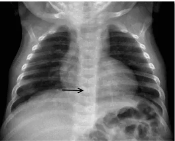
At 13 weeks old, our patient was admitted to the gastroenterology clinic because of jaundice. Alagille syn-drome was diagnosed due to the findings of persistent jaundice, peripheral pulmonary artery stenosis, typical facies and butterfly vertebrae (Fig. 1). The diagnosis was established with a de novo mutation of the protein-encoding JAG1-region of chromosome 20.
In addition to clinical follow-up, liver US was performed every 6 months. At the age of 10 months the child developed biliary cirrhosis with hepato-splenomegaly, portal hypertension and oesophageal varices grade I. At the age of 36 months nodular changes of the liver parenchyma were noted. There was a hypoechoic, 2.7-cm well-defined liver lesion in segment VI/VII and another large, isoechoic nodular lesion in segment V displacing the adjacent vessels (Fig. 2).
Since there were new focal liver lesions with different sonographic echotextures and elevated alpha fetoprotein (AFP) levels, these lesions were further characterized with a standard paediatric MRI-liver protocol and dynamic
S. C. Wetli
:
E. S. Gralla:
E. Stranzinger (*)Division of Diagnostic, Interventional and Pediatric Radiology, Department of Radiology, Neuroradiology and Nuclear Medicine, Bern University Children’s Hospital,
Freiburgstrasse 10, 3010 Bern, Switzerland
e-mail: enno.stranzinger@insel.ch S. Schibli
Division of Pediatric Gastroenterology,
Hepathology and Nutrition, Bern University Children’s Hospital, Bern, Switzerland
Pediatr Radiol (2010) 40:1696–1698 DOI 10.1007/s00247-010-1784-6
contrast-enhanced sequences with gadolinium using a 1.5-T magnet (Figs.3and 4).
The liver lesion in segment VI/VII showed slight hyperin-tense signal on the T2-W sequence and hypoinhyperin-tense signal on the T1-W sequence. The lesion was characterized by early enhancement in the arterial phase and a wash-out phenome-non in the late phases on the dynamic contrast-enhanced images. This enhancement pattern was consistent with a malignant lesion, most likely a HCC. The larger liver lesion in segment V had similar intensity to liver parenchyma on all sequences and the T1-W dynamic contrast-enhanced images showed isointense signal in all phases with central hypo-intense signal and a prominent central vessel. This enhance-ment pattern was consistent with a HNH. A biopsy of the likely malignant lesion confirmed a moderately differentiated HCC.
As the only curative option, orthotopic liver transplan-tation was performed and histological investigation of the liver confirmed the diagnosis of HCC of the smaller lesion and regenerating nodule of the larger lesion.
Discussion
There are only a few reported cases in the literature of associated liver tumours (HCC and HNH) in Alagille syndrome and the youngest child described is a 17-month-old boy [2–5].
US is the imaging method of choice for the first evaluation and routine follow-up examination of the liver in children. An inhomogeneous and echogenic liver paren-chyma may be seen due to steatosis, fibrosis and/or cirrhosis of the liver. Blane et al. [6] described that liver lesions in children cannot be characterized using US alone and further imaging methods must be performed. Dynamic contrast-enhanced US (CEUS) imaging of focal liver lesions is a promising technique, but still not approved for children.
Fig. 2 US of the liver at 36 months of age. A new 2.7-cm hypoechoic liver lesion (arrow) and a large isoechoic liver lesion (asterisk) are detected
Fig. 1 Chest radiograph at 13 weeks of age with a normal cardiothymic silhouette. Butterfly vertebrae of the lower thoracic spine are noted (arrow)
Fig. 3 MRI of the liver at 36 months of age. a Axial T2-W and (b) axial T1-W sequences of the liver. The liver lesion in segment VI/VII (arrow) shows slight hyperintense signal in the T2-W image and hypointense signal in the T1-W image. The larger lesion in segment V (asterisk) has almost similar in-tensity to the liver parenchyma in both sequences. A prominent central vessel is noted
Contrast-enhanced dynamic techniques are used to differentiate among vascular lesions, HNH or malignant lesions demonstrating a characteristic enhancement pattern (reported sensitivity of HCC >1 cm with dynamic contrast imaging is up to 87% with a specificity of 79% [7]. For benign liver lesions the accuracy is 83.1% [8].)
MRI and US are both methods without ionizing radiation and with good soft-tissue contrast. The advantage of MRI is a significantly better overview of the liver and the detection of several lesions at the same time, while in CEUS only part of the liver per administration of contrast medium may be continuously imaged. On the other hand, there is usually no need for sedation or anaesthesia when performing CEUS.
The MRI findings in our case showed a different contrast-enhancement pattern between the malignant (early enhancement and wash-out) and the benign liver lesion. The regenerating nodule showed an isointense enhancement pattern, a central hypointense scar and a prominent central vessel that has been described in the literature to be consistent with HNH [5].
In summary, Alagille syndrome is a rare disease resulting in cirrhosis of the liver in children. Complications include focal liver lesions such as HCC and US follow-up studies are mandatory to detect them as early as possible. Contrast-enhanced dynamic imaging of the liver is recommended to
characterize these lesions and plays an important role for further management.
References
1. Lowe LH, Schlesinger AE (2008) Congenital abnormalities, Alagille syndrome. In: Slovis TL et al (eds) Caffey’s pediatric diagnostic imaging, 11th edn. Mosby, Philadelphia, p 1861 2. Wegmann W, Evison J, Schaub N et al (1996) Liver cell carcinoma
as a late complication of Alagille syndrome (arterio-hepatic dysplasia). Leber Magen Darm 26:157–158, 161-153
3. Bhadri VA, Stormon MO, Arbuckle S et al (2005) Hepatocellular carcinoma in children with Alagille syndrome. J Pediatr Gastro-enterol Nutr 41:676–678
4. Tajima T, Honda H, Yanaga K et al (2001) Hepatic nodular hyperplasia in a boy with Alagille syndrome: CT and MR appearances. Pediatr Radiol 31:584–588
5. Kim B, Park SH, Yang HR et al (2005) Hepatocellular carcinoma occurring in alagille syndrome. Pathol Res Pract 201:55–60 6. Blane CE, Jongeward RH Jr, Silver TM (1983) Sonographic features
of hepatocellular disease in neonates and infants. AJR 141:1313–1316 7. Choi SH, Lee JM, Yu NC et al (2008) Hepatocellular carcinoma in liver transplantation candidates: detection with gadobenate dimeglumine-enhanced MRI. AJR 191:529–536
8. Strobel D, Seitz K, Blank W et al (2009) Tumor-specific vascularization pattern of liver metastasis, hepatocellular carcino-ma, hemangioma and focal nodular hyperplasia in the differential diagnosis of 1,349 liver lesions in contrast-enhanced ultrasound (CEUS). Ultraschall Med 30:376–382
Fig. 4 MRI of the liver at 36 months of age. T1-W dynamic contrast-enhanced sequences with gadolinium. The lesion in segment VI/VII (arrow) shows hypointense signal without contrast agent (a), early enhancement in the arterial phase (b) and a wash-out phenomenon in
the late phases (c and d). The larger liver lesion in segment V (asterisk) shows almost isointense signal in all phases and a central hypointense signal. The prominent vessel is best visible in the arterial phase (b)