Sebastian Leschka Paul Stolzmann Hans Scheffel Simon Wildermuth André Plass Michele Genoni Borut Marincek Hatem Alkadhi Received: 13 March 2008 Revised: 30 April 2008 Accepted: 5 May 2008 Published online: 19 July 2008
# European Society of Radiology 2008
Prevalence and morphology of coronary
artery ectasia with dual-source CT
coronary angiography
Abstract To assess the prevalence and morphological characteristics of coronary artery ectasia (CAE) with CT coronary angiography (CTCA) in comparison to conventional catheter-angiography (CCA). Dual-source CTCA examinations from 677 con-secutive patients (223 women; median age 57 years) were retrospectively evaluated by two blinded observers for the presence of CAE defined as a diameter enlargement≥1.5 times the diameter of adjacent normal coronary segments. Vessel diameters and con-trast attenuation within and proximal to ectatic segments were measured. CCA was used to compare measure-ments obtained from CTCA with the coronary flow velocity by using the thrombolysis in myocardial infarction (TIMI) frame count. CTCA identified CAE in 20 of 677 (3%) patients. CCA was performed in ten of these patients.
CAE diameter measurements with CTCA (10.0±5.4 mm) correlated sig-nificantly (r=0.92, p<0.001) with the CCA measurements (8.8±4.9 mm), but had higher diameters (levels of agreement:−1.0 to 3.4 mm). Contrast attenuation was significantly lower in the ectatic (343±63 HU) than in the proximal (394±60 HU) segments (p< 0.01). The attenuation difference sig-nificantly correlated with the CAE ratio (r=0.67, p<0.01) and the TIMI frame count (r=0.58, p<0.05). The prevalence of CAE in a population examined by CTCA is around 3%. Contrast attenuation measurements with CTCA correlate well with the flow alterations assessed with CCA.
Keywords Coronary artery ectasia . Coronary artery anatomy .
CT coronary angiography . Catheter coronary angiography
Introduction
Coronary artery ectasia (CAE) is characterized by an inappropriate dilatation of the coronary vessel [1] and has a reported prevalence of 1–5% [2–4]. The etiology and pathology of CAE and its clinical significance remains poorly understood. Although the literature supports the notion that no difference in survival exists between patients with and those without CAE [2], there is a growing body of evidence suggesting that CAE is not a benign condition or normal variant of coronary artery anatomy. Ectatic arteries have been shown to be more prone to spasm [5], exercise-induced myocardial ischemia [6], thrombosis [7], dissec-tion [8], or rupture [9]. Moreover, the severity of
myocar-dial ischemia has been shown to significantly correlate with the degree of luminal enlargement [6]. The detection of CAE might be important as those patients might benefit from medical treatment with antiplatelet and anticoagula-tion agents or surgery [10]. The latter treatment is recommended in patients with CAE complications and for saccular CAE because of the higher risk of thrombosis and rupture [10].
In conventional catheter angiography (CCA), CAE is usually defined as an enlargement of a coronary artery segment with the diameter of the ectatic segment being more than 1.5 times larger than an adjacent normal-appearing vessel segment [1, 3]. Falsetti and Carroll [1] further subdivided CAE into simple ectasia (a 1.5–2-fold S. Leschka . P. Stolzmann .
H. Scheffel . B. Marincek . H. Alkadhi (*)
Institute of Diagnostic Radiology, University Hospital Zurich, Raemistrasse 100, 8091 Zurich, Switzerland e-mail: [email protected] Tel.: +41-1-2553662 Fax: +41-1-2554443 S. Leschka . S. Wildermuth Institute of Radiology, Kantonsspital St. Gallen,
St. Gallen, Switzerland A. Plass . M. Genoni
Clinic for Cardiovascular Surgery, University Hospital Zurich, Zurich, Switzerland
segment dialation) and an aneurysm (a >2-fold segment dilatation when compared to a normal segment). Further signs include delayed antegrade dye filling, segmental back flow, and local dye stasis in the ectatic coronary segment, which represent disturbances in blood flow filling and washout [6,11].
Recent advances in multi-detector row computed tomo-graphy coronary angiotomo-graphy (CTCA) have continuously increased its role for non-invasive imaging of the coronary arteries [12–16]. The most recent technical innovation is dual-source CT, which is characterized by a high and consistent temporal resolution of 83 ms through simulta-neous acquisition of data with two X-ray tubes and detectors [17–19]. CTCA is now routinely performed as a non-invasive filter test for demonstrating or excluding coronary artery stenosis in patients with a low to interme-diate pre-test probability of CAD [20–22]. Thus, a variety of associated cardiac abnormalities are increasingly encountered in patients for the first time with CTCA. To date, only two anecdotal reports [23,24] have demonstra-ted imaging findings of CAE with CTCA, and the morphological features and the prevalence of CAE with CTCA have not been described in a larger series.
The purpose of this study was to assess the prevalence and morphologic characteristics of CAE with CTCA in a large patient population in comparison to conventional catheter angiography (CCA). In addition, we sought to
correlate the contrast attenuation measurements obtained with CTCA, with the hemodynamic flow alterations within ectatic segments in CCA.
Materials and methods
Our institutional review board approved this retrospective study; written informed consent was waived.
We retrospectively reviewed all CTCA examinations performed in a 12-month period from July 2006 to June 2007 at our institute. Within the study time period, 677 consecutive patients (223 women, 454 men; mean age 57±13 years; age range 39 to 83 years) were scheduled for a clinically indicated CTCA examination. All of these patients suffered from chest pain, had a negative or equivocal stress test, and an intermediate pre-test probability of CAD according to the scoring method of Morise et al. [25] (i.e., 9 to 15 points). Patient characteristics are summarized in Table1. From these 677 patients, 278 individuals (93 women, 185 men; mean age 59±14 years; age range 44 to 83 years) had an additional CCA workup within 6 months of the CT examination. The median time interval between CTCA and CCA was 28 days (range 1 to 113 days).
Exclusion criteria for CTCA were renal dysfunction (serum creatinine level >1.5 mg/dl), previous reaction to iodinated contrast agent, and pregnancy.
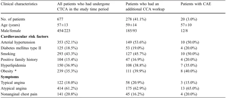
Table 1 Clinical characteristics of the study population
Clinical characteristics All patients who had undergone CTCA in the study time period
Patients who had an additional CCA workup
Patients with CAE
No. of patients 677 278 (41.1%) 20 (3.0%)
Age (years) 57±13 59±14 57±10
Male/female 454/223 185/93 12/8
Cardiovascular risk factors
Arterial hypertension 353 (52.1%) 149 (53.6%) 10 (50.0%)
Diabetes mellitus type II 125 (18.5%) 53 (19.0%) 4 (20.0%)
Smoking 293 (43.3%) 127 (45.7%) 10 (50.0%)
Positive family history 104 (15.4%) 47 (16.9%) 4 (20.0%)
Hyperlipidemia 150 (36.9%) 108 (38.8%) 7 (35.0%)
Obesity * 239 (35.3%) 111 (39.9%) 8 (40.0%)
Symptoms
Typical angina 122 (18.0%) 58 (20.9%) 3 (15.0%)
Atypical angina 414 (61.2%) 175 (62.9%) 13 (65.0%)
Nonanginal chest pain 141 (20.8%) 45 (16.2%) 4 (20.0%)
*Obesity was defined as body mass index >27 kg/m2according to the scoring method of Morise et al. [25]
Dual-source CT protocol and image reconstruction
The patients were examined on a dual-source CT system (Somatom Definition, Siemens Medical Solutions, For-chheim, Germany). All patients received a single dose of 2.5 mg isosorbiddinitrate s. l. (Isoket, Schwarz Pharma, Monheim, Germany). The amount (71±6 ml; range 65 to 80 ml) of contrast material (iodixanol, Visipaque 320, 320 mg/ml, GE Heathcare, Buckinghamshire, UK) was adjusted to the individual CT data acquisition time and to the body mass index for each patient. It was injected at a flow rate of 5 ml/s followed by 50 ml of a 20% contrast agent/80% saline solution mixture. The contrast agent application was controlled by bolus tracking in the ascending aorta (signal attenuation threshold 140 HU). CT was performed in the cranio-caudal direction from the level of the carina to the diaphragm, using the following parameters: detector collimation 2×32×0.6 mm, slice collimation 2×64×0.6 mm by means of a z-flying focal spot, gantry rotation time 330 ms, pitch of 0.2–0.5 depending on the heart rate, tube current time product 350 mAs per rotation, and tube potential 120 kV. Electro-cardiography pulsing for radiation dose reduction was used in all patients [26]: with mean heart rates below 60 bpm, the full tube current was applied from 60 to 70%, at 61– 70 bpm from 50 to 80%, and with heart rates above 70 bpm an RR-interval from 30 to 80% was used. The CT data were reconstructed with a slice thickness of 0.75 mm, a reconstruction increment of 0.5 mm, and using a soft-tissue convolution kernel (B26f) during mid-diastole with 60% to 70% in 5% steps of the R-R interval. When motion artifacts were present in these datasets, additional recon-structions were performed in 5% steps within the full tube current window. The estimated radiation dose using this CTCA protocol was approximately 7–9 mSv [27]. CTCA data analysis
CTCA data analysis was performed by two observers in consensus who were both unaware of the clinical history of the patients and blinded to the findings in CCA. Coronary artery segments were defined according to a scheme from the American Heart Association [28]. The right coronary artery was defined to include segments 1–4, the left anterior descending artery to include segments 5–10, and the left circumflex artery to include segments 11–15.
First, the observers assessed in consensus each coro-nary segment for the presence or absence of CAE according to the criteria of Falsetti and Carroll [1]. A simple ectasia is defined as a 1.5–2-fold dilatation and an aneurysm as a >2-fold dilatation of the segment in comparison with an adjacent proximal normal segment. A normal segment was defined as a coronary artery
segment without arteriosclerotic plaques, stenosis, or ectasia/aneurysm.
Second, both observers measured independently the ectatic diameters as the maximum diameter of each ectatic segment and the difference to the mean of the diameter measurements in normal segments proximal to the CAE. From these measurements, the ectasia ratio (the ratio of the diameter of the ectatic segment to the mean diameter of adjacent normal segments) was calculated. Vessel dia-meters were measured on reconstructions perpendicularly oriented to the vessel centerline using an electronic caliper tool. According to the ectasia ratio, CAE were divided into two groups: ectasia with a ratio of 1.5- to 2.0 and aneurysms with a ratio larger than 2.0 [1].
Third, both observers independently placed a region of interest in the ectatic segment and in the normal adjacent proximal coronary segment in order to estimate distur-bances in blood flow. The region of interest was defined as large as possible, carefully avoiding calcifications, plaques, and stenoses. The extent of the flow disturbance was defined as the difference in contrast enhancement between the ectatic and that in an adjacent normal-appearing proximal coronary segment. Figure 1 demonstrates the measurements of diameter and the contrast attenuation.
According to Markis et al. [11], the distribution of CAE was classified by both observers in consensus in each patient into four distinct types: type I, diffuse ectasia of two or three vessels; type II, diffuse disease in one vessel and localized disease in another vessel; type III, diffuse ectasia of one vessel only; type IV, localized or segmental ectasia. Ectatic segments were classified as localized when they involved a discrete portion of the segment with another portion being of normal vessel diameter within that segment, and diffuse when the entire segment was ectatic with no normal portion within the segment [11]. CAE was further classified according to the anatomical shape of the ectatic segment into a saccular type (i.e., spherical shape with the transverse dimension longer than the longitudinal dimension) and a fusiform type (i.e., spindle shape with the longitudinal dimension larger than the transverse dimension) [11].
Both observers assessed (in consensus) the CTCA data of patients with CAE with respect to the presence or absence of significant coronary artery stenosis. Significant stenosis was defined as luminal diameter reduction exceeding 50% [13–16].
Catheter coronary angiography
CCA was performed by the Judkins technique. All patients undergoing CCA received intracoronary glyceryl trinitrate prior to dye injection. Cineangiograms were filmed at 30 frames per second. At least two views in different planes were obtained for quantitative analysis. The films of all
patients with evidence of CAE in CTCA were reviewed by one experienced observer (who was not involved in the read-out of the CTCA data) with regard to the presence of CAE without knowledge of the clinical findings or the results of CTCA. The maximum diameter of each ectatic segment was measured in the view in which it appeared the largest using an electronic caliper. Similar to CTCA, the diameter of an adjacent normal-appearing proximal coro-nary segment was used as reference diameter. The tip of the coronary catheter was used for calibration of diameter measurements. Coronary segments were defined according to the scheme of the American Heart Association [28], similar to CTCA.
For evaluation of coronary flow velocity in CAE, we determined the “thrombolysis in myocardial infarction " (TIMI) frame-count method for each coronary artery with CAE as described by Gibson et al. [29]. The first frame used for counting was the frame in which dye was initially seen to enter the artery. The last frame was defined as the one in which the dye first entered the end-point branch of the target artery. Then, measurements of the frame count for each artery were made by subtracting the first from the last frame. The commonly longer TIMI frame counts of the
left anterior descending artery were corrected for by dividing the value through 1.7, as recommended [29].
The presence or absence of coronary artery stenosis in patients with CAE was assessed in CCA in the same fashion as described above for CTCA.
Statistical analysis
The statistical analysis was performed using SPSS software (version 12.0, SPSS Inc., IL). The continuous variables were presented as means ± standard deviation, and the categorical variables as percentages. The quantitative values were compared using the two-sided Student’s t test. Pearson’s correlation coefficients were used to test for inter-reader agreement regarding the diameter and attenu-ation measurements in CTCA. The Bland-Altman analysis was used to illustrate the relation between CTCA and CCA measurements of ectatic segment diameters. A one-sample t-test was used to determine whether the resulting mean difference was significant from zero, representing a significant under- or overestimation with CTCA. Pearson’s correlation test was used to analyze the relation between Fig. 1 CTCA in a 48-year-old female patient with isolated coronary
artery ectasia of the distal circumflex artery (CX). On the left side, a curved-planar reformation shows the ectasia in the distal CX with normal-appearing adjacent coronary segments. The inlay demon-strates the relation of ectasia (arrow) to the first postero-lateral branch
(PL1). Perpendicular views indicated by a and b show diameter measurements of the normal proximal coronary segment (a1) and the ectatic segment (b1). In the same planes, the attenuation was measured by placing a region of interest in both segments (a2, b2)
the contrast attenuation difference for each CAE and the coronary artery ectasia ratio with CTCA and to analyze the relation between contrast attenuation differences in CTCA and the TIMI frame count in CCA. AP value of less than 0.05 was considered significant.
Results
Prevalence of CAE with CTCA
CAE (i.e., both ectasias and aneurysms) were identified in 20 of the 677 patients (3.0%; 8 women, 12 men; median age 57 years; age range 39 to 82 years) with a total of 27 coronary arteries and 45 coronary segments involved. CCA was performed in ten of these patients (50%). Both CCA and CTCA measurements were available for 15 coronary arteries (55.6%; 15/27) and 21 coronary segments (46.7%; 21/45) with CAE.
With CTCA, five patients (25%) had an isolated CAE with vessel enlargement being the only pathological finding in otherwise normal coronary arteries. Eleven patients (55%) with CAE had associated non-significant stenosis (≤50%) in other segments, while 4 patients (20%) with CAE had significant stenosis (>50%) in other segments. In both patients with significant stenosis and in eight patients with non-significant stenosis with CTCA, CCA was available and confirmed the CTCA diagnosis.
Location and type of CAE with CTCA
CAEs (i.e., both ectasias and aneurysms) were most frequently found in the right coronary artery (48.1%, 13/27), and less often in the left anterior descending (29.6%, 8/27) and circumflex artery (22.3%, 6/27). CAE was segmental in 35.6% (16/45) and diffuse in 64.4% (29/45) of the ectatic segments. Saccular type CAE was found in 40% (18/45) and fusiform type in 60% of the ectatic segments (27/45).
According to the Markis classification [11], type I was present in 20% of the patients (4/20), type II in 10% (2/20), type III in 25% (5/20), and type IV in 45% (9/20). CAE affected only one coronary artery in 75% of patients (15/20), while CAE was present in two or more coronary arteries in the remaining 25% of patients (5/20). Fifty percent of isolated CAEs that involved only one vessel were found in the right coronary artery (7/14).
Diameter of CAE and coronary ectasia ratio with CTCA and CCA
For CTCA measurements, intra-reader agreement was excellent for diameter measurements of adjacent normal
coronary artery segments (mean difference 0.2±0.3 mm, range 0–0.7 mm, r=0.93, p<0.01) and CAE measurements (mean difference 0.3±0.4 mm, range 0–0.7 mm, r=0.95, p<0.01). Therefore, the mean of diameter measurements from both observers were used for further calculations.
The mean diameter of all 45 CAE segments (i.e., both ectasias and aneurysms) with CTCA was 8.2±4.2 mm (range 3.9–26.2 mm). Compared to normal adjacent coronary segments (mean 3.1±0.7 mm, range 2.1– 4.8 mm), the mean diameter difference between ectatic and normal segments with CTCA was 5.1±4.2 mm (range 1.8–23.2 mm).
For the 21 CAE visualized with both CTCA and CCA, the mean diameter of the ectatic segments at CTCA was 10.0±5.4 mm (range 5.4–26.2 mm). The mean diameter of normal coronary segments was 2.9±0.6 mm (range 2.1– 4.4 mm), with a mean diameter difference of 7.1±5.5 mm (range 2.4–23.2 mm) between ecstatic and normal coronary segments. In comparison, diameter measurements with CCA in the same coronary segments were 8.8±4.9 mm (range 4.3–24.9 mm) for ectatic segments, 2.8±0.5 mm (range 2.0–4.2 mm) for normal adjacent coronary seg-ments, with a mean diameter difference of 6.0±5.0 mm (range 1.7–22.2 mm). There was a significant correlation for diameter measurements between CTCA and CCA (r= 0.92, p<0.001; Fig. 2a). The Bland-Altman analysis revealed a tendency towards higher measurements with CTCA (mean 1.2±1.1 mm; levels of agreement: −1.0 to 3.4; Fig.2b), with a significant overestimation of CTCA as compared to CCA (p<0.001). Table2 summarizes diam-eter measurements of CAE with both modalities.
According to the coronary ectasia ratio calculated from CTCA measurements in all CAE (mean 2.8±1.6, range 1.6– 8.7), ectasias were present in 18 segments (40%; mean ectasia ratio 1.8±0.2, range 1.6–2.0) and aneurysms in 27 segments (60%; mean ectasia ratio 3.4±1.8, range 2.1–8.7). Difference in attenuation values with CTCA
in comparison to CCA
The intracoronary attenuation measurements showed excellent inter-reader agreement for both normal segments (mean difference 15±11 HU, range 4–29 HU, r=0.92, p< 0.01) and CAE (mean difference 18±12 HU, range 9–27 HU, r=0.91, p<0.01). For all 45 CAE (i.e., both ectasia and aneurysms), the mean intracoronary attenuation within the affected segments (376 ± 78 HU; range 135–523 HU) was significantly lower than the mean attenuation in adjacent proximal normal coronary segments (418±70 HU; range 200–559 HU; p<0.01), resulting in a mean attenuation difference of 42±21 HU (range 9–92 HU) between the affected and non-affected segments. For CAE visualized with both modalities, the attenuation in 21 CAE segments was 343±63 HU (range 135–450 HU), the attenuation in
the adjacent proximal normal coronary segments was 394± 60 HU (range 200–487 HU), and the mean attenuation difference was 51±25 HU (range 9–92 HU). There was a significant correlation between the attenuation differences with the coronary ectasia ratio (r=0.67, p<0.01) as determined with CTCA.
The attenuation difference measured by CTCA signifi-cantly correlated with the coronary flow velocity evaluated by using the TIMI frame count (rB=0.58, p<0.05; Fig.3).
The attenuation difference was significantly lower in ectasia (i.e., 1.5–2-fold dilatation) with 34±12 HU (range 9–50 HU) in comparison to aneurysms (i.e., >2-fold dilatation) with 61±24 HU (range 25–118 HU; p<0.05). The same was valid for the TIMI frame count with 36.9± 6.1 frames (range 27.5–47.1 frames) in coronary arteries with ectasia and 44.6 ± 7.8 frames (range 33–55 frames) in aneurysms (p<0.05).
Discussion
This study investigated the prevalence and morphology of CAE in a series of 677 consecutive patients who were referred to CTCA for non-invasive imaging of their coronary arteries. This study demonstrates the feasibility of CTCA to identify and to evaluate the morphology of CAE. Moreover, this study is the first to estimate the flow disturbances in CAE by calculating the contrast attenuation differences between the ectatic and the adjacent normal coronary segments followed by a data correlation with the TIMI frame-count method.
Using CTCA for the diagnosis of CAE, our incidence of 3.0% is comparable to what was reported in CCA studies of 1–5% [2–4]. Only 0.6% of our patients had a CAE with no significant CAD in other segments, confirming that CAE is a rare isolated finding [6]. Although all coronary arteries can be affected by CAE, the disease is unilateral in approximately 75% of the patients [30]. Our findings were similar to those of previous reports [30,31], where the right coronary artery is the most commonly affected vessel. In addition, the distribution of Markis’ types of CAE in our study was comparable to a recent publication, showing type II to be the least frequent variant [4].
Although the literature supports the notion that no survival difference exists between patients with and those without CAE [2], there is a growing body of evidence suggesting that CAE is not a benign condition or normal variant of coronary artery anatomy. Ectatic arteries have been shown to be more prone to spasm [5], exercise-induced myocardial ischemia [6], thrombosis [7], dissec-tion [8], or rupture [9]. Moreover, the severity of myocar-dial ischemia has been shown to significantly correlate with the degree of luminal enlargement [6]. As presented in our study, measurements of vessel diameters with CTCA and CCA showed an excellent correlation. However, a signif-icant tendency towards higher diameter measurements with CTCA as compared to CCA was found. This is most likely explained by the foreshortening of true vessel dimensions with CCA, which is usual for projection imaging, but not for cross-sectional imaging techniques. Another reason could be the difference in the action profile of nitroglycerin leading to different effects on the vessel wall when using two different administration routes (i.e., sublingually for CT and intracoronary for CCA).
Fig. 2 (a) The linear regression plot between the mean diameters of the ectatic segments (n=21) measured by CCA (y-axis) and CTCA (x-axis). The dotted lines represent 95% confidence limits. There is a significant correlation between measurements with CCA and CTCA (Pearson correlation, r=0.92; p<0.001). (b) The Bland-Altman analysis between the mean diameter measurements of the ectatic segments (n=21) by CCA and CTCA. The dotted lines represent the limits of agreement (mean ±2 SD). There is a tendency towards higher diameter values with CTCA when compared to CCA (mean 1.2±1.1 mm; levels of agreement:−1.0 to 3.4)
In general, one of the major weaknesses of CTCA is the lack of hemodynamic information on blood flow direction and velocity [32]. Explained by the physical law of Hagen-Poiseuille, disturbances in flow are an inherent character-istic of changes in the cross-section of the vessel as demonstrated by the equation of Bernoulli. To quantify coronary blood flow with CCA, the TIMI frame-count method has been introduced [29]. As CAE is associated with reduced blood flow velocity [33, 34], a significant relationship has been observed between increased TIMI
frame count and increased diameter of the ectatic segment [35] and coronary ectasia ratio [35,36]. With CTCA, there is no means to measure the velocity of coronary blood flow velocity because CTCA is not a time-resolved imaging technique. Instead, the angiographic signs of delayed filling, segmental back flow, and local deposition of contrast medium could theoretically epitomize reduced intracoronary contrast attenuation when compared to the contrast attenuation of normal proximal segments. As a matter of fact, in this study we have observed that the attenuation difference as measured with CTCA signifi-cantly correlated with the TIMI frame count as measured with CCA. Thus, attenuation difference measurements in CAE with CTCA might serve as indirect evidence for dynamic flow alterations that otherwise cannot be assessed with CTCA.
There are some study limitations. First, our retrospective survey has revealed only 20 patients with CAE representing a low number for evaluation of morphological character-istics. In addition, data were incomplete because CCA was performed in only half of the patients presenting with CAE in CTCA. Therefore, our results are preliminary, and the small sample size possibly limits the transfer of these results to clinical practice. Second, the prevalence of CAE in our study of patients who had CTCA does not represent that of the general population. Finally, we correlated the contrast attenuation differences to estimate flow disturbances in CAE with the TIMI frame-count method, although Doppler guide-wire investigations might have been more appro-priate for flow velocity measurements.
In conclusion, CTCA is feasible to identify and to evaluate the morphology of CAE. The prevalence of CAE in a patient population that had CTCA is around 3.0%. Vessel diameter measurements of ectatic segments with CTCA significantly correlate with measurements with Fig. 3 The linear regression plot of contrast attenuation differences
measured with CTCA (y-axis) and the thrombolysis in myocardial infarction (TIMI) frame count (x-axis) as measured with CCA (n= 21). The dotted lines represent 95% confidence limits. A significant correlation between the contrast attenuation differences and the TIMI frame count is shown (Pearson correlation,r=0.58; p<0.05)
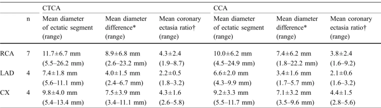
Table 2 Diameter measurements of the 21 segments with CAE that were visualized with both CTCA and CCA
CTCA CCA n Mean diameter of ectatic segment (range) Mean diameter difference* (range) Mean coronary ectasia ratio† (range) Mean diameter of ectatic segment (range) Mean diameter difference* (range) Mean coronary ectasia ratio† (range) RCA 7 11.7±6.7 mm 8.9±6.8 mm 4.3±2.4 10.0±6.2 mm 7.4±6.2 mm 3.8±2.4 (5.5–26.2 mm) (2.6–23.2 mm) (1.9–8.7) (4.5–24.9 mm) (1.8–22.2 mm) (1.6–9.2) LAD 4 7.4±1.8 mm 4.0±1.5 mm 2.2±0.5 6.6±2.0 mm 3.4±1.6 mm 2.1±0.6 (5.6–11.1 mm) (2.4–6.7 mm) (1.8–3.2) (4.3–9.9 mm) (1.7–5.7 mm) (1.6–3.2) CX 4 9.8±4.0 mm 7.5±3.9 mm 4.3±1.6 9.2±3.3 mm 7.1±3.2 mm 4.4±1.5 (5.4–13.4 mm) (3.4–11.1 mm) (2.6–5.8) (5.5–11.7 mm) (3.5–9.6 mm) (2.8–5.6) *Mean diameter difference between ectatic and normal proximal segments
†Mean coronary ectasia ratio calculated as ratio between the diameter of the ectatic segment and the diameter of the adjacent normal proximal segment
Abbreviations: CTCA, computed tomography coronary angiography; CCA, catheter coronary angiography; RCA, right coronary artery; LAD, left anterior descending artery; CX, left circumflex artery
CCA. Moreover, attenuation differences between ectatic and normal coronary segments with CTCA significantly correlate with flow alterations obtained through TIMI frame-count measurements with CCA.
Acknowledgements This research was supported by the National Center of Competence in Research, Computer-Aided and Image-Guided Medical Interventions of the Swiss National Science Foundation, Switzerland.
References
1. Falsetti HL, Carrol RJ (1976) Coronary artery aneurysm. A review of the literature with a report of 11 new cases. Chest 69:630–636
2. Swaye PS, Fisher LD, Litwin P et al (1983) Aneurysmal coronary artery disease. Circulation 67:134–138 3. Manginas A, Cokkinos DV (2006)
Coronary artery ectasias: imaging, functional assessment and clinical im-plications. Eur Heart J 27:1026–1031 4. Lam CS, Ho KT (2004) Coronary
artery ectasia: a 10-year experience in a tertiary hospital in Singapore. Ann Acad Med Singapore 33:419–422 5. Suzuki H, Takeyama Y, Hamazaki Y et
al (1994) Coronary spasm in patients with coronary ectasia. Cathet Cardio-vasc Diagn 32:1–7
6. Kruger D, Stierle U, Herrmann G, Simon R, Sheikhzadeh A (1999) Exer-cise-induced myocardial ischemia in isolated coronary artery ectasias and aneurysms (“dilated coronopathy”). J Am Coll Cardiol 34:1461–1470 7. Perlman PE, Ridgeway NA (1989)
Thrombosis and anticoagulation thera-py in coronary ectasia. Clin Cardiol 12:541–542
8. Huikuri HV, Mallon SM, Myerburg RJ (1991) Cardiac arrest due to spontane-ous coronary artery dissection in a patient with coronary ectasia-a case report. Angiology 42:148–151 9. Satoda M, Tatsukawa H, Katoh S
(1998) Images in cardiovascular medi-cine. Sudden death due to rupture of coronary aneurysm in a 26-year-old man. Circulation 97:705–706 10. Ramappa P, Kottam A, Kuivanemi H,
Thatai D (2007) Coronary artery ectasia - is it time for a reappraisal? Clin Cardiol 30:214–217
11. Markis JE, Joffe CD, Cohn PF, Feen DJ, Herman MV, Gorlin R (1976) Clinical significance of coronary arte-rial ectasia. Am J Cardiol 37:217–222 12. Leber AW, Knez A, von Ziegler F et al
(2005) Quantification of obstructive and nonobstructive coronary lesions by 64-slice computed tomography: a comparative study with quantitative coronary angiography and intravascular ultrasound. J Am Coll Cardiol 46:147– 154
13. Leschka S, Alkadhi H, Plass A et al (2005) Accuracy of MSCT coronary angiography with 64-slice technology: first experience. Eur Heart J 26:1482– 1487
14. Mollet NR, Cademartiri F, van Mie-ghem CA et al (2005) High-resolution spiral computed tomography coronary angiography in patients referred for diagnostic conventional coronary angi-ography. Circulation 112:2318–2323 15. Ong TK, Chin SP, Liew CK et al
(2006) Accuracy of 64-row multide-tector computed tomography in detect-ing coronary artery disease in 134 symptomatic patients: influence of cal-cification. Am Heart J 151:1323 e1321–1323 e1326
16. Raff GL, Gallagher MJ, O’Neill WW, Goldstein JA (2005) Diagnostic accu-racy of noninvasive coronary angiog-raphy using 64-slice spiral computed tomography. J Am Coll Cardiol 46:552–557
17. Achenbach S, Ropers D, Kuettner A et al (2006) Contrast-enhanced coronary artery visualization by dual-source computed tomography-initial experi-ence. Eur J Radiol 57:331–335 18. Flohr TG, McCollough CH, Bruder H
et al (2006) First performance evalua-tion of a dual-source CT (DSCT) system. Eur Radiol 16:256–268 19. Scheffel H, Alkadhi H, Plass A et al
(2006) Accuracy of dual-source CT coronary angiography: first experience in a high pre-test probability population without heart rate control. Eur Radiol 16:2739–2747
20. Budoff MJ, Achenbach S, Blumenthal RS et al (2006) Assessment of coronary artery disease by cardiac computed tomography: a scientific statement from the American Heart Association Com-mittee on Cardiovascular Imaging and Intervention, Council on Cardiovascu-lar Radiology and Intervention, and Committee on Cardiac Imaging, Coun-cil on Clinical Cardiology. Circulation 114:1761–1791
21. Fox K, Garcia MA, Ardissino D et al (2006) Guidelines on the management of stable angina pectoris: executive summary: the Task Force on the Management of Stable Angina Pectoris of the European Society of Cardiology. Eur Heart J 27:1341–1381
22. Hendel RC, Patel MR, Kramer CM et al (2006) ACCF/ACR/SCCT/SCMR/ ASNC/NASCI/SCAI/SIR 2006 appro-priateness criteria for cardiac computed tomography and cardiac magnetic res-onance imaging: a report of the Amer-ican College of Cardiology Foundation Quality Strategic Directions Committee Appropriateness Criteria Working Group, American College of Radiolo-gy, Society of Cardiovascular Com-puted Tomography, Society for Car-diovascular Magnetic Resonance, American Society of Nuclear Cardiol-ogy, North American Society for Car-diac Imaging, Society for Cardiovas-cular Angiography and Interventions, and Society of Interventional Radiolo-gy. J Am Coll Cardiol 48:1475–1497 23. Wells TA, Peebles CR, Gray HG (2008)
Giant left anterior descending coronary artery aneurysm. Int J Cardiol 126(2): e27–e28 Epub 2007 Mar 28
24. Goz M, Cakir O (2007) Multiple cor-onary artery aneurysms that cause thrombosis: 22-month follow-up results with multi-slice spiral computerized tomography without surgery. Int J Cardiol 119(2):e48–e50 Epub 2007 May 2
25. Morise AP, Haddad WJ, Beckner D (1997) Development and validation of a clinical score to estimate the proba-bility of coronary artery disease in men and women presenting with suspected coronary disease. Am J Med 102:350– 356
26. Leschka S, Scheffel H, Desbiolles L et al (2007) Image quality and recon-struction intervals of dual-source CT coronary angiography: recommenda-tions for ECG pulsing windowing. Invest Radiol 42:543–549
27. Stolzmann P, Scheffel H, Schertler T et al (2008) Radiation dose estimates in dual-source computed tomography coronary angiography. Eur Radiol 18:592–599
28. Austen WG, Edwards JE, Frye RL et al (1975) A reporting system on patients evaluated for coronary artery disease. Report of the Ad Hoc Committee for Grading of Coronary Artery Disease, Council on Cardiovascular Surgery, American Heart Association. Circula-tion 51:5–40
29. Gibson CM, Cannon CP, Daley WL et al (1996) TIMI frame count: a quanti-tative method of assessing coronary artery flow. Circulation 93:879–888 30. Aintablian A, Hamby RI, Hoffman I,
Kramer RJ (1978) Coronary ectasia: incidence and results of coronary by-pass surgery. Am Heart J 96:309–315 31. Hartnell GG, Parnell BM, Pridie RB
(1985) Coronary artery ectasia. Its prevalence and clinical significance in 4993 patients. Br Heart J 54:392–395
32. Frauenfelder T, Boutsianis E, Schertler T et al (2007) In-vivo flow simulation in coronary arteries based on computed tomography datasets: feasibility and initial results. Eur Radiol 17:1291– 1300
33. Akyurek O, Berkalp B, Sayin T, Kumbasar D, Kervancioglu C, Oral D (2003) Altered coronary flow proper-ties in diffuse coronary artery ectasia. Am Heart J 145:66–72
34. Hamaoka K, Onouchi Z, Kamiya Y, Sakata K (1998) Evaluation of coronary flow velocity dynamics and flow re-serve in patients with Kawasaki disease by means of a Doppler guide wire. J Am Coll Cardiol 31:833–840
35. Papadakis MC, Manginas A, Cotileas P et al (2001) Documentation of slow coronary flow by the TIMI frame count in patients with coronary ectasia. Am J Cardiol 88:1030–1032
36. Kosar F, Acikgoz N, Sahin I, Topal E, Aksoy Y, Cehreli S (2005) Effect of ectasia size or the ectasia ratio on the thrombosis in myocardial infarction frame count in patients with isolated coronary artery ectasia. Heart Vessels 20:199–202