O R I G I N A L A RT I C L E
Jameel M. Inal
Complement C2 receptor inhibitor trispanning: from man
to schistosome
Received: 4 April 2005 / Accepted: 20 May 2005 / Published online: 19 October 2005 # Springer-Verlag 2005
Abstract Horizontal gene transfer (HGT), in relation to genetic transfer between hosts and parasites, is a little described mechanism. Since the complement inhibitor CRIT was first discovered in the human Schistosoma parasite (the causative agent of Bilharzia) and in Trypanosoma cruzi (a parasite causing Chagas’ disease), it has been found to be distributed amongst various species, ranging from the early teleost cod to rats and humans. In terms of evolutionary distance, as measured in a phylogenetic analysis of these CRIT genes at nucleotide level, the parasitic species are as removed from their human host as is the rat sequence, suggesting HGT. The hypotheses that CRIT in humans and schistosomes is orthologous and that the presence of CRIT in schistosomes occurs as a result of host-to-parasite HGT are presented in the light of empirical data and the growing body of data on mobile genetic elements in human and schistosome genomes. In summary, these data indicate phylogenetic proximity between Schistosoma and human CRIT, identity of function, high nucleotide/amino acid identity and secondary protein structure, as well as identical genomic organization.
CRIT is found in the platyhelminth human parasiteSchistosoma
Complement C2 receptor inhibitor trispanning (CRIT) represents a novel family of receptors, the first member of which was found in Schistosoma haematobium and, subsequently, Schistosoma mansoni [14]. Schistosome is a platyhelminth worm and is the causative agent of Bilharzia. Around 250 million people worldwide, mainly in tropical countries, are afflicted
J. M. Inal ())
Immunonephrology, Department of Research, University Hospital Basel, Basel, Switzerland e-mail: [email protected] . Fax: +44-20-71332184
J. M. Inal
Department of Health and Human Sciences, London Metropolitan University, 166-220 Holloway Road, London, N7 8DB, UK
with the disease. The intermediate host is a fresh waterborne snail. The infectious stage, which is released from the snail into the water, is called the cercaria. Infection is by means of penetration of the skin by a combination of proteolytic action and vigorous movement of the cercarial tail. Upon infection and shedding of the tail, the cercaria transforms into the larval stage, termed the schistosomulum. The mature worms, which live in copulo, settle in mes-enteric venules of the intestine or those surrounding the urinary bladder. The pathology of the disease is due to the granulomatous response against eggs which, having been shed by the female worm, become trapped in the liver, intestinal wall, spleen or other organs [4,26].
CRIT was found by screening an adult worm cDNA library of S. haematobium with a‘vaccination serum’ obtained from baboons that had been vaccinated with γ-irradiated cercariae. This serum was passively protective to challenge infection with S. mansoni cercariae. Therefore, antigens that were recognized by this serum, especially those that were found on the surface of the parasite, were deemed good vaccine candidate antigens. In such a screening, aλgt11 clone containing the CRIT cDNA was found and sequenced [14]. At this point, an affinity-purified polyclonal antibody against a 27-mer synthetic peptide based on the N-terminal putative first extracellular domain 1 (ed1) region (Fig.1) was generated to reveal the native CRIT protein as an approximately 32-kDa protein. The CRIT mRNA transcript at 1.35 kb meant that the open reading frame (ORF) of 0.86 kb was approximately of full length, thereby confirming the size of the native protein.
The parasite’s sensitivity to complement (C) is dependent on its life cycle stage. The infectious cercariae, which are free-swimming, having been shed from the intermediate host,
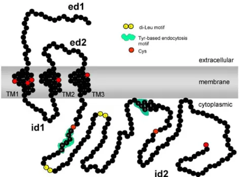
Fig. 1 Schematic topology prediction of CRIT showing N-terminal extracellular domains (ed1 and ed2), three N-terminal transmembrane domains (TM1, TM2 and TM3), two intracytoplasmic domains (id1) and the cytoplasmic tail (id2). Also indicated are tyrosine-based endocytosis motifs in id2, as well as cysteine residues shown in red
which is a fresh waterborne snail, are C-sensitive but become C-resistant upon infection and transformation to larval schistosomulae living within their host [6]. We found that CRIT expression at the mRNA level was greater in the C-resistant schistosomulae than in the adult worm [14], although we do not know if this translates to an increased level of CRIT expression at the protein level. Interestingly, others have found the mRNA expression of CRIT to be still higher in the eggs [11], which is not surprising as eggs, too, need to be protected en route to the environment ex vivo. The C-resistant schistosomulae and mature adult worm are able to evade complement by expressing proteins that mimic known C-regulators and by acquiring at least one of the host’s C-regulatory proteins. By acquiring DAF from human erythrocytes, the adult worm can disassemble the classical and alternative pathway C3 convertases. By mimicking human CD59, the Schistosoma SCIP-1 protein binds the late complement components C8 and C9 of the multiple attack complex, thereby preventing C-mediated lysis[33]. CRIT is a recently described complement regulatory receptor, which appears to target the early complement pathway. By sharing antigenic determinants with the C2-binding C component C4 and by acting as a decoy C2-binding receptor, CRIT regulates C activation on the surface of schistosomulae by limiting the amount of C3 convertase formation.
Using scanning electron microscopy, we showed CRIT to be located on the surface of the many pits and channels in the tegument of the adult worm. These pits and channels (Fig.2) are filled with host blood and, by increasing the surface area of the interface between the parasite and the host blood, aid in the quicker absorption of nutrients from the blood. It is therefore likely that part(s) of the extracellular domains of CRIT interacts with component(s) in human blood. CRIT in humans is also found on the surface—in this case on the plasma membrane of a monocytic cell, as seen in Fig.2b.
The membrane topology of CRIT, as predicted by several algorithms, shows a molecule with an extracellular N-terminus and a cytoplasmic tail (Fig.1). Since this is the typical topology of a type IIIb membrane protein [38] (extracellular N-terminus, several trans-membrane domains and no cleavable signal peptide), we went on to find a likely ligand for this putative receptor. Besides this prediction, we had empirical data for this topology and so, by using extracellular domains (ed1 or ed2), we tried to isolate the ligand from normal human serum by affinity chromatography. This formed the basis of a simple ‘receptor affinity chromatography’ experiment [20] in which we arbitrarily used a synthetic peptide, chosen to be ed1, coupled to an epoxy-activated Sepharose affinity column through those amino acid side chains containing NH2, -OH or -SH groups. Amongst the few serum proteins
spe-Fig. 2 Scanning electron micrographs of anti-CRIT-ed1/immunogold-labelled aSchistosoma haematobium surface tegument and b human monocyte cell surface. CRIT protein is expressed in the surface pits and channels found in the Schistosoma tegument, which presents an interface with the host blood. CRIT is also found on the plasma membrane of the monocyte from where it may be able to undergo endocytosis
cifically bound to the matrix, after extensive washing and then elution from the column with low-pH glycine, a protein identified by N-terminal sequencing and confirmed by spe-cific recognition with a monoclonal antibody was human complement C2. With this finding, our initial working hypothesis was that the C2-binding CRIT receptor could play a role in regulating the activation of CP (Classical Pathway) on the parasite surface. To this end, we and others found that the synthetic peptide CRIT-ed1 inhibited CP activation both in vitro [18,31] and in vivo [19,32].
However, CRIT was also found to be phosphorylated on tyrosine and to have a plethora of putative binding sites on the cytoplasmic tail or intracellular domain 2 (id2) (Fig.1) for a host of signalling molecules, including tyrosine kinases such as Fyn. In schistosomes, such molecules are believed to play a role in cell growth and development, with a Fyn-like tyrosine kinase having been found in schistosomes [23]; this strongly suggests, as was speculated recently [15], that CRIT may play similar roles, in addition to the regulation of complement. In its cytoplasmic tail, CRIT has characteristic Tyr-X-X-Leu and Tyr-X-X-Ile motifs as well as two dileucine motifs (Fig. 1), which could be involved in receptor endocytosis that is mediated via the clathrin-coated pit pathway. Although endocytosis has not been shown in schistosomes, a homologue of a rat component of the adaptor complex that links clathrin to receptors in coated vesicles exists in Schistosoma japonicum [11], as does an S. mansoni orthologue of a Caenorhabditis elegans clathrin coat assembly protein [40]. S. mansoni also expresses a clathrin heavy-chain protein and dynamin [40], which is associated with endocytic sorting of proteins. The cytoplasmic tail of CRIT also has tyrosine-based motifs, which, if phosphorylated, could be consensus motifs for binding to SH2 domains of various cytoplasmic tyrosine kinases, including Fyn. In mammals, amongst other functions, fyn is involved in T-cell receptor (TCR) signalling, binding to the CD3– TCR complex and being activated when the TCR is crosslinked. Fyn is also involved in platelet-derived growth factor receptor mitogenic signalling and, from studies of fyn−/− mice, it participates in the differentiation of myelin-producing oligodendrocytes as well as keratinocytes. Fyn also participates in cell-adhesion-mediated signalling [34].
CRIT is a novel complement regulator in humans
Human CRIT was recently cloned from testis and pancreas cDNA [16]. This study confirmed CRIT binding to C2 by ligand blotting and flow cytometry, and addressed the mechanism by which CRIT regulates the classical pathway. CRIT expression at the protein level was looked at in various tissues. CRIT was expressed strongly in glandular epithelial cells of proliferating —but not secretory—endometrium and pancreatic islet cells (probably insulin-producing β-cells). In the kidney, where the activation of complement is believed to expedite the progression of chronic renal damage [17], CRIT (in terms of protein expression and CRIT mRNA transcription) was uniquely confined to podocyte cells within the kidney glomerulus. This constitutive expression was confirmed in a more recent investigation (to be described in greater detail elsewhere), which showed CRIT upregulation in glomeruli in membranous nephropathy. In the testis, CRIT was expressed in cells in testicular tubules attached to the basement membrane, which were presumably Sertoli cells. CRIT was also found on a range of haemopoietic cells as well as on certain endothelial cells.
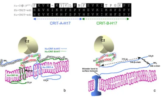
When C2 is bound to CRIT-ed1 peptide [18] or native CRIT on the cell surface [16], the cleavage of C2 by C1s is inhibited. This is neither the case with the homologous C4β212–232 peptide, based on the so-called CRIT-ed1 domain of C4 [18], nor with native C4, of course. It could be that both CRIT-ed1 and the CRIT-ed1 domain from C4 bind C2, but it is only when CRIT-ed1 binds that there is a conformational change in the structure of C2, preventing cleavage at the C1s site or a steric interference that prevents C1s-mediated cleavage. In another work reported elsewhere, we have confirmed theβ-chain of C4 as possessing an important binding site for C2 and have begun to reveal the role of the vWFA1 domain of C2 in this interaction. CRIT appears to exist as a dimer, possibly a homodimer, as it is phosphorylated on tyrosine, primarily in the dimeric form (unpublished observations), and C2 binds both to the monomeric and dimeric forms, as shown in far Western blot analysis [16]. Based on this, we compared (Fig.3a) the sequences of CRIT-H17 motifs from two CRIT molecules, CRIT‐A and CRIT‐B, laid contiguously with the two CRIT‐H17 motifs (F231–N240 and F241–Y251) comprising the C4β‐chain (F231–Y251). As shown in Fig.3a there is a 58% identity and 73% similarity across the region. We have also presented a schematic model diagram of CRIT and how we might expect it to exist in the membrane as a homodimer (Fig.3b). We also show, by comparison, the CRIT-ed1 domain in the C4 β-chain. The two CRIT-H17 motifs NH2-FEVKKYVLPN-CO2H and NH2
-FEVKITPGKPY-Fig. 3 Alignment of amino acid sequence of the CRIT-ed1 domain of the human C4 β-chain F231 –Y251
with S. haematobium and human CRIT-ed1. a The C-terminal 11-amino-acid CRIT-ed1, the so-called CRIT-H17 motif, is shown. In this schematic, two such motifs have been placed contiguously to represent how two such regions may come into close proximity in a CRIT homodimer made up of CRIT-A and CRIT-B. b A schematic of theβ-chain of C4, with emphasis on the predicted C2-binding ed1 domain. c A schematic of two CRIT molecules constituting a homodimer in which two CRIT-H17 motifs might be brought together in such a way as to represent the equivalent of the CRIT-ed1 domain in the C4β-chain
CO2H, of which the CRIT-ed1 domain in the C4β-chain is composed, are separated by a Pro239-inducedβ-turn, and Fig.3c illustrates how they might be expected to lie.
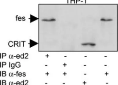
An interesting feature of the human CRIT molecule and its parasite orthologue, compared with the membrane regulators of complement activation (mRCA, membrane Regulators of Complement Activation), including DAF, CD59, CR1 and MCP, is its unusually long cytoplasmic tail at 163 residues, suggesting further roles besides complement regulation. In a submitted article describing the downstream molecular associations of the CRIT cytoplasmic tail in greater detail, human CRIT was found to be phosphorylated on tyrosine as was previously shown for its Schistosoma homologue [14]. CRIT also associates with the cytoplasmic tyrosine kinase fes (Fig.4), which is believed to play a role in the terminal differentiation of myeloid cells. At least in certain cell types, it thus appears likely that CRIT functions in cellular signalling.
CRIT phylogeny suggests that human andSchistosoma CRIT are orthologous
Horizontal gene transfer (HGT), as may occur between species, can dramatically alter the evolution of eukaryotic genomes. The main driving force for this is the presence of mobile genetic elements, principally long terminal repeat (LTR) retrotransposons and retroviruses, within a genome and the later acquisition of envelope protein genes that could then render such retroviruses infectious, thereby enabling HGT.
On comparing the genetic organization of CRIT in different species, it is clear that they all possess a single exon structure. Phylogenetic analyses at both the nucleotide level (Fig.5) and at the amino acid level (not shown) suggest that schistosome CRIT (which is absent from the nematode worm C. elegans) has a closer phylogenetic relationship to human CRIT than to rat CRIT. Furthermore, besides pairwise alignment showing these proteins to be 87.5% identical at the amino acid level, they have almost identical secondary structures (not shown) and share some cross-reactive epitopes [16]. The high degree of identity between human and Schistosoma CRIT is particularly notable in the immunologically important part of the receptor, namely the ligand binding ed1, which shows 92% identity at the amino acid level, as well as ed2 (88% identity). Whether one considers CRIT on a haematopoietic cell or on the surface of a Schistosoma adult worm, both molecules possess the identical function of interacting via ed1 with the same protein in normal human serum, this being human C2. The transmembrane domains also have a high degree of similarity, with the greatest sequence difference being in the N-terminal region of the cytoplasmic tail, proximal to the third transmembrane domain (TM3). This is hardly surprising as the intracellular milieu of cytoplasmic signalling molecules possibly interacting with the CRIT cytoplasmic tail is likely to differ between the species. In summary, the available data, which include both CRIT molecules being receptors for an identical ligand, nucleotide identity (including similarity of genomic organization), amino acid identity (and closeness of phylogenetic relationship) and secondary structure identity, point toward the principally haemopoietic, expressed human CRIT and Schistosoma CRIT genes being orthologous. That the CRIT receptor shares no significant overall sequence homology with other receptors—or, indeed, any other proteins in the database—and that there is significant sequence homology between Schistosoma and human CRIT suggest that Schistosoma CRIT arose as a result of a rare host-to-parasite HGT. A recently described example of another orthologous gene in Schistosoma and human, also
presumed to be transferred from the host to the parasite through the intervention of mobile genetic elements, is that of the S. mansoni aspartic protease gene cathepsin D [28]. In schistosomes, this digests host haemoglobin from ingested human blood [1]. Although human lysosomal cathepsin D appears to also digest haemoglobin, in this case, its action is part of the recycling of old erythrocytes, which occurs in the liver and spleen.
Considering host complement regulators, which could disguise the parasite as‘host’ in terms of the immune system, molecular mimicry in the parasite–host relationship can be achieved in three ways. The first mechanism involves directly adsorbing such proteins (e.g., human DAF) onto the schistosome surface [7], in this case from human erythrocytes. In the second mechanism, homologues of human proteins, such as DAF and CD59 are normally expressed by trypanosomes [22, 30] and schistosomes [33], respectively. The third mechanism, reviewed by Damian [3], involves naturally occurring retroviral vectors, which, having captured the host gene, can mediate HGT on infection of the parasite. Retroviral-related sequences were first found in schistosomes in preliminary studies in which antibodies against BALB virus 2 envelope glycoprotein (gp70) reacted with Schistosoma adult worms and in which homologous DNA sequences to the gag and pol regions of the ecotropic murine leukaemia virus were detected in Southern blots of Schistosoma adult worm DNA [39]. These rather tenuous findings were supported by later works in which in situ hybridization was used to show the transcription of host (mouse)-related DNA sequences in the adult worm Fig. 4 CRIT associates with the cytoplasmic tyrosine kinase fes. Immunoprecipitation with the anti-CRIT-ed2 of Jurkat (T lymphocyte) and THP-1 (monocyte) cells and immunoblotting with anti-fes show CRIT to be associated with the cytoplasmic tyrosine kinase fes in THP-1 cells. No such association was shown in Jurkat cells, which possess CRIT but lack the fes tyrosine kinase (not shown)
Fig. 5 Unrooted phylogenetic tree based on nucleotides 1–846 of the human CRIT. These were compared with the nucleotides of rat CRIT and those from the human parasites S. haematobium and T. cruzi. The topology algorithm used was that of the European Molecular Biology Laboratory, European Bioinformatics Institute Molecular Biology Server. The evolutionary distance is represented by the length of the line segments, which is indicated in brackets 0 0.02 0.04 0.06 0.08 0.1 0.12 evolutionary distance TcCRIT (0.005) ShCRIT (0.003) HuCRIT (0.005) RatCRIT (0.064)
and infectious cercarial stages of S. japonicum and S. mansoni. Using32P-labelled probes of mouse type C and type A retroviruses and of the env-specific region of the mouse ecotropic type C retrovirus, hybridization was observed in subtegumental and inner tissues of S. japonicum and S. mansoni adults, respectively, but interestingly not in cercariae of either species [21]. In later confirmatory studies, it was found that mouse type A and type C retroviral sequences could be transmitted horizontally from the host to the schistosomes [12]. Others have also suggested the acquisition of host genes by parasites [36]; such genetic exchanges could explain the high sequence homology between the parasite (particularly Schistosoma) and the host. In other supportive works using in situ polymerase chain reaction (PCR) and hybridization, the histocompatibility complex (major histocompatibility complex or MHC) class I sequence of the mouse was found in the genome of S. mansoni [12], indicating the horizontal transmission of class I MHC from the host to the parasite [13]. Using similar techniques, the mouse type 2 Alu sequence (B2), a repetitive DNA sequence in the mouse genome, was found in the adult worm body of S. mansoni and S. japonicum as were mouse retrovirus-related sequences.
The best support for horizontal gene transmission between the host and the parasite would be to find a mobile genetic element; to this aim, other workers more recently identified a retrovirus-like LTR retrotransposon, called Boudicca [2]. Retrotransposons are mobile genetic elements that transpose by reverse transcription. They may be subdivided into LTR (long terminal repeats) retrotransposons, which include retroviruses, and non-LTR retro-transposons or long interspersed nucleotidic elements, which lack LTRs. Some LTR transposons have been misclassified as transposable elements, which, having been subsequently found to be infectious, should be reclassified as endogenous retroviruses. Similarly, it has been speculated for some time that today’s retrotranposons originated from ancient endogenous retroviruses that lost their infectivity. Approximately 20% of the Schistosoma genome of about 270 Mbp [37] is believed to be made up of retrotransposons [25]. Recently, DeMarco et al. [5] described four new ones: Saci-1, Saci-2 and Saci-3, which are of the LTR variety, and one non-LTR-expressed retrotransposon Perere, which is integrated into the S. mansoni genome. Characterization of the retrotransposon Boudicca revealed three likely ORFs with 5′ and 3′ LTRs of 328 bp. ORF1 consists of a retrovirus-like major homology region and a Cys/His box motif, as is typically found in the Gag polyprotein of similar retrotransposons and retroviruses. ORF2 contains contiguous aspartic protease, reverse transcriptase, RNAase H and integrase enzymatic domains. This is a structure similar to that of a retrovirus polyprotein, as might be found in the gypsy/Ty3 retrotransposons; indeed, when the reverse transcriptase sequence of ORF2 was used to search the database, Boudicca was confirmed as a gypsy-like retrotransposon and shown to be closely related to CsRn1 from the oriental liver fluke Clonorchis sinensis and to kabuki from Bombyx mori. The third ORF at the 3′ end was deemed to encode an envelope protein, which, if confirmed, would confer the ability of host cell entry, enabling Boudicca to undergo vertical as well as horizontal transmission. This study also found over a thousand copies of Boudicca to be dispersed throughout the Schistosoma genome, which accounted for up to 4% of the genome of S. mansoni. The active transcription of Boudicca was confirmed in the sporocyst, cercaria and adult worm stages of S. mansoni by reverse transcription PCR.
CRIT phylogeny and parasite tropism for blood cells: implications for transmission of CRIT gene from host to parasite
A phylogenetic analysis at the nucleotide level of the sequences of human and rat CRIT and the parasite S. haematobium and Trypanosoma cruzi (Fig.5) shows that the parasite cluster is no less related than rat CRIT is to human CRIT, implying that the most likely origin of parasite (Schistosoma or Trypanosoma) CRIT is indeed the host. Although host-to-parasite HGT has not been reported much, one example is that of the protease inhibitor chagasin of T. cruzi [35] another one occurring in the semiparasitic mite Proctolaelaps regalis of the fruit fly Drosophila [10]. As parasite genomes begin to be deciphered, more parasite homologues of host proteins are discovered; indeed, the latest transcriptome analysis of S. japonicum [11] and S. mansoni [40] parasites has revealed many receptors for growth factors, hormones and cytokines shared with humans.
The CRIT gene in Schistosoma is presumably of human origin. Despite being apparently intronless, although we cannot as yet rule out possible intron(s) in the 5′ and/or 3′ un-translated regions, CRIT is, like many G-protein-coupled receptors, not a pseudogene. As verification of the functional status of the transferred CRIT gene, we can confirm the expression status of the Schistosoma CRIT gene by the presence of an expressed sequence tag (EST) for CRIT in the Schistosoma EST database. This replaces the need for confirmatory expression data using other empirical methods, such as primer extension analysis. That CRIT is expressed as a functional protein [14] and that it is therefore not a pseudogene are further shown by the recognition of the native 32-kDa Schistosoma protein with anti-CRIT-ed1 in a lysate of the adult worm. In other experiments, Schistosoma CRIT ORF was expressed as a recombinant protein recognized by the vaccination serum VBabS [14]. Human CRIT was expressed using a cell-free in vitro transcription/translation system; here, a∼32-kDa protein was recognized by anti-CRIT-ed1, anti-ed2 and anti-id2 [16].
As well as in Schistosoma, the CRIT gene is found in the kinetoplastid parasite T. cruzi [14,16]. The trypomastigote stage of this parasite invades cells (typically of the heart muscle or liver) and white blood cells, and reproduces as amastigote. On cell death, some amastigotes may infect new cells, but others will transform into trypomastigotes, remaining in the bloodstream until the vector takes another blood meal. With the presence of T. cruzi trypomastigotes in peripheral blood, it is clear why they need to express CRIT. This may be found to be even more apparent in the related T. brucei, the causative agent of African sleeping sickness, because here the trypomastigotes replicate asexually solely in the peri-pheral blood. On these grounds, we can also understand the need for CRIT expression on the schistosome larval stage, especially the adult worm, which may live for up to 5 years in the host vasculature, as well as the released eggs.
Further evidence that the Schistosoma CRIT gene and probably that of T. cruzi were acquired independently via HGT is the lack of ubiquity amongst human parasites living in the vasculature. For example, CRIT is not found in Plasmodium falciparum. We were unable to find a CRIT homologue of this parasite by Western blot analysis using anti-ed1 antibody, or by PCR using degenerate oligonucleotides (unpublished data). More recently, this has been confirmed by similarity searches of the virtually completely sequenced P. falciparum genome. P. falciparum parasites are also believed to acquire host genes by HGT; in the case of CRIT, it is interesting to speculate that its absence in this parasite may be because P. falciparum spends a large part of its life cycle replicating as a merozoite in the protected environment of the host’s erythrocytes, which, curiously, do not express CRIT [16]. CRIT is
also absent from Leishmania major, another protozoan of the order Kinetoplastida. This parasite reproduces as an amastigote within circulating macrophages, which also lack CRIT expression [16]. For now, whether CRIT is absent from parasites that spend only a limited part of their life cycle in the peripheral blood of the host, or is absent due to a lack of mobile genetic elements, or is absent because the CRIT gene has been lost from certain parasites, remains open to speculation.
Hitherto, gene transfers amongst eukaryotes were thought to occur rarely, although this perception is changing. The plethora of new eukaryotic genome sequences becoming avail-able is only now providing material for phylogenomic analysis. Whilst HGTs are relatively rare amongst multicellular eukaryotes, this is not the case with regard to unicellular eu-karyotes, such as Trypanosoma [9], which lacks a separate germ line and thus must tolerate the acquisition of foreign genes to survive.
CRIT is found in the teleost cod fishGadhus morhua
The CRIT gene was found in cod ([16] and further work to be reported elsewhere), suggesting that CRIT genes may have evolved from a common ancestral gene, at least dating as far back as the earliest teleosts. CRIT is a receptor for mammalian C2; but in teleost fish, in particular in cod, although there is a classical pathway [27], there appears to be functional redundancy between alternative and classical pathways and, recently, fB/C2 genes have been isolated from carp [29], zebrafish [8] and medaka fish [24]. In evolutionary terms, the teleosts show the first compartmentalization of the immune system, including the first appearance of an important lymphoid organ—the thymus. Within the thymus, CRIT appears to be expressed in some thymocytes and macrophages, and possibly in Hassall’s bodies as determined by immunohistochemistry using anti-CRIT-ed1 antibody and by in situ hybridization using an antisense CRIT probe (Lange, S., unpublished data). As in humans, by immunoblotting, we found CRIT to be present on cod lymphocytes. Further work will be needed to establish whether CRIT has a complement regulatory or as yet unidentified role in fish.
Conclusions
CRIT is a complement regulatory receptor in humans, which, in either its monomeric or dimeric form, attempts to regulate the amount of complement activation on the autologous cell surface by competing with C4b for binding C2. Furthermore, C2 bound to CRIT cannot be activated by C1s cleavage, so a‘variant’ C3 convertase cannot be formed. CRIT is also found in the Schistosoma parasite, located in blood-filled pits and channels within the surface tegument of the adult worm. CRIT is also found in T. cruzi, although it has not been localized and we suppose from a phylogenetic analysis at the nucleotide level that the CRIT gene was acquired by these human parasites by the so far rarely described process of HGT from the host. Interestingly, CRIT is not found in the free-living nematode worm C. elegans and appears not to exist in Drosophila melanogaster. In both Schistosoma and humans, CRIT is phosphorylated on tyrosine, with the molecule having nine tyrosine residues in its cyto-plasmic tail and interacting with the cytocyto-plasmic tyrosine kinase fes, which is possibly associated with terminal myeloid cell differentiation. In evolutionary terms, CRIT is found at
least as far back as the early teleosts, having been cloned from the cod fish; this begs the question as to whether it is also found in the more primitive echinoderms. Looking at the CRIT family as a whole, besides the described function in complement regulation, we believe that there are alternative functions relating to the receptor’s signalling capacity within the cell. It will be interesting to study these in relation to the various CRIT species along the phylogenetic tree.
References
1. Brindley PJ, Kalinna BH, Wong JYM (2001) Proteolysis of human haemoglobin by schistosome cathepsin D. Mol Biochem Parasitol 112:103
2. Copeland CS, Brindley PJ, Heyers O et al (2003) Boudicca, a retrovirus-like long terminal repeat retrotransposon from the genome of the human blood fluke Schistosoma mansoni. J Virol 77:6153 3. Damian RT (1997) Parasite immune evasion and exploitation: reflections and projections. Parasitology
115:S169
4. deJesus AR, Silva A, Santana LB (2002) Clinical and immunological evaluation of 31 patients with acute schistosomiasis mansoni. J Infect Dis 185:98
5. DeMarco R, Kowaltowski A, Machado AA et al (2004) Saci-1, -2, and -3 and Perere, four novel retrotransposons with high transcriptional activities from the human parasite Schistosoma mansoni. J Virol 78:2967
6. Deng J, Gold D, LoVerde PT et al (2003) Inhibition of the complement membrane attack complex by Schistosoma mansoni paramyosin. Infect Immun 71:6402
7. Fatima M, Horta M, Ramalho-Pinto EJ (1991) Role of human decay-accelerating factor in the evasion of Schistosoma mansoni from the complement-mediated killing in vitro. J Exp Med 174:1399
8. Gongora, Figueroa F, Klein J (1998) Independent duplications of Bf and C3 complement genes in the zebrafish. Scand J Immunol 48:651
9. Hannaert V, Saavedra E, Duffieux F et al (2003) Plant-like traits associated with metabolism of Trypanosoma parasites. Proc Natl Acad Sci U S A 100:1067
10. Houck MA, Clark JB, Peterson KR et al (1991) Possible horizontal transfer of Drosophila genes by the mite Proctolaelaps regalis. Science 253:1125
11. Hu W, Yan Q, Shen DK et al (2003) Evolutionary and biomedical implications of a Schistosoma japonicum complementary DNA resource. Nat Genet 35:139
12. Imase A, Kobayashi K, Ohmae H et al (2001) Horizontal and vertical transmission of mouse class I sequence in Schistosoma mansoni. Parasitology 123:163
13. Imase A, Matsuda H, Irie Y et al (2003) Existence of host DNA sequences in schistosomes—horizontal and vertical transmission. Parasitol Int 52:369
14. Inal JM (1999) Schistosoma TOR (trispanning orphan receptor), a novel, antigenic surface receptor of the blood-dwelling, Schistosoma parasite. Biochim Biophys Acta 1445:283
15. Inal JM (2004) Parasite interaction with host complement: beyond attack regulation. Trends Parasitol 20:407
16. Inal JM, Hui KM, Miot S et al (2005) Complement C2 receptor inhibitor trispanning: a novel human complement inhibitory receptor. J Immunol 174:356
17. Inal JM, Pascual M, Lesavre P et al (2003) Complement inhibition in renal diseases. Nephrol Dial Transplant 18:237
18. Inal JM, Schifferli JA (2002) Complement C2 Receptor Inhibitor Trispanning and theβ-chain of C4 share a binding site for complement C2. J Immunol 168:5213
19. Inal JM, Schneider B, Armanini M et al (2003) A peptide derived from the parasite receptor, Complement C2 Receptor Inhibitor Trispanning, suppresses immune complex-mediated inflammation in mice. J Immunol 170:4310
20. Inal JM, Sim RB (2000) A Schistosoma protein, Sh-TOR, is a novel inhibitor of complement which binds human C2. FEBS Lett 470:131
21. Irie Y, Iwamura Y (1993) Host-related DNA sequences are localized in the body of schistosome adults. Parasitology 107:519
22. Joiner KA, da Silva WD, Rimoldi MT et al (1988) Biochemical characterization of a factor produced by trypomastigotes of Trypanosoma cruzi that accelerates the decay of complement C3 convertases. J Biol Chem 263:11327
23. Kapp K, Schussler P, Kunz W et al (2001) Identification, isolation and characterization of a Fyn-like tyrosine kinase from Schistosoma mansoni. Parasitology 122:317
24. Kuroda N, Wada H, Naruse K et al (1996) Molecular cloning and linkage analysis of the Japanese medaka fish complement Bf/C2 gene. Immunogenetics 44:459
25. Laha T, Brindley PJ, Verity CK et al (2002) Pido, a non-long terminal repeat retrotransposon of the chicken repeat 1 family from the genome of the Oriental blood fluke, Schistosoma japonicum. Gene 284:149
26. Lambertucci JR (1993) Schistosoma mansoni: pathological and clinical aspects. In: Jordan P, Webbe G, Sturrock RF (eds) Human schistosomiasis. CAB International, Wallingford, UK, pp 195–235 27. Magnadottir B (2000) The spontaneous haemolytic activity of cod serum: heat insensitivity and other
characteristics. Fish Shellfish Immunol 10:731
28. Morales ME, Kalinna BH, Heyers O et al (2004) Genomic organization of the Schistosoma mansoni aspartic protease gene, a platyhelminth orthologue of mammalian lysosomal cathepsin D. Gene 338:99 29. Nakao N, Matsumoto M, Nakazawa M et al (2002) Diversity of complement factor B/C2 in the common carp (Cyprinus carpio): three isotypes of B/C2-A expressed in different tissues. Dev Comp Immunol 26:533
30. Norris KA, Bradt B, Cooper NR et al (1991) Characterization of a Trypanosoma cruzi C3 binding protein with functional and genetic similarities to the human complement regulatory protein, decay-accelerating factor. J Immunol 147:2240
31. Oh K-S, Kweon M-H, Rhee KI-H et al (2003) Inhibition of complement activation by recombinant Sh-CRIT-ed1 analogues. Immunology 110:73
32. Oh K-S, Na DK, Kweon MH et al (2003) Expression and purification of the anticomplementary peptide Sh-CRIT-ed1 (formerly Sh-TOR-ed1) as a tetramultimer in Escherichia coli. Protein Expr Purif 27:202 33. Parizade M, Arnon R, Lachmann PJ, et al (1994) Functional and antigenic similarities between a 94-kD
protein of Schistosoma mansoni (SCIP-1) and human CD59. J Exp Med 179:1625 34. Resh MD (1998) Fyn, a Src family tyrosine kinase. Int J Biochem Cell Biol 30:1159
35. Rigden DJ, Monteiro ACS, Fatima M et al (2001) The protease inhibitor chagasin of Trypanosoma cruzi adopts an immunoglobulin-type fold and may have arisen by horizontal gene transfer. FEBS Lett 504:41 36. Salzet M, Capron A, Stefano GB (2000) Molecular crosstalk in host–parasite relationships: schistosome–
and leech–host interactions. Parasitol Today 16:536
37. Simpson AJ, Sher A, McCutchan TF (1982) The genome of Schistosoma mansoni: isolation of DNA, its size, bases and repetitive sequences. Mol Biochem Parasitol 6:126
38. Singer SJ (1990) The structure and insertion of integral proteins in membranes. Annu Rev Cell Biol 6:247
39. Tanaka M, Iwamura Y, Amanuma H et al (1989) Integration and expression of murine retrovirus-related sequences in schistosomes. Parasitology 99:31
40. Verjovski-Almeida S, DeMarco R, Martins EA et al (2003) Transcriptome analysis of the coelomate human parasite Schistosoma mansoni. Nat Genet 35:148