INTERVENTIONAL NEURORADIOLOGY
Selective inferior petrosal sinus sampling without venous
outflow diversion in the detection of a pituitary adenoma
in Cushing
’s syndrome
Lukas Andereggen&Gerhard Schroth&Jan Gralla&Rolf Seiler&Luigi Mariani&
Jürgen Beck&Hans-Rudolf Widmer&Robert H. Andres&Emanuel Christ&
Christoph Ozdoba
Received: 19 April 2011 / Accepted: 7 July 2011 / Published online: 2 August 2011 # Springer-Verlag 2011
Abstract
Introduction Conventional MRI may still be an inaccu-rate method for the non-invasive detection of a micro-adenoma in adrenocorticotropin (ACTH)-dependent
Cushing’s syndrome (CS). Bilateral inferior petrosal sinus
sampling (BIPSS) with ovine corticotropin-releasing hormone (oCRH) stimulation is an invasive, but accurate, intervention in the diagnostic armamentarium surround-ing CS. Until now, there is a continuous controversial debate regarding lateralization data in detecting a micro-adenoma. Using BIPSS, we evaluated whether a highly selective placement of microcatheters without diversion of venous outflow might improve detection of pituitary microadenoma.
Methods We performed BIPSS in 23 patients that met clinical and biochemical criteria of CS and with equivocal MRI findings. For BIPSS, the femoral veins were catheterized bilaterally with a 6-F catheter and the inferior petrosal sinus bilaterally with a 2.7-F micro-catheter. A third catheter was placed in the right femoral
vein. Blood samples were collected from each catheter to determine ACTH blood concentration before and after oCRH stimulation.
Results In 21 patients, a central-to-peripheral ACTH gradient was found and the affected side determined. In 18 of 20 patients where transsphenoidal partial hypophy-sectomy was performed based on BIPSS findings, micro-adenoma was histologically confirmed. BIPSS had a sensitivity of 94% and a specificity of 67% after oCRH stimulation in detecting a microadenoma. Correct
localiza-tion of the adenoma was achieved in all Cushing’s disease
patients.
Conclusion BIPSS remains the gold standard in the detection of a microadenoma in CS. Our findings show that the selective placement of microcatheters without venous outflow diversion might further enhance better recognition to localize the pituitary tumor.
Keywords Angiography, Digital subtraction . Cushing disease . Petrosal sinus sampling . Pituitary gland . Magnetic resonance imaging
Introduction
Cushing’s syndrome (CS) is a hormonal disorder charac-terized by an increase in circulating cortisol. Endogenous hypercortisolism may be adrenocorticotropin (ACTH) dependent (pituitary or an ectopic source) or independent
(related to the anatomy of the adrenal glands) [1, 2].
ACTH-dependent pituitary disease (Cushing’s disease, CD) is responsible for about two thirds of Cushing’s syndrome
[3–5]. It is most frequently caused by a microadenoma
(diameter<10 mm) [5, 6], less frequently by a
macro-L. Andereggen
:
G. Schroth:
J. Gralla:
C. Ozdoba (*) University Institute of Diagnostic and Interventional Neuroradiology, Bern University Hospital,Freiburgstrasse 4, 3010 Bern, Switzerland
e-mail: [email protected]
L. Andereggen
:
R. Seiler:
L. Mariani:
J. Beck:
H.-R. Widmer:
R. H. AndresDepartment of Neurosurgery, Bern University Hospital, Bern, Switzerland
E. Christ
Division of Endocrinology, Diabetology and Clinical Nutrition, Bern University Hospital,
adenoma (diameter>10 mm) or hyperplasia of
corticotro-phin cells [5,7,8], and rarely by carcinoma of the pituitary
gland [9–11]. In contrast to pituitary-dependent CS, ectopic
CS is a rare condition [12].
Differentiating between CD and ectopic ACTH syn-drome (EAS) is a diagnostic challenge as they show similar
clinical and biochemical features [13]. Due to the small
size, a pituitary lesion can only be detected in about 50% of
imaging studies [12,14–17]. Furthermore, the differential
diagnosis of a pituitary incidentaloma vs. an
ACTH-secreting microadenoma can be challenging [13,18,19].
Since the majority of patients with CD can be treated by transsphenoidal microsurgery and has a remission rate of
80–90% [5], it is critical to confirm the pituitary etiology of
hypercortisolism and precisely localize the pituitary micro-adenoma preoperatively.
Digital imaging studies are often not sufficient for the unambiguous identification of microadenomas; both com-puterized tomography (CT) and magnetic resonance imag-ing (MRI) with standard sequences have a rather low
sensitivity for these tumors: CT scan 15–60% [20,21] and
MRI 60%, respectively [22]. Conventional spin echo
gadolinium-enhanced MRI with fine cuts through the sella turcica is nevertheless indicated in all patients with
ACTH-dependent Cushing syndrome [13,23] and allows detection
of a discrete adenoma in 50% to 60% of patients [22].
Spoiled gradient-recalled acquisition sequences can
increase this sensitivity to approximately 80% [4]. Dynamic
contrast-enhanced MRI may improve imaging accuracy
[24–26] given that adenomas in comparison to normal
pituitary gland parenchyma tend to be slowly enhancing. Furthermore, coronal postcontrast SPGR-MRI images showed to better differentiate microadenoma than conven-tional SE-MRI imaging protocols of the pituitary gland in
patients with suspected Cushing disease [27]. Recently,
Portocarrero et al. showed that a half-dose gadolinium protocol in dynamic 3-T MRI may further increase the sensitivity of MRI detection of ACTH-secreting pituitary
tumors [28]. Nevertheless, false-negative MR results due to
microadenomas that are still too small to detect by current imaging are common. In addition, approximately 10% of the normal population without any symptoms had
inciden-talomas in radiological series [29]. Therefore, bilateral
inferior petrosal sinus sampling (BIPSS) before and following corticotropin-releasing hormone (oCRH) stimu-lation remains the gold standard to distinguish CD from
EAS [30,31]. This procedure is included in the diagnostic
algorithm for CS in current guidelines [32]. Whether BIPSS
is capable to localize the correct side of the pituitary
adenoma—in addition to confirm the central etiology of
hypercortisolism—remains a controversial issue [33–35].
Thus, the aim of the current study was to evaluate the influence of microcatheters for the petrosal sinus sampling
without flow diversion in the accurate detection of the origin of ACTH secretion.
Material and methods Study patients
We retrospectively analyzed the accuracy of a highly selective placement of microcatheters without diversion of venous outflow in patients with Cushing syndrome and ambiguous conventional MRI findings, who were referred to our institution from June 1997 to January 2010. All patients fulfilled the diagnostic criteria of Cushing’s syndrome, specifically the clinical features, biochemical tests with measurement of plasma cortisol, 24-h free cortisol in the urine, ACTH values, and a high-dose dexamethason suppression test before BIPSS.
Study procedures
Magnetic resonance imaging
In all patients, a pituitary conventional 1.5-T MRI was performed. The standard protocol included a PD/T2-weighted whole-brain study with 5-mm slice thickness.
Both unenhanced and contrast-enhanced overlapping 3 mm studies in sagittal and coronal plane over the sellar region were conducted.
BIPSS
BIPSS was performed when the MRI was suspected as normal or inconclusive for a pituitary process and when the biochemical results indicated Cushing’s disease.
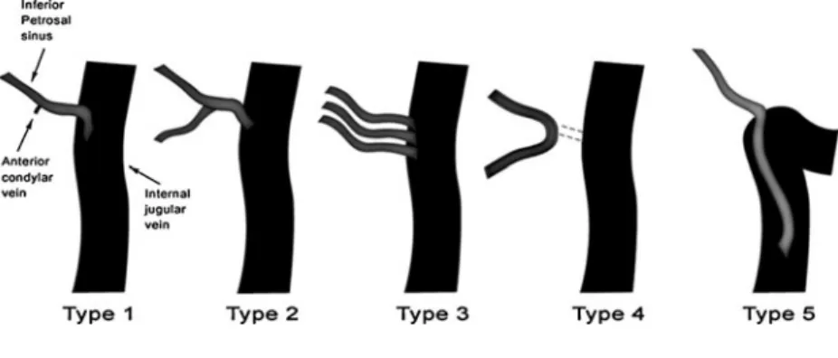
Twenty patients were examined using neuroradiological angiography with a biplane system; in three patients, the angiographic examination was conducted using an Axiom Artis Zee including 3D rotational angiography. All procedures were performed by two senior interventional neuroradiologists. In all but one patient, local anesthesia at the femoral puncture sites was used prior to the BIPSS procedure. In patient no. 13, general anesthesia was required because of the young age at examination date. The femoral veins were catheterized bilaterally with a 6-F catheter and heparin was used in the flushing fluid for systemic anticoagulation. The tips of these catheters were placed in the internal jugular veins. The 2.7-F microcatheters were guided under fluoro-scopic control into both petrosal sinuses. This was possible in 15 out of 16 patients; however, in one case, the right catheter remained in the proximal internal jugular vein. The pattern of venous drainage was determined on each side after contrast injection. The venous drainage of the petrosal
sinus in the jugular vein was classified as one of five
drainage patterns including four common variants [36] and
a fifth rather rare variant [37], where the inferior petrosal
sinus connects far below the jugular bulb into the jugular
vein (Figs.1 and2).
In seven patients, the exact type of venous drainage could only be identified from the written report because of loss of the angiography films. The position of the catheter tip was controlled before and after the venous sinus petrosal
sampling (Fig.3).
Venous blood was sampled simultaneously from both petrosal sinuses and the peripheral vein. Two baseline values were taken followed by the administration of a
bolus of oCRH (1 μg/kg body weight). In all three
catheters, i.e., both petrosal sinuses and the peripheral vein, venous blood was sampled simultaneously for the measurement of ACTH at 3, 5, and 10 min after oCRH injection.
After the procedure, patients were observed neurologi-cally for 24 h in our department.
ACTH gradients
The average ratio of ACTH values before and after oCRH administration was used to calculate the gradients needed for detecting the source of cortisol depletion. A
central-to-peripheral gradient ≥2.0 at baseline, or ≥3.0 after oCRH
stimulation was regarded as indicating a pituitary source of
ACTH production according to Oldfield et al. [31]. An
intersinus gradient≥1.4 was evidence of right–left
lateral-ization, an intersinus gradient <1.4 showed either bilateral
disease or a midline lesion [38].
Results Patients
Briefly, the study included 23 patients (18 women, five men; mean age 47 years, age range 7–70 years). A full
description of the study patients can be found in Table1.
MRI studies
In eight out of the 23 patients, MRI identified the microadenoma correctly compared with the histopatho-logical findings after surgery as standard of reference (patient no: 1, 4, 7, 8, 17, 18, 22, and 23). In one patient (no. 6), a pituitary adenoma was recognized by MRI but the side of the pathologic process could not be identified. The pituitary was correctly recognized as normal (true-negative result) in two patients (no. 3 and 14). In 12 patients, a false result was identified. There were four false-positive findings (no. 2, 9, 11, and 15) and eight false-negative results (cases 5, 10, 12, 13, 16, 19, 20, and 21). These findings resulted in 52.9% sensitivity and 33.3% specificity for MRI to detect a pituitary microadenoma compared with the histopatho-logcial findings.
Catheterization procedure
The variants of the venous drainage that describe the pattern of flow from the petrosal sinus into the jugular vein
are shown in Figs.1and2. Twenty-one of 23 patients were
correctly diagnosed by BIPSS in terms of the central to peripheral gradient. In all cases with a central lesion, a correct lateralization was made. However, in one patient (no. 17) the required value of 1.4 after oCRH stimulation was marginally beneath the required cut-off, namely 1.339, yet we decided to take this result as sufficient for the lateralization.
In patient no. 3, the result of the central to peripheral gradient showed a false-positive value compared with the histopathological result. In the latter, no microadenoma could be identified postoperatively, therefore an adrenalec-tomy was performed.
Patient no. 4 (false-negative result) had a central to peripheral vein ratio less than needed for the diagnosis of CD. Nevertheless, pituitary surgery was performed because of a strong clinical suspicion of CD and an MRI result that revealed a suspicious pathological intrasellar lesion inter-preted as a microadenoma on the right side. At surgery, a Fig. 1 Schematic representation
of the variations of inferior petrosal sinus drainage into the internal jugular vein: types 1–4 modified after Shiu et al. [36] and type 5 variant described by Benndorf and Campi [37]
small tumor was found and histopathologically confirmed
(Table1).
Of the 20 patients with CD, a central to peripheral gradient suggesting CD was already obtained before oCRH in 17 patients, two additional patients showed a gradient greater than 3.0 after CRH application (no. 10 and 15). In
one patient (no. 4), a false-negative result was obtained (see previous description above).
Three patients showed an ectopic localization of the ACTH source regarding BIPSS result (no. 2, 4, and 14): patient no. 2 and 14 showed a true-negative result whereas patient no. 4 had a false-negative result. In the CD group of 20 patients, an intersinus gradient >1.4 was found (16 patients before oCRH application and in four patients after (no. 10, 17, 22, and 23)).
No serious complications occurred during and after the invasive procedure.
Surgical technique
In 20 patients with suspected CD as a result of BIPSS, transsphenoidal surgery for adenoma extirpation was performed. Briefly, surgery was performed via a unilateral transseptal transsphenoidal approach while in the semi-sitting position. Above all, especially in cases where a hyperplasia in the histological examination was found, the intraoperative adenoma tissue was frequently difficult to differentiate or a suspicious brighter tissue could be demarcated. In these cases, according the result of BIPSS, one third of the lateral adenoma tissue was extirpated. Afterwards, the cavity was filled with gelatin foam soaked in fibrin glue, and the sellar opening was covered with a Fig. 2 Angiography of drainage variant type 5. Uncommonly low
origin of the inferior petrosal sinus from the right jugular vein on the right side. The intercavernous sinus is depicted as well. Awareness of this infrequent venous pattern may help to better identify the successful approach to the cavernous sinus
Fig. 3 BIPSS procedure. A Digital subtraction angiography of the inferior petrosal sinus shows 6-F guiding catheters in the internal jugular veins (white arrows) and 2.7-F microcath-eters in the petrosal sinuses (black arrows). B Similar an-giogram as shown in (A) with simultaneous contrast filling of both inferior petrosal sinus as well as the intercavernous sinus. Images (C and D) depict the same venous pattern in a 3D rotational angiography
T able 1 Results of patients with BIPSS Patient no. A g e/ se x V enous drainage a right/left Pituitary MRI b Centr ./peri. G. before/after CRH Lateral. b/after CRH BIPSS results Sur gery Pathology results Outcome 1 51/F I/III R adenoma +/+ +/+ R central T otal exstirpation Adenoma Cure 2 43/F (n.a.) L fullness − /−− /− Ectopic Lung lobe resection ACTH-expression Cure 3 53/M I/I Normal +/+ +/+ L central Exploration of sella Normal Adrenalectomy 4 41/F III/III R adenoma − /− +/+ Ectopic c Resection of right third ACTH-hyperplasia Cure 5 49/F (n.a.) Normal +/+ +/+ R central Gamma knife – d 6 49/F (n.a.) Adenoma +/+ +/+ L central Resection of left third ACTH-expression No remission 7 45/F I/IV R adenoma +/+ +/+ R central Adenoma exstirpation ACTH-expression Cure 8 40/F (n.a.) R fullness +/+ +/+ R central Resection of right third Adenoma Resid adenoma=>reop. 9 52/F (n.a.) R fullness +/+ +/+ L central Resection of left part Adenoma Cure 10 58/F Distal/I Normal − /+ − /+ L central Adenoma exstirpation Adenoma Cure 1 1 37/F I/I L fullness +/+ +/+ R central Resection of right third ACTH-hyperplasia No remission 12 50/F V/I Normal +/+ +/+ R central Resection of right third Adenoma Cure 13 7/M (n.a.) Normal +/+ +/+ L central Resection of left third Gamma knife 14 65/F (n.a.) Normal − /−− /− Ectopic Adrenal gland ectomy Partial regression 15 63/F III/III R adenoma − /+ +/+ L central Resection of left part ACTH-hyperplasia Regression 16 42/M III/IV Normal +/+ +/+ R central Resection of right third A d en o m a/ hyperplasia N o remission 17 42/F I/I R adenoma +/+ − /− (R) central Adenoma exstirpation Adenoma Cure 18 64/M II/II L adenoma +/+ +/+ L central Adenoma exstirpation Adenoma 19 40/F I/II Normal +/+ +/+ R central Adenoma exstirpation Adenoma Cure 20 43/F III/III Normal +/+ +/+ R central Adenoma exstirpation Adenoma (Cure) 21 48/F I/III R adenoma +/+ +/+ L central Adenoma exstirpation Adenoma Cure 22 38/F I/I Central +/+ − /+ Central Adenoma exstirpation Adenoma Cure 23 70/M I/I R adenoma +/+ − /+ R central Adenoma exstirpation Adenoma Cure R right, L left, CRH corticotropin-releasing hormone, MRI magnetic resonance imaging, (n.a.) not available aT ype I– IV according to Shiu et al. [ 36 ]; V : variant described by Benndorf and Campi [ 37 ] b MRI finding “adenoma ” means suspected micr oadenoma c T reated as central (see details in text) d Relapse of pituitary adenoma after pituitary operation
synthetic absorbable vicryl patch as described in the
literature [39].
Histopathological results
In the 20 patients where a central adenoma was suspected using BIPSS, a transsphenoidal surgery was performed. Hyperplasia of ACTH-producing cells or an adenoma was observed in 18 of the 20 histopathological samples. In one patient (no. 13), a histological evaluation was not made because of loss of the histological material.
In the patient with highly suspected CD, gamma knife irradiation of the pituitary gland was performed and the patient had a favorable outcome.
In the three patients with an ectopic source of ACTH expression (no. 2, 4, and 14), histology confirmed the localization of ACTH expression: one patient (no. 2) had a lung lobe resection and ACTH expression was identified by histology, another patient had a double-sided adrenalectomy (no. 14) causing a remission, and the treatment of the third patient (no. 4, false-negative result) has been previously discussed. Of note, histopath-ological confirmation of an ACTH-secreting adenoma
strongly correlates with remission [40].
Clinical outcomes
The clinical outcome of the 23 patients analyzed with BIPSS was favorable in the majority of cases: 15 patients showed postoperative normalization of the hypercortisolism with favorable clinical development, one patient showed partial regression (no. 14), and three patients showed no remission (no. 6, 11, and 16). Two patients had a gamma knife treatment with no available long-term result to date (no. 5 and 13). Patient no. 3 had adrenalectomy because the BIPSS results revealed a pituitary process, but surgery showed no adenoma and finally, patient no. 8 had a reoperation because of a residual adenoma of the pituitary. Statistical analyses
The BIPSS procedure allowed the correct differentiation between CD and EAS in 82.4% and 95.0% of patients before and after oCRH stimulation, respectively. The specificity of the BIPSS procedure before and after oCRH stimulation was 66.7% respectively. Correct lateralization could be made in all cases of CD (100%).
Discussion
There is considerable controversy in regards to the accuracy
of the side of pituitary lesion using BIPSS data [41]. In the
present study, BIPSS was able to correctly localize the side of the adenoma in all patients with CD. Of the examined 20 patients with CD, an intersinus gradient >1.4 was achieved in 80% of the cases before and in 100% after oCRH stimulation even if an asymmetric venous drainage was documented. This is consistent with the study of Oliverio et
al. [42]. They reported in a series of 17 patients that
bilateral cavernous sinus sampling (BCSS) enabled the identification of the correct side in all patients, yielding a positive predictive value of 100%. The author concluded that this result might be due to the potential of cavernous sinus sampling to more accurately lateralize the lesion.
However, Doppman et al. [43] reported correct
lateraliza-tion in patients with CD using BIPSS in 60% of the cases without and in 73% of the cases with oCRH stimulation.
Newell-Price et al. [34] found that CD can be distinguished
from EAS with a diagnostic accuracy close to 100% when BIPSS is combined with oCRH stimulation. On the other hand, the intersinus gradient correctly predicted the localization of the pituitary adenoma in only 78% of the cases.
These controversial findings may be related to the
different venous drainage pattern [41]. Lefournier et al.
[44] reported that three parameters influenced the
laterali-zation result, i.e., venous drainage pattern, sampling site,
and oCRH stimulation. Friedman et al. [45] performed the
procedure in adults with CD and healthy volunteers and found that the correct lateralization side was the side of the dominant petrosal sinus.
However, in the current study asymmetric venous drainage did not influence the lateralization data. Due to the fact that we detected 80% accuracy before and 100% accuracy after stimulation, we assume that compensatory and regulatory mechanism on the contra-lateral side of the adenoma may have influenced the hormone levels. Fur-thermore, for all patients we selectively placed the micro-catheter in the petrosal sinus, which does not disturb the venous blood flow in both petrosal sinus and the cavernous sinus. In contrast, the placement of catheters with relatively large diameter influences the drainage pattern by obstruct-ing the venous outflow around the catheter. This may result in retrograde blood flow through the intercavernous sinus leading to false-positive values on the opposite side of the lesion. In addition, digital subtraction angiography was performed to assure the correct catheter placement and to document the anatomical venous pattern drainage. Micro-catheters allow a normal blood flow without influencing the venous blood flow even in asymmetric drainages. This also may minimize complications such as thromboembolism or perforations. We conclude that the use of microcatheters result in significantly improved measured ACTH concen-trations on the effective side of the lesion. We propose that digital subtraction angiography should be performed to
assure the correct catheter placement for documenting the exact anatomical venous pattern drainage. This allows for a critical and comprehensive analysis of the anatomy with the obtained biochemical cortisol levels.
Additionally, it is established that the diagnostic perfor-mance of BIPSS is superior to pituitary imaging in ACTH-dependent hypercortisolism to distinguish between CD and ectopic ACTH-dependent hypercortisolism. The current
findings are consistent with previous results [30, 31, 46,
47] showing a superior sensitivity and specificity of BIPSS
compared with MRI (95% to 52.9% and 66.7% to 33.3%, respectively). Most pituitary macroadenomas can be detected by MRI. CD, however, is mostly caused by microadenomas so that it is often difficult or even impossible to determine a pathologic intrasellar process.
There were no severe complications in the current study suggesting that this invasive procedure is safe in experi-enced hands. However, in previous studies several major
complications such as pulmonary thromboembolism [15,
41,42] and lower extremity deep venous thrombosis [48]
have been reported. In addition, Lefournier et al. [44]
described two cases of transient sixth cranial nerve palsy during BCSS. They attributed these incidents to the guide wire of the catheter and suggest that BCSS might be less
safe than BIPSS. Graham et al. [49] performed cavernous
sinus sampling (CSS) with 2.7-F microcatheters and compared it with results of inferior petrosal sinus sampling. They found that CSS was superior to IPSS for intrapituitary tumor localization and no complication occurred in their series of 70 patients. Recent results suggest that BIPSS is not inferior to BCSS with regard to correct lateralization of a microadenoma. This view is consistent with the studies of
Doppman [43], Mamelak [41], and Lefournier [44]. In a
recent study published by Gandhi et al. [50], one serious
complication of irreversible brain stem injury in a study of 44 patients who underwent BIPSS for evaluation of Cushing’s disease was described. In this rare case, there was radiographic evidence of venous outflow variance that may have contributed to the injury. They concluded that venous hypertension as a result of catheter-related occlusion of a venous channel or from the pressure or toxicity of contrast injection probably resulted in the pontine infarction.
Study limitations
In seven out of 23 subjects, no angiogram films were available so that the type of drainage had to be estimated from the written report which limits exact reconfirmation of catheter position.
We had no control group in our study comparing selective catheterization versus a more proximal non-selective venous blood sampling. This would allow direct
comparison of the influence of microcatheters in this specific setup and would also be technically possible; however, two major concerns limit this approach. First, measurement of ACTH concentrations 3, 5, and 10 min after oCRH stimulation had to be measured two times with an already attained peak of ACTH concentration after the first stimulation. Second, prolonged catheter placement in the venous system bears the danger of possible thrombus formation with consecutive thromboembolism even under continuous heparin infusion during the procedure. Addi-tionally, our patient collective still represents a relatively small sample and would not be definitive.
Conclusion
BIPSS remains the gold standard in the detection of a microadenoma in CS. Supporting selective placement of microcatheters without venous outflow diversion may play an important role in accurate detection of the origin of ACTH secretion. Therefore, this super-selective catheteri-zation technique provides a firm basis in the assessment of CS caused by a non-detectable microadenoma of the pituitary.
Acknowledgments We especially thank Josef Rohrer from the Department of Neuroradiology for his excellent technical assistance to the interventional procedures; Daniela Miescher, Department of Neurosurgery, for her wonderful drawing of the different drainage patterns; Verena Grimm from the Department of Endocrinology for collecting blood samples; and Susan Wieting for medical editing of the final version of this manuscript, Department of Neurosurgery, Publications Office, Bern Switzerland.
Conflict of interest We declare that we have no conflict of interest.
References
1. Howlett TA, Drury PL, Perry L, Doniach I, Rees LH, Besser GM (1986) Diagnosis and management of ACTH-dependent Cushing's syndrome: comparison of the features in ectopic and pituitary ACTH production. Clin Endocrinol (Oxf) 24:699–713
2. Imöhl MSA, Krieg M (2003) Diagnose und differentialdiagnose des Cushing-Syndroms. J Lab Med 27(1/2):43–52
3. Findling JW, Doppman JL (1994) Biochemical and radiologic diagnosis of Cushing's syndrome. Endocrinol Metab Clin North Am 23:511–537
4. Kaye TB, Crapo L (1990) The Cushing syndrome: an update on diagnostic tests. Ann Intern Med 112:434–444
5. Mampalam TJ, Tyrrell JB, Wilson CB (1988) Transsphenoidal microsurgery for Cushing disease. A report of 216 cases. Ann Intern Med 109:487–493
6. Fahlbusch R, Buchfelder M, Muller OA (1986) Transsphenoidal surgery for Cushing's disease. J R Soc Med 79:262–269 7. Cushing H (1932 (1994)) The basophil adenomas of the pituitary
body and their clinical manifestations (pituitary basophilism). Obes Res 2:486–508
8. Kruse A, Klinken L, Holck S, Lindholm J (1992) Pituitary histology in Cushing's disease. Clin Endocrinol (Oxf) 37:254–259 9. Gabrilove JL, Anderson PJ, Halmi NS (1986) Pituitary pro-opiomelanocortin-cell carcinoma occurring in conjunction with a glioblastoma in a patient with Cushing's disease and subsequent Nelson's syndrome. Clin Endocrinol (Oxf) 25:117–126
10. Kaiser FE, Orth DN, Mukai K, Oppenheimer JH (1983) A pituitary parasellar tumor with extracranial metastases and high, partially suppressible levels of adrenocorticotropin and related peptides. J Clin Endocrinol Metab 57:649–653
11. Nawata H, Higuchi K, Ikuyama S, Kato K, Ibayashi H, Mimura K, Sueishi K, Zingami H, Imura H (1990) Corticotropin-releasing hormone and adrenocorticotropin-producing pituitary carcinoma with metastases to the liver and lung in a patient with Cushing's disease. J Clin Endocrinol Metab 71:1068–1073
12. Trainer PJ, Grossman A (1991) The diagnosis and differential diagnosis of Cushing's syndrome. Clin Endocrinol (Oxf) 34:317– 330
13. Boscaro M, Arnaldi G (2009) Approach to the patient with possible Cushing's syndrome. J Clin Endocrinol Metab 94:3121– 3131
14. Dickstein G, DeBold CR, Gaitan D, DeCherney GS, Jackson RV, Sheldon WR Jr, Nicholson WE, Orth DN (1996) Plasma corticotropin and cortisol responses to ovine corticotropin-releasing hormone (CRH), arginine vasopressin (AVP), CRH plus AVP, and CRH plus metyrapone in patients with Cushing's disease. J Clin Endocrinol Metab 81:2934–2941
15. Newell-Price J, Perry L, Medbak S, Monson J, Savage M, Besser M, Grossman A (1997) A combined test using desmopressin and corticotropin-releasing hormone in the differential diagnosis of Cushing's syndrome. J Clin Endocrinol Metab 82:176–181 16. Orth DN (1995) Cushing's syndrome. N Engl J Med 332:791–803 17. Trainer PJBG (1995) Cushing’s syndrome. The Bart’s endocrine
protocols. Churchill Livingstone, London, In, pp 84–92 18. Kaskarelis IS, Tsatalou EG, Benakis SV, Malagari K, Komninos I,
Vassiliadi D, Tsagarakis S, Thalassinos N (2006) Bilateral inferior petrosal sinuses sampling in the routine investigation of Cushing's syndrome: a comparison with MRI. AJR Am J Roentgenol 187:562–570
19. Tabarin A, Laurent F, Catargi B, Olivier-Puel F, Lescene R, Berge J, Galli FS, Drouillard J, Roger P, Guerin J (1998) Comparative evaluation of conventional and dynamic magnetic resonance imaging of the pituitary gland for the diagnosis of Cushing's disease. Clin Endocrinol (Oxf) 49:293–300
20. Buchfelder M, Nistor R, Fahlbusch R, Huk WJ (1993) The accuracy of CT and MR evaluation of the sella turcica for detection of adrenocorticotropic hormone-secreting adenomas in Cushing disease. AJNR Am J Neuroradiol 14:1183–1190 21. Saris SC, Patronas NJ, Doppman JL, Loriaux DL, Cutler GB Jr,
Nieman LK, Chrousos GP, Oldfield EH (1987) Cushing syn-drome: pituitary CT scanning. Radiology 162:775–777
22. Boscaro M, Barzon L, Fallo F, Sonino N (2001) Cushing's syndrome. Lancet 357:783–791
23. Arnaldi G, Angeli A, Atkinson AB, Bertagna X, Cavagnini F, Chrousos GP, Fava GA, Findling JW, Gaillard RC, Grossman AB, Kola B, Lacroix A, Mancini T, Mantero F, Newell-Price J, Nieman LK, Sonino N, Vance ML, Giustina A, Boscaro M (2003) Diagnosis and complications of Cushing's syndrome: a consensus statement. J Clin Endocrinol Metab 88:5593–5602
24. Bartynski WS, Lin L (1997) Dynamic and conventional spin-echo MR of pituitary microlesions. AJNR Am J Neuroradiol 18:965– 972
25. Gao R, Isoda H, Tanaka T, Inagawa S, Takeda H, Takehara Y, Isogai S, Sakahara H (2001) Dynamic gadolinium-enhanced MR imaging of pituitary adenomas: usefulness of sequential sagittal and coronal plane images. Eur J Radiol 39:139–146
26. Jagannathan J, Sheehan JP, Jane JA Jr (2007) Evaluation and management of Cushing syndrome in cases of negative sellar magnetic resonance imaging. Neurosurg Focus 23:E3
27. Batista D, Courkoutsakis NA, Oldfield EH, Griffin KJ, Keil M, Patronas NJ, Stratakis CA (2005) Detection of adrenocorticotropin-secreting pituitary adenomas by magnetic resonance imaging in children and adolescents with Cushing disease. J Clin Endocrinol Metab 90:5134–5140
28. Portocarrero-Ortiz L, Bonifacio-Delgadillo D, Sotomayor-Gonzalez A, Garcia-Marquez A, Lopez-Serna R (2010) A modified protocol using half-dose gadolinium in dynamic 3-Tesla magnetic resonance imaging for detection of ACTH-secreting pituitary tumors. Pituitary 13:230–235
29. Hall WA, Luciano MG, Doppman JL, Patronas NJ, Oldfield EH (1994) Pituitary magnetic resonance imaging in normal human volunteers: occult adenomas in the general population. Ann Intern Med 120:817–820
30. Findling JW, Kehoe ME, Shaker JL, Raff H (1991) Routine inferior petrosal sinus sampling in the differential diagnosis of adrenocorticotropin (ACTH)-dependent Cushing's syndrome: early recognition of the occult ectopic ACTH syndrome. J Clin Endocrinol Metab 73:408–413
31. Oldfield EH, Doppman JL, Nieman LK, Chrousos GP, Miller DL, Katz DA, Cutler GB Jr, Loriaux DL (1991) Petrosal sinus sampling with and without corticotropin-releasing hormone for the differential diagnosis of Cushing's syndrome. N Engl J Med 325:897–905
32. Newell-Price J, Bertagna X, Grossman AB, Nieman LK (2006) Cushing's syndrome. Lancet 367:1605–1617
33. Lad SP, Patil CG, Laws ER Jr, Katznelson L (2007) The role of inferior petrosal sinus sampling in the diagnostic localization of Cushing's disease. Neurosurg Focus 23:E2
34. Newell-Price J, Trainer P, Besser M, Grossman A (1998) The diagnosis and differential diagnosis of Cushing's syndrome and pseudo-Cushing's states. Endocr Rev 19:647–672
35. Swearingen B, Katznelson L, Miller K, Grinspoon S, Waltman A, Dorer DJ, Klibanski A, Biller BM (2004) Diagnostic errors after inferior petrosal sinus sampling. J Clin Endocrinol Metab 89:3752–3763
36. Shiu PC, Hanafee WN, Wilson GH, Rand RW (1968) Cavernous sinus venography. Am J Roentgenol Radium Ther Nucl Med 104:57–62
37. Benndorf G, Campi A (2002) Aberrant inferior petrosal sinus: unusual transvenous approach to the cavernous sinus. Neuroradi-ology 44:158–163
38. Oldfield EH, Chrousos GP, Schulte HM, Schaaf M, McKeever PE, Krudy AG, Cutler GB Jr, Loriaux DL, Doppman JL (1985) Preoperative lateralization of ACTH-secreting pituitary micro-adenomas by bilateral and simultaneous inferior petrosal venous sinus sampling. N Engl J Med 312:100–103
39. Seiler RW, Mariani L (2000) Sellar reconstruction with resorbable vicryl patches, gelatin foam, and fibrin glue in transsphenoidal surgery: a 10-year experience with 376 patients. J Neurosurg 93:762–765
40. Prevedello DM, Pouratian N, Sherman J, Jane JA Jr, Vance ML, Lopes MB, Laws ER Jr (2008) Management of Cushing's disease: outcome in patients with microadenoma detected on pituitary magnetic resonance imaging. J Neurosurg 109:751–759
41. Mamelak AN, Dowd CF, Tyrrell JB, McDonald JF, Wilson CB (1996) Venous angiography is needed to interpret inferior petrosal sinus and cavernous sinus sampling data for lateralizing adrenocorticotropin-secreting adenomas. J Clin Endocrinol Metab 81:475–481
42. Oliverio PJ, Monsein LH, Wand GS, Debrun GM (1996) Bilateral simultaneous cavernous sinus sampling using
corticotropin-releasing hormone in the evaluation of Cushing disease. AJNR Am J Neuroradiol 17:1669–1674
43. Doppman JL, Nieman LK, Chang R, Yanovski J, Cutler GB Jr, Chrousos GP, Oldfield EH (1995) Selective venous sampling from the cavernous sinuses is not a more reliable technique than sampling from the inferior petrosal sinuses in Cushing's syndrome. J Clin Endocrinol Metab 80:2485–2489
44. Lefournier V, Martinie M, Vasdev A, Bessou P, Passagia JG, Labat-Moleur F, Sturm N, Bosson JL, Bachelot I, Chabre O (2003) Accuracy of bilateral inferior petrosal or cavernous sinuses sampling in predicting the lateralization of Cushing's disease pituitary microadenoma: influence of catheter position and anatomy of venous drainage. J Clin Endocrinol Metab 88:196–203
45. Friedman TC, Yanovski JA, Nieman LK, Doppman JL, Cutler GB Jr, Oldfield EH, Gold PM, Chrousos GP, Kalogeras KT (1996) Inferior petrosal sinus arginine vasopressin concentrations in normal volunteers and patients with Cushing's disease. J Clin Endocrinol Metab 81:3068–3072
46. Joshi SM, Hewitt RJ, Storr HL, Rezajooi K, Ellamushi H, Grossman AB, Savage MO, Afshar F (2005) Cushing's disease in children and adolescents: 20 years of experience in a single neurosurgical center. Neurosurgery 57:281–285
47. Semple PL, Vance ML, Findling J, Laws ERJ (2000) Transsphenoidal surgery for Cushing's disease: outcome in patients with a normal magnetic resonance imaging scan. Neurosurgery 46:553–559 48. Bonelli FS, Huston J 3rd, Carpenter PC, Erickson D, Young WF
Jr, Meyer FB (2000) Adrenocorticotropic hormone-dependent Cushing's syndrome: sensitivity and specificity of inferior petrosal sinus sampling. AJNR Am J Neuroradiol 21:690–696
49. Graham KE, Samuels MH, Nesbit GM, Cook DM, O'Neill OR, Barnwell SL, Loriaux DL (1999) Cavernous sinus sampling is highly accurate in distinguishing Cushing's disease from the ectopic adrenocorticotropin syndrome and in predicting intra-pituitary tumor location. J Clin Endocrinol Metab 84:1602–1610 50. Gandhi CD, Meyer SA, Patel AB, Johnson DM, Post KD (2008) Neurologic complications of inferior petrosal sinus sampling. AJNR Am J Neuroradiol 29:760–765