ABSTRACT. During the last decade, a systemat-ic effort to develop a pharmacologsystemat-ical treatment for Alzheimer’s disease (AD) resulted in three drugs being registered for the first time in the US and Europe. All three compounds are cholinesterase inhibitors (ChEI). The major ther-apeutic effect of ChEI on AD patients is to main-tain cognitive function at a stable level during a 6-month to 1-year period of treatment, as com-pared to placebo. Additional drug effects are to slow down cognitive deterioration and improve be-havioral and daily living activity. Recent studies show that in many patients the cognitive stabi-lization effect can be prolonged up to 24 months. This long-lasting effect suggests a mechanism of action other than symptomatic, and directly cholinergic. In vitro and in vivo studies have con-sistently demonstrated a link between cholinergic activation and amyloid precursor protein (APP) metabolism. Lesions of cholinergic nuclei cause a rapid increase in cortical APP and cholinergic synaptic function; the effect of such lesions can be reversed by ChEI treatment. A reduction in cholin-ergic neurotransmission, experimental or patho-logical, leads to amyloidogenic metabolism and contributes to the development of neuropatholo-gy and cognitive dysfunction. To explain the long-term effect of ChEI, for which evidence is available on an experimental as well as clinical level, a mechanism based on beta-amyloid metabolism is postulated. The question whether cholinergic sta-bilization implies simply slowing down progression of disability or also involves delay of disease pro-gression is discussed.
(Aging Clin. Exp. Res. 13: 247-254, 2001)
©2001, Editrice Kurtis
CHOLINERGIC THERAPY OF ALZHEIMER’S DISEASE. HOW DID IT START?
There are presently three possible modalities of pharmacological treatment of Alzheimer’s disease (AD) (Fig. 1); the most explored has been the cholin-ergic approach.
Basic as well as clinical knowledge of the cholinergic system, its normal function, and dysfunction form the theoretical base of the therapy of AD with cholinesterase inhibitors (ChEI) (1). This strategy was developed considering that: a) cholinergic neurons and synapses undergo early and selective damage, as demonstrated by a decrease in acetylcholine (ACh) synthesis and nicotinic receptor binding (1); b) a steady-state rise in synaptic ACh levels following cholinesterase (ChE) inhibition produces a symp-tomatic short-term cognitive benefit which can be demonstrated experimentally in animals and humans; c) because this effect is short-lasting and symptomat-ic in nature, effsymptomat-icacy of ChEI should not persist beyond cessation of the central nervous system (CNS)-ACh el-evation resulting from ChE inhibition; d) based on these three conditions, one can predict that early, mild-ly affected patients should respond best to ChEI ther-apy, while severely impaired patients with extensive cholinergic dysfunction would not benefit from this treatment; e) finally, clinical benefits should be limited to cognitive (memory), non-behavioral symptomatic improvement (Table 1).
Recent clinical data seem to contradict such as-sumptions (Table 2). First, neocortical cholinergic deficits such as decrease in choline-acetyltransferase (ChAT) and acetylcholinesterase (AChE) enzymatic ac-tivity, which are characteristic of severely demented pa-tients, are not clearly apparent in individuals with
Is anti-cholinesterase therapy of Alzheimer’s disease
delaying progression?
E. Giacobini
University Hospitals of Geneva, Department of Geriatrics, University of Geneva Medical School, Thonex-Geneva, Switzerland
Key words: Alzheimer’s disease, amyloid precursor protein (APP), beta-amyloid, cholinergic stabilization, cholinesterase inhibitors (ChEI). Correspondence: E. Giacobini, M.D., University Hospital of Geneva, Department of Geriatrics, University of Geneva Medical School,
CH-1226 Thonex-Geneva, Switzerland. E-mail: Ezio.Giacobini@hcuge.ch Received and accepted April 10, 2001.
early mild AD (2). Significant cholinergic enzymatic deficits are not demonstrable until relatively late in the course of the disease (2). On the other hand, de-creases in ChAT activity in the frontal and parietal cor-tex and in the hippocampus correlate to losses of cog-nitive domain scores (MDRS, Mattis Dementia Rating Scale and MMSE, Mini Mental State Examination) (3). The clinical effect of ChEI seen at early and mild stages of the disease suggests that the cholinergic system may be hypo-functional, or that parameters other than enzyme activity levels are impaired. Demonstrated deficits are: reduced ACh biosynthesis; decreased storage and impaired release of ACh, and cholinergic receptor defective either in number (nico-tinic) or in function (muscarinic) (1).
To support adequate synthesis and hydrolysis of ACh under non-physiological conditions, choliner-gic enzymes are in excess concentrations in the brain (1). This implies that patients at severe stages of the disease showing extensive cholinergic damage would constitute a target for cholinergic treatment. Trials
de-signed specifically to investigate indications for asymp-tomatic early-phase or severe late-phase patients are in progress. The preliminary data available suggest that pharmacological interventions can improve cogni-tive function in the range from very mild and minimally cognitively impaired AD patients (cases showing memory impairment only) to severe cases (MMSE<10). Treatment duration can be extended to two-three years, which correspond approximately to one third of the natural history of the disease, thus raising the question: is such a prolonged effect of ChEI only symptomatic, or are ChEI disease-modify-ing drugs? Recent data, which are summarized in this paper, support the second alternative. The clini-cal effect seems to be stronger in more advanced cases; moreover, treatment benefit seems to be more pronounced and more long-lasting at higher doses. The clinical effect of ChEI is measurable three-four weeks following interruption of the treatment (wash-out period) despite the fact that ChE inhibition is strongly diminished or no longer present (4). In this case, new clinical effect can be demonstrated as an im-provement in ADAS-Cog scores when treatment is re-sumed after being suspended for several weeks (3-6 weeks). It seems difficult to explain this stabilizing effect in terms of symptomatic mechanisms (Table 2). Evidence accumulated from experiments in animals and neuronal cell lines suggests a different explanation (5-7).
CLINICAL DATA SUPPORT A STABILIZING EFFECT OF CHOLINESTERASE INHIBITORS
The benefit of ChEI treatment was previously con-sidered to be exclusively symptomatic and cognitive. It has now been demonstrated that improvement in-volves cognitive as well as behavioral symptoms (8, 9). The cognitive improvement is significant up to 12 months (Fig. 2), and several clinical studies have
Table 1 - Basic assumptions for a cholinergic strategy. Are
they correct?
1. Brain cholinergic neurons and synapses are damaged very ear-ly in the disease process which results in a decreased level of acetylcholine.
2. Early, mildly-affected patients will benefit maximally from ChEI treatment.
3. Treatment benefit will be short-term, symptomatic and mainly cognitive.
4. Drug effect will not persist beyond cessation of ChE inhibition. ChEI: Cholinesterase Inhibitors; ChE: Cholinesterase.
Table 2 - Evidence that ChEI clinical effect may not only be
symptomatic.
1. Cognitive deterioration progresses more slowly in treated than in untreated patients. This effect is dose-dependent.
2. The clinical effect may last for several weeks after drug discon-tinuation while cholinesterase inhibition in CSF is low or even ab-sent.
3. The clinical effect is seen also in early, mildly affected patients with little cholinergic impairment.
4. The strongest clinical effect is seen in rapidly progressing patients. 5. Relatively more advanced patients respond better than early mild
cases.
ChEI: Cholinesterase Inhibitors; CSF: Cholinergic Synaptic Function. TREATMENT OF ALZHEIMER’S DISEASE:
WHICH WAY TO GO? TWO WAYS TO GO:
ACETYLCHOLINE
BETA-AMYLOID
THE THIRD WAY: ESTROGENS ANTI-OXIDANTS ANTI-INFLAMMATORIES CHE-INHIBITORS
MUSCARINICS OR NICOTINICS
Beta-and gamma-secretase inhibitors Beta-sheet breakers
IMMUNIZATION (anti-beta-A-1-42)
Figure 1 - Pharmacological treatment of Alzheimer’s disease
demonstrated that the drug effect can be seen for as long as 2 years in many patients (Table 3). This long-term effect translates into an improved activity of daily living for the patient, and a reduced emotional impact for the caregiver, as well as a reduction in care costs.
The six-month data available for six ChEI suggest that patients treated with the active compound change little cognitively from baseline at the beginning of the trial to the end of the trial (19, 20). Studies that approximate a randomized-start design suggest that treatment with ChEI may delay cognitive deterioration (21). Two-year open-label data from a donepezil trial reveal a decline in ADAS-Cog from baseline that is 50% lower than the predicted value; untreated patients progress more rapidly than treated ones, and the treatment effect seems to be related to the dose (21). Average annual rate of decline for patients with a high-er dose of rivastigmine is almost 50% lowhigh-er (4.5
ADAS-Cog points/year) than that of patients treated with a lower dose (8.2 ADAS-Cog points/year) (21). Increasing the dose of rivastigmine reduces the rate of cognitive decline over a 3-year period, sug-gesting a reduction in the rate of progression of cog-nitive deterioration (10, 21). Clinical data also indicate that rapidly progressing patients show the strongest drug effect, therefore, both disease stage and dose of the ChEI seem to play a role in altering the course of the disease (21).
Stabilization of cognitive deterioration suggests ei-ther a protective and structural effect, or a long-term improvement in the cholinergic synaptic function. The gradual return to the predicted deterioration-line after drug wash-out also suggests additional non-cholinergic effects. The long-term clinical effect could be related either to cholinesterase inhibition through the active site of the enzyme (5-7), or to non-cholin-ergic properties through interaction with a site close to the peripheral anionic binding site of the enzyme (22).
LONG-TERM STABILIZING EFFECTS OF CHOLINESTERASE INHIBITORS
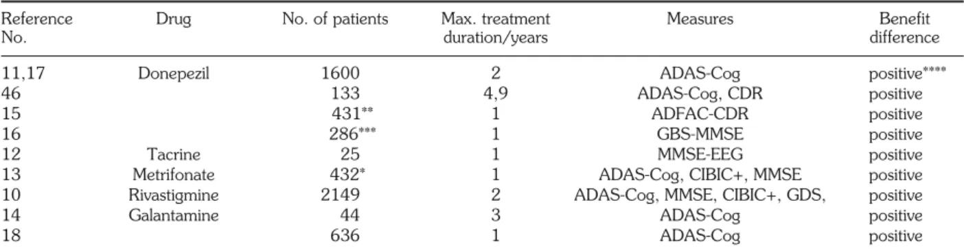
Recent data from 12-24 month open trials and one randomized placebo-control trial suggest that opti-mization and maintainance of clinical effects for one year or more is a feasible goal in many patients (Table 3). Figure 2 reports the effect on the mean change in ADAS-Cog score of a 12-month treat-ment with three ChEI presently in clinical use: donepezil (17), galantamine (18) and rivastigmine (10). The data at twelve months show either a small or no difference from the baseline. The results of several clinical studies (placebo-controlled and open label) for periods longer than one year (up to 3 years) are
re-Table 3 - Long-term efficacy of five cholinesterase inhibitors in AD patients.
Reference Drug No. of patients Max. treatment Measures Benefit
No. duration/years difference
11,17 Donepezil 1600 2 ADAS-Cog positive****
46 133 4,9 ADAS-Cog, CDR positive
15 431** 1 ADFAC-CDR positive
16 286*** 1 GBS-MMSE positive
12 Tacrine 25 1 MMSE-EEG positive
13 Metrifonate 432* 1 ADAS-Cog, CIBIC+, MMSE positive
10 Rivastigmine 2149 2 ADAS-Cog, MMSE, CIBIC+, GDS, positive
14 Galantamine 44 3 ADAS-Cog positive
18 636 1 ADAS-Cog positive
Bernhardt and Woelk, 2000 (13)*, Winblad et al., 1999 (15)**, and Mohs et al., 1999 (16)*** are prospective, placebo-controlled, double-blind studies. Total number of patients: 4258. ****Positive indicates statistically significant clinical improvement from baseline.
Mean change from baseline in ADAS-Cog score 3 2 1 0 -1 -2 -3 12 TIME (WEEKS) 18 26 38 52 IMPROVED Galantamine (24 mg) Donepezil (10 mg) Rivastigmine (6-12 mg) Metrifonate (50 mg)
Figure 2 - Stabilization effect of 12-month treatment with four
cholinesterase inhibitors. The patients change little cognitively from baseline during this period (10, 13, 15, 18).
ported in Table 3 (10-18). These data indicate that benefit differences can be maintained in a number of patients for up to 12-24 months by five different in-hibitors (donepezil, tacrine, metrifonate, rivastigmine and galantamine). In terms of global improvement in the ADAS-Cog score, this may sum up to a total 15-20 point gain, which represents an 18-24 month difference in disease history from placebo-treated pa-tients. How to interpret this improvement? Is it the re-sult of slowing down the increase of disability, or is it an expression of delaying progression of the dis-ease? This question is similar to that asked in reference to the effect of MAO-B inhibitors such as selegyline with regard to Parkinson disease.
BASIC RESEARCH DATA SUPPORTING NON-SYMPTOMATIC EFFECT OF
CHOLINESTERASE INHIBITORS
The amyloid precursor protein (APP) pathway that generates beta-amyloid (beta-A) is regulated by the se-quential action of three enzymes (alpha, beta and gamma secretases). Alpha secretase cleaves APP within the beta-A sequence, and releases soluble N-ter-minal non-aggregating fragments (sAPP) (Fig. 3). Nu-merous studies have shown that the stimulation of sAPP release is associated with reduced formation of amyloidogenic peptides. Muscarinic-agonist-induced sAPP secretion through activation by carbachol of m1 and m3 (but not m2 and m4) receptor subtypes in-creases sAPP release in human embryonic cell lines (23). Activation of the pathway that cleaves APP de-creases the release of beta-A fragments, and may slow down amyloid formation in the brain. On the ba-sis of the results obtained in superfused rat cortex slices demonstrating an increased release of sAPP in re-sponse to muscarinic stimulation, we proposed an ef-fect of ChEI on sAPP secretion acting through the same pathway (5) (Fig. 3).
Racchi et al. (6) using neuroblastoma cells, and Pakaski et al. (7) using primary cultures of rat basal forebrain neurons have shown that short-term treat-ment with reversible and irreversible ChEI such as am-benomium, and metrifonate or its metabolite DDVP, increases sAPP release into the conditioned media, and elevates protein kinase C (PKC) levels. These studies demonstrated that this effect on APP is con-sistent with AChE inhibition, and with indirect mus-carinic-mediated cholinergic stimulation. In addition, short-term or long-term stimulation does not result in changes in APP mRNA expression either in cortical slices or neuroblastoma cells (6, 20), nor in a down-regulation of the response to cholinergic stimulation of muscarinic receptors (6). These results suggest that
ChEI promote the non-amyloidogenic route of APP processing through a stimulation of alpha-secretase ac-tivity mediated by PKC (Fig. 3).
This demonstrated feature of ChEI and of mus-carinic agonists, and their ability to enhance the re-lease of non-amyloidogenic soluble derivatives of APP in vitro and in vivo suggests a slowing down in the formation of amyloidogenic compounds in the brain (1).
EFFECT OF CHOLINERGIC STIMULATION ON BETA-A BRAIN METABOLISM?
Selective muscarinic (m1) direct activation of al-pha-secretase activity accelerates APP processing, and consequently decreases the generation of beta-A peptides in cellular model systems (6, 7, 23). A re-duction in both total beta-A and beta-1-42 peptide lev-els in the cholinergic synaptic function (CSF) is seen in AD patients treated for 4 weeks with either AF102B,
AChE A ACh -SECR APP M1 PKC
Figure 3 - Secretion of sAPP can be increased in the brain by
di-rect stimulation of m1 muscarinic receptors, or indirectly
through cholinesterase inhibition producing an increase in acetylcholine and subsequent protein phosphorylation (PKC). This effect might concomitantly decrease the production of potentially amyloidogenic Beta-A peptides and slow down pro-gression of the disease.
ACh: Acetylcholine; PKC: Protein Kinase C; APP: Amyloid Precursor Protein; -Secr: Alfa-Secretases; -A: beta-Amlyoid; M1: m1muscarinic receptors.
or talsaclidine in placebo-controlled trials (24). Both drugs are selective m1agonists. The decrease in total CSF beta-A is in the range of 10-40% in 80% of pa-tients (24). No changes are seen in either total tau or phosphorylated tau CSF levels. Levels of total beta-A do not change following treatment with the ChEI physostigmine, or with the anti-inflammatory drug hydroxychloroquine. It is interesting to note that physostigmine, in contrast to metrifonate, does not show any effect on sAPP secretion in cell lines (25). In rat cortical slices, the physostigmine effect is dose-de-pendent (5). Tacrine, on the other hand, was shown to increase sAPP release and decrease levels of beta-A, beta-1-40 and beta-1-42 in both cell lines and cortical slices (25, 26).
Lahiri et al. (25) demonstrated that levels of soluble beta-1-42 were reduced by 20-25% when human neuroblastoma cells were treated with either 3,4 di-aminopyridine, metrifonate or tacrine, but were un-changed with physostigmine. In these experiments, lev-els of shorter beta-A species were found to be 10-fold higher than longer and potentially amyloidogenic be-ta-1-42 species (25). Metrifonate treatment resulted in the lowest percent accumulation of the beta-1-42 species relative to the total secreted into the conditioned media than any other drug tested. It was also found that the effects of ChEI on sAPP release do not depend on their selectivity for either AChE or BuChE.
On the basis of the results obtained in vitro and in vivo, ChEI can be classified into two groups: the first group with little or no effect on APP processing as ex-emplified by physostigmine, and the second group rep-resented by drugs such as tacrine and metrifonate that increase APP release and decrease beta-A levels. The fact that anti-amyloid properties are not common to all ChEI poses the question of optimization of cholinergic properties combined with anti-amyloid effects as a challenge for future drug development (1).
ChE BRAIN INHIBITION AND CLINICAL EFFECT: A CRITICAL RELATION
The cognitive effect (ADAS-Cog) seen with most ChEI becomes statistically significant after 2-3 weeks of treatment (Fig. 2). This delay may be due to at-tenuation of the placebo effect. In most patients, some decrease in the clinical effect is observed fol-lowing a period of 30-36 weeks of treatment. Does this decrease depend on progressive patient deterio-ration, or on other factors such as lower clinical effi-cacy of ChEI medication? How is the long-term clin-ical effect maintained if enzyme inhibition is pro-gressively decreasing?
The data suggest that tolerance to repeated doses of ChEI, combined with patient deterioration, may contribute to attenuate the clinical effect of the drug. There is a vast literature addressing the phenomenon of tolerance to both single and repeated doses of ChEI (27). Adaptation due to decreased effect of ChEI is supported by behavioral as well as toxicolog-ical studies (27). Tolerance to repeated doses of ChEI might be explained by two mechanisms (27). First, a reduction in sensitivity due to ACh elevation causes a decrease in the number (down-regulation) of mus-carinic and/or nicotinic receptors. Alzheimer patients at advanced disease stages show a deficit in synthesis and levels of Ach, and a decrease in nicotinic (but not muscarinic) receptor number (28).
A second, more likely explanation, is the induction of new enzyme synthesis by increased AChE gene ex-pression at nerve terminals (29). Direct stimulation of m1 muscarinic receptors or ACh itself may be the signal for increased gene expression. Von der Kammer et al. (29) demonstrated activation of m1 increased tran-scription from Egr-dependent promoters, including the AChE promoter. This effect is reflected by an increased level of AChE activity (not an inhibition!) in the CSF of long-term treated patients (1, 10, 30-33) (Table 4).
Table 4 - Effect of ChEI on CSF and RBC AChE activity (1).
Ref. No. Drug Route of administration (dose) AChE activity (percent) Time
CSF RB
1 physostigmine icv (1 μg) 30 100 5 minutes
1 physostigmine icv (8 μg) 15 100 5 minutes
10 rivastigmine oral (3 mg) 70 90 2 hours
30 rivastigmine oral (12 mg) 60 21** 12 months
31 tacrine oral (80-160 mg) 150 75 12 months
32 metrifonate oral (2.9 mg/kg w.) 40-100 33 6 months
33 metrifonate oral (2.9 mg/kg w.) 92 35 24 months*
* Open label (33), ** ChE activity in plasma (30).
Stimulation of AChE release from CNS neurons in-to the CSF by the action of ChEI has been invoked by Bareggi and Giacobini (34). Increased AChE activity in the CSF was demonstrated by Mattio et al. (35) in dogs chronically administered high doses of physostig-mine intraventricularly. An example of upregulation of AChE activity is seen in the CSF of tacrine-treated pa-tients (80-160 mg for 12 months), as reported by Nordberg et al. (31) (Table 4). In this study, no change was observed in AChE inhibition in RBC, while a 50% increase (sic) in AChE (but not BuChE) activity was seen in the CSF. Other observations indicate that an upregulated synthesis of both AChE and BuChE may occur in dogs and rats treated with vari-ous types of ChEI (1). Metrifonate, an organophos-phate ChEI, does not produce AChE upregulation in the brain or RBC of rats treated for 12 weeks (36). In AD patients, RBC AChE inhibition is the same at 2, 8 and 12 weeks following oral administration (0.65-2.0 mg/kg/day) of metrifonate (33). This is in agree-ment with the data on CSF following rivastigmine treatment, but in contrast with the effect of tacrine on CSF ChE (30) (Table 4).
In conclusion, there seems to be a difference in the production of tolerance and enzymatic induction among different compounds. It is surprising that in cer-tain cases clinical efficacy can be maincer-tained despite low enzymatic effect, implying mechanisms other than ChE inhibition.
HOW TO EXPLAIN A POSSIBLE DISEASE-MODIFYING EFFECT OF CHOLINESTERASE INHIBITORS?
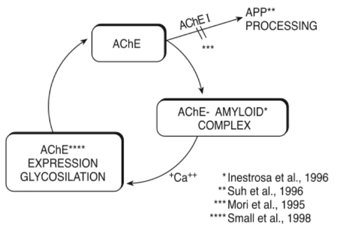
Table 5 summarizes the special relationship be-tween AChE and beta-amyloid in the brain of AD pa-tients (1). Accordingly, AChE, which is present in a glycosylated form associated with the amyloid core of neuritic plaques (37), is stimulated in its synthesis by
beta-amyloid. AChE stimulates beta-amyloid accu-mulation in or near to the plaque (22, 40) (Fig. 4).
One first explanation for a long-term neuropro-tective effect of ChEI is an interaction of the beta-A cy-cle with ChEI, promoting the release of soluble forms of beta-APP, and the reduction of amyloidogenic forms. Such a mechanism implies the regulation of al-pha secretase activity as the common final effector of PKC-dependent modulation of APP metabolism act-ing through a muscarinic m1receptor, as supported by experimental evidence (5-7, 20, 23). Secretion of APP soluble forms related to ChE inhibition has been demonstrated in vitro and in vivo in the CSF of NBM lesioned rats (rivastigmine and phenserine) (44), neuroblastoma cells for metrifonate (6), rat cortex slices (metrifonate) (5), rat basal forebrain cell cul-tures (metrifonate and ambenomium) (7), and in hu-man neuroblastoma cells for several ChEI (25, 45).
A second hypothesis is based on the data showing that the AChE molecule may interact with beta-amy-loid through a hydrophobic site close to the peripheral anionic binding site (PAS), promoting amyloid fibril for-mation (22). In addition, AChE is incorporated into se-nile plaques in vitro by forming macromolecular complexes with the growing beta-amyloid fibrils (44). AChEI binding to PAS may reduce this formation. Both hypotheses need clinical confirmation utilizing CSF markers to monitor brain beta-amyloid levels and metabolism in patients treated with ChEI for a long term. Studies are in progress to verify such an ef-fect in patients.
Table 5 - Acetylcholinesterase-beta-amyloid relationship in AD
brain.
ACETYLCHOLINESTERASE:
1. Is associated with the amyloid core of neuritic plaques (37). The hydrophobic site of aggregation is distinct from the enzy-matic active site (38).
2. Is abnormally glycosylated (39, 40) and its expression is stimu-lated by beta-amyloid (23, 40).
3. Is increased within and around the amyloid plaques (37) and pro-motes aggregation of amyloid beta-peptide fragments (41). 4. Is increased in the brain of transgenic mice expressing the
c-ter-minal fragment of the beta-amyloid precursor protein (42). 5. Nucleus basalis lesions increase synthesis of beta-amyloid (43).
AChE APP** PROCESSING AChEI *** +Ca++ Inestrosa et al., 1996 Suh et al., 1996 Mori et al., 1995 Small et al., 1998 AChE- AMYLOID* COMPLEX * ** *** **** AChE**** EXPRESSION GLYCOSILATION
Figure 4 - Proposed beta-amyloid cycle: AChE co-localizes with
beta-amyloid and accelerates beta-amyloid formation and de-position in AD brain. Beta-amyloid increases AChE in the brain. Inhibition of AChE activity inhibiting APP release reduces beta-amyloid deposition. This mechanism could contribute to the patient long-term cognitive stabilization seen during AChEI treatment.
The longest clinical study on the effect of a ChEI was performed as an extension trial lasting almost 5 years. The results demonstrate treatment benefits on cognition and global function in patients progressing from mild to moderate AD, and from moderate to se-vere AD over a period of 4, 9 years (46). The mean annual rate of decline in ADAS-Cog and CDR (Clin-ical Dementia Rating) of patients receiving the ChEI was significantly less over the first through the third year of treatment than might be predicted had this co-hort of patients not been treated (46). This long-term efficacy supports the concept of a disease-de-laying effect of this class of drugs.
CONCLUSIONS
In vitro as well as in vivo studies have consistent-ly demonstrated a link between cholinergic activa-tion and APP metabolism. A lesion of the cholinergic nuclei (nucleus basalis Meynert) causes a rapid in-crease in APP in the neocortex and CSF of rats (43). The effect of such lesions can be reversed by treatment with ChEI (44). A reduction in cholinergic neuro-transmission, experimental or pathological, leads to amyloidogenic metabolism in the brain, and con-tributes to the neuropathology and cognitive dys-function. Cholinesterase inhibitors may interact with both mechanisms, stabilizing the patient. This inter-pretation establishes a relationship between the effects of cholinergic drugs with a muscarinic mechanism of action, such as ChEI, and muscarinic agonists with treatments specifically targeted to decrease the beta-amyloid burden in the brain (Fig. 1).
REFERENCES
1. Giacobini E.: Cholinesterase inhibitors: from the calabar bean to Alzheimer therapy. In: Giacobini E. (Ed.), Cholinesterase
and cholinesterase inhibitors. From molecular biology to therapy. Martin Dunitz, London, pp. 181-222, 2000.
2. Davis K.L., Mohs R.C., Marin D.: Cholinergic markers in el-derly patients with early signs of Alzheimer Disease. JAMA 281: 1401-1406, 1999.
3. Pappas B.A., Bayley P., Bui B.K., Hansen L.A., Thal L.J.: Choline acetyltransferase activity and cognitive domain scores of Alzheimer’s therapy. Neurobiol. Aging 21: 11-17, 2000. 4. Doody R.S., Pratt R.D., Perdomo C.A.: Clinical benefits of donepezil: results from a long-term Phase III extension trial.
Neurology 52 (Suppl. 2): A174, 1999.
5. Mori F., Lai C.C., Fusi F., Giacobini E.: Cholinesterase in-hibitors increase secretion of APPs in rat brain cortex. Neurol.
Rep. 6: 633-636, 1995.
6. Racchi M., Schmidt B., Koenig G.: Treatment with metrifonate promotes soluble amyloid precursor protein release from SH-SY5Y neuroblastoma cells. Alzheimer Dis. Assoc. Disord. 13: 679-688, 1999.
7. Pakaski M., Rakonczay Z., Kasa P.: Reversible or irreversible cholinesterase inhibitors cause changes in neuronal amyloid percursor processing and protein kinase C level in vitro.
Neurochem. Int. 38: 219-226, 2001.
8. Cummings J.L.: Changes in neuropsychiatric symptoms as out-come measures in clinical trials with cholinergic therapies for Alzheimer disease. Alzheimer Dis. Assoc. Disord. 11: S1-S9, 1997.
9. Raskind M.A., Cyrus P.A., Ruzicka B.B.: The effects of Met-rifonate on the cognitive, behavioral, and functional perfor-mance of Alzheimer’s Disease patients. J. Clin. Psychiatry 60: 318-325, 1999.
10. Anand R., Hartman R., Messina J.: Long-term treatment
with rivastigmine continues to provide benefits for up to one year. Fifth Int. Geneva/Springfield Symposium on Advances in
Alzheimer Therapy, Geneva, 1998, p.18 (Abstract). 11. Rogers S., Friedhoff L.: Long-term efficacy and safety of
donepezil in the treatment of Alzheimer’s disease. Eur.
Neu-ropsychopharmacol. 8: 67-77, 1998.
12. Jelic V., Amberla K., Almkvist O.: Long-term tacrine
treat-ment slows the increase of theta power in the EEG of mild Alzheimer patients compared to untreated controls.
Fifth Int. Geneva/Springfield Symposium on Advances in Alzheimer Therapy, Geneva, 1998, p. 147 (Abstract). 13. Bernhardt T., Woelk H.: Metrifonate demonstrates
sus-tained improvement in cognition and global functioning in a 12-month, double blind placebo-controlled trial. European
Neurology Society Meeting, Jerusalem, 2000, p. 36 (Ab-stract).
14. Rainer M., Mucke H.A.M.: Long-term cognitive benefit from Galanthamine in Alzheimer’s disease. Int. J. Geriatr.
Psy-chiatry 1: 197-201, 1999.
15. Winblad B., Engedal K., Soininen H.: Donepezil enhances
global function, cognition and activities of daily living compared with placebo one year. 12thCongress of ECNP, London, 1999, Abstract 30.
16. Mohs R., Doody R., Morris J.: Donepezil preserves func-tional status in Alzheimer’s disease patients. Eur. Neuropsych. (Suppl. 5), Abstract S328, 1999.
17. Matthews H.P.: Donepezil in Alzheimer’s Disease: eighteen-month results from Southampton memory clinic. Int. J.
Geri-atr. Psychiatry 15: 713-720, 2000.
18. Raskind M.A., Peskind E.R., Wessel T.: Galantamine in AD. A sixth-month randomized, placebo-controlled trial with a 6-month extension. Neurology 54: 2261-2268, 2000. 19. Giacobini E.: Cholinesterase Inhibitors for Alzheimer’s Disease
therapy, from tacrine to future applications. Neurochem.
Int. 32: 413-419, 1998.
20. Giacobini E.: Cholinesterase inhibitors do more than inhibit cholinesterase. In: Becker R., Giacobini E. (Eds.), Alzheimer
Disease: From molecular biology to therapy. Birkhäuser,
Boston, 1996, pp. 187-204.
21. Farlow M.: New approaches in assessing delay of progression
of AD. Symp. Pivotal Research, World Alzheimer Congress
2000, Washington D.C., 2000, pp. 10-11 (Abstract). 22. Inestrosa N., Alvarez A., Perez C.A.: Acetylcholinesterase
accelerates assembly of amyloid-beta-peptides into Alzheimer’s fibrils: possible role of the peripheral site of the enzyme.
Neuron 16: 881-891, 1996.
23. Nitsch R.M.: Release of Alzheimer amyloid precursor deriva-tives stimulated by activation of muscarinic acetylcholine re-ceptors. Science 258: 304-307, 1992.
24. Nitsch R.: Muscarinic agonists reduce CSF levels of amyloid
beta-peptides in patients with Alzheimer’s disease. XVth
In-ternational Symposium of Medicinal Chemistry, Bologna, 2000, Abstract Ml-41, p. 52.
25. Lahiri D.K., Farlow M.R., Hintz N., Utsuki T., Greig N.H.: Cholinesterase inhibitors, beta-amyloid precursor protein and amyloid beta-peptides in Alzheimer’s Disease. Acta Neurol.
Scand. (Suppl. 176): 60-67, 2000.
26. Svensson A.L., Giacobini E.: Cholinesterase inhibitors do more than inhibit cholinesterase. In: Giacobini E. (Ed.),
Cholinesterases and Cholinesterase Inhibitors. Martin
Dunitz, London, 2000, pp. 227-235.
27. Becker E., Giacobini E.: Mechanisms of cholinesterase inhi-bition in senile dementia of the Alzheimer type. Drug Dev.
Res. 12: 163-195, 1988.
28. Giacobini E., Desarno P., Clark B.: The cholinergic receptor system of the human brain - Neurochemical and pharmaco-logical aspects in aging and Alzheimer. In: Nordberg A., Fuxe K., Holmstedt B. (Eds.), Progress in Brain Research. El-sevier Ed., Amsterdam, 1989, pp. 335-343.
29. Von Der Kammer H., Mayhaus M., Albrecht C.: Muscarinic acetylcholine receptors activate expression of the Erg gene family of transcription factors. J. Biol. Chem. 273: 10-17, 1998.
30. Taher D.S.: Long-term rivastigmine treatment produces
persistent inhibition of acetyl- and butyrylcholinesterase ac-tivity in CSF. World Alzheimer Congress 2000, Washington
DC, Abstract 1111.
31. Nordberg A., Hellstrom-Lindahl E., Almqkvist O.: Acetyl-cholinesterase activity in CSF of Alzheimer’s patients after treatment with tacrine. Alzheimer’s Report 2: 347-352, 1999.
32. Unni L., Vicari S., Moriearty P.: The recovery of cerebrospinal fluid acetylcholinesterase activity in Alzheimer’s disease patients after treatment with metrifonate. Methods Find. Exp. Clin.
Pharmacol. 22: 57-61, 2000.
33. Cummings M.D., Cyrus P.A., Bieber F.: Metrifonate treatment of the cognitive deficits in Alzheimer’s Disease. Neurology 50: 1214-1221, 1999.
34. Bareggi S.R., Giacobini E.: Acetylcholinesterase activity in ven-tricular and cisternal CSF of dogs. J. Neurosc. Res. 3: 33-43, 1978.
35. Mattio T., McIlhany M., Giacobini E.: The effects of physostig-mine on acetylcholinesterase activity of CSF, plasma and brain. Neuropharmacol. 25: 1167-1177, 1986.
36. Hinz V.C., Kolb J., Schmidt B.: Effects of subchronic ad-ministration of metrifonate on cholinergic neurotransmission in rats. Neurochem. Res. 23: 931-938, 1998.
37. Geula C., Mesulam M.: Special properties of cholinesterases in the cerebral cortex of Alzheimer’s disease. Brain Res. 498: 185-189, 1989.
38. Inestrosa N.C., Alvarez A., Reyes A., De Ferrari G.V.: Acetyl-cholinesterase -amyloid -peptide interaction and Wnt signaling involvement in A-beta neurotoxicity. Acta Neurol. Scand. (Suppl. 176): 53-59, 2000.
39. Saez-Valero J., Sberna G., McLean C.A., Small D.H.: Molec-ular isoform distribution and glycosylation of acetyl-cholinesterase are altered in brain and cerebrospinal fluid of pa-tients with Alzheimer’s disease. J. Neurochem. 72: 1600-1608, 1999.
40. Small D.H., Sberna G., Li Q.X.: The beta-amyloid protein
in-fluence acetylcholinesterase expression, assembly and gly-cosylation. Sixth International Conference on Alzheimer’s
Dis-ease and Related Disorders, Amsterdam, Abstract 880, S.209, 1998.
41. Alvarez A.: Acetylcholinesterase promotes the aggregation of amyloid B-peptide fragments by forming a complex with the growing fibrils. J. Mol. Biol. 272: 348-361, 1997. 42. Sberna G., Saez-Valero J., Li Q.X.: Acetylcholinesterase is
in-creased in the brains of transgenic mice expressing C-terminal fragment of the beta-amyloid protein precursor of Alzheimer’s disease. J. Neurochem. 71: 723-731, 1998.
43. Wallace W.C., Bragin V., Robakis N.K., Sambamurti K., VenderPutten D., Merril C.R., Davis K.L., Santucci A.C., Haroutunian V.: Increased biosynthesis of Alzheimer amyloid precursor protein in the cerebral cortex of rats with lesions of the nucleus basalis Meynert. Brain Res. Mol. Brain Res. 10: 173-178, 1991.
44. Haroutunian V., Wallace W.C., Greig N.: Induction, secretion
and pharmacological regulation of beta-APP in animal model systems. Sixth Int. Stockholm/Springfield
Sympo-sium on Advances in Alzheimer Therapy, 2000, p. 81 (Ab-stract).
45. Suh Y-H., Chong Y.H., Kim S-H.: Molecular Physiology, Biochemistry and Pharmacology of Alzheimer’s Amyloid Pre-cursor Protein (APP). Ann. N.Y. Acad. Sci. 786: 169-183, 1996.
46. Rogers S.L.: Long-term efficacy and safety of donezepil in the treatment of Alzheimer’s Disease: final analysis of a US mul-ticentre open-label study. Eur. Neuropsychopharmacol. 10: 195-203, 2000.