HAL Id: hal-02909846
https://hal-amu.archives-ouvertes.fr/hal-02909846
Submitted on 14 Dec 2020HAL is a multi-disciplinary open access archive for the deposit and dissemination of sci-entific research documents, whether they are pub-lished or not. The documents may come from teaching and research institutions in France or abroad, or from public or private research centers.
L’archive ouverte pluridisciplinaire HAL, est destinée au dépôt et à la diffusion de documents scientifiques de niveau recherche, publiés ou non, émanant des établissements d’enseignement et de recherche français ou étrangers, des laboratoires publics ou privés.
1,2H hyperfine spectroscopy and DFT modeling unveil
the demethylmenasemiquinone binding mode to E. coli
nitrate reductase A (NarGHI)
Maryam Seif Eddine, Frédéric Biaso, Julia Rendon, Eric Pilet, Bruno
Guigliarelli, Axel Magalon, Stéphane Grimaldi
To cite this version:
Maryam Seif Eddine, Frédéric Biaso, Julia Rendon, Eric Pilet, Bruno Guigliarelli, et al.. 1,2H hyperfine spectroscopy and DFT modeling unveil the demethylmenasemiquinone binding mode to E. coli nitrate reductase A (NarGHI). Biochimica biophysica acta (BBA) - Bioenergetics, Elsevier, 2020, 1861 (8), pp.148203. �10.1016/j.bbabio.2020.148203�. �hal-02909846�
1
1,2
H hyperfine spectroscopy and DFT modeling unveil the
demethylmenasemiquinone binding mode to E. coli nitrate reductase A
(NarGHI)
Maryam Seif Eddine, a,1 Frédéric Biaso, a Julia Rendon, a,2 Eric Pilet,a Bruno Guigliarelli, a Axel Magalon, b Stéphane Grimaldi a,*
a
Aix Marseille Univ, CNRS, BIP, Marseille, France
b
Aix Marseille Univ, CNRS, LCB, Marseille, France
1
Present address: Imperial College London, Department of Chemistry, Molecular Sciences Research Hub, W12 0BZ, London, UK
2
Present address: Univ. Grenoble Alpes, CEA, Department of the Interfaces for Energy, Health and Environment, Laboratoire SYMMES, Grenoble, France
*
Correspondence to: S. Grimaldi, Aix Marseille University, CNRS, Bioenergetics and Protein Engineering (BIP UMR7281), 31 chemin Joseph Aiguier, CS70071, 13402 Marseille cedex 09, France.
E-mail address: stephane.grimaldi@univ-amu.fr
Keywords
Bioenergetics • Quinones • HYSCORE • Metalloenzyme • Electron Transfer
Abbreviations
(b)RC, (bacterial) reaction center ; Cw, continuous wave; E’m,7, midpoint redox potential at
pH = 7; DFT, density functional theory ; (D)MK, (D)MSK, (D)MKH2,
(demethyl)menaquinone, (demethyl)menasemiquinone, (demethyl)menaquinol; (D)MSKD,
USQD, (demethyl)menasemiquinone and ubisemiquinone formed at the quinol oxidation site
(QD) from EcNarGHI, respectively; EcNarGHI, membrane-bound nitrate reductase from
Escherichia coli; ESEEM, electron spin echo envelope modulation; hf(c)(c)(s), hyperfine
(coupling) (constant)(s); hfi, hyperfine interaction; EPR, electron paramagnetic resonance; HYSCORE, hyperfine sublevel correlation; IMVs, inner membrane vesicles; nq(cc), nuclear quadrupole (coupling constant) ; nqi, nuclear quadrupole interaction; Q, SQ, QH2, quinone,
2 Abstract
The quinol oxidation site QD inE. coli respiratory nitrate reductase A (EcNarGHI) reacts with
the three isoprenoid quinones naturally synthesized by the bacterium, i.e. ubiquinones (UQ), menaquinones (MK) and demethylmenaquinones (DMK). The binding mode of the demethylmenasemiquinone (DMSK) intermediate to the EcNarGHI QD quinol oxidation site
is analyzed in detail using 1,2H hyperfine (hf) spectroscopy in combination with H2O/D2O
exchange experiments and DFT modeling, and compared to the menasemiquinone one bound to the QD site (MSKD)previouslystudied by us. DMSKD and MSKD are shown to bind in a
similar and strongly asymmetric manner through a short (~1.7 Å) H-bond. The origin of the specific hf pattern resolved on the DMSKD field-swept EPR spectrum is unambiguously
ascribed to slightly inequivalent contributions from two β-methylene protons of the isoprenoid side chain. DFT calculations show that their large isotropic hf coupling constants (Aiso ~ 12 and 15 MHz) are consistent with both (i) a specific highly asymmetric binding
mode of DMSKD and (ii) a near in-plane orientation of its isoprenyl chain at C relative to the
aromatic ring, which differs by ~ 90° to that predicted for free or NarGHI-bound MSK. Our results provide new insights into how the conformation and the redox properties of different natural quinones are selectively fine-tuned by the protein environment at a single Q site. Such a fine-tuning most likely contributes to render NarGHI as an efficient and flexible respiratory enzyme to be used upon rapid variations of the Q-pool content.
3 1. Introduction
Isoprenoid quinones are lipophilic electron mediators which play key roles in bioenergetics processes leading to efficient harvesting of environmental energy and its conversion into a transmembrane chemiosmotic potential. While they all bear a ring moiety with two oxygen atoms at positions 1 and 4, their chemical structure and electrochemical properties can differ significantly. The vast majority of biological isoprenoid quinones belong to either the low-potential naphthoquinones, e.g. phylloquinones (PhQ or vitamin K1) and
menaquinone (MK or vitamin K2), or to the higher potential benzoquinones, e.g. ubiquinone
(UQ) and plastoquinone (PQ) (Fig. 1) [1]. The crucial role of isoprenoid quinones in bioenergetics relies mainly on (i) their soluble character in lipid bilayers conferred by their apolar hydrophobic side chain which can vary in length, in degree of saturation and in the presence of additional groups, and (ii) on the redox properties of their aromatic ring which can easily and reversibly oscillate under physiological conditions between three different oxidation states with different protonation levels: the oxidized deprotonated quinone state (Q), the intermediate semiquinone (SQ) form which can be anionic (SQ•-) or neutral (SQH•), and the fully reduced and protonated quinol state (QH2).
4
Fig. 1. Chemical structures of some naturally occurring isoprenoid low- and high-potential quinones. The type I and type II classification is based on the nature of the C2 substituting group, namely a methyl group for type I and a hydrogen atom for type II [2]. The atom numbering scheme used in the text is shown at the bottom. (D)MK-8, (demethyl)menaquinone-8, PhQ, phylloquinone, UQ-8, ubiquinone-8, PQ-9, plastoquinone-9. The digit indicates the number of prenyl units which may vary in a given organism or between different species. The α-protons are directly bound to the aromatic rings, and the β-protons are found within the methyl group and the first CH2 group of the quinone prenyl (or
phytyl for PhQ) chain.
Isoprenoid quinones interact with bioenergetic complexes within well-defined protein sites (called Q sites) where they transiently bind and exchange with the Q-pool, or act as permanently bound cofactors possibly involved in intramolecular electron transfer and proton exchange. Several studies on photosynthetic and respiratory complexes have shown that the (electro)chemical properties of the protein-bound quinones and therefore the reactivity towards quinones can be drastically modulated in a Q site, hence contributing to define the electron transfer directionality in a particular enzyme or its specificity towards quinones [3-6].
The ability of some facultative anaerobes such as E. coli to synthesize both low potential (i.e. MK with E’m,7 MK/MKH2 = -70 10 mV) and high potential quinones (i.e.
UQ with E’m,7 UQ/UQH2 = + 100 10 mV) [1] opens up the possibility to investigate in vivo
the specificity of quinone utilization by respiratory complexes [7]. In this context, the respiratory nitrate reductase complex NarGHI from E. coli (EcNarGHI) is an excellent model to address this issue. EcNarGHI is a membrane-bound heterotrimeric complex which couples the oxidation of quinols at a periplasmically-oriented Q site (QD) [8] to the cytoplasmic
two-electron reduction of nitrate into nitrite [5, 9, 10]. EcNarGHI turnover induces a net translocation of protons across the membrane which contributes to maintaining the transmembrane proton gradient that drives, for instance, ATP synthesis. The NarG catalytic subunit holds the Mo-bisPyranopterin Guanosine Dinucleotide cofactor [11] and a FeS cluster [10, 12, 13]. The electron transfer subunit NarH harbors four FeS clusters [9, 14]. Finally, the cytoplasmically exposed NarGH subunits are connected to the membrane-integral NarI, which has two b-type hemes termed bD and bP according to their respective distal and proximal
positions with respect to the nitrate reducing site [10, 15, 16]. The metal cofactors form a chain of electron transfer relays from the quinol oxidation site QD in NarI to the molybdenum
5
EcNarGHI has been shown to react with the three quinones synthesized by the
bacterium, namely MK, UQ and demethylmenaquinone (DMK) [17-19]. DMK differs from MK by the lack of the methyl group at the C2 position of the quinone ring and by its ~ 60 mV higher redox potential (E’m,7 DMK/DMKH2 = +36 [20] or -7 mV [21]) (Fig. 1). Using
EPR-monitored redox titrations on E. coli inner membrane vesicles (IMVs) enriched in NarGHI, we have previously shown that the EcNarGHI QD site stabilizes the semiquinone intermediate
of each of the three endogenous quinones. Noteworthy, a specific hf interaction with a 14N nucleus was similarly detected to each of the three corresponding protein-bound radicals using HYSCORE spectroscopy [19, 22, 23]. Based on multifrequency 14,15N ESEEM/HYSCORE measurements [23], site-directed mutagenesis studies [22, 24] and selective 15N labeling [25], this interacting nucleus has been ascribed to the N imidazole nitrogen of the heme bD axial ligand His66. Combining the use of H2O/2H2O exchange
experiments, selective 2H labeling of the ring methyl protons and 1,2H hyperfine spectroscopies, we have shown that MSKD is stabilized thanks to a short in-plane H-bond to
the quinone oxygen O1, leading to a highly asymmetric distribution of the electron spin density over the semiquinone ring in comparison with the unbound species [25, 26].
Intriguingly, EPR-monitored redox titrations indicated that, in contrast to MSK and USQ [22, 27], the two-electron midpoint potential of EcNarGHI-bound DMK is decreased by at least 30 mV with respect to that of the unbound species [25]. This shift likely results from a redox-dependent differential binding of DMK at the QD site corresponding to a ~10 fold
tighter binding of DMK than DMKH2 to this site. In addition, in contrast to USQD and MSKD,
the DMSKD EPR signal exhibits a resolved hf structure from one or several nearby non
exchangeable protons [19] whose origin was unclear. Its linewidth has been shown to decrease upon H2O/2H2O exchange, revealing the existence of at least one exchangeable
proton weakly coupled to DMSKD [25].
To fully resolve the DMSKD electronic structure and address the role of the C2
substituent on the naphthoquinone ring in binding to the EcNarGHI QD site, we performed a
detailed spectroscopic analysis of DMSKD using 1,2H hyperfine pulsed EPR spectroscopy in
combination with H2O/2H2O exchange experiments and DFT modeling. Our results show that,
in spite of a conserved asymmetric binding mode through a short H-bond, both DMSKD and
MSKD exhibit markedly different orientations of their isoprenyl side-chain. This sheds new
light on how a single Q site enables the binding of different quinones and modulate their structural and redox properties.
6 2. Material and methods
2.1. Sample preparations
NarGHI was expressed in an E. coli nitrate reductase-deficient strain JCB4023ubiE (RK4353, ΔnapA-B, narG::ery, narZ::Ω, SpcR, ΔubiE::KanR) [19, 28] which contains demethylmenaquinone as sole respiratory quinone. pVA700 plasmid (AmpR) [14], which encodes for the narGHJI operon under control of the tac promoter, was used in all experiments. Cells were grown in Terrific Broth under semi-anaerobic conditions at 37 °C as described in [19, 24] with ampicillin (100 g.ml-1) and spectinomycin (50 g.ml-1) included in the growth medium.
Purified E. coli NarGHI-enriched inner membrane vesicles (IMVs) were used for this study, allowing to maintain an unmodified lipid environment and to study the interaction of NarGHI with its endogenous demethylmenaquinol substrate. For this purpose, purified E. coli NarGHI-enriched IMVs were isolated by differential centrifugation and sucrose gradient steps as described in [24] using a buffer containing 100 mM MOPS and 5 mM EDTA at pH 7.5. Deuterium-exchanged samples were prepared using the same membrane extraction protocol with a buffer containing 2H2O (99.9 % atom 2H) instead of 1H2O. The functionality of
NarGHI in our samples was confirmed spectrophotometrically by measuring the quinol:nitrate oxidoreductase activity.Stabilization of the semiquinone at the QD site was achieved through
redox titrations under the same conditions as those described in [19, 24, 27] except that samples were prepared in a glove box. They were redox poised at a potential allowing to maximize the DMSK concentration with respect to the two other redox species (~ - 60 to -90 mV) [19]. Redox potentials are given in the text with respect to the standard hydrogen electrode.
2.2. EPR and HYSCORE spectroscopy
The instrumentation for X- and Q-band CW EPR measurements was previously described [24]. The instrumentation, pulse sequences, and spectral processing for X-band 1D 4-pulse ESEEM (/2--/2-t--t-/2--echo) and 2D 4-pulse ESEEM (HYSCORE) (/2- -/2-t1--t2-/2--echo) were also as described [26]. Spectral simulations were carried out
using the EasySpin package (release 5.01.1) under Matlab (The MathWorks, Inc., US) [29]. Simulations of 1,2H HYSCORE spectra were carried out assuming axial hfi tensors. Principal axes of hf tensors were all assumed to be collinear with the g-tensor principal axes unless explicitly stipulated otherwise.
7
2.3. Hyperfine and nuclear quadrupole interactions
A hfc between a S = ½ radical and a nucleus with nuclear spin value I consists in general of (i) the isotropic contribution Aiso = 20gegnen|0(0)|2/3h where |0(0)|2 is the
electron spin density at the nucleus, ge and gn are electron and nuclear g-factors, respectively,
e and n are Bohr and nuclear magnetons, respectively, h is Planck’s constant, and (ii) the
anisotropic contribution described by the traceless dipolar coupling tensor T. In most cases, T can be assumed to be axial, with principal values (–T, –T, 2T).
The hfccs of different isotopes of the same element are proportional to a very good approximation to the corresponding gn values. In this study, the direct and simultaneous
determination of Aiso and T of the protons interacting with DMSK were derived from the
analysis of 1H HYSCORE cross-peak contours as described in the Supporting Information [30].
A 2H nucleus has a quadrupole moment which interacts with the electric field gradient (EFG) at the nucleus. The components of the EFG tensor are defined in its principal axis system and ordered according to qZZ qYY qXX. This traceless tensor can then be fully
described by only two parameters: (i) the 2H nuclear quadrupole coupling constant = |e2qZZQ/h|, where e is the charge of an electron, Q is the 2H nuclear electric quadrupole
moment, (ii) the asymmetry parameter = |qYY-qXX/qZZ|. is a measure of the strength of the
interaction between the nuclear quadrupole moment and the EFG at the 2H nucleus site due to anisotropic charge distribution around the nucleus whereas is a measure of the deviation of this distribution from axial symmetry. Thus, the EFG is related to the specific binding geometry. Its components can, therefore, be used to obtain detailed information on hydrogen bonds. In this study, parameters and of the 2H nucleus interacting with DMSKD were
estimated by simulating X-band 2H HYSCORE spectra.
2.4. DFT calculations
All calculations have been performed with Orca 3.0 quantum chemistry package [31] at a DFT level of theory, using the B3LYP hybrid functional (the Becke’s three parameters hybrid exchange functional with 20% of Hartree-Fock admixture and the Lee-Yang-Parr non local correlation functional). A restricted geometry optimization has been performed in vacuo on each model with the def2-SVP basis set [32, 33]. The resolution of identity with the appropriate auxiliary basis sets was used to accelerate the calculations [34]. The optimized
8
structures have then been used as input for electronic and magnetic properties calculations, using the EPR-II basis set [35] and employing the conductor-like screening model COSMO [36] with a dielectric constant = 4.0 to replicate electrostatic effects of the protein surrounding. 1H hfi and 2H nqi coupling constants of the methyl group were calculated by averaging the raw matrices corresponding to the three positions of the methyl protons and the eigenvalues and eigenvectors were determined.
3. Results and discussion
3.1. Field swept Electron Spin Echo (ESE) EPR spectra of DMSK
To specifically study the interaction of EcNarGHI with its natural DMK substrate, NarGHI-enriched IMVs were purified from an E. coli ubiE- strain containing DMK as its sole
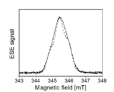
respiratory quinone. The sample was titrated and redox poised to stabilize the maximal amount of DMSK, and studied by EPR methods at 90 K. At this temperature, the radical can be specifically probed without spectral contamination from other faster relaxing paramagnetic centers such as EPR-active cofactors in EcNarGHI [19]. X-band field swept ESE spectra of NarGHI-enriched IMVs recorded at 90 K from samples prepared either in H2O (solid line) or
in 2H2O (dotted line) are shown in Fig. 2. The EPR signal is characterized by an average
g-value gav ~ 2.0045. This signal is not present in NarGHI-deficient IMVs prepared and titrated
in similar conditions [19]. The characteristic structure of DMSKD is best resolved in the ESE
spectrum recorded using the 2H2O-exchanged sample due to the decrease of the linewidth
upon replacement of H2O by 2H2O [19]. This decrease, which results from downscaling of
hyperfine coupling constants (hfccs) of exchangeable 2H nuclei by a factor of ~ 6.5 compared to the 1H one’s, indicates that at least one nearby exchangeable proton is coupled to the radical [19]. In the following, spectral resolution was further increased by using 2D pulsed hyperfine spectroscopy.
9
Fig. 2. Field swept ESE spectra of NarGHI-bound DMSK prepared in H2O (solid line) and 2
H2O (dotted line). Microwave frequency, 9.700 GHz (H2O), 9.686 GHz (2H2O). A magnetic
field offset of -4.5 G was applied to the spectrum measured in 2H2O. Spectra are normalized
to the same amplitude.
3.2. Analysis of 1H resonances in HYSCORE spectra of DMSK in H2O or 2H2O buffer
To provide detailed insights into the DMSKD electronic structure and accurate
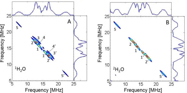
information about its proton environment, HYSCORE experiments were performed at 90 K using the EcNarGHI samples studied in previous section. Fig. 3A shows a representative 1H HYSCORE spectrum of DMSKD prepared in H2O buffer. It has been recorded with = 204 ns
to suppress the strong diagonal peak at (1H) = 14.7 MHz contributed by matrix protons. Five pairs of cross-features located symmetrically relative to the diagonal can be distinguished in this spectrum. They are designated 1-5 in Fig. 3A and arise from protons that are magnetically coupled with the unpaired electron spin of DMSKD. Among them, ridges 5, 5' exhibit the
largest hf splitting estimated to ~ 15 MHz by measuring the distance between their maxima. They appear therefore well separated from the others which are contributed by protons with hfccs smaller than ~ 6 MHz. Notably, the ~ 15 MHz splitting is nearly three times larger than the largest one previously detected on the corresponding 1H HYSCORE spectrum of MSKD
and assigned to the three equivalent methyl protons with Aiso ~ 5.5 MHz [25, 26].
Cross-ridges 3-3' possess the most extended anisotropic contour, with the largest deviation from the diagonal, whereas cross-peaks 4-4' deviate significantly from the normal to the diagonal. These two features indicate a significant anisotropic component of the corresponding hfi tensor(s). Cross-peaks 2-2' and 4-4' partially overlap. Contours 1-1' and 2-2' are
10
approximately normal to the diagonal, suggesting a smaller anisotropic component of the corresponding 1H hfi tensors. To discriminate cross-features arising from exchangeable or non-exchangeable protons, HYSCORE measurements were carried out on the sample prepared in 2H2O buffer using similar conditions to those used for the sample prepared in H2O
(Fig. 3B). Cross-peaks 3-3' and 4-4' completely disappear in the DMSKD 1H HYSCORE
spectrum of the sample prepared in 2H2O buffer. This shows that they arise from at least one
exchangeable proton, in consistency with the decrease of the linewidth observed in the field-swept EPR spectra upon H2O/2H2O buffer exchange. In contrast, the other cross-peaks are not
affected by buffer exchange, indicating that they are produced by nonexchangeable protons. No other feature than those discussed above were detected in 1H HYSCORE spectra recorded using other values.
Fig. 3. 1H HYSCORE spectra of DMSKD in H2O (A) or in 2H2O (B). Microwave frequency,
9.6944 GHz (A) and 9.7045 GHz (B); magnetic field, 345.2 mT (A) and 345.9 mT (B). Other experimental parameters are as given in Material and methods, time between first and second pulses τ = 204 ns.
1
H HYSCORE spectra were quantitatively analyzed using the square frequency plot to assess the number of coupled protons and determine their respective hyperfine characteristics [30]. The α2 versus β2 plot of the cross-features analyzed from the DMSKD1H HYSCORE
spectra shown in Fig. 3 is depicted in Fig. 4 together with linear regressions of ridges 1-5 and assuming axial symmetry for all proton hyperfine interaction (hfi) tensors. The corresponding slopes and intercepts are given in Table S1 with the two possible sets of (Aiso, T) that satisfy
11
Eq. S2 and S3. The results indicate that ridges 1-1' and 2-2' arise from at least two distinct non exchangeable protons, hereafter referred to as HA and HB, respectively, whereas 3-3' and 4-4'
are the sub-ridges of cross-features belonging to a single additional exchangeable proton named HC characterized by an anisotropic contribution T(HC) ~ 5.6 MHz. This value is
consistent with the one calculated from the shift of the single sum combination line from 2I(1H) measured in four-pulse 1H ESEEM spectra of DMSKD prepared in H2O (see
supplementary information and Fig. S1). In contrast to HA and HB, selection of the right (Aiso,
T) for proton HC from the two alternatives has been achieved based on numerical simulations
of HYSCORE spectra. Indeed, the experimental relative intensities of ridges 3-3’ and 4-4’ can only be well reproduced using Aiso = ∓0.2 MHz and T = ±5.8 MHz (Fig. 5A and Table 1).
A peculiar feature of 1H HYSCORE spectra of DMSKD prepared either in H2O or in 2
H2O is the presence of cross peaks 5-5’ indicating that these features arise from at least one
non exchangeable proton strongly coupled to the radical (Fig. 3). In contrast to the other HYSCORE features, the points measured along the 5-5’ ridges do not align on a straight line in the square frequency plot, rendering the simple analysis presented above not satisfactorily applicable. However, ridges 5-5’ are well accounted for by a linear regression assuming that they are due to partially overlapping cross-features originating from two protons with distinct axially symmetric hfi tensors and that each of them mainly contributes to opposite sides (referred to 5A-5'A and 5B-5'B in Fig. 4) of the same ridge. Using this approach, the anisotropic
components of the hf tensors of these two protons (hereafter referred to as HD and HE) are
found to be similar, namely T ~ 2.0-2.1 MHz while their isotropic components are slightly different, i.e. Aiso (HD) = 12 or 14 MHz, and Aiso (HE) = 15 or 17 MHz (Table S1).
12
Fig. 4. Plot of cross-peaks detected in 1H HYSCORE spectra of DMSK in the versus coordinate system. Straight lines show the linear fit of plotted data points. The dashed line is defined by = and corresponds to the diagonal in the (+, +) quadrant of HYSCORE spectra. The lowermost contours were used to measure the coordinates of arbitrary points along the ridge of each proton cross-peak in HYSCORE spectra of DMSKD in H2O (3’, 4) or 2
H2O (1’, 2’, 5’).
3.3. Characterization of the exchangeable deuteron by 2H HYSCORE
Further details concerning exchangeable protons coupled to DMSKD were obtained
through the use of 2H HYSCORE spectroscopy. Given that the largest exchangeable proton splitting in the DMSKD HYSCORE spectra measured at different values is ~ 6 MHz and
that 2H nuclei give rise to weak quadrupole splittings (typically a few tenths of MHz) [26, 37], a splitting not greater than 1 MHz is predicted in DMSKD2H HYSCORE spectra. However,
this feature is expected to be hardly detectable because of the intense and broad diagonal peak at I(2H) ~ 2.3 MHz arising from weakly coupled matrix 2H which dominates
ESEEM/HYSCORE spectra of 2H2O exchanged protein samples [26, 38]. To suppress this
intense signal, a HYSCORE spectrum of DMSKD prepared in 2H2O buffer has been measured
using a -value of 416 ns and is shown in Fig. 5B. It is compared to the corresponding spectrum of MSKD measured using similar conditions (Fig. 5C). Complete suppression of the
13
symmetrically around I(2H) which are very similar for the two radicals. This peculiar shape
for a powder HYSCORE spectrum of a (S = ½, I = 1) spin coupled system is well simulated by downscaling for 2H nucleus the HC hf parameters obtained from analysis of the 1H
HYSCORE spectra, assuming that a single 2H nucleus contributes (Fig. 5B). In addition, our numerical simulations indicated that the anisotropic hfi is mainly responsible for the elongation of the ridges perpendicularly to the diagonal (not shown) whereas the nqi is known to induce a splitting of the ridges parallel to the diagonal [39, 40], allowing these two contributions to be well distinguished from each other. Whereas the simulated spectrum is very sensitive to the magnitude of the nqcc , variations of the asymmetry parameter in the [0, 1] range have negligible effects. Therefore, a value of (HC) = 0.17 ± 0.02 MHz can be
estimated (Table 1). This value is consistent with that determined from analysis of the DMSKD Q-band 2H Mims ENDOR spectrum measured at ~ 34 GHz (Fig. S2). Eventually,
variations of the relative angle between the unique axes of hyperfine and nuclear quadrupole tensors have a modest effect on the simulated spectrum. This angle is therefore estimated to 0° ± 25°. Consequently, hyperfine and quadrupole tensors were assumed to be collinear in the simulations shown in Figure 5B and 5C.
Fig. 5. Contour plot of experimental and simulated (red contours) HYSCORE spectra from the exchangeable 1H (A) or 2H (B, C) coupled to DMSKD (A,B) or MSKD (C) in EcNarGHI
prepared in H2O (A) and in 2H2O buffer (B, C). Simulation parameters are given in Table 1,
and rescaled for 2H nuclei when relevant. Other experimental parameters are: microwave frequency, 9.691 GHz (A), 9.686 GHz (B) and 9.693 GHz (B), τ-value, 204 ns (A) and 416 ns (B, C).
14
1
H hyperfine and 2H nuclear quadrupole tensors of the exchangeable proton/deuteron coupled to DMSKD used for the simulation of 1H and 2H HYSCORE spectra. 1H hyperfine tensors
were initially derived from linear regression of 1H HYSCORE spectra in square frequency plot. Selection of the set of (Aiso, T) (Table S1) and evaluation of errors are based on
numerical simulations while the signs of the couplings are selected according to DFT calculations. g-, hfi- and nqi tensors are assumed to be collinear. 2H nuclear quadrupole tensors are derived from simulations of 2H HYSCORE (Fig. 5) and Q-band 2H Mims ENDOR spectra (Fig. S2).
Semiquinone species 1H (Aiso, T) [MHz] 2H ( [MHz],) References
DMSKD -0.20 ± 0.06, 5.8 ± 0.1 0.17 ± 0.02, 0.2 This work
MSKD +0.06, 5.73 0.176, 0.2 [25], [26]
3.4. The exchangeable proton coupled to DMSKD is involved in a short hydrogen bond to the
radical
Interestingly, comparative analysis of the 1,2H HYSCORE, four-pulse ESEEM and Q-band 2H Mims ENDOR spectra measured on DMSKD and MSKD reveals similar hfi and nqi
characteristics for the detected exchangeable 1,2H nucleus (HC) coupled to the two
protein-bound radical intermediates (Table 1). On the basis of its hf characteristics, this exchangeable proton is assigned to a proton involved in H-bonding to one of the quinone carbonyl oxygens [26]. This proton combines both an almost zero Aiso value and a relatively large T value (T
~ 5.6 MHz) for a proton H-bonded to a protein-bound semiquinone. A small Aiso is expected
when the H-bond lies in the quinone ring plane due to small overlap between the hydrogen 1s orbital and the oxygen 2pz orbital forming part of the semiquinone SOMO. In addition, the large T value of HC is predicted to account for a short hydrogen bond length with substantial
covalent character. In this context, DFT calculations have shown that the H-bond length rO-H
can be more reliably evaluated from the nqcc of the corresponding substituted 2H using the following empirical relation [41]:
= a – b/r3O-H (Eq. 1)
where a and b are empirical parameters. The value = 0.17 ± 0.02 MHz determined for DMSKD is similar to that obtained for MSKD, confirming a similar hydrogen bond geometry
of both radical species. A distance rO-H ~ 1.6 ± 0.1 Å can be estimated using Eq. 1, a = 319
15
biological systems. For instance, it is similar to that formed from the carbonyl oxygen O4 of the ubisemiquinone QA.- in reaction centers from Rhodobacter sphaeroides R-26 to the
imidazole nitrogen N of His M219 (rO-H = 1.60 ± 0. 04 Å [42]. In addition, 14N HYSCORE
spectroscopy has revealed a similar through-bond interaction of both MSKD and DMSKD with
a nitrogen nucleus that, in the case of MSKD, has been unambiguously assigned to the N
imidazole nitrogen of the heme bD axial ligand His66 [22, 23, 25]. Therefore, the
exchangeable proton HC coupled to DMSKD is similarly attributed to a H-bond between one
of the SQ oxygen atoms and this nitrogen atom.
3.5. Assignment of 1H hyperfine couplings to DMSKD by DFT modeling
A remarkable characteristic of DMSKD1H HYSCORE spectra is the detection of cross
peaks associated with unusually strong hf couplings arising from two non-exchangeable protons (HD and HE). Notably, the isotropic hfcc of HE (i.e. ~ 16 MHz) is the largest one
measured so far for a protein-bound semiquinone [43]. Importantly, spectral simulations (Fig. 6) show that the resolved hf structures detected on the cw X- and Q-band EPR spectra of DMSKD which are best seen in samples prepared in 2H2O are well accounted for by the
contribution of protons HD and HE using the hfc tensors given in Table 2 derived from
16
Fig. 6. A) Experimental and simulated (red contour plot) HYSCORE cross features of DMSKD in 2H2O buffer. B) Experimental (black solid line) and simulated (red dotted line)
continuous wave EPR spectra of DMSKD in 2H2O measured at X- (B) and Q-band (C)
frequencies. Simulations have been performed by considering the contributions of HA, HB, HD
and HE with individual hfccs as given in Table 2. Experimental conditions: redox potential,
-65 mV (A,B) and -70 mV (C), temperature, 60 K (B) and 150 K (C), microwave frequency, 9.482 GHz (B) and 34.122 GHz (C), microwave power, 0.1 mW (B) and 5 W (C), modulation amplitude, 0.3 mT, modulation frequency, 100 kHz. The signal indicated by an asterisk in Fig. 6C is due to paramagnetic impurities in the Q-band resonator. Experimental spectrum in A) corresponds to that shown in Fig. 3B.
To identify the origin of the hf couplings, we assume that DMSKD and MSKD display
a similar strongly asymmetric binding mode to NarGHI. Indeed, this assumption is based on the detection of similar hf and nq characteristics for the single exchangeable 1,2H nucleus and of the 14N nucleus coupled to DMSKD and MSKD. In our previous work on MSKD, these
nuclei were shown to be involved in a short hydrogen bond to the quinone carbonyl oxygen O1 which induces a strong decrease (~ 30 %) of the spin density on the Cα at the 2-position in comparison to a symmetrical hydrogen bonding situation to both carbonyl oxygens [25, 26, 44]. This model was supported by DFT calculations performed on a simple molecular model referred to as Im-MSK involving a deprotonated MSK hydrogen bonded via its carbonyl oxygen O1 to the N of an imidazole ring, with an isoprenoid chain truncated after the fourth carbon atom. Structural parameters inherent to this simple model were chosen to be consistent with available spectroscopic and crystallographic data. These include an O1-H distance rO-H of
1.7 Å between the imidazole ring and the quinone carbonyl oxygen O1, an in-plane hydrogen bond angle = 0°, an out-of-plane hydrogen bond angle = 30°, and a twist angle between SQ and imidazole rings = 30° (angles , and are defined in Fig. S3) [25]. In particular, this model accounts well for the unusually small value of the isotropic hfcc to the three equivalent methyl protons substituting the MSKD aromatic ring (i.e. Aiso ~ 5.5 MHz).
17 Table 2
Comparison between principal values of the g-tensor and 1H hyperfine coupling tensors of DMSKD determined from analysis of HYSCORE spectra, or calculated by DFT methods
using the Im-DMSK model with a C2C3CβC dihedral angle = 10°. Proton numbering is according to molecular positions given in Fig. 1. Principal values of the axial hyperfine tensors are defined as [Aiso-T, Aiso-T, Aiso+2T] using the values given in Table S1, unless
otherwise specified, and the corresponding signs of Aiso and T are selected according to DFT
calculations.
Experimental Calculated
g-tensor principal values
2.0061, 2.0050, 2.0023 2.0064, 2.0051, 2.0022 1
H hyperfine coupling tensors [MHz]
Proton numbering Assignment
Aiso [A, A, A//] Aiso [AX, AY, AZ] H2 HB -5.41 [-8.4, -8.4, 0.6] 1,2 -5.4 [-7.9, -8.4, -0.1] H-CH2 HD 11.51 [9.4, 9.4, 15.7] 1,3 12.1 [11.1, 15.6, 9.7] HE 16.01 [14.0, 14.0, 20.1] 1,3 17.8 [16.8, 15.7, 21.0] H HA -1.1 [-3.0, -3.0, 2.7] 0.2 [-0.4, -0.8, 1.8] H5 -0.4 [2.3, -2.3, -1.1] H6 -2.6 [-3.2, -4.3, -0.3] H7 -1.6 [-2.3, -2.8, 0.2] H8 -1.3 [-1.5, -3.1, 0.7]
18
2 Simulations performed with Euler angles between the g- and the hfi tensors optimized to (60°, 90°, 0°) 3 Simulations performed with Euler angles between the g- and the hfi tensors optimized to (75°, 90°, 0°)
Therefore, assuming a similar binding mode for DMSKD and MSKD, the redistribution of the
spin density in the ring-localized SOMO of the strongly asymmetrically bound DMSK with respect to a symmetrical hydrogen bonding situation results in its increase at carbon C3 to which the CH2 group is attached (Fig. 7). This is hence expected to lead to a concomitant
increase of the isotropic hfcc Aisoβ-H of these protons, according to the McLachlan formula:
Aisoβ-H = (B0 + B1 cos2θ) ρC3π (Eq. 2)
ρC3π is the spin density on carbon C3, θ is the angle between the CH bond and the axis of the
pz carbon orbital both projected in a plane orthogonal to the C-C bond (Fig. 8), B0 is a
constant that is close to zero, and B1 is also a constant with a value in the range [120 – 212]
MHz [45].
Fig. 7. Comparison of electronic spin density distribution between symmetrically hydrogen-bond (e.g. free) (D)MSK (left) and asymmetrically hydrogen-hydrogen-bond (e.g. NarGHI-bound) (D)MSK (right), with truncated isoprenyl chain. Spin density populations are represented by red circles whose radius is proportional to the spin density carried by the atom at the center of the circle (cut off = 0.01). The red arrow emphasizes the shift of the spin density from C2 to C3 due to the asymmetric (D)MSKD binding mode. H-bond partners are omitted on the left
19
Further, the nature of the substituent at the C2 carbon atom has been shown to be a crucial factor determining the value for semiquinones solubilized in various solvents [2, 46, 47]. In the lowest energy conformation of quinones with methyl-substituted C2 carbon (referred to as type I quinones, see Fig. 1) like MK, PhQ and UQ, the two β-CH2 protons have
Aiso β-CH2 values ~ 1-4 MHz, corresponding to (θ1, θ2) ~ (+60°, -60°) or ~ (+120°, -120°) [46,
48, 49] (Figure 8A and B). This translates into a C2C3CβC dihedral angle γ for the first isoprenyl unit relative to the quinone ring of ~ 90° or 270°, respectively, leading to a perpendicular head-to-tail orientation [2]. In contrast, type II quinones with proton-substituted C2 carbon like plastoquinones exhibit larger β-CH2 couplings (e.g. Aiso β-CH2 ~ 7.0 MHz for
PQ-9 in 2-propanol), corresponding to (θ1, θ2) ~ (+30°, +150°) or (-30°, -150°) for these two
protons (Figure 8C and D) [2, 50-52]. The corresponding C2C3CβC dihedral angle γ is then 0° or 180°, respectively, placing the isoprenyl chain in the plane of the quinone ring while minimizing steric repulsions between the carbonyl oxygen and the C substituents. These conclusions have been further substantiated by DFT calculations [47] and molecular dynamics simulations on PQ and UQ radical anion models [53, 54] showing that the presence or absence of the methyl group at the C2 carbon is indeed a crucial factor for determining the rotational arrangements of the quinones.
Fig. 8. Schematic conformation of the -CH2 protons and of the isoprenyl side chain relative
20
Newman projections along the Cβ-C3 axis are shown. The inset shows the definitions of angles and used in the text. angles for the two -CH2 protons are 120° (A), and 60°
(B), 30° and 150° (C), -30° and -150° (D).
Based on these previous works and given that DMSK is a type II quinone, we assigned HD and HE to the two β-CH2 protons of the highly asymmetrically bound DMSKD side chain
with an orientation suitable to generate large hyperfine coupling constants for these protons. In addition, this orientation is predicted to differ from that of MSKD, explaining the apparent
differences in the magnitude of the CH2 hf couplings between MSKD and DMSKD inferred
from analysis of their respective 1H HYSCORE spectra. To further support this assignment and compare the conformation of the DMSKD and MSKD side chain relative to the quinone
ring, isotropic -CH2 hfccs were computed for all possible values for DMSK (Fig. 9A) and
MSK (Figure 9B) using the Im-DMSK and Im-MSK models, respectively. These orientations were explored through a systematic variation of the C2C3CβCγ dihedral angle γ from -180° to 180°, in 5° or 10° step. At first, it can be noticed that Aisoβ-CH2 values as high as ~20 MHz are
calculated using these models, supporting the assignment of HD and HE to the two β-CH2
protons of DMSKD. Further, comparison between calculated -CH2 isotropic hfccs and
corresponding experimental values for HD and HE (i.e. Aiso (HD) ~ 12 MHz and Aiso (HE) ~ 16
MHz) determined from spectral analysis reveals that the best agreement is found for γ values close to 10° (Fig. 9A). This indicates that the isoprenyl CβCγ bond of the DMSKD side
chain is nearly coplanar to the aromatic ring (Fig. 10A).
Fig. 9. Influence of the C2C3CβC dihedral angle γ on the isotropic hyperfine coupling constant Aiso β-CH of the two β-CH2 protons (red and black squares, respectively) in the
21
consistent with HYSCORE data while light blue vertical bars show the corresponding ranges of γ values where calculated hfccs best agree with experimental data.
The principal values of the hfi tensors for non-exchangeable protons coupled to DMSK calculated using the Im-DMSK model with ~ +10° are reported in Table 2 where they are compared to the axially symmetric hfi tensors determined for protons HA, HB, HD and
HE. The calculated isotropic hfcc for the C2 α-proton H2 (Aiso ~ -5.4 MHz) is in very good
agreement with the value Aiso (HB) ~
-
5.4 MHz inferred from the simulation of EPR andHYSCORE spectra (Table 2). Therefore, HB is assigned to the DMSKD C2 substituting
-proton H2. Notably, its Aiso value is similar to that determined for the three equivalent
methyl protons on the MSKD ring, i.e. Aiso ~ 5.5 MHz [26]. This provides further support for
the similar asymmetric binding mode of DMSKD andMSKD to EcNarGHI.
Eventually, the calculated Aiso values for the other DMSK ring protons as well as that
of the γ-proton of the side-chain lie in the range [0.2 – 2.6] MHz (Table 2), the largest coupling being due to the C6 substituting proton H6. Aiso values ~ 1.0 MHz determined for
HA lie within this range, indicating that several protons contribute to the observed 1-1’ cross
peaks and that HA cannot be attributed to a single proton.
3.6. The conformations of the DMSKD and MSKD isoprenyl chains relative to the quinone
head differ by ca. 90°.
Examination of calculated -CH2 isotropic hfccs for MSKD indicates that out-of-plane
conformations with = ±90° (±5°) best account for our spectroscopic data on MSKD [26].
Indeed, these two situations lead to calculated Aiso β-CH2 values ~ 4-4.6 MHz for the two
protons using the Im-MSK model (Fig. 9B). These are consistent with MSKD HYSCORE
spectra in which no proton resonance with hfcc > 5 MHz is detected other than those clearly assigned to the methyl protons and to the exchangeable proton discussed above [26].
Beside, hfccs of 3 MHz and 1.5 MHz were resolved in 1H ENDOR spectra of MSK dissolved in 2-propanol and assigned to the -CH2 protons [55]. The magnitude of these
couplings was shown to be indicative of the perpendicular head-to-tail orientation of the menaquinone radical anion in solution [47]. Therefore, the conformation of the MSKD side
chain appears to be similar to that of the unbound species i.e. MSK in solution. Finally, the in-plane conformation of the isoprenyl chain relative to the semiquinone head of DMSKD differs
22
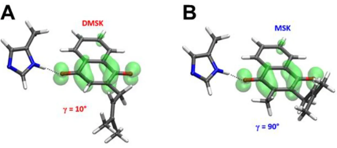
Fig. 10. Proposed conformations for DMSKD (A) and MSKD (B) inferred from this work.
Stick representations using the Im-MSK and Im-DMSK models are shown together with the distribution of the electron spin density over the semiquinone rings (cut off = 0.003). C2C3CβCγ dihedral angle γ is 10° (A) or 90° (B).
3.7. The protein environment in the NarGHI QD site finely tunes the side chain orientation of
the accommodated demethylmenaquinone.
Despite their simplicity, the Im-(D)MSK models account well for the EPR spectroscopic data collected on MSKD and DMSKD without relying on any protein constraint
on the side chain of the modeled quinones. In Fig. 11, the computed potential energy curves for rotation about the C3Cβ bond are compared for the Im-MSK and the Im-DMSK models. For the Im-MSK model, the potential displays two well-defined single minima at C2C3CβCγ dihedral angles γ ~ +90° and -100° as expected for type I quinones (Fig. 11, red squares). These orientations match with those determined above for MSKD, indicating that the protein
environment has no significant influence on the orientation of the head-to-tail orientation of the bound menasemiquinone. The two highest energy conformations arise from steric repulsion between the Cγ substituents and the C2 methyl group protons (γ ~ 0°) or O4 (γ ~ 180°) and lead to a calculated energy barrier of ~ 5.3-5.5 kcal/mol. This barrier agrees very well with the 5-7 kcal/mol estimate obtained from linewidth effects in -proton EPR measurements on vitamin K1 anion radicals [46].
The rotational energy plot for the Im-DMSK model in which the quinone C2 methyl group is substituted by a hydrogen atom exhibits three minima at angles γ ~ -100°, ~ 20° and ~ 110° with a low barrier ~ 1.6 kcal.mol-1, between these three nearly perpendicular orientations (Fig. 11, black squares). Energy differences between these minima do not exceed
23
0.6 kcal.mol-1, indicating that in the absence of protein constraints on the quinone side chain, DMSK can easily oscillate between these three energetically nearly equivalent conformations, leading most likely the quinone moiety to perform a wagging motion in solution, as proposed for free plastoquinones [47]. In contrast, our 1H hfc measurements show that the DMSKD
aromatic ring and the isoprenyl CβCγ bond of the side chain are nearly coplanar, a situation corresponding closely to the calculated secondary energy minimum at γ ~ 20° using the Im-DMSK model. This suggests that the protein environment in the EcNarGHI QD site finely
tunes the conformation of the DMSK side chain, selecting one of the three low energy conformers found in the absence of protein constraints on the side chain. Eventually, the
EcNarGHI QD site appears to be able to accommodate various types of naphthoquinones with
markedly different head-to-tail orientations corresponding most likely to one of the lowest energy conformations of their native semiquinones in solution.
Fig. 11 Head-to-tail rotational energy plot (kcal/mol) of the MSK (red squares) and Im-DMSK (black squares) models. The insert shows the Newman projection of quinone models along the C3-Cβ axis and the definition of and angles.
3.8. Role of the protein environment in tuning the head-to-tail orientation of the bound (semi)quinones.
The present work shows that the protein environment in the EcNarGHI QD site can
have a significant influence on the accessible conformation of the first isoprenoid unit of the side chain of the bound quinone. This property can be discussed in light of previous spectroscopic and structural studies on the well-studied primary electron acceptor QA in bRC
24
or in plant photosystem II (PSII). Using temperature-dependent special TRIPLE experiments in 2H2O-exchanged sample, the spectral contributions of two distinct -CH2 protons coupled
to the reduced plastosemiquinone QA- generated in PSII could be distinguished [2].
Comparison with corresponding data obtained on free PSQ-9 in solution led to the proposal that the conformation of the PSQ isoprenyl chain at C relative to the aromatic ring is conserved upon binding to PSII. Further, on the basis of comparison with available hf data obtained on type I (i.e. with a CH3 group at C2) and type II semiquinone anions in solution
together with analysis of the dihedral angle γ of the type I menaquinone or ubiquinone bound to the QA site of different bacterial RCs (bRCs), it has been proposed that this angle for type II
plastoquinone in the QA site of higher plant PSII differs from that of MK or UQ in bRC by ca.
90°. Interestingly, quinone replacement experiments have highlighted a high specificity of the QA site in PSII and in bRC for their respective endogenous quinone [56, 57] and this has been
proposed to arise from the evolution of different reaction center protein structures surrounding the isoprenyl/quinone head junction to accommodate the favoured low energy conformers of type I and type II semiquinones [2].
However, it is worth to mention that the conformations of the plastoquinone side-chain within the PSII QA site resolved in the available X-ray crystal structures appear very diverse,
ranging from an almost in-plane conformation with = -10° in the PSII from
Thermosynechococcus elongatus (pdb code 5MX2) [58], to a perpendicular head-to-tail
conformation with = 88°- 89° in that from Pisum sativum (pdb code 5XNL) [59], via intermediate situations with angles in the range of 32° to 70° in the PSII from
Thermosynechococcus vulcanus (pdb code 5B66) [60] or Thermosynechococcus elongatus. In
line with our results on the NarGHI QD site, this shows that the protein environment can tune
the conformation of the bound quinones. In the case of MSKD, the steric interactions between
the isoprenyl chain and the substituting methyl group on C2 are stronger than the constraints due to the protein environment, and the perpendicular orientation of the isoprenyl chain with respect to the aromatic ring is maintained. In contrast, in DMSKD, the steric interactions of
the isoprenyl chain with the C2 substituent are weaker and the protein constraints are sufficient to enable a conformation selection leading to an isoprenyl chain parallel to the ring. Such an influence of the protein surrounding on the quinone conformation could play a role in either optimizing enzyme functioning and/or in tuning the specificity of a given Q site towards quinones [2].
25
3.9. Implications of the presence/absence of a ring methyl group on the structure and function of Q sites.
While it has been shown in vivo that several bioenergetics complexes can react with both MK and DMK, e.g. E. coli cytochrome bo3 and cytochrome bd[61], detailed molecular
investigations aimed at comparing the binding mode and the functional properties of these two different quinones bound to the same Q site are still lacking. In a previous study on PSI, it has been shown that the inactivation of the gene (i.e. menG) responsible for transferring the methyl group to 2-phytyl-1,4-naphthoquinone in the biosynthetic pathway of phylloquinone (a type I quinone, Fig. 1) results in the incorporation of a 2-phytyl-1,4-naphthoquinone (i.e. a type II quinone) into the A1 site normally occupied by a phylloquinone [62]. Continuous wave
and time-resolved EPR measurements have shown that the corresponding light-induced semiquinone radical has the same orientation as phylloquinone and shows the same distance to the P700+ primary donor. In addition, the internal electron transfer is affected in a way qualitatively consistent with the expected change in midpoint potential of the bound quinones. Notably, no -CH2 proton coupling to the A1 semiquinone has been identified by EPR
methods. This is most likely due to some distribution of the corresponding hfccs consecutive to small variations of the -angle [49, 63] and precluded any detailed analysis of side chain conformational changes of semiquinones bound to the PSI A1-site upon removal of the
phylloquinone ring methyl group.
In the case of EcNarGHI, in addition to the ~ 90° rotation of the quinone side chain relative to the ring in the QD site when going from MSK to DMSK, substitution of the C2 ring
methyl group by a hydrogen atom on the naphthoquinone ring is associated to a downshift of the two-electron midpoint potential of the NarGHI-bound DMK by ~ 30 mV with respect to the unbound species. This phenomenon was neither observed for MSKD nor for USQD [19]
and reveals a ~10 fold tighter binding of DMK than DMKH2 to the QD site. Notably, varying
the orientation of the quinone side-chain in our simple Im-(D)MSK model does not significantly affect the semiquinone electronic structure. This indicates that this orientation itself does not significantly influence the quinone redox properties, in contrast to that of the ubiquinone methoxy groups [64]. Therefore, the origin of this shift is most likely due to protein environment effects that have to be considered to elucidate the factors that fine-tune the DMSKD side chain conformation and redox properties. This will require further detailed
26 4. Conclusions
EcNarGHI QD site stabilizes the semiquinone radical of MK and DMK via a similar
short H-bond between the semiquinone O1 oxygen and the imidazole N of the heme bD axial
ligand His66 while essentially maintaining the markedly different side-chain orientations of the unbound species. This adaptability of the EcNarGHI QD site could contribute to the
metabolic flexibility of NarGHI, a key player in the energy metabolism of the bacterium upon variations of the quinone content of the Q-pool consecutive to variations of the E. coli niche. In mammalian intestines, this niche indeed alternates between microaerobic and anaerobic, conditions in which electron flow to NarGHI via MK and DMK contributes a major colonization advantage [65, 66].
Acknowledgements
This work was supported by the A*MIDEX project MicrobioE funded by the “Investissements d’Avenir” French Government program (ANR-11-IDEX-0001-02), the CNRS “Mission pour l’interdisciplinarité” program (project “Instrumentations aux limites”), and the French EPR network (RENARD, IR3443). DFT calculations were performed by using computing resources from the “Centre Régional de Compétences en Modélisation Moléculaire” (CRCMM, Marseille). We thank Dr. Emilien Etienne and Dr. Guillaume Gerbaud for their assistance in the use of the Aix-Marseille EPR facility. MSE was supported by a PhD fellowship from the Lebanese Government and by the A*MIDEX programme for Aix-Marseille technological platforms. JR was supported by a CNRS/Région Provence-Alpes-Côte d’Azur PhD fellowship.
Appendix A. Supplementary data
Supplementary data to this article available. References
[1] M.D. Collins, D. Jones, Distribution of isoprenoid quinone structural types in bacteria and their taxonomic implication, Microbiol. Rev. 45 (1981) 316-354.
[2] M. Zheng, G.C. Dismukes, The conformation of the isoprenyl chain relative to the semiquinone head in the primary electron acceptor (QA) of higher plant PSII
(plastosemiquinone) differs from that in bacterial reaction centers (ubisemiquinone or menasemiquinone) by ca. 90 degrees, Biochemistry 35 (1996) 8955-8963.
[3] E. Maklashina, G. Cecchini, S.A. Dikanov, Defining a direction: Electron transfer and catalysis in Escherichia coli complex II enzymes, Biochim. Biophys. Acta 1827 (2013) 668-678.
27
[4] N. Srinivasan, J.H. Golbeck, Protein-cofactor interactions in bioenergetic complexes: the role of the A1A and A1B phylloquinones in Photosystem I, Biochim. Biophys. Acta 1787 (2009) 1057-1088.
[5] S. Grimaldi, B. Schoepp-Cothenet, P. Ceccaldi, B. Guigliarelli, A. Magalon, The
prokaryotic Mo/W-bisPGD enzymes family: a catalytic workhorse in bioenergetic, Biochim. Biophys. Acta 1827 (2013) 1048-1085.
[6] B. Soballe, R.K. Poole, Microbial ubiquinones: multiple roles in respiration, gene regulation and oxidative stress management, Microbiology 145 (1999) 1817-1830.
[7] B.J. Wallace, I.G. Young, Role of quinones in electron transport to oxygen and nitrate in
Escherichia coli. Studies with a ubiA- menA- double quinone mutant, Biochim. Biophys.
Acta 461 (1977) 84-100.
[8] M.G. Bertero, R.A. Rothery, N. Boroumand, M. Palak, F. Blasco, N. Ginet, J.H. Weiner, N.C. Strynadka, Structural and biochemical characterization of a quinol binding site of
Escherichia coli nitrate reductase A, J. Biol. Chem. 280 (2005) 14836-14843.
[9] F. Blasco, B. Guigliarelli, A. Magalon, M. Asso, G. Giordano, R.A. Rothery, The coordination and function of the redox centres of the membrane-bound nitrate reductases, Cell. Mol. Life Sci. 58 (2001) 179-193.
[10] M.G. Bertero, R.A. Rothery, M. Palak, C. Hou, D. Lim, F. Blasco, J.H. Weiner, N.C. Strynadka, Insights into the respiratory electron transfer pathway from the structure of nitrate reductase A, Nat. Struct. Biol. 10 (2003) 681-687.
[11] J. Rendon, F. Biaso, P. Ceccaldi, R. Toci, F. Seduk, A. Magalon, B. Guigliarelli, S. Grimaldi, Elucidating the Structures of the Low- and High-pH Mo(V) Species in Respiratory Nitrate Reductase: A combined EPR, N-14,N-15 HYSCORE, and DFT Study, Inorg. Chem. 56 (2017) 4422-4434.
[12] R.A. Rothery, M.G. Bertero, R. Cammack, M. Palak, F. Blasco, N.C. Strynadka, J.H. Weiner, The catalytic subunit of Escherichia coli nitrate reductase A contains a novel [4Fe-4S] cluster with a high-spin ground state, Biochemistry 43 (2004) 5324-5333.
[13] P. Lanciano, A. Savoyant, S. Grimaldi, A. Magalon, B. Guigliarelli, P. Bertrand, New method for the spin quantitation of [4Fe-4S]+ clusters with S = 3/2. Application to the FS0 center of the NarGHI nitrate reductase from Escherichia coli, J. Phys. Chem. B 111 (2007) 13632-13637.
[14] B. Guigliarelli, A. Magalon, M. Asso, P. Bertrand, C. Frixon, G. Giordano, F. Blasco, Complete coordination of the four Fe-S centers of the beta subunit from Escherichia coli nitrate reductase. Physiological, biochemical, and EPR characterization of site-directed mutants lacking the highest or lowest potential [4Fe-4S] clusters, Biochemistry 35 (1996) 4828-4836.
[15] R.A. Rothery, F. Blasco, A. Magalon, J.H. Weiner, The diheme cytochrome b subunit (Narl) of Escherichia coli nitrate reductase A (NarGHI): structure, function, and interaction with quinols, J. Mol. Microbiol. Biotechnol. 3 (2001) 273-283.
[16] A. Magalon, D. Lemesle-Meunier, R.A. Rothery, C. Frixon, J.H. Weiner, F. Blasco, Heme axial ligation by the highly conserved His residues in helix II of cytochrome b (NarI) of
Escherichia coli nitrate reductase A, J. Biol. Chem. 272 (1997) 25652-25658.
[17] U. Wissenbach, A. Kroger, G. Unden, The specific functions of menaquinone and demethylmenaquinone in anaerobic respiration with fumarate, dimethylsulfoxide,
trimethylamine N-oxide and nitrate by Escherichia coli, Arch. Microbiol. 154 (1990) 60-66. [18] U. Wissenbach, D. Ternes, G. Unden, An Escherichia coli mutant containing only demethylmenaquinone, but no menaquinone: effects on fumarate, dimethylsulfoxide, trimethylamine N-oxide and nitrate respiration, Arch. Microbiol. 158 (1992) 68-73.
[19] J. Rendon, E. Pilet, Z. Fahs, F. Seduk, L. Sylvi, M.H. Chehade, F. Pierrel, B. Guigliarelli, A. Magalon, S. Grimaldi, Demethylmenaquinol is a substrate of Escherichia coli nitrate
28
reductase A (NarGHI) and forms a stable semiquinone intermediate at the NarGHI quinol oxidation site, Biochim. Biophys. Acta 1847 (2015) 739-747.
[20] R. Hollander, Correlation of the function of demethylmenaquinone in bacterial electron transport with its redox potential, FEBS Lett. 72 (1976) 98-100.
[21] P. Infossi, E. Lojou, J.-P. Chauvin, G. Herbette, M. Brugna, M.-T. Giudici-Orticoni,
Aquifex aeolicus membrane hydrogenase for hydrogen biooxidation: Role of lipids and
physiological partners in enzyme stability and activity, Int. J. Hydrog. Energy 35 (2010) 10778-10789.
[22] R. Arias-Cartin, S. Lyubenova, P. Ceccaldi, T. Prisner, A. Magalon, B. Guigliarelli, S. Grimaldi, HYSCORE evidence that endogenous mena- and ubisemiquinone bind at the same Q site (QD) of Escherichia coli nitrate reductase A, J. Am. Chem. Soc. 132 (2010) 5942-5943.
[23] S. Grimaldi, R. Arias Cartin, P. Lanciano, S. Lyubenova, B. Endeward, T.F. Prisner, A. Magalon, B. Guigliarelli, Direct evidence for nitrogen ligation to the high-stability
semiquinone intermediate in Escherichia coli nitrate reductase A, J. Biol. Chem. 285 (2010) 179-187.
[24] P. Lanciano, A. Magalon, P. Bertrand, B. Guigliarelli, S. Grimaldi, High-stability
semiquinone intermediate in nitrate reductase A (NarGHI) from Escherichia coli is located in a quinol oxidation site close to heme bD, Biochemistry 46 (2007) 5323-5329.
[25] M. Seif Eddine, F. Biaso, R. Arias-Cartin, E. Pilet, J. Rendon, S. Lyubenova, F. Seduk, B. Guigliarelli, A. Magalon, S. Grimaldi, Probing the menasemiquinone binding mode to nitrate reductase A by selective 2H & 15N labelling, HYSCORE spectroscopy and DFT modeling, ChemPhysChem 18 (2017) 1-12.
[26] S. Grimaldi, R. Arias-Cartin, P. Lanciano, S. Lyubenova, R. Szenes, B. Endeward, T.F. Prisner, B. Guigliarelli, A. Magalon, Determination of the proton environment of high stability Menasemiquinone intermediate in Escherichia coli nitrate reductase A by pulsed EPR, J. Biol. Chem. 287 (2012) 4662-4670.
[27] S. Grimaldi, P. Lanciano, P. Bertrand, F. Blasco, B. Guigliarelli, Evidence for an EPR-detectable semiquinone intermediate stabilized in the membrane-bound subunit NarI of nitrate reductase A (NarGHI) from Escherichia coli, Biochemistry 44 (2005) 1300-1308.
[28] L.C. Potter, P. Millington, L. Griffiths, G.H. Thomas, J.A. Cole, Competition between
Escherichia coli strains expressing either a periplasmic or a membrane-bound nitrate
reductase: does Nap confer a selective advantage during nitrate-limited growth?, Biochem. J. 344 (1999) 77-84.
[29] S. Stoll, R.D. Britt, General and efficient simulation of pulse EPR spectra, Phys. Chem. Chem. Phys. 11 (2009) 6614-6625.
[30] S.A. Dikanov, M.K. Bowman, Cross-peak lineshape of two-dimensional ESEEM spectra in disordered S = 1/2, I = 1/2 spin systems, J. Magn. Reson. A 116 (1995) 125-128.
[31] F. Neese, The ORCA program system, Wiley Interdisciplinary Reviews-Computational Molecular Science 2 (2012) 73-78.
[32] A. Schafer, H. Horn, R. Ahlrichs, Fully optimized contracted Gaussian-basis sets for atoms Li to Kr, J. Chem. Phys. 97 (1992) 2571-2577.
[33] A. Schafer, C. Huber, R. Ahlrichs, Fully optimized contracted Gaussian basis sets of triple zeta valence quality for atoms Li to Kr, J. Chem. Phys. 100 (1994) 5829-5835. [34] F. Neese, An improvement of the resolution of the identity approximation for the formation of the Coulomb matrix, J. Comput. Chem. 24 (2003) 1740-1747.
[35] V. Barone, Structure, Magnetic Properties and Reactivities of Open-Shell Species from Density Functional and Self-consistent Hybrid Methods, in: D.P. Chong (Ed.) Recent Advances in Density Functional Methods, World Scientific, 1995, pp. 287-334.
29
[36] A. Klamt, G. Schuurmann, COSMO - A new approach to dielectric screening in solvents with explicit expressions for the screening energy and its gradient, J. Chem. Soc. Perkin Trans. 2 (1993) 799-805.
[37] S.A. Dikanov, Y.D. Tsvetkov, Electron spin echo envelope modulation (ESEEM) spectroscopy, CRC Press, Boca Raton, 1992.
[38] S.A. Dikanov, R.I. Samoilova, D.R. Kolling, J.T. Holland, A.R. Crofts, Hydrogen bonds involved in binding the Qi-site semiquinone in the bc1 complex, identified through deuterium
exchange using pulsed EPR, J. Biol. Chem. 279 (2004) 15814-15823.
[39] A. Poppl, R. Bottcher, Cross peak intensities in two-dimensional four-pulse electron spin echo modulation spectra of deuteriums in single crystals, Chem. Phys. 221 (1997) 53-66. [40] A. Poppl, M. Hartmann, W. Bohlmann, R. Bottcher, Coordination geometry of the copper-pyridine complex in frozen solution as studied by proton and deuterium two-dimensional hyperfine sublevel correlation electron spin resonance spectroscopy, J. Phys. Chem. A 102 (1998) 3599-3606.
[41] S. Sinnecker, E. Reijerse, F. Neese, W. Lubitz, Hydrogen bond geometries from electron paramagnetic resonance and electron-nuclear double resonance parameters: density functional study of quinone radical anion-solvent interactions, J. Am. Chem. Soc. 126 (2004) 3280-3290.
[42] M. Flores, R. Isaacson, E. Abresch, R. Calvo, W. Lubitz, G. Feher, Protein-cofactor interactions in bacterial reaction centers from Rhodobacter sphaeroides R-26: II. Geometry of the hydrogen bonds to the primary quinone formula by 1H and 2H ENDOR spectroscopy, Biophys. J. 92 (2007) 671-682.
[43] S.A. Dikanov, Resolving protein-semiquinone interactions by two-dimensional ESEEM spectroscopy, Electron Paramagnetic Resonance v.23, RSC Publishing, 2013, pp. 103-179. [44] W. Lubitz, G. Feher, The primary and secondary acceptors in bacterial photosynthesis. III. Characterization of the quinone radicals QA-. and QB-. by EPR and ENDOR, Appl. Magn.
Reson. 17 (1999) 1-48.
[45] H.M. McConnell, Indirect Hyperfine Interactions in the Paramagnetic Resonance Spectra of Aromatic Free Radicals, J. Chem. Phys. 24 (1956) 764-766.
[46] M.R. Das, H.D. Connor, D.S. Leniart, J.H. Freed, An Electron Nuclear Double
Resonance and Electron Spin Resonance Study of Semiquinones Related to Vitamins K and E, J. Am. Chem. Soc. 92 (1970) 2258-2268.
[47] F. Himo, G.T. Babcock, L.A. Eriksson, Conformational analysis of quinone anion radicals in photosystem II and photosynthetic bacteria, J. Phys. Chem. A 103 (1999) 3745-3749.
[48] G. Feher, R.A. Isaacson, M.Y. Okamura, W. Lubitz, ENDOR of Semiquinones in RCs from Rhodopseudomonas sphaeroides, in: M.E. Michel-Beyerle (Ed.) Antennas and Reaction Centers of Photosynthetic Bacteria, Springer-Verlag, Place Published, 1985, pp. 174-189. [49] C. Teutloff, R. Bittl, W. Lubitz, Pulse ENDOR Studies on the Radical Pair P700+.A1.- and the Photoaccumulated Quinone Acceptor A1.- of Photosystem I, Appl. Magn. Reson. 26 (2004) 5-21.
[50] F. MacMillan, F. Lendzian, W. Lubitz, EPR and ENDOR characterization of
semiquinone anion radicals related to photosynthesis, Magn. Reson. Chem. 33 (1995) S81-S93.
[51] F. MacMillan, F. Lendzian, G. Renger, W. Lubitz, EPR and ENDOR investigation of the primary electron acceptor radical anion QA.- in iron-depleted photosystem II membrane
fragments, Biochemistry 34 (1995) 8144-8156.
[52] S.E. Rigby, P. Heathcote, M.C. Evans, J.H. Nugent, ENDOR and special TRIPLE resonance spectroscopy of QA.- of photosystem 2, Biochemistry 34 (1995) 12075-12081.