HAL Id: hal-01175463
https://hal.archives-ouvertes.fr/hal-01175463
Submitted on 27 Oct 2015HAL is a multi-disciplinary open access archive for the deposit and dissemination of sci-entific research documents, whether they are pub-lished or not. The documents may come from teaching and research institutions in France or abroad, or from public or private research centers.
L’archive ouverte pluridisciplinaire HAL, est destinée au dépôt et à la diffusion de documents scientifiques de niveau recherche, publiés ou non, émanant des établissements d’enseignement et de recherche français ou étrangers, des laboratoires publics ou privés.
Influence of the synthetic method on the properties of
two-photon-sensitive mesoporous organosilica
nanoparticles
Jonas Croissant, Olivier Mongin, Vincent Hugues, Mireille Blanchard-Desce,
Xavier Cattoën, Michel Wong Chi Man, Vanja Stojanovic, Clarence Charnay,
Marie Maynadier, Magali Gary-Bobo, et al.
To cite this version:
Jonas Croissant, Olivier Mongin, Vincent Hugues, Mireille Blanchard-Desce, Xavier Cattoën, et al.. Influence of the synthetic method on the properties of two-photon-sensitive mesoporous organosilica nanoparticles. Journal of Materials Chemistry B: Materials for Biology and Medicine, Royal Society of Chemistry, 2015, 3 (26), pp.5182–5188. �10.1039/c5tb00787a�. �hal-01175463�
Influence of the Synthetic Method on the Properties of Two-Photon-Sensitive Mesoporous Organosilica Nanoparticles.
Jonas Croissant*,a Olivier Mongin,b Vincent Hugues,c Mireille Blanchard-Desce*,c Xavier
Cattoën,d Michel Wong Chi Man,a Vanja Stojanovic,e Clarence Charnay,a Marie Maynadier,e,f
Magali Gary-Bobo,e Marcel Garcia,e Laurence Raehm,a and Jean-Olivier Durand*a
a) Institut Charles Gerhardt Montpellier, UMR-5253 CNRS-UM2-ENSCM-UM1cc 1701, Place Eugène Bataillon F-34095 Montpellier cedex 05, France. Fax: +33-467-143-852, E-mails: durand@um2.fr, jonasc@chem.ucla.edu
b) Institut des Sciences Chimiques de Rennes, CNRS UMR 6226 Université Rennes 1 Campus Beaulieu F-35042 Rennes Cedex, France.
c) Université Bordeaux, Institut des Sciences Moléculaires, UMR CNRS 5255, 351 Cours de la Libération, F-33405 Talence Cedex, France. E-mail: m.blanchard-desce@ism.u-bordeaux1.fr. d) Institut NEEL, CNRS, and Université Grenoble Alpes, F-38042 Grenoble, France.
e) Institut des Biomolécules Max Mousseron UMR 5247 CNRS; UM 1; UM 2 - Faculté de Pharmacie, 15 Avenue Charles Flahault, 34093 Montpellier Cedex 05, France.
f) NanoMedSyn, Faculté de Pharmacie, Montpellier cedex 05, France.
Keywords: Mesoporous silica nanoparticles, two-photon, cancer cells, imaging, cross-section.
Abstract: Herein we report the modulation of the properties of mesoporous organosilica
nanoparticles (NPs) via various synthetic approaches. Three types of elaborations were compared, one in aqueous media at 25 °C, and the other two at 80 °C in water or in a water/ethanol mixture. For all these methods, an alkoxysilylated two-photon photosensitizer (2PS) was co-condensed with tetraethylorthosilicate (TEOS) in the presence of cetyltrimethylammonium bromide (CTAB), leading to five two-photon-sensitive mesoporous
organosilica (M2PS) NPs. The M2PS NPs porous structure could be tuned from radial to wormlike and MCM-41 types of organization. Besides, the 2PS precursor spatial dispersion was found to be highly dependent of both the 2PS initial concentration and the elaboration process. As a result, two-photon properties were modulated by the choice of the synthesis, the best results being found in aqueous media at 25 or 80°C (Scheme 1). Finally, the M2PS NPs were used for
in-vitro two-photon imaging of cancer cells.
Introduction :
Over the past few years, two-photon-sensitive NPs have attracted a lot of attention for theranostic nanomedicine.1-16 Near-infrared (NIR) two-photon excitation is indeed very attractive
for its deep-tissue penetration and 3D-spatiotemporal accuracy, which are crucial for site-specific cancer treatment.17 Among various nanoplatforms, mesoporous silica nanoparticles (MSN) are
particularly suited nanomedical devices for their low cytotoxicity, excretion18, 19, selective
endocytosis through the enhanced permeability and retention effect (EPR), and multifunctionality.20-25 The diversity of MSN features arises from their large surface areas,
tunable pore size, controllable shape and morphology, and known silicon chemistry.26, 27 Besides,
conversely to other non-porous inorganic NPs applied for two-photon nanomedicine, the sol-gel elaboration enables the co-condensation of versatile species with silica precursors to obtain organosilica NPs with high porosity.28, 29
Hence, two-photon fluorophores and photosensitizers (2PS) could be doped8, 13, 30 or
covalently bound in the material.2, 31 Phase segregation, low doping efficiency of 2PS moieties,
as well as the photosensitizer diffusion out of the nanomaterial often make the chemical grafting more attractive.32 The 2PS concentration and spatial distribution in the NPs will be crucial for the
final two-photon properties of each chromophore.33 The control of many different parameters is
thus required for the construction of efficient M2PS NPs.
Herein, we report a study on M2PS NPs designed from three different elaboration processes, in order to investigate the influence of the synthetic method on the properties of the M2PS NPs and on their two-photon properties (Scheme 1). The absorption and emission of
fluorescence, two-photon cross-sections and fluorescence quantum yields are systematically compared in five M2PS nanocarriers. It was found that the choice of the synthetic approach is crucial and can induce either the enhancement or the collapse of the two-photon absorption cross-sections of the M2PS NPs. Finally, the performances of the designed M2PS NPs is demonstrated for in-vitro fluorescence imaging.
Scheme 1. Modulation of the two-photon properties of M2PS NPs via various synthetic pathways. Typical TEM images of the NPs porous framework are presented for each reaction.
Results and discussion
Firstly, the M2PS nanomaterials were designed via various synthetic approaches. Three procedures were compared which involved the co-condensation of a previously reported tetraalkoxysilylated two-photon photosensitizer (2PS)2-4 with the TEOS silica precursor, through sodium hydroxide catalysis with a template of cetyltrimethylammonium bromide. The first approach involved modified Mann’s conditions,34 the six minutes reaction in aqueous media at 25°C. Two materials are compared from this procedure, M2PS-1 and M2PS-2, with 9 and 18 weight percent (wt%) of 2PS respectively (see Table 1). In the second approach, the synthesis
was carried out at 80°C in a water/ethanol mixture (5:2, v:v) for 30 minutes; the related compound will be called M2PS-3. The third approach is a modified Lin’s reaction,35 which was performed in aqueous media at 80°C for 1 hour 30 minutes. Higher concentrations of 2PS precursor were used in this procedure, with 44 and 20 wt% of 2PS were obtained for M2PS-4 and M2PS-5 respectively.
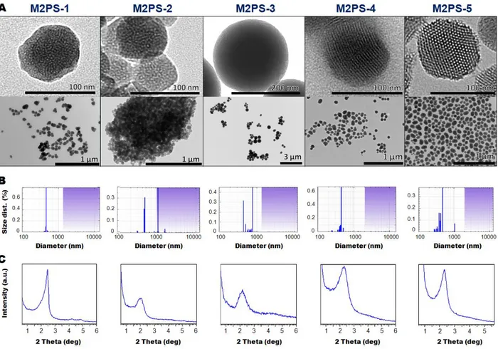
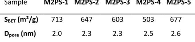
Secondly, the structure and morphology of the M2PS nanomaterials were characterized. Transmission electron microscopy (TEM) images depicted nearly 80 nm nanospheres for all types of M2PS particles, except the 200 nm nanospheres obtained from the water/ethanol mixture for M2PS-3 (Fig. 1A). Dynamic light scattering (DLS) size distributions showed that the best approaches to obtain monodisperse non-aggregated M2PS NPS were in aqueous media at 25°C for low 2PS concentration, or in aqueous media at 80°C for higher concentrations (Fig. 1B). Besides, electron micrographs revealed different types of porous frameworks for each reaction (see Scheme 1 and Fig. 1A). Typically, MCM-41 P6mm structure was obtained with the aqueous route at 25°C for M2PS-1, but the pore organization was very sensitive to the 2PS content. Comparing the same procedure in M2PS-1 and M2PS-2, the increase of the 2PS content from 8 to 16 wt% led to an important structural disorganization, as testified with the small angle X-ray diffraction (XRD) patterns (Fig. 1C). Conversely, the third strategy performed at 80°C retained the MCM-41 hexagonal array of the pores for the M2PS-4 and M2PS-5 materials at high 2PS concentration, as displayed by TEM and XRD patterns, but enlargement of the Bragg peak and the TEM image for M2PS-4 indicated a more slightly disordered structure. Alternatively, a radial porosity was obtained on M2PS-3 NPs in the water/ethanol mixture (Fig. 1A, 1C). The mesoporous structure of the materials was validated by the nitrogen-adsorption-desorption technique. The BJH transform indicated pore diameters from 2.1 to 2.6 nm (see Table 2); high BET surface areas were found for all samples, ranging from 500 to 700 m²/g (Fig. S1 to S5). Thus, both the size and the porosity of the M2PS nanomaterials make them suitable nanocarriers for nanomedicine applications.
Figure 1. TEM images (A), DLS size distributions (B), and small angles XRD patterns of M2PS-1 to M2PS-5 NPs (C).
Table 1. 2PS weight per cent determination in M2PS NPs.
Sample Method NPs N wt% [a] NPs 2PS wt% [b] Reactant 2PS wt% M2PS-1 25 °C/H2O 1.3 7.7 8 M2PS-2 M2PS-3 M2PS-4 M2PS-5 25 °C /H2O 80 °C/H2O:EtOH 80 °C/H2O 80 °C/H2O 2.7 3.5 7.3 3.4 16.1 20.8 43.5 20.2 12 13 38 10
[a] Elemental analysis by combustion measurements of the NPs. [b] Determination based on the nitrogen wt% in the condensed 2PS moieties (O1.5Si-R-SiO1.5).
Thirdly, the efficiency of the 2PS encapsulation in the nanomaterials was found to be highly dependent on the synthetic route. UV-Visible spectroscopy demonstrated the successful encapsulation of the 2PS molecules in the silica framework of all M2PS NPs (Fig. S6 and S7). Solid state nuclear magnetic resonance (NMR) 29Si and 13C CPMAS spectra (Fig. S8) further supported that assertion. Besides, as shown in Table 1 with the comparison of the 2PS wt% in the NPs and in the reactants, the efficacy of the 2PS condensation was higher than that of TEOS. Interestingly, highly organically functionalized M2PS NPs could be designed with both syntheses at 80°C. However, the usefulness of the M2PS-3 nanocarriers (water/ethanol mixture) is greatly impoverished, as we shall see with the photophysical properties.
Table 2. Textural properties of M2PS NPs.
Sample M2PS-1 M2PS-2 M2PS-3 M2PS-4 M2PS-5 SBET (m²/g) 713 647 603 503 677
Dpore (nm) 2.0 2.3 2.3 2.5 2.6
The 2PS spatial distribution in the silica was then studied by absorption and emission of fluorescence, since this factor affects directly the photophysical properties of the NPs. The emission spectra (Fig. 2A) and the fluorescence quantum yields (Table 3) of the M2PS compounds were measured and compared to those of the 2PS molecular reference (2PS-Ref, see structure Fig. S9). The most fluorescent NPs were found to be M2PS-1, M2PS-4, and M2PS-5, the latter having a quantum yield two to four times larger than the formers. A bathochromic effect was also observed on the 2PS band, as seen in the normalized spectra (Fig. 2B). The most red-shifted band corresponds to the synthesis performed in the water/ethanol mixture, with an absorption maximum at 469 nm; then follows the aqueous mixture at 25°C (max = 453-455 nm),
and finally the aqueous reaction at 80°C (max ≈ 448-450 nm). Such an observation was
consistent with the absorption spectra of the M2PS NPs (Fig. S7). These results are the direct outcome of the spatial dispersion of the 2PS molecules in the mesoporous silica matrices, with an important aggregation36 of the 2PS for the M2PS-3 material. Thus we expected to obtain poorer two-photon properties on material having the most red-shifted 2PS emission bands. Indeed, the
aggregation of 2PS moieties via - stacking interactions or condensation generally leads to increased non-radiative de-excitation rates and therefore to a quenching of the fluorescence, as well as to a decrease of the two-photon absorption properties.37
Figure 2. Fluorescence emission spectra of M2PS NPs (A), and the associated normalized spectra in ethanol (B).
Finally, the two-photon photophysical properties of the M2PS nanocarriers were investigated. The two-photon absorption cross-sections (2) were determined between 700 and
900 nm by investigating their two-photon excited fluorescence, and as anticipated M2PS-1,
M2PS-4, and M2PS-5 displayed the highest cross-sections (see Table 3, and Fig. 3). It should be
noticed that M2PS-1, M2PS-4, and M2PS-5 also exhibit the largest quantum yields (11, 26, and 41%, respectively), while their maximum cross-sections ranged from 110 to 200 GM. In the case of the synthesis at 25°C, the increase of the 2PS concentration from 8% (M2PS-1) to 16% in
cross-section, from 11 to 8%, from 110 to 80 GM. On the other hand, comparing M2PS-3 (reaction in water/ethanol) with M2PS-5 (synthesis in water at 80°C), which both have 20% of photosensitizer, drastically different properties were obtained. Indeed, the latter exhibits a two-photon brightness (max F) of 82 GM, which is 40 times larger than that of the former (2 GM,
see Fig. 3B). Furthermore, for a given reaction, the 2PS threshold for optimum properties was different, in the first synthetic approach, 8% was the maximum, whereas in the third approach the 2PS content could be increased at least up to 25 % without quenching the properties. Consequently, the modulation of the two-photon properties of M2PS NPs could be done by an appropriate choice of the elaboration process.
Table 3. Photophysical properties of M2PS NPs in ethanol.
Sample abs/em (nm) F [a] max[b] (GM) maxF (GM) M2PS-1 386/455 0.11 110 12 M2PS-2 M2PS-3 M2PS-4 M2PS-5 388/453 420/469 385/450 384/448 0.08 0.03 0.26 0.41 80 60 180 200 6 2 46 82
[a] Quinine bisulfate standard at 0.5 M in H2SO4. [b] Maximum two-photon absorption cross-section per chromophore. [c] Maximum two-photon action cross-section (two-photon brightness) per chromophore.
Figure 3. Two-photon absorption cross sections of M2PS NPs (A), and the corresponding two-photon brightness spectra in ethanol (B).
The two-photon potential of our M2PS NPs for fluorescence imaging was eventually tested in vitro on MCF-7 breast cancer cells. First, M2PS-3 NPs were found to have low cytotoxicity in cells up to 100 g.mL-1 (see Fig. S10). The two-photon laser excitation was then
carried out using a Carl Zeiss two-photon confocal microscope at a low laser power (3% of the laser input) and at ex = 760 nm. M2PS-3 NPs were incubated for 24 h with MCF-7 cancer cells
at 40 g.mL-1 in 35 mm glass bottom dishes. In order to avoid artifact signals, we work on living
cells without fixation or permeabilization of the membrane. The membranes of the cells were stained using a membrane marker (cell mask) for 15 min before imaging experiments. Two-photon confocal images correspond to a thin slice of cell (0.62 m) without an out-of-focus signal, thus allowing us to determine which part of the signal is inside the cells (arrows). Even with the lowest two-photon absorption cross-section and quantum yield, M2PS-3 NPs were detected (Fig. 4), showing the successful endocytosis of these nanoparticles in cancer cells.
Figure 4. Two-photon in vitro imaging of M2PS-3 NPs on living MCF-7 cells demonstrating the cellular uptake of the nanocarriers. Arrows pointed nanoparticles located inside the cells. Scale bar 20 m.
Conclusions
In summary, we report a study of the properties of organically modified mesoporous silica NPs for two-photon excitation. The modulation of the two-photon properties was found to be highly dependent on the synthetic approach used, which directly affected the spatial distribution of the photosensitizers. Furthermore, the threshold of the photosensitizer maximum concentration varied according to the elaboration method, the best results being observed with a modified Mann’s reaction in aqueous media at 25°C for low concentration, whereas the Lin’s method at 80°C in water was preferred at higher concentrations. Hence, M2PS nanomaterial was used for in-vitro two-photon fluorescence imaging. This study highlights the intricacies implied in the design of efficient NIR two-photon-sensitive nanomaterials for various applications such as nanomedicine.
Experimental
Materials
Tetraethoxysilane, cetyltrimethylammonium bromide, sodium hydroxide, ammonium nitrate and tetrahydrofuran were purchased from Sigma-Aldrich. Absolute ethanol was purchased from Fisher Chemicals. R. Norma Pure.
Apparatus
Absorption spectra were recorded on a Hewlett-Packard 8453 spectrophotometer and fluorescence data were collected on an Edinburgh Instruments (FLS920) fluorimeter. Fluorescence quantum yields were measured in ethanol using quinine bisulfate in aqueous H2SO4
(0.5 M) as a reference (F = 0.546). Mass spectrometry was carried out at the Laboratoire de
Spectrometrie de Masse (Lyon, France) with a Thermo-Finnigan MAT95 apparatus in electronic impact ionization mode. Dynamic light scattering analyses were performed using a Cordouan Technologies DL 135 Particle size analyzer instrument. 29Si and 13C CPMAS solid state NMR sequences were recorded with a VARIAN VNMRS300, using Q8MH8 and adamantane references respectively. TEM analysis performed on a JEOL 1200 EXII instrument. SEM analysis performed on a FEI Quanta FEG 200 instrument.
Synthesis
M2PS-1 NPs. A mixture of cetyltrimethylammonium bromide (345 mg, CTAB), and sodium
hydroxide (20 mL, 0.2 M) was stirred at room temperature during 50 minutes at 700 rpm in a 250 mL three neck round bottom flask. Then, the stirring speed was changed to 1000 rpm. TEOS (1.6 mL) was added and after 40 seconds water (260 mL) was poured out. Afterwards, an ethanolic solution of the 2PS precursor (n0 = 9.0 10-5 mol, in 1 mL EtOH) was added. The
solution was then heated using a hair drier (from T0=25 °C to T’=27 °C over 1-2 minutes), in
order to trigger the condensation process. After 5 minutes 30 seconds of reaction, a solution of hydrochloric acid (0.2 M, ca 36 mL ) was added to reach pH 6.9. Fractions were gathered in polypropylene tubes and collected by centrifugation for 15 minutes at 21 krpm. The sample was then extracted twice with an ethanolic solution of ammonium nitrate (6 g.L-1), and washed three times with ethanol, water, and ethanol. Each extraction involved a sonication step of 30 minutes at 50 °C; the collection was carried out in the same manner. The as-prepared material was dried under air flow for few hours.
M2PS-2 NPs. A mixture of cetyltrimethylammonium bromide (345 mg, CTAB), and sodium
250 mL three neck round bottom flask. Then, the stirring speed was changed to 1000 rpm, TEOS (1.6 mL) was added along with an ethanolic solution of the 2PS precursor (n0 = 1.3*10-4 mol, in
1 mL EtOH). After 40 seconds, water (260 mL) was poured out. The solution was then heated using a hair drier (from T0=25 °C to T’=27 °C over 1-2 minutes), in order to trigger the
condensation process. After 5 minutes 30 seconds of reaction, a solution of hydrochloric acid (0.2 M, ca 36 mL) was added to reach a pH of 6.9. Fractions were gathered in polypropylene tubes and collected by centrifugation for 15 minutes at 21 krpm. The sample was then extracted twice with an ethanolic solution of ammonium nitrate (6 g.L-1), and washed three times with
ethanol, water, and ethanol. Each extraction involved a sonication step of 30 minutes at 50 °C in order to remove the CTAB surfactant; the collection was carried out in the same manner. The as-prepared material was dried under air flow for few hours.
M2PS-3 NPs. A mixture of cetyltrimethylammonium bromide (640 mg, CTAB), deionized
water (100 mL), and ethanol (40 mL) was stirred at 80 °C for 40 minutes at 650 rpm in a 250 mL three neck round bottom flask. Then, an ethanolic solution of 2PS (n0 = 9.0*10-5 mol, in 1 mL
EtOH) was added to the stirred solution. A delay of 5 minutes was used to homogenize the solution, and TEOS (1.2 mL) was added via a syringe then the stirring speed was changed to 1000 rpm. The reaction was conducted for 30 minutes, then the solution was neutralized with hydrochloric acid (0.2 M), and the mixture was cooled down to room temperature. Fractions were gathered in polypropylene tubes and collected by centrifugation for 15 minutes at 21 krpm. The sample was then extracted twice with an ethanolic solution of ammonium nitrate (6 g.L-1),
and washed three times with ethanol, water, and ethanol. Each extraction involved a sonication step of 30 minutes at 50 °C; the collection was carried out in the same manner. The as-prepared material was dried under air flow for few hours.
M2PS-4 NPs. A mixture of cetyltrimethylammonium bromide (250 mg, CTAB), distilled
water (120 mL), and sodium hydroxide (875 µL, 2 M) was stirred at 80 °C for 50 minutes at 700 rpm in a 250 mL three neck round bottom flask. Then, TEOS (1.0 mL) was added along with the two-photon photosensitizer (n0 = 2.0*10-4 mol, in 1 mL of dry THF), and the condensation
process was conducted for 1 hour 30 minutes. Afterwards, the solution was cooled to room temperature while stirring; fractions were gathered in polypropylene tubes and collected by centrifugation for 15 minutes at 21 krpm. The sample was then extracted twice with an ethanolic
solution of ammonium nitrate (6 g.L-1), and washed three times with ethanol, water, and ethanol. Each extraction involved a sonication step of 30 minutes at 50 °C; the collection was carried out in the same manner. The as-prepared material was dried under air flow for few hours.
M2PS-5 NPs. A mixture of cetyltrimethylammonium bromide (250 mg, CTAB), distilled
water (120 mL), and sodium hydroxide (875 µL, 2 M) was stirred at 80 °C during 50 minutes at 700 rpm in a 250 mL three neck round bottom flask. Then, TEOS (1.0 mL) was added along with the two-photon photosensitizer (n0 = 6.4*10-5 mol, in 1 mL of dry THF), and the
condensation process was conducted for 1 hour 30 minutes. Afterwards, the solution was cooled to room temperature while stirring; fractions were gathered in polypropylene tubes and collected by centrifugation for 15 minutes at 21 krpm. The sample was then extracted twice with an ethanolic solution of ammonium nitrate (6 g.L-1), and washed three times with ethanol, water, and ethanol. Each extraction involved a sonication step of 30 minutes at 50 °C; the collection was carried out in the same manner. The as-prepared material was dried under air flow for few hours.
TPE fluorescence and measurements of the two-photon absorption cross-sections.
Two-photon excited fluorescence spectroscopy was performed using a mode-locked Ti:sapphire laser generating 150 fs wide pulses at a 76 MHz rate, with a time-averaged power of several hundreds of mW (Coherent Mira 900 pumped by a 5 W Verdi). The laser light is attenuated using a combination of half-wave plates and a Glan-laser polariser and the excitation power is further controlled using neutral density filters of varying optical density mounted in a computer-controlled filter wheel. After five-fold expansion through two achromatic doublets, the laser beam is focussed by a microscope objective (10X, NA 0.25, Olympus, Japan) into a standard 1 cm stirred absorption cuvette containing the sample. The applied average laser power arriving at the sample was between 0.5 and 15 mW, leading to a time-averaged light flux in the focal volume on the order of 0.1-1 mW/μm2. The generated fluorescence is collected in epifluorescence mode, through the microscope objective, and reflected by a dichroic mirror (675dcxru, Chroma Technology Corporation, USA). Residual excitation light is removed using a barrier filter (e650-2p, Chroma) and the fluorescence is coupled into a 600 μm multimode fiber by an achromatic doublet. The fiber is connected to a compact CCD-based spectrometer
(BTC112-E, B&WTek, USA), which measures the two-photon excited emission spectrum. The emission spectra are corrected for the wavelength-dependence of the detection efficiency using correction factors established through the measurement of reference compounds having known fluorescence emission spectra. Briefly, the set-up allows for the recording of corrected fluorescence emission spectra under multiphoton excitation at variable excitation power and wavelength. Absolute values for the two-photon excitation action cross sections σ2ΦF were
obtained according to the method described by Xu et al. (J. Opt. Soc. Am. B 1996, 13, 481). using 10-4 M fluorescein in 10-2 M aqueous NaOH as a reference, applying corrections for the
refractive index of the solvent (M. H. V. Werts et al., Photochem. Photobiol. Sci. 2005, 4, 531). In the 700-720 nm excitation range, refined reference values for fluorescein were used. (C. Katan
et al., J. Phys. Chem. B 2007, 111, 9468).
Two-photon imaging. The day prior to the experiment, MCF-7 human breast cancer cells
(purchased from ATCC) were seeded onto bottom glass dishes (World Precision Instrument, Stevenage, UK) at a density of 106 cells.cm-2. Then, adherent cells were washed once and incubated in 1 mL medium containing M2PS NPs at a concentration of 40 μg.mL-1 for 20 h. 15 minutes before the end of incubation, cells were loaded with Cell Mask (Invitrogen, Cergy Pontoise, France) for membrane staining at a final concentration of 5 μg.mL-1. Before
visualization, cells were washed gently with phenol red-free Dulbecco’s modified Eagle’s medium (DMEM). Cells were then scanned with a LSM 780 LIVE confocal microscope (Carl Zeiss, Le Pecq, France), at 760 nm with a slice depth (Z stack) of 0.62 μm.
Electronic Supplementary Information (ESI) available: [Characterization of the prepared MSN, photophysical properties]. See DOI: 10.1039/xxxxxx
Acknowledgements
We thank ANR P2N Mechanano for funding. Technological support from the Rio Imaging Platform is gratefully acknowledged.
References
1. M. Gary-Bobo, Y. Mir, C. Rouxel, D. Brevet, I. Basile, M. Maynadier, O. Vaillant, O. Mongin, M. Blanchard-Desce, A. Morère, M. Garcia, J.-O. Durand and L. Raehm,
Angew. Chem. Int. Ed., 2011, 123, 11627-11631.
2. J. Croissant, A. Chaix, O. Mongin, M. Wang, S. Clément, L. Raehm, J.-O. Durand, V. Hugues, M. Blanchard-Desce, M. Maynadier, A. Gallud, M. Gary-Bobo, M. Garcia, J. Lu, F. Tamanoi, D. P. Ferris, D. Tarn and J. I. Zink, Small, 2014, 10, 1752–1755.
3. J. Croissant, D. Salles, M. Maynadier, O. Mongin, V. Hugues, M. Blanchard-Desce, X. Cattoën, M. Wong Chi Man, A. Gallud, M. Garcia, M. Gary-Bobo, L. Raehm and J.-O. Durand, Chem. Mater., 2014, 26, 7214–7220.
4. J. Croissant, M. Maynadier, A. Gallud, H. Peindy N'Dongo, J. L. Nyalosaso, G. Derrien, C. Charnay, J.-O. Durand, L. Raehm, F. Serein-Spirau, N. Cheminet, T. Jarrosson, O. Mongin, M. Blanchard-Desce, M. Gary-Bobo, M. Garcia, J. Lu, F. Tamanoi, D. Tarn, T. M. Guardado-Alvarez and J. I. Zink, Angew. Chem. Int. Ed., 2013, 125, 14058-14062. 5. S. S. Banerjee and D.-H. Chen, Nanotechnology, 2009, 20, 185103.
6. T. Zhao, K. Yu, L. Li, T. Zhang, Z. Guan, N. Gao, P. Yuan, S. Li, S. Q. Yao, Q.-H. Xu and G. Q. Xu, ACS Appl. Mater. Interfaces, 2014, 6, 2700-2708.
7. S.-H. Cheng, C.-C. Hsieh, N.-T. Chen, C.-H. Chu, C.-M. Huang, P.-T. Chou, F.-G. Tseng, C.-S. Yang, C.-Y. Mou and L.-W. Lo, Nano Today, 2011, 6, 552-563.
8. S. Kim, T. Y. Ohulchanskyy, H. E. Pudavar, R. K. Pandey and P. N. Prasad, J. Am.
Chem. Soc., 2007, 129, 2669-2675.
9. J. Croissant, M. Maynadier, O. Mongin, V. Hugues, M. Blanchard-Desce, A. Chaix, X. Cattoën, M. Wong Chi Man, A. Gallud, M. Gary-Bobo, M. Garcia, L. Raehm and J.-O. Durand, Small, 2015, 11, 295–299.
10. Z. Zhu, X. Zhao, W. Qin, G. Chen, J. Qian and Z. Xu, Sci. China Chem., 2013, 56, 1247-1252.
11. J.-L. Li, H.-C. Bao, X.-L. Hou, L. Sun, X.-G. Wang and M. Gu, Angew. Chem. Int. Ed., 2012, 51, 1830-1834.
12. T. M. Guardado-Alvarez, L. Sudha Devi, M. M. Russell, B. J. Schwartz and J. I. Zink, J.
Am. Chem. Soc., 2013, 135, 14000-14003.
13. V. Lebret, L. Raehm, J. O. Durand, M. Smaihi, M. H. V. Werts, M. Blanchard-Desce, D.Methy-Gonnod and C. Dubernet, J. Sol-Gel Sci. Technol., 2008, 48, 32-39.
14. J. Park, A. Estrada, K. Sharp, K. Sang, J. A. Schwartz, D. K. Smith, C. Coleman, J. D.Payne, B. A. Korgel, A. K. Dunn and J. W. Tunnell, Opt. Exp., 2008, 16, 1590-1599. 15. C. Mauriello-Jimenez, J. G. Croissant, M. Maynadier, X. Cattoën, M. W. C. Man,
J.Vergnaud, V. Chaleix, S. Vincent, M. Garcia and M. Gary-Bobo, J. Mater. Chem. B, 2015, 10.1039/C5TB00315F.
16. J. Lu, J. Croissant,J.-O. Durand, T. Guardado-Alvarez, J. I. Zink and F. Tamanoi, Cancer
Res., 2014, 74, LB-9-LB-9.
17. M. Pawlicki, H. A. Collins, R. G. Denning and H. L. Anderson, Angew. Chem. Int. Ed., 2009, 48, 3244-3266.
18. Q. He, Z. Zhang, F. Gao, Y. Li and J. Shi, Small, 2011, 7, 271-280.
19. J. S. Souris, C. H. Lee, S. H. Cheng, C. T. Chen, C. S. Yang, J. A. A. Ho, C. Y. Mou and L. W. Lo, Biomaterials, 2010, 31, 5564-5574.
20. Z. Li, J. C. Barnes, A. Bosoy, J. F. Stoddart and J. I. Zink, Chem. Soc. Rev., 2012, 41, 2590-2605.
21. M. W. Ambrogio, C. R. Thomas, Y.-L. Zhao, J. I. Zink and J. F. Stoddartt, Acc. Chem.
Res., 2011, 44, 903-913.
22. P. Yang, S. Gai and J. Lin, Chem. Soc. Rev., 2012, 41, 3679-3698.
23. S. Dib, M. Boufatit, S. Chelouaou, F. Sadi-Hassaine, J. Croissant, J. Long, L. Raehm, C. Charnay and J. O. Durand, RSC Adv., 2014, 4, 24838-24841.
24. J. Croissant and J. I. Zink, J. Am. Chem. Soc., 2012, 134, 7628-7631.
25. C. Argyo, V. Weiss, C. Bräuchle and T. Bein, Chem. Mater., 2013, 26, 435-451.
26. I. I. Slowing, J. L. Vivero-Escoto, B. G. Trewyn and V. S. Y. Lin, J. Mater. Chem., 2010,
20, 7924-7937.
27. K. K. Coti, M. E. Belowich, M. Liong, M. W. Ambrogio, Y. A. Lau, H. A. Khatib, J. I. Zink, N. M. Khashab and J. F. Stoddart, Nanoscale, 2009, 1, 16-39.
28. B. G. Trewyn, I. I. Slowing, S. Giri, H.-T. Chen and V. S. Y. Lin, Acc. Chem. Res., 2007,
40, 846-853.
29. J. Kecht, A. Schlossbauer and T. Bein, Chem. Mater., 2008, 20, 7207-7214.
30. E. Chelebaeva, L. Raehm, J. O. Durand, Y. Guari, J. Larionova, C. Guerin, A. Trifonov, M. Willinger, K. Thangavel, A. Lascialfari, O. Mongin, Y. Mir and M. Blanchard-Desce,
J. Mater. Chem., 2010, 20, 1877-1884.
31. T. M. Guardado-Alvarez, L. S. Devi, J.-M. Vabre, T. A. Pecorelli, B. J. Schwartz, J.-O. Durand, O. Mongin, M. Blanchard-Desce and J. I. Zink, Nanoscale, 2014, 6, 4652-4658. 32. P. Couleaud, V. Morosini, C. Frochot, S. Richeter, L. Raehm and J. O. Durand,
Nanoscale, 2010, 2, 1083-1095.
33. X. Wang, A. R. Morales, T. Urakami, L. Zhang, M. V. Bondar, M. Komatsu and K. D. Belfield, Bioconjugate Chem., 2011, 22, 1438-1450.
34. C. E. Fowler, D. Khushalani, B. Lebeau and S. Mann, Adv. Mater., 2001, 13, 649-652. 35. H.-P. Lin and C.-P. Tsai, Chem. Lett., 2003, 32, 1092-1093.
36. K. Natte, T. Behnke, G. Orts-Gil, C. Würth, J. F. Friedrich, W. Österle and U. Resch-Genger, J. Nanopart. Res., 2012, 14, 1-10.
37. H. Y. Woo, B. Liu, B. Kohler, D. Korystov, A. Mikhailovsky and G. C. Bazan, J. Am.
Chem. Soc., 2005, 127, 14721-14729.
35. H.-P. Lin and C.-P. Tsai, Chem. Lett., 2003, 32, 1092-1093.
36. K. Natte, T. Behnke, G. Orts-Gil, C. Würth, J. F. Friedrich, W. Österle and U. Resch-Genger, Journal of Nanoparticle Research, 2012, 14, 1-10.
37. H. Y. Woo, B. Liu, B. Kohler, D. Korystov, A. Mikhailovsky and G. C. Bazan, J. Am.