HAL Id: tel-02320973
https://tel.archives-ouvertes.fr/tel-02320973
Submitted on 20 Oct 2019
HAL is a multi-disciplinary open access
archive for the deposit and dissemination of sci-entific research documents, whether they are pub-lished or not. The documents may come from teaching and research institutions in France or abroad, or from public or private research centers.
L’archive ouverte pluridisciplinaire HAL, est destinée au dépôt et à la diffusion de documents scientifiques de niveau recherche, publiés ou non, émanant des établissements d’enseignement et de recherche français ou étrangers, des laboratoires publics ou privés.
Nicolas Tardif
To cite this version:
Nicolas Tardif. Mechanosignaling through Caveolae : A New Role for the Control of JAK-STAT Signaling. Biochemistry, Molecular Biology. Université Paris-Saclay, 2018. English. �NNT : 2018SACLS337�. �tel-02320973�
Mechanosignaling through
caveolae :
A new role for the control of
JAK-STAT signaling
Thèse de doctorat de l'Université Paris-Saclay préparée à l’université Paris Sud
École doctorale n°568 signalisations et réseaux intégratifs en biologie (Biosigne), aspect moléculaire et cellulaire de la biologie
Thèse présentée et soutenue à Paris, le 19 octobre 2018, par
Nicolas Tardif
Composition du Jury : Marc Le Maire
Professeur emerite, institut de biologie intégrative de la cellule Président
Corinne Albiges-Rizo
Directeur de recherche, institut pour l’avancée des biosciences Rapporteur
Benjamin Nichols
Directeur de recherche, MRC-LMB Cambridge Rapporteur
Gideon Schreiber
Professeur, Weizmann institute Examinateur
Christophe Lamaze
Directeur de recherche, institut Curie Directeur de thèse
Cédric Blouin NNT : 2 0 1 8 S A CL S 33 7
Joachim Du Bellay
«Ici on taille pas des bambous»
As Ulysse, I am delighted with my amazing journey, which was as challenging as thrilling. This adventure wouldn’t have been possible without the support of my supervisor and the whole team, in which I always felt very proud to be part of. Hence, in first place I would like thank my thesis supervisor, Christophe a.k.a. “the boss” for putting all his trust in me with this project. I wish all PhD students could have a supervisor as supportive as you, and I’m glad we made it possible. Then I would like to thank Cedric, the man in the shadow, you are a wise man, always of good advice and very supportive. I would like to acknowledge our collaborators from IINS Bordeaux: Grégory Giannone, Olivier Rossier and R.K. Finally, I thank Ludger Johannes for his precious feedback during joined meeting.
I would like to thank my thesis jury members, Corinne Albiges-Rizo and Ben Nichols for the time they dedicated to review my manuscript and their pertinent feedback. I couldn’t be more proud having reviews by such scientists. I would like to thank Gideon Schreiber for nicely accepting my invitation and last but not least, Marc Le Maire who drove me through my university course until the wonderful world of plasma membrane and cell signaling and will now preside my jury.
I would like to thank also the team members that contributed to the everyday pleasure I had to sit at my desk/bench such as Christophe, you really contributed to strengthen my self-confidence and participated to the pleasure I had to work every single day for this project. You are a man of extremes, endearing with whom we like to share. Then the second leader of this team (and babysitter), most likely carrying the Lamaze lab legacy on his shoulders: Cédric (papy mougeot) for all his benevolence for “les grumeaux” and other “babies” of the team. Thank you for inculcating me the taste of excessively overpriced watches and other techs, which I could never afford as a PhD student. You are such a great mentor. Last but not least, thank you for allowing me to stay in the office whenever some of my worst jokes come up. I would like to thank my sidekick, wing-woman, bench-neighbor and desk-neighbor: Melissa. Thank you for your support, I’m so grateful I could share this adventure with you. Counter-thank you for deliberately ignoring that singing (or
harboring a poker face just to act like they were not funny although they were… (Christine would agree… and Cédric (on Fridays)). Hence, thank you Christine for sharing my humor. Thank you for your patience and your wise lab advice and mostly for all the fun we shared; vinos vomitas. I would like to have a special “tachacor” for Massi. Without you I wouldn’t speak fluent Persian lv2. Thank you for being that supportive and for your life and lab advice. I enjoyed having highly intellectual conversations with you, now I’m pretty sure everybody knows DNA is double strand. During these four years both the Jedi and the padawan fought the general Koss. I thank Estelle for being such a good badminton partner and for all the fun she puts in the lab. Carlos our spanish sound-box – desk vibrator, thank you for your support. I thank Satish for your gaming advice and above all for your chicken. Alison for actively contributing to the nice atmosphere we have in the office and for teaching me rude Italian words. I apologize to Ewan for winning the word cup. I guess it did not come home… Xièxiè Changting. I acknowledge Manon for her patience (a key asset to work in this office). I would like to thank Christian and all other members of the Johannes team for their wise advice and feedback on this project. To finish I thank all the past members who actively contributed to my lab life: Steph, Natacha, Henri, Weiwei and Daniela.
Finally I warmly acknowledge the people who constitute the pillars of my life: my closest friends, Camille, Jérémie (la bande de Bazoches) and the team rocket Francky, Flo, Michou and Baba for their unswerving support and loyalty. I thank my whole family especially Mariette and Bernard for transmitting me the great interest I have for sciences since my early childhood. I am also grateful to Yann and Evon for their precious help starting my life in science. Behind every man success there is woman. The story does not tell whether behind small success there is a short woman. Anyway, thank you Anna for sharing my everyday life and making it a bit sweeter everyday. Thank you for your unbreakable support and for standing by my side these last months regardless how grumpy I could be. To finish, I dedicate this manuscript to my parents for their support and for believing in me since ever. They play a key role in the reach of my life goals.
Table of Contents
ABBREVIATIONS ... 3 LIST OF FIGURES ... 7 THESIS SUMMARY ... 9 RÉSUMÉ DE LA THÈSE ... 10 INTRODUCTION ... 131 CELL MECHANICS AND MECHANOTRANSDUCTION ... 13
1.1 CELL AND TISSUE MECHANICS ... 13
1.1.1 Force generation (may the force be with you) ... 13
1.1.2 Biochemical versus mechanical signal ... 14
1.1.3 The dark side of the force ... 15
1.1.3.1 Cardiovascular and muscular pathologies ... 15
1.1.3.2 Cancer ... 16
1.1.3.2.1 Mechanical forces in the context of tumor progression ... 16
1.1.3.2.2 Tumor physical microenvironment influences cancer progression ... 17
1.2 SIGNAL MECHANOTRANSDUCTION ... 18
1.2.1 Mechanosignaling at the plasma membrane ... 19
1.2.2 Mechanosignaling in the cytosol ... 19
1.2.3 Nuclear mechanotransduction ... 21
2 THE CAVEOLAE: SPECIALIZED PLASMA MEMBRANE STRUCTURES ... 23
2.1 STRUCTURE AND COMPOSITION ... 23
2.1.1 Caveolar proteins ... 23 2.1.1.1 Caveolins ... 23 2.1.1.2 Cavins ... 27 2.1.1.3 Accessory proteins ... 28 2.1.2 Lipid composition ... 29 2.1.3 Caveolar ultrastructure ... 30
2.2 CAVEOLAE BIOGENESIS: FROM PROTEIN SYNTHESIS TO THE CAVEOLAR BULB ... 30
2.2.1 Caveolin-1: Tale of a journey from the ER to the plasma membrane ... 31
2.2.2 Cavin recruitment and caveolae morphogenesis ... 31
2.2.3 Recruitment of accessory proteins ... 32
2.3 CAVEOLAE FUNCTIONS ... 33
2.3.1 Caveolae mediated endocytosis and trafficking ... 33
2.3.2 Lipid homeostasis ... 34
2.3.3 Mechanoprotection ... 35
2.3.4 Cell signaling ... 36
2.3.4.1 Indirect regulation of signaling ... 36
2
2.3.4.4 Remaining controversies on Cav1 signaling ... 38
2.4 CAVEOLAE PATHOPHYSIOLOGY ... 40
2.4.1 Lipodystrophy ... 40
2.4.2 Vascular dysfunction ... 41
2.4.3 Muscular dystrophies and cardiomyopathies ... 42
2.4.4 Cancer ... 43
3 TYPE I INTERFERON-INDUCED JAK-STAT SIGNALING ... 44
3.1 INTERFERONS ... 44
3.1.1 Classification ... 45
3.1.2 Specificity ... 45
3.2 INTERFERONS RECEPTORS ... 46
3.2.1 IFNAR1, IFNAR2 and their isoforms ... 46
3.2.2 Structure and mechanism of activation ... 47
3.3 JAK:JUST ANOTHER KINASE... ... 48
3.3.1 Structural features ... 48
3.3.2 Mechanism of activation ... 49
3.4 SIGNAL TRANSDUCERS AND ACTIVATORS OF TRANSCRIPTION ... 50
3.5 JAK-STAT REGULATION ... 51
3.5.1 Upstream regulation ... 51
3.5.2 Downstream regulation ... 51
3.6 JAK-STAT IN TUMOR PROGRESSION ... 52
RESULTS ... 56
4 CAVEOLAE MECHANICS CONTROL JAK-STAT SIGNALING ... 56
4.1 OBJECTIVES AND SUMMARY ... 56
4.2 ARTICLE ... 58
DISCUSSION ... 93
5 DISCUSSION AND PERSPECTIVES ... 93
5.1 CAVEOLAR CAV1 VERSUS “FREE”CAV1 ... 93
5.2 CSD DEPENDENT REGULATION OF JAK1 ... 94
5.3 SIGNAL SPECIFICITY ... 97
5.4 CAVEOLAE MECHANOTRANSDUCTION: ROLE IN TUMOR PROGRESSION ... 98
REFERENCES ... 100
ANNEX 1 ... 131
ANNEX 2 ... 135
Abbreviations
aa: amino acid
AMFR: Autocrine Motility Factor Receptor Bcl-xL: B cell Lymphoma-extra Large bFGF: basic Fibroblast Growth Factor
BSCL: Berardinelli-Seip-Congenital Lipodistrophy c-Myc: cellular Myelocytosis
Ca2+: Calcium
CAF: Cancer Associated Fibroblast Cas: Crk associated substrate Cav1: Caveolin-1
Cav2: Caveolin-2 Cav3: Caveolin-3
CCL2: Chemokine (C-C) Ligand 2 CD: Circular Dichroism
cGAS: cyclic GMP-AMP Synthase CKα: Casein Kinase α
CLIC: CLathrin Independent Carrier cPLA2: cytosolic PhosphoLipase A2
CRAC: Cholesterol Recognition/interaction Amino acid Consensus CREB: cAMP Responsive Element Binding
CSD: Caveolin Scaffolding Domain CTxB: Cholera Toxin subunit B DPC: Dodecyl-PhosphatidylCholine ECD: ExtraCellular Domain
ECM: ExtraCellular Matrix
EGFR: Epidermal Growth Factor Receptor EMT: Epithelial to Mesenchymal Transition eNOS: endothelial Nitric Oxide Synthase ER: Endoplasmic Reticulum
4 ER-α: Estrogen Receptor α
ERK: Extracellular signal-Regulated protein Kinase EV: Extracellular Vesicle
FA: Focal Adhesion
FAK: Focal Adhesion associate Kinase FERM: Four point one Ezrin, Radixin, Moesin FNIII: Fibronectin III
G3BP2: Ras GTPase-activating protein-binding 2 GAP: GTPase Activating Protein
GAS: IFN Gamma Activated Sequence
GEEC: GPI-AP-enriched Early Endosomal Compartment GSL: GlycoSphingoLipid
hCR: helical Cytokine Receptor
HER2: Human Epidermal growth factor Receptor 2 HO: Heme Oxygenase
IFN: Interferon
IFNAR: InterFeroN Alpha Receptor IFNGR: InterFeroN Gamma Receptor IL6: InterLeukin 6
IRF2: IFN-Regulatory Factor 2 IRF9: IFN-Regulatory Factor 9 IRS-1: Insulin Response Substrate-1 ISG: IFN-Stimulated Gene
ISRE: IFN-Sensitive Response Element JH: JAK Homology
KIR: Kinase Inhibitory Region Klf-8: Kruppel-like factor 8
LATS: LArge Tumor Suppressor LDL: Low-Density Lipoprotein
LINC: Linker of Nucleoskeleton and Cytoskeleton LOX: 5-LipOXygenase
MAPK: Mitogen Activated Protein Kinase MDR: MultiDrug Resistance
MG53: MitsuGumin 53
MSC: Mechano-Sensitive Channel MsC: Mensenchymal stem Cells
MT1-MMP: Membrane Type 1 Matrix MetalloProteinase MURC: Muscie-Related Coiled coil protein
MVB: Multi Vesicular Bodies
NMR: Nuclear Magnetic Resonance NO: Nitric Oxide
P2X7: P2X purinoreceptor 7 PDK2: Protein kinase D2
PEST: Proline glutamic acid Serine Threonine PI(3)K: PhosphoInositide-3 Kinase
PIAS: Protein Inhibitor of Activated STAT PiP2: Phosphatidylinositol (4,5) bisPhosphate
PKCα: Protein Kinase C α PKCδ: Protein Kinase C δ POPC: PhOsPhatidylCholine
PRKCDB: PRotein Kinase C Delta-Binding protein Ptdser: Phosphatidylserine
PTEN: Phosphatase and TENsin homologue PTP1B: Protein Tyrosine Phosphatase 1B
PTRF: Polymerase 1 and Transcript Release Factor Rap1: Ras-related protein 1
ROCK: Rho associate protein Kinase
SDPR: Serum Deprivation Protein Response Ser80: Serine 80
SFK: Src Family Kinase
SHP1: Src homology region 2 domain-containing phosphatase 1 SHP2: Src homology region 2 domain-containing phosphatase 2 SOCS: Suppressor Of Cyrokine Signaling
SOX2: Sex determining region on Y box 2
SRBC: SDPR-Related gene product that Binds to C kinase Src: Sarcoma protein kinase
6 STING: STimulator of INterferon Genes
SV40: Simian Virus 40 TAZ: TAfaZzin
TEAD: TEA Domain family member
TGFBR1: Transforming Growth Factor β Receptor type 1 TMD: TransMembrane Domain
TRIM: TRI-partite Motif
TRP: Transient Receptor Potential channel TWIST1: Twist related protein 1
Tyr14: Tyrosine 14
USP18: Ubiquitin-Specific Peptidase 18
VIP21: Vesicular Integral-membrane Protein of 21 kDa YAP: Yes Associated Protein
List of figures
Figure 1. Mechanical forces and mechanoreciprocity Figure 2. Biochemical versus mechanical signal
Figure 3. Tissue rheology and pathological development Figure 4. Mechanosensitive channels
Figure 5. Mechanotransduction at the level of the protein and protein complex Figure 6. Schematic representations of mechanical stimuli influencing YAP and TAZ subcellular localization and activity
Figure 7. Mechanosensitive calcium signaling in the nucleus
Figure 8. Proposed model of mechanosensitive nucleoplasmic shuttling
Figure 9. Visualization of the caveolar coat at the plasma membrane of myotubes Figure 10. Caveolin domains and insertion in the plasma membrane
Figure 11. Cavins structure and caveolae morphogenesis
Figure 12. Putative model of caveolar coat assembly and organization Figure 13. Model of caveolae assembly and biosynthetic trafficking of Cav1 Figure 14. Model for the assembly of the cavin coat
Figure 15. Caveolae mechanical disassembly Figure 16. Cav1 signaling hypothesis
Figure 17. Molecular and cellular consequences of caveolar flattening induced by mechanical stress
Figure 18. Potential role of caveolae in tumor progression
Figure 19. Structure and dynamics of IFN-IFNAR ternary complex formation Figure 20. JAKs general structure and regulation
Figure 21. JAK-STAT signal transduction
Figure 22. STAT domains structure and protein binding sites Figure 23. Negative regulation of JAK-STAT signaling
Figure 24. JAKs and STATs with associated cytokines and phenotypes Figure 25. Therapeutic inhibitors of JAKs and STATs
Figure 26. Proposed topology of free Cav1
Figure 27. Cav1 tyrosine phosphorylation upon hypo-osmotic shock Figure 28. CBM localization on JAK1 structure
8 Figure 29. CBM1-mutated JAK1 pulldown
Figure 30. Sequence alignment of SOCS1 and SOCS3 KIR domain with caveolins Figure 31. Specific inhibition of STAT3 by Cav1 in human breast cancer cells
Figure 32. IFN-α-induced STAT3 activation of breast cancer cells upon hypo-osmotic shock
Figure 33. IFN-α-induced STAT3 activation of breast cancer cells under compression
Thesis summary
Caveolae are small cup-shaped plasma membrane invaginations. These multifunctional organelles play a key role in cell mechanoprotection and cell signaling. Indeed our laboratory reported that caveolae have the ability to flatten out upon membrane tension increase, protecting cells from mechanical strains (Sinha et al., 2011). Since caveolae play a key role in cell signaling we hypothesized that the mechano-dependent cycle of caveolae disassembly/reassembly may constitute a mechanical switch for signaling pathways (Nassoy and Lamaze, 2012). In this project, we elucidated the molecular mechanism underlying the control of JAK-STAT signaling by caveolae mechanics. The fate of caveolar components upon caveolae disassembly remains elusive. We showed that caveolin-1 (Cav1), the essential structural component of caveolae, is released and become highly mobile at the plasma membrane under mechanical stress. Considering that caveolae are important signaling hubs at the plasma membrane, we addressed the effects of the mechanical release of Cav1 on cell signaling. Using high throughput screening, we identified the JAK-STAT signaling pathway as a candidate. To further dissect the molecular mechanism underlying the control of JAK-STAT signaling by caveolae mechanics, we addressed the role of Cav1 in the control of JAK-STAT signaling stimulated by IFN-α. We found that Cav1 was a specific negative regulator of the JAK1 dependent STAT3 phosphorylation. Furthermore, the level of Cav1 interaction with JAK1 depended on mechanical stress. We could show that Cav1-JAK1 interaction was mediated by the Caveolin Scaffolding domain (CSD), abolishing JAK1 kinase activity, hence, interfering with STAT3 activation upon IFN-α stimulation. Interestingly, STAT1 activation by IFN-α was not affected by caveolae mechanics. Altogether our results show that caveolae are mechano-signaling organelles that disassemble under mechanical stress, releasing non-caveolar Cav1, which binds to the JAK1 tyrosine kinase and inhibits its catalytic activity, preventing thereby JAK-STAT signal transduction.
10
Résumé de la thèse
Les cavéoles sont des invaginations en forme de coupelle à la membrane plasmique. Ces organelles multifonctionnelles jouent entre autres, un rôle clé dans la mécanoprotection et la signalisation cellulaire. En effet, notre laboratoire a démontré que les cavéoles ont la faculté de s’aplanir en réponse à l’augmentation de la tension membranaire, afin de protéger la cellule des contraintes mécaniques (Sinha et al., 2011). Les cavéoles jouant un rôle clé dans la signalisation cellulaire, nous avions émis l’hypothèse que le cycle mécano-dépendent de désassemblage/réassemblage des cavéoles constitue un interrupteur mécanique de certaines voies de signalisation. Ce projet consiste à élucider le mécanisme moléculaire responsable du contrôle de la voie de signalisation JAK-STAT par la mécanique des cavéoles. Le devenir des constituants cavéolaires lors du désassemblage des cavéoles, reste à ce jour inconnu. Dans ces travaux, nous avons pu démontré que la cavéoline-1 (Cav1), un constituant essentiel des cavéoles, est libérée et devient hautement mobile au niveau de la membrane plasmique. Considérant les propriétés de signalisation de Cav1, nous avons testé l’effet du désassemblage des cavéoles sur la signalisation cellulaire. En effectuant un criblage à haut débit, nous avons identifié la voie de signalisation JAK-STAT stimulée par l’IFN-α comme voie modèle pour cette étude. En effet, la transduction du signal JAK-STAT induit par l’IFN-α est modulée par la mécanique des cavéoles. Afin de disséquer le mécanisme moléculaire responsable du control de la signalisation JAK-STAT par la mécanique des cavéoles, nous avons déterminé le rôle de Cav1 dans le contrôle de la signalisation JAK-STAT stimulée par l’IFN-α. Nous avons observé que Cav1 est un régulateur négatif de la phosphorylation de STAT3 dépendante de la kinase JAK1. De plus, nous avons démontré que Cav1 interagit avec JAK1 en fonction de la tension membranaire. Nous avons également démontré que cette interaction Cav1-JAK1 fait intervenir le « scaffolding domain » de Cav1 (CSD), et que celui-ci est responsable de l’abolition de l’activité kinase de JAK1. Par conséquent, l’interaction de Cav1 avec JAK1 empêche l’activation de STAT3 par la kinase JAK1. De manière surprenante, STAT1 échappe à cette régulation. En effet, l’activation de STAT1 par l’IFN-α n’est pas affectée par la mécanique des cavéoles. Ces résultats démontrent que les cavéoles sont des organelles de mécanosignalisation, qui, lors d’un stress mécanique,
libèrent de la Cav1 non cavéolaire capable d’inactiver la kinase JAK1, empêchant ainsi, la transduction du signal JAK-STAT induite par une stimulation à l’IFN-α.
12
- INTRODUCTION -
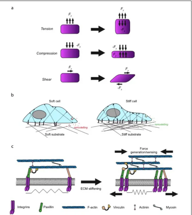
Figure 1. Mechanical forces and mechanoreciprocity
(a) Tension stress is applied perpendicular to the cell and leads to expansion. Compression stress is
applied perpendicular to the cell and leads to compaction. Shear stress is applied parallel to the surface of the cell (adapted from Butcher et al., 2009). (b) The cell senses soft matrix and deforms the ECM by contracting. The cell has only few focal adhesions (purple sticks) and actin fibers (black arrows). It can also loosen the matrix by secreting metalloproteinases that digest the ECM (red dots) (left). The same cell on stiff matrix that cannot be deformed, the number of focal adhesions increases. The secretion of crosslinking factors (green dots) can mediate ECM stiffening. Substrate stiffening results in more and thicker actin stress fibers (black arrows) leadind to cell spreading and stiffening (right). (c) The basic machinery that senses and responds to ECM-generated mechanical signal. The cell surveys its mechanical environment with periodic contractions of stress fibers, which are attached to integrins that pull against matrix. Immature focal adhesions cannot sense matrix stiffness or exert strong mechanical forces on the ECM (left). While mature focal adhesions recruits myosin that allows force generation in response to matrix stiffening and recruits vinculin and actinin that increase F-actin number and crosslink the filaments to enlarge and strengthen focal adhesions and generates more contraction force (right) (based on Janmey et al., 2011).
13
Introduction
1 Cell mechanics and mechanotransduction
1.1 Cell and tissue mechanics
Of course, as every element of our universe, cells are subjected to the laws of physics and the renewal of the study of mechanics applied to the cell with the emergence of a mechanobiology field has already changed our understanding of most of fundamental biological processes.
1.1.1 Force generation (may the force be with you)
Cells are continuously subjected to mechanical forces including tensile force, compression, shear stress or hydrostatic pressure. Cells accommodate them by modifying their behavior and remodeling their microenvironment (Fig. 1). The interplay between cells and their microenvironment contribute through the resulting mechanical strains to fundamental biological processes but also to the development of pathologies (Egeblad et al., 2010; Kai et al., 2016). Tissues are composed of multiple constituents, among those; one can find different cell types and the extra cellular matrix (ECM). Each of these components can be characterized by several mechanical properties such as elasticity, viscosity, plasticity and stiffness (Janmey and Miller, 2011). All these physical properties govern how a tissue senses, transmits and responds to mechanical cues. For example, ECM stiffness can modulate essential biological processes at the level of individual cell such as differentiation, motility and morphology (Weaver et al., 1997; Engler et al., 2006; Vogel and Sheetz, 2009). Pelman and Wang, who used collagen-coated beads embedded in substrate with different stiffness, demonstrated that increasing substrate stiffness leads to the modulation of the size and dynamics of focal adhesion and therefore cell locomotion (Pelham and Wang, 1998). On the other hand, cells can generate mechanical strains
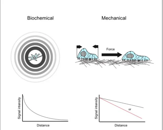
Figure 2. Biochemical versus mechanical signal
Biochemical signal diffuses and form a gradient from a point of source and can be re released into the circulation (upper left). The strength of the signal decreases with the distance from its origin at a rate of 1/r2 (bottom left). Mechanical signals are transmitted through forces directionally applied to a target cell, directly or indirectly (through cell-cell contacts or matrix deformation) (upper right). Mechanical signals do not significantly decrease with the distance (bottom right, black). How ever signal intensity might be altered by the physical properties of the transmitting substance (cell or ECM) such as elasticity. Therefore the full force of the initial deformation might not be transmitted (red) (adapted from Janmey et al., 2011).
14 on the ECM and surrounding cells (Fig. 1b, c). Indeed, cells could wrinkle a soft rubber sheet on which they were cultured (Harris et al., 2008). This phenomenon occurs through active processes including actin assembly and ROCK dependent actomyosin contraction. The cytoskeleton appears to play a central role in the mechanobiology of the cell. It allows the cells to exert forces in the nanoNewton range on their surrounding environment and sense the mechanics of cells or substrate around them (du Roure et al., 2005). The overall cytoskeletal organization of cells, including actin filaments, intermediate filaments and microtubules, in interplay with the ECM components dictate the viscoelastic property of a tissue and the resulting forces. ECM is highly dynamic since cells constantly remodel their microenvironment with the secretion of either metalloproteinases leading to ECM degradation (Mrkonjic et al., 2017), or in contrast, of ECM components (Lu et al., 2011) and crosslinking enzymes such as lysyl oxidases and transglutaminases that stiffen the ECM (Lee et al., 2015). By using inner active processes such as cytoskeletal reorganization together with the secretion of ECM degrading/crosslinking enzymes, cells are able to adapt to their physical microenvironment to reach the mechanical equilibrium. Thus, all cells respond to mechanical forces by a mechanism called “mechanoreciprocity” (Fig. 1) (Ding et al., 2013).
1.1.2 Biochemical versus mechanical signal
The major difference between these two types of signal is their way of spreading. Indeed, chemical messengers passively diffuse from the source of production to the target receptor (Fig. 2). This implies that the signal is not directed and equally irradiates to all directions from the production site. However, in the context of chemotaxis, the interpretation of the signal by the target is directed but limited in terms of distances. The diffusion of first messengers upon paracrine and autocrine signaling occurs through the microenvironment, therefore, it is subjected to environmental forces such as flow. Thus, it rapidly decays over time (at the rate of 1/radius2) and depends on the rate of production over the rate of neutralization (Fig. 2). Moreover, chemical molecules can only signal across few tens of micrometers. On the contrary, mechanical forces can be directed in a specific direction through fibers within the cells or the ECM (Janmey and Miller, 2011). A meshwork of
filaments such as those composing the ECM or the cytoskeleton can align in the direction of the stress to convert filament bending into filament stretching (Onck et al., 2005). Unlike chemical signals, mechanical signals decay linearly and therefore can rapidly propagate up to hundreds of micrometers away from its point of generation (Winer et al., 2009). Transduction of mechanical signals is a rapid process generated from the perturbation of the mechanical equilibrium between cells and their microenvironment. While biochemical signaling is generally a slower process with timescales of seconds or minutes, (except for processes such as synaptic transduction), mechanotransduction is believed to be a fast process. For example, Sarcoma kinase (Src) activation by mechanical cues occurs in less than 0.3s while chemokine mediated activation of Src requires more than 12s (Na et al., 2008).
1.1.3 The dark side of the force
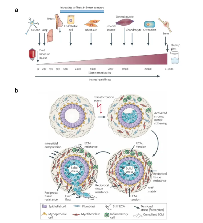
As discussed above, the cell microenvironment and its mechanical properties govern a wide range of biological processes. For example, substrate stiffness can control cell fate (Fig. 3a). Mesenchymal stem cells (MsCs) can differentiate into three cell types depending on their substrate stiffness. MsCs cultured on substrates mimicking brain (0.15-0.30 kPa), muscle (8-14 kPa) or bone stiffness (25-45 kPa) respectively differentiate into neuronal, muscle-like or bone cells (Engler et al., 2006). Therefore, any abnormality in the ECM that could modify its mechanical properties such as stiffness can perturb essential biological events by generating abnormal mechanical signal. These processes may trigger non-physiological conditions where tissue mechanics alter programmed cell functions leading to pathologies. Indeed, mechanical stress is involved in a myriad of diseases such as liver, renal, muscular diseases and cancer. The relationship between mechanical deregulation and pathological behavior in the context of cardiovascular, muscular pathologies and cancer are described below.
1.1.3.1 Cardiovascular/and/muscular/pathologies/
Figure 3. Tissue rheology and pathological development
(a) All cells are exposed to mechanical forces that are generated by cell-cell or cell-ECM interactions.
These mechanical forces influence cell function. Each cell type is specifically tuned to the specific tissue in which it resides. Following transformation, breast tissue becomes progressively stiffer and tumor cells become significantly more contractile and hyper-responsive to mechanical cues. (b) Transformation (blue cells) resulting from accumulation of genetic and epigenetic alterations in the epithelium along with an altered stromal matrix leads to unchecked proliferation and enhanced survival of luminal epithelial cells, which compromises normal ductal structure. The abnormal pre-neoplastic luminal mammary epithelial cells eventually expand to fill the breast ducts exerting outwards projecting compression forces on the basement membrane and adjacent myoepithelium. The damaged pre-neoplastic tissue produces soluble factors that stimulate cell infiltration and activation of fibroblast to induce a dramatic reorganization and stiffening of the ECM over time. The rigid parenchyma exerts a progressively greater inward projecting resistance force on the expanding pre-neoplastic duct. Over time, the number of myoepithelial cells surrounding the pre-neoplastic mass decreases and the basement membrane thins, probably owing to increased matrix metalloproteinases (from Butcher et al., 2009).
a
microenvironment preventing the myocyte normal functions is highly deleterious. For example, myocardial infarct leads to fibrotic stiffening that impairs cardiac output (Cecelja and Chowienczyk, 2009). Indeed, embryogenic myocytes grown on a heart-like substrate stiffness of approximately 11 kPa normally beat, while those grown on a myocardial scar-like substrate stiffness (35-70 kPa) exhibit a drastic decrease of beat frequency (Engler et al., 2008).
An increased stiffness of muscle fibers most likely due to ECM stiffening by collagen crosslinking, is responsible for diastole in the context of congenital heart disease (Chaturvedi et al., 2010). Similarly, aortic stiffness increases the risk of heart failure and strokes (DeLoach and Townsend, 2008).
More recently our lab discovered that caveolin-3 mutations involved in muscular dystrophies prevent caveolae formation at the plasma membrane. Hence myotubes carrying these mutations are more prone to plasma membrane disruption under mechanical stress due to a defect of caveolae mechanoprotection (developed in 2.3.3). It also exhibits a defect of IL6/STAT3 mechanosignaling (Dewulf et al., 2018 under revision, see annex 3).
1.1.3.2 Cancer/
1.1.3.2.1
Mechanical-forces-in-the-context-of-tumor-progression-Some cancer clinical diagnoses rely on the detection of abnormal mechanical properties of the tissue. Indeed, detection of stiffen tissues is proceeded by palpation, X-ray, and ultrasound techniques. Most of the solid tumors are stiffer than surrounding healthy tissues. For example, normal breast tissue stiffness is around 0.2 kPa while breast tumors stiffness is increased by approximately 20 folds (Levental et al., 2007; Baker et al., 2009). Similar stiffness increase is found for pancreatic and colorectal cancer (Butcher et al., 2009; Kai et al., 2016). Abnormal tissue stiffness most likely originates from the imbalance between ECM deposition and its degradation increasing the total ECM quantity. Tissue stiffening is not only the result of pathological processes; it also promotes their establishment (Fig. 3b). Collagen crosslinking and stiffening induce the invasiveness of oncogene activated
17 epithelial cells by generating larger focal adhesion sites (Levental et al., 2009). Secretion of several metalloproteinases at the level of the invadosome such as MT1-MMP loosen and reduce matrix stiffness to counteract this crosslinking process (Willis et al., 2013) in response to mechanical cues (Mrkonjic et al., 2017). Stromal cells also exert considerable forces on the ECM. Cancer associated fibroblasts (CAFs) deform the extracellular matrix via strong actomyosin contractions, contributing to ECM stiffening (Laklai et al., 2016). The increase of cell density also promotes tissue stiffening. Cancer cells are usually stiffer than healthy cells (Fabry et al., 2001). Moreover “jamming” of cancer cells prevents their spatial reorganization to adapt to mechanical strains therefore contributing to tissue stiffening. Ultimately, in the context of metastasis, tumor cells have to squeeze through matrix fibers, travel through the organism and establish in a new tissue with different mechanical properties, which are as many processes that subject the cells to different mechanical stresses with a variety of biological consequences on tumor cells.
1.1.3.2.2
Tumor-physical-microenvironment-influences-cancer-progression-Tissue stiffening is not only symptomatic of cancer development; it also actively drives tumor progression. As discussed above mechanical forces influence a wide range of biological processes. Hence, throughout evolution, devices named mechanotransducers have been conserved to convert mechanical cues into interpretable biochemical information (detailed in 1.2). Most of pathological situations emerge from altered signal transduction such as irrelevant pathway activation or inactivation. Similarly to aberrant biochemical signaling such as growth hormone signaling, aberrant mechanical signaling also promotes tumor progression. For example abnormally stiff ECM drives epithelial to mesenchymal transition (EMT) and metastasis through the TWIST1-G3BP2 mechanotransduction pathway (Wei et al., 2015a). The most well-known mechanosignaling hubs are focal adhesions. The direct anchorage to the ECM and their link with the cytoskeleton, place these structures at the perfect place to integrate mechanical cues provided by ECM deformation. Several focal adhesion components such as integrins, talin, paxillin and Cas are sensitive to mechanical strains. Focal adhesions regulate a myriad of signaling molecules of major cancer pathways such as Src, FAK, Rho, ERK, PTEN (Kandoth et al., 2013; Mouw et al., 2014). Together with filamentous actin, integrins
also control the mechanical activation of the YAP/TAZ oncogenes (Dupont et al., 2011; Lamar et al., 2012). In three dimensional situation, studies have emphasized the role of integrins clustering in the transduction of mechanical cues (Harunaga and Yamada, 2011). Tumor cells are subjected to mechanical stretch at the periphery of the tumor but cells in the deep interior of the tumor are subjected to compressive forces (Fig. 3b). Mechanical forces are known to control cell cycle and apoptosis therefore promoting growth heterogeneity within the tumor (Chang et al., 2008; Lien et al., 2013). Cells sense and adapt to the microenvironment physical strains through the reorganization of their cytoskeleton, however such a process also implies plasma membrane topological reorganization to achieve mechanoadaptation and mechanosensation. Indeed, some mechanoreceptors engaged in cancer evolution reside inside the lipid bilayer. For example, stretch sensitive channel Piezo 1 and 2 that triggers calcium signaling and other TRP ion channels are involved in tumor pogression (Li et al., 2015; Wu et al., 2017; Pardo-Pastor et al., 2018).
1.2 Signal mechanotransduction
Cells can generate precise three-dimensional structure through morphogenic movements and ensure the cell/organism structural stability. As gravity and muscle contraction shape bones, shear stress from flowing blood influences the heart vasculature and many other examples show that the ability of cells to convert mechanical cues into biochemical signal is essential. Failure of these mechanisms contributes to a wide breadth of pathologies (Janmey and Miller, 2011) as discussed in 1.1.3. Therefore, considerable efforts are currently directed toward determining how a variety of mechanical stimuli lead to the regulation of gene expression.
Mechanotransduction is achieved through a rich set of mechanisms. In this introduction, only a few of them will be presented as examples. One of the best-studied mechanisms of signal mechanotransduction is the conversion of physical forces into biochemical signal through protein structural refolding. In general, proteins adopt the conformation that favors the lowest free energy. Therefore, changes in the energy landscape induced by physical strains lead to the reshaping of the protein (Orr et al., 2006). This process is comparable to other post-translational modifications
Figure 4. Mechanosensitive channels
(a) Mechanism of activation of mechanosensitive channels (MSCs). The channel occupies a restricted
area in resting, while proceeding to rearrangement into open conformation to decrease energy cost due to membrane tension and exposition of its hydrophobic part induced by membrane thinning. (b) Schematic structure of Piezo1 viewed from above, showing the three “propeller blades” surrounding a centrale pore (left). Side view of the structure of Piezo1 (center). When the cell is not submitted to to pressure, Piezo1 bends to make a dome-like structure pointing inside the cell, and the channel is closed. When the membrane is stretched the complex flattens, openning the channel (Chesler et al., 2018).
such as phosphorylation. Phosphorylation introduces a negatively charged phosphate group, inducing protein refolding due to charge repulsion in the inner structure of the protein. These modifications will then induce structural changes that will modulate the catalytic activity or the interaction ability to transduce a signal.
1.2.1 Mechanosignaling at the plasma membrane
The best example of plasma membrane mechanotransducers are mechanosensitive channels (MSCs) such as voltage gating mechanochannels (Martinac, 2004). These channels can sense membrane stretch and curvature. Pressure sensitive channels such as Piezo 1 and 2 are also an emerging important category of MSCs (Coste et al., 2011). Increasing the membrane tension, increases the probability to open the channel (Martinac and Hamill, 2002). In this context, MSCs opened conformation occupies a greater space in the stretched lipid bilayer, favoring lower free energy. Hence the opening of this gate induces membrane permeability to ions (Fig. 4). The inner leaflet of the plasma membrane is negatively charged therefore the arrival of positively charged ions into the cytoplasm leads to membrane depolarization and generates an electrical signal. Moreover Ca2+ entry through MSCs also leads to the activation of Ca2+ sensitive molecules such as calmodulin domain-containing proteins. Piezo and the other MSCs are involved in many biological processes such as hearing, touch, nociception and proprioception.
1.2.2 Mechanosignaling in the cytosol
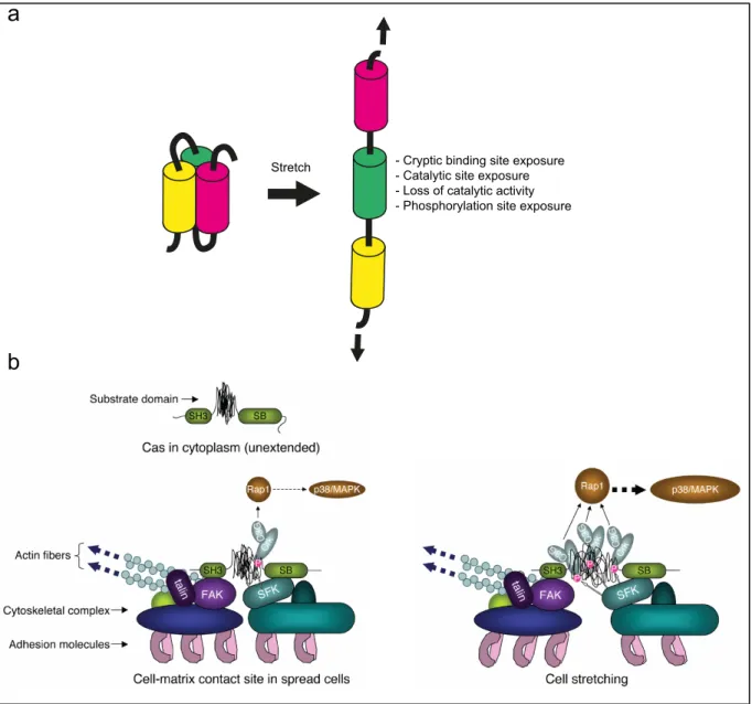
On the other hand, direct application of a stalling force against a molecular motor or an enzyme inhibits its catalytic activity, most likely by unfolding its catalytic domain (Finer et al., 1994; Yin et al., 1995; Ingber, 2006). Similarly, some proteins unfold in a stepwise manner when they are mechanically extended. This unfolding may lead to the exposure of cryptic binding or phosphorylation sites for potential signaling downstream effectors (Ingber, 2006) (Fig. 5a). In addition, mechanical strains can also influence signal transduction by strengthening or weakening protein-protein interaction (Evans and Calderwood, 2007).
Figure 5. Mechanotransduction at the level of the protein and protein complex
(a) Stress on mechanosensors can induce conformational changes generating a biochemical signal
(based on Ding et al., 2013). (b) Cas molecule with unextended configuration of substrate domain and with moderate extension of substrate domain at the cell-matrix contact site of spread cells, respectively (left). Extension-dependent phosphorylation of Cas substrate domain by SFK and enhancement of its downstream signaling. SH3 and SB represent the SH3 and Src-binding domain of Cas, respectively (right) (Sawada et al., 2006).
- Cryptic binding site exposure - Catalytic site exposure - Loss of catalytic activity - Phosphorylation site exposure Stretch
a
Focal adhesion sites (FA) are probably the most studied mechanosignaling platforms. Its direct link with the cell cytoskeleton and the ECM put this structure in perfect position to transduce mechanical signals. Mechanical forces directly control the shape, size and dynamics of FA (Galbraith et al., 2002). Indeed, clusters of integrins (focal adhesion) can generate forces on the overall cell through the cytoskeleton. Nevertheless, focal adhesion can sense mechanical forces (Grashoff et al., 2011). For example shear stress-induced cytoskeletal reorganization is controlled by integrins activation balance (Macek Jilkova et al., 2014). Integrins are connected to the cytoskeleton through several adaptor proteins such as talin, vinculin, zyxin, Cas etc., which are as many components susceptible to transduce mechanical cues.
For example, talin directly links integrins to the actin filaments. Upon ECM deformation or actomyosin contractions, talin rods are stretched and deformed by the mechanical strain. The unfolding of talin unveils cryptic binding sites for vinculin interaction (Fig 5a). Hence, talin unfolding leads to the recruitment of vinculin that strengthen the link with filamentous actin and cluster the integrins into focal adhesion sites (del Rio et al., 2009).
Similarly, p130Cas (Crk associated substrate) binds to the focal adhesion kinase (FAK) and the Src family of protein kinase (SFK). Upon cell stretch, Cas substrate domain unfolds exposing phosphorylation sites (Fig. 5b). Their phosphorylation by the SFK kinase triggers the recruitment of signaling partners and the activation of the p38/MAPK pathway through Ras associated protein 1 (Rap1) activation (Sawada et al., 2006).
Another example of cytosolic mechanotransduction is the yes associated protein/taffazin (YAP/TAZ) pathway that controls processes such as proliferation and differenciation (Dupont et al., 2011; Mosqueira et al., 2014). YAP/TAZ is an emerging mechanotransduction pathway. It transduces mechanical signals from the cytoskeleton to the nucleus. YAP/TAZ are transcriptional co-factors that are well studied in the context of the Hippo signaling, where upstream large tumor suppressor kinases (LATS) phosphorylate YAP and TAZ leading to their cytosolic retention (Low et al., 2014). There are clear evidences linking YAP/TAZ regulation to cell
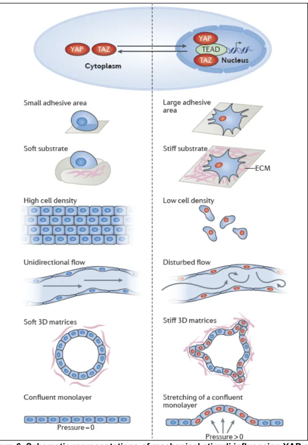
Figure 6. Schematic representations of mechanical stimuli influencing YAP and TAZ subcellular localization and activity
When YAP and TAZ are mechanically activated (red), they translocate to the nucleus, where they interact with TEA domain family factors (TEAD) to regulate gene expression (top). Schematics illustrating how different matrix, geometry and physical conditions influence YAP and TAZ localization and activity: the left panels show conditions in which YAP and TAZ are inhibited and localized to the cytoplasm, whereas the right panels show conditions that promote YAP and TAZ nuclear localization (indicated by red coloring of cell nuclei) (Panciera et al., 2017).
cell mechanics and matrix physical properties (Fig. 6). For example cells cultured on soft substrate exhibit a cytosolic YAP/TAZ distribution, while those grown on stiff microenvironment exhibit a nuclear YAP/TAZ localization and those residing in an “in-between” microenvironment stiffness possess an evenly distributed YAP/TAZ localization. Therefore, substrate stiffness but also cell shape controls YAP/TAZ distribution (Dupont et al., 2011). Yet, how mechanical cues regulate YAP/TAZ localization and activity is still unknown, however it is tightly dependent on the F-actin organization, contraction and cytoskeletal mechanics (Dupont et al., 2011; Aragona et al., 2013). Propagation of tensile forces through the cytoskeleton induced by actomyosin network contraction or ECM deformation pulling on cytoskeleton-linked integrins leads to YAP and TAZ activation (Taniguchi et al., 2015; Hu et al., 2017). The cytoskeleton is also connected to the nucleus; therefore, force propagation through the cytoskeleton may also reach the nucleus thereby directly affecting nuclear mechanics and the regulation of YAP/TAZ nuclear translocation as detailed in the next chapter.
1.2.3 Nuclear mechanotransduction
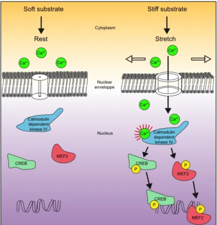
Mechanotransduction is not a process only restricted to “surface” receptors. An array of biological processes such as cell migration, programmed necrosis or infection trigger nucleus deformation (swelling, squeezing and stretching). For example, a transient decrease of extracellular osmotic pressure promotes nuclear swelling (Irianto et al., 2013). External mechanical strains can also exert direct mechanical forces on the nucleus since it is connected to the cell surface through the cytoskeleton via the linker of nucleoskeleton and cytoskeleton (LINK) complex. In a metastatic context, tumor cells have to squeeze through the extracellular matrix fibers subjecting their nucleus to deformations. Interestingly, the nucleus appears to be a good mechanotransduction organelle. Indeed, the nuclear envelope participate less to cell trafficking, therefore its overall area fluctuates less. Moreover its membrane is more loose and easy to stretch, thus it is more prone to detect external strains (Bigay and Antonny, 2012). The nuclear envelope possesses a dynamic structure that can rapidly adapt to tensile force by adjusting its stiffness via the phosphorylation of proteins of the nuclear envelope such as emerin (Guilluy et al.,
Figure 7. Mechanosensitive calcium signaling in the nucleus
Figure 8. Proposed model of mechanosensitive nucleoplasmic shuttling (Elosegui-Artola et al., 2017)
2014). The nuclear envelope serves as scaffold for the activation of important peripheral proteins. Hence, nuclear expansion induced by cell swelling leads to the recruitment of the phospholipase enzyme cPLA2 and the 5-lipoxygenase (LOX) at the inner face of the nuclear membrane. This promotes LOX-dependent generation of pro-inflammatory eicosanoids such as leukotrienes, which are key factors in inflammatory diseases such as asthma (Peters-Golden and Brock, 2001). Nuclear mechanics can also directly control gene expression. Indeed, some MSCs are found in the nuclear envelope and mediate Ca2+ release in response to nuclear and endoplasmic reticulum stretch induced by cell spreading. For example, the nuclear mechanical release of Ca2+ regulates gene expression through the cAMP responsive element binding (CREB) and myocyte enhancer factor 2 (MEF2) transcription factor phosphorylation by the calmodulin dependent kinase IV (Itano et al., 2003) (Fig. 7). Nucleus deformation also alters the integrity of the nuclear pores. Indeed, the crossing of molecules through the nuclear envelope via these nuclear pores is a key and tightly regulated step of many signaling cascades to achieve the regulation of gene expression and by extension the cellular responses. The nucleus deformation stretches the nuclear pores thereby decreasing the energy requirement for molecular transport through these structures. Nuclear deformation and the consecutive nuclear pores stretch have been shown to trigger YAP nuclear translocation and might be a more general mechanism for other signaling molecules (Elosegui-Artola et al., 2017) (Fig. 8). Finally, harsh nuclear deformation induced by nuclear squeezing during cell migration, can result in nuclear membrane damages releasing DNA in the cytoplasm. This situation promotes the activation of damage-sensing pathways including cyclic GMP-AMP synthase and stimulator of interferon genes (cGAS-STING) pathway and triggers the expression of immunomodulatory genes such as interferons (Raab et al., 2016).
To summarize, mechanical forces generated by external strains can modify protein folding and larger complex organization inside the cell, thereby changing their biochemical properties and therefore transforming mechanical cues into biochemical signals. Nevertheless, studies demonstrated that larger structure such as plasma membrane nanodomains called caveolae can also respond to mechanical strains applied to the plasma membrane (Sinha et al., 2011).
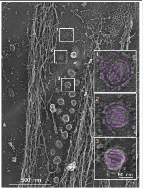
Figure 9. Visualization of the caveolar coat at the plasma membrane of myotubes
Survey view of the cytoplasmic surface of an unroofed myotube presenting caveolae at the plasma membrane. Different types of caveolae structures are apparent, ranging from flat (1), circular (2), to fully budded (3). Scale 500 nm. Scale bar in insets: 50 nm (from Lamaze et al., 2017)
2 The caveolae: specialized plasma membrane structures
More than sixty years ago, fine plasma membrane structures identified by electron microscopy as “caves” or “cave-like indentation of the plasma membrane” thereby named caveolae, were first visualized in blood capillaries and mouse gall bladder by George E. Palade and Eichi Yamada (Palade, 1953; Yamada, 1955). These plasma membrane invaginations are smaller than clathrin coated pits since their diameter varies between 50 and 80 nm and they present a striated coat (Rothberg et al., 1992) (Fig. 9). Caveolae are also found as interconnected caveolar structures named “rosette” (Pelkmans and Zerial, 2005). Adipocytes, endothelial and muscle cells are particularly enriched in caveolae, yet almost all mammalian cell types possess caveolae except neurons and lymphocytes despite their expression of caveolin-1.
2.1 Structure and composition
The first step to fully understand the function of these “cave-like” plasma membrane structures was to elucidate their molecular composition. Therefore, since the discovery of caveolae, a lot of effort has been put toward the identification of their molecular composition and organization.
2.1.1 Caveolar proteins
2.1.1.1 Caveolins/
Almost forty years after the first visualization of caveolae, structural studies of the caveolae inner face (facing the cytoplasm) revealed a striated coat composed of “filaments”. These filaments have been first identified as a 22 kDa substrate for v-src tyrosine kinase in virus-transformed chick embryo fibroblasts and therefore called caveolin (Rothberg et al., 1992). The same year, a vesicular integral-membrane protein of 21 kDa (VIP21) was also found by another team to be an important
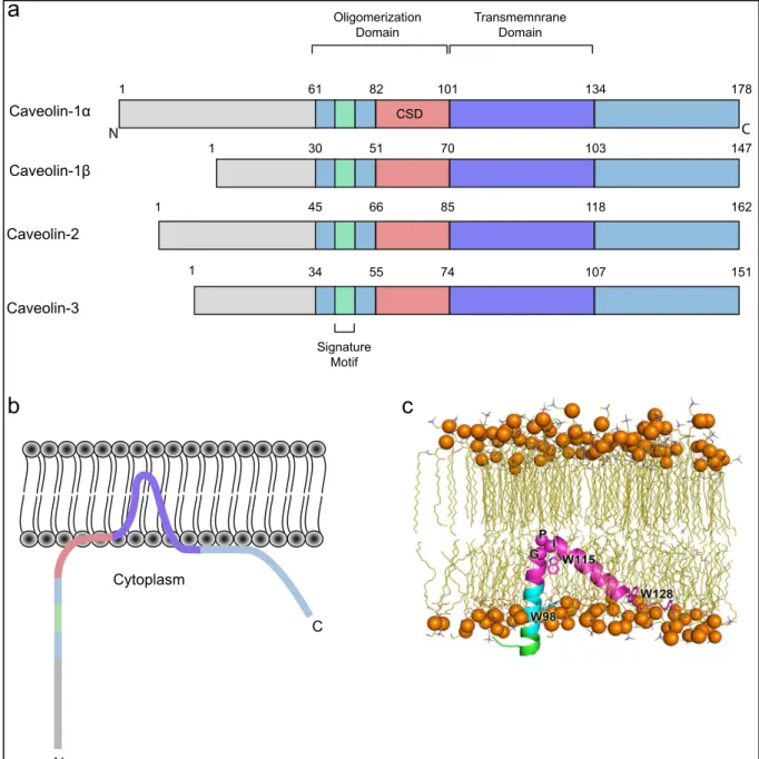
Figure 10. Caveolins domains and insertion in the plasma membrane
(a) Conserved domains of the caveolin protein family. An oligmerization domain encompassing an 8
amino acids stretch signature motif (FEDVIAEP) and the caveolin scaffolding domain followed by the transmembrane domain. Length variability of caveolins -1β, -2 and -3 is the consequence of truncated N-terminal part. (b) Proposed model of caveolin-1 (Cav1) topology within the lipid bilayer. Cav1 is inserted in the plasma membrane inner leaflet via its TMD conferring a hairpin-shaped topology. Both N- and C-termini are facing the cytoplasm (based on Parton and del Pozo, 2013). (c) Computational analysis of Cav1 insertion within the plasma membrane. Snapshot of the insertion of Cav182-136is shown in the illustration with the CRAC motif in cyan and putative TMD in magenta as well as the N-terminus in green. The Cαatoms of G108-P110 are shown in spheres. W98, W115 and W128 are
shown in sticks. Cholesterol molecules and, ions and water are hidden for clarity (Liu et al., 2016).
1 61 82 101 134 178 1 30 51 70 103 147 1 45 66 85 118 162 1 34 55 74 107 151 N C Oligomerization Domain Transmemnrane Domain CSD Signature Motif Caveolin-1α Caveolin-1β Caveolin-2 Caveolin-3
a
b
c
N C Cytoplasmdiscovered caveolar proteins appeared to be the same (Glenney and Soppet, 1992). Hence, it has been decided that both v-src substrate in Rous sarcoma virus-transformed fibroblasts and VIP21 would be renamed as caveolin. This is how the first member of the caveolin family has been identified thus named as caveolin-1 (Cav1), shortly followed, by the identification of two homologues: caveolin-2 (Cav2) and the muscle restricted isoform: caveolin-3 (Cav3) (Way and Parton, 1995).
Cav1 molecules (and Cav3 in muscle cells) are essential components of caveolae. Indeed, these two proteins are required for caveolae biogenesis. Lack of Cav1 or Cav3 leads to mis-invagination of the caveolae. Caveolins can form structures of higher complexity by homo and hetero-oligomerization into 300 kDa complexes (Sargiacomo et al., 1995). All the caveolin isoforms share a signature motif of eight amino acids 68FEDVIAEP75 localized in their oligomerization domain (Fig. 10a).
Cav1 is the most studied member of the caveolin family. Cav1 is an integral plasma membrane protein of 178 amino acids with both N- and C-termini facing the cytoplasm (Monier et al., 1995; Aoki et al., 2010) (Fig. 10b). Its structure remains debated and mainly relies on predictions based on circular dichroism (CD), nuclear magnetic resonance (NMR) and computational analysis. From these studies, we know that Cav1 crosses only the inner leaflet of the plasma membrane through a putative hairpin-shaped transmembrane domain (TMD) predicted to be from L102 to I134 (Razani et al., 2002a). The TMD adopts a helix-break-helix topology (Lee and Glover, 2012) (Fig. 10c). Indeed the two α-helices are separated by three residues linker regions containing a proline 110 that induces a 50° angle between those two helices (Root et al., 2015). The TMD adopts a U-shaped conformation (Aoki et al., 2010) and plays a key role in the oligomerization ability of Cav1 and Cav2 (Das et al., 1999). The insertion of the TMD within the inner leaflet of the plasma membrane thereby displacing lipids of the inner layer has been proposed to induce membrane curvature (McMahon and Gallop, 2005). The Cav1 C-terminal end (K135-I178) is involved in plasma membrane attachment, trans-Golgi localization and oligomer-oligomer interaction (Song et al., 1997; Schlegel and Lisanti, 2000) . Moreover, it presents three palmitoylation sites on C133, C153 and C156, which are not required
25 for Cav1 anchorage to the plasma membrane but may influence Cav1 oligomerization (Dietzen et al., 1995; Monier et al., 1996).
The most prominent domain of Cav1 is the caveolin scaffolding domain (CSD) (Fig. 10a). Indeed this domain is involved in Cav1 oligomerization, protein-protein interaction, and cholesterol recognition/binding (Li et al., 1996; Couet et al., 1997; Schlegel et al., 1999). However, the role of the CSD in the control of biological processes such as signaling remains somehow controversial (developed in 2.3.4.4 and discussion)(Collins et al., 2012). The CSD topology is not clear, and was first proposed to be a fully amphipathic α-helix that partially lies inside the plasma membrane inner leaflet (Le Lan et al., 2006), then later appeared to be a mixture of β and α structures (Hoop et al., 2012). More recently, the CSD was proposed to adopt a dynamic structure that can be either fully helical or partially unstructured (Liu et al., 2016). Interestingly, Cav1 also exhibits a high affinity for cholesterol (Murata et al.,
1995). Cholesterol recognition is achieved through the cholesterol
recognition/interaction amino acid consensus (CRAC) motif within the CSD. Since Cav1 has been first discovered as a tyrosine kinase substrate, it was later shown that Cav1 can be phosphorylated on its tyrosine 14. More recently, serine 80 has been found to be phosphorylated. Yet, the precise function of these two post-translational modifications is not well understood. Tyr14 phosphorylation has been recently proposed to mediate signaling (Joshi et al., 2012) and Cav1 conformation changes (further developed in the discussion) to regulate the CSD accessibility (Shajahan et al., 2012; Meng et al., 2017). The Ser80 phosphorylation is required for proper caveolae formation (Ariotti et al., 2015) and triggers its binding to the endoplasmic reticulum (ER) membrane in the context of regulated secretion of pancreatic cells (Schlegel et al., 2001). Finally, Cav1 can be ubiquitinated, a process that mediates Cav1 lysosomal degradation (Hayer et al., 2010a).
Cav1 encompasses two isoforms: a full-length α isoform (α-Cav1) and a truncated β-isoform (β-Cav1). The β-isoform lack the first thirty-three amino acids, hence it cannot be phosphorylated on Tyr14.
Few years after the identification of Cav1, a homologue of α-Cav1 was discovered by nucleotide sequence alignment. Cav3 shares common features with
Cav1 such as the caveolin signature motif, an oligomerization domain, an hydrophobic 102-134 TMD, a CSD and palmitoylation sites (Way and Parton, 1995; Tang et al., 1996) (Fig. 10a). Moreover, Cav1 and Cav3 can form hetero-oligomeric complexes with Cav1 in cardiac myocytes (Volonte et al., 2008). Cav3 also lacks the first twenty-seven aa, hence, neither Cav3 undergoes Tyr14 phosphorylation.
The purification of membrane fractions enriched in caveolae revealed the existence of a last member of the caveolin family: caveolin-2 (Cav2). Indeed, this isoform shares the common signature motif in its N-terminal part. It has been first described to differ from Cav1 from its inability to interact with the heterotrimeric G protein (Scherer et al., 1996). Moreover, Cav2 colocalizes with Cav1 indicating that Cav2 is a component of the caveolar coat (Scherer et al., 1996; Tang et al., 1996). There are two Cav2 isoforms: α-Cav2 and β-Cav2 that are respectively shortened by 16 and 29 aa compared to α-Cav1 (Fig. 10a). Likewise, Cav2 is not phosphorylated on its Tyr14. Unlike the other members of the caveolin family, Cav2 cannot homo-oligomerize, but it can dimerize and hetero-oligomerize with other caveolins (Scherer et al., 1996, 1997). α-Cav2 can be tyrosine phosphorylated on its residues 19 and 27 and serine phosphorylated on its residues 23 and 36. In addition Cav2 can be fatty acylated. However, the function of these post-translational modifications remains poorly understood. Cav2 serine phosphorylation participates to caveolae morphogenesis (Sowa et al., 2003). On another hand both Cav2 fatty acylation and phosphorylation have been reported to play a role in insulin signaling (Kwon et al., 2009; Kwon and Pak, 2010; Kwon et al., 2015). For example Cav2 phosphorylation prevent the interaction between the insulin receptor and a signal terminator (Kwon and Pak, 2010).
To conclude, caveolins are integral plasma membrane proteins possessing a characteristic hairpin shaped topology with both N- and C-termini facing the cytoplasm. They all share a common signature motif in their N-terminal part and an oligomerization domain allowing the formation of complex oligomerized structures that compose an essential part of the caveolar coat.
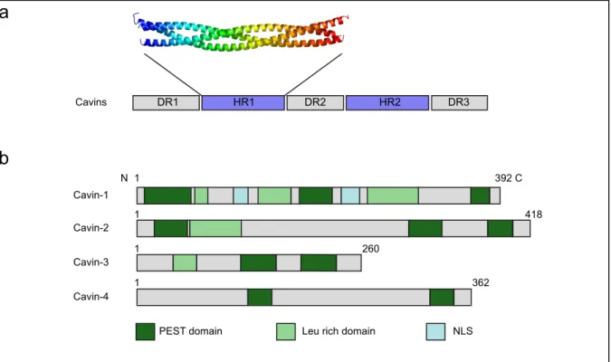
Figure 11. Cavins structure and caveolae morphogenesis
(a) General structural organization of cavins. Three disordered regions (DR) are separated by two
coiled coil (ordered) regions (HR) (adapted from Kovtun et al., 2014). (b) Cavins domains. PEST domain mediates cavins degradation and Leu-rich domain mediates protein-protein interaction (based on Bastiani et al., 2009). Cavin sizes are heterogenous. Cavin-1 is the only member possessing nuclear localization sequences.
DR1 HR1 DR2 HR2 DR3 Cavins Cavin-1 Cavin-2 Cavin-3 Cavin-4
PEST domain Leu rich domain NLS
N 1 392 C 1 418 1 260 1 362
a
b
c
2.1.1.2 Cavins/
Cavins are a family of cytosolic proteins which comprises four members: PTRF, SDPR, SRBC and MURC. Their caveolar localization and function pushed the scientists to gather these proteins under the name of “cavins” and rename them from cavin-1 to cavin-4 (Vinten et al., 2005; Bastiani et al., 2009; McMahon et al., 2009). As the caveolin family, the last member of the cavin family (cavin-4) is restricted to muscle cells (Ogata et al., 2008). Initially, polymerase 1 and transcript release factor (PTRF) was first identified as a regulator of RNA polymerase 1 (Jansa et al., 2001). Vinten and colleagues identified a 60 kDa protein associated with the caveolar coat in adipocytes. The protein was therefore called cavP60 (Vinten et al., 2001). Three years later PTRF has been reported to be enriched at the caveolar coat (Aboulaich et al., 2004), shortly after, PTRF and cavP60 appeared to be the same protein and thus were renamed cavin (Vinten et al., 2005).
SDPR was first described as a phosphatidylserine binding protein thus named PS-p68 (Burgener et al., 1990). When it was involved in serum deprivation response, it was renamed serum deprivation-response protein (SDPR) (Gustincich and Schneider, 1993). SDPR was shown to associate together with the protein kinase C α (PKCα) to the caveolar coat (Mineo et al., 1998). The SDPR-related gene product that binds to c-kinase (SRBC) was also involved in the serum deprivation response and first identified as a binding partner of PKCδ and thus named protein kinase C delta-binding protein (PRKCDBP) (Izumi et al., 1997). Both SRBC and SDPR were found in the mass spectrometry analysis that identified cavin-1 (Aboulaich et al., 2004).
Cavin-1 is required for caveolae formation (Hill et al., 2008; Liu et al., 2008). Cavins are proteins from 31 to 47 kDa with 261 to 425 amino acids. The four proteins share the same topology (Hansen et al., 2009). In silico analysis revealed that cavins possess two conserved α helical regions (HR) that are basic and positively charged. These HR regions are separated by acidic and negatively charged disordered regions (DR) (Fig. 11a). The HR1 region mediates cavin trimeric oligomerization (Kovtun et al., 2014). Indeed, cavin-1 can form trimers with either two cavin-1 or with cavin-1 and cavin-2 or with cavin-1 and cavin-3 (Fig. 14). All members of the cavin
28 family except cavin-4 carry a leucine zipper motif in the HR1 region that mediates protein-protein interaction (Fig. 11b). These leucine zipper domains are required for cavin recruitment at the plasma membrane (Wei et al., 2015b). Moreover all four cavins exhibit a short half-life due to the presence of PEST motifs that mediate proteasomal degradation (Fig 11b). Cavins have a basic C-terminal domain that participates to their membrane anchorage (Parton and del Pozo, 2013). Finally cavin-1 possesses two nuclear localization signals (Hansen and Nichols, 20cavin-10).
All cavins exhibit multiple phosphorylation sites. For example cavin-1 possesses more than twenty tyrosine and serine phosphorylation sites (Kovtun et al., 2014). Cavin-1 phosphorylation has been linked to Cav1 phosphorylation in the context of insulin signaling (Kruger et al., 2008) and have been suggested to participate to the control of cavin-1 fragmentation at the caveolar coat (Aboulaich et al., 2004). Cavins binds to phosphatidylserine (Ptdser) (Burgener et al., 1990). In addition cavin-1 and cavin-2 exhibit a high affinity for phosphatidyl 4,5 bisphosphate (PiP2) (Kovtun et al., 2014).
In summary, cavins are cytosolic proteins associated to the caveolar coat. All four cavins present a similar strucuture with a first coiled coil helical region that mediates trimeric association and a second coiled coil helical region that mediates hetero-association of cavin trimers.
2.1.1.3 Accessory/proteins/
Several non-essential caveolar components have been also identified since then. The F-BAR protein PACSIN2 (Protein Kinase C and Casein Substrate In Neurons) also called syndapin 2, which regulates and senses membrane curvature and participates to caveolae morphogenesis (Hansen et al., 2011; Senju et al., 2011). The dynamin2 GTPase and the dynamin-like ATPase Eps15 homology-domain containing protein 2 (EHD2) oligomers localize at the caveolar neck (Oh et al., 1998; Stoeber et al., 2012; Ludwig et al., 2013). While dynamin2 is involved in caveolae internalization, EHD2 controls caveolar dynamics and stability at the plasma membrane (Morén et al., 2012). In addition EHD2 and other EHDs (1 and 4)