HAL Id: hal-01846993
https://hal.umontpellier.fr/hal-01846993
Submitted on 31 May 2021
HAL is a multi-disciplinary open access archive for the deposit and dissemination of sci-entific research documents, whether they are pub-lished or not. The documents may come from teaching and research institutions in France or abroad, or from public or private research centers.
L’archive ouverte pluridisciplinaire HAL, est destinée au dépôt et à la diffusion de documents scientifiques de niveau recherche, publiés ou non, émanant des établissements d’enseignement et de recherche français ou étrangers, des laboratoires publics ou privés.
Distributed under a Creative Commons Attribution - NonCommercial| 4.0 International
novel mutation in two sisters
F Uettwiller, G. Sarrabay, P Rodero, I Rice, E Lagrue, Y Marot, K Deiva, I.
Touitou, Y Crow, P. Quartier
To cite this version:
F Uettwiller, G. Sarrabay, P Rodero, I Rice, E Lagrue, et al.. ADA2 deficiency: case report of a new phenotype and novel mutation in two sisters. RMD Open : Rheumatic & Musculoskeletal Diseases, EULAR ; BMJ, 2016, 2 (1), �10.1136/rmdopen-2015-000236�. �hal-01846993�
CLINICAL CASE
ADA2 deficiency: case report of a new
phenotype and novel mutation in two
sisters
F Uettwiller,1,2G Sarrabay,3M P Rodero,4G I Rice,5E Lagrue,6,7Y Marot,8 K Deiva,9I Touitou,3Y J Crow,4,5P Quartier1
To cite: Uettwiller F, Sarrabay G, Rodero MP,et al. ADA2 deficiency: case report of a new phenotype and novel mutation in two sisters. RMD Open 2016;2:e000236. doi:10.1136/rmdopen-2015-000236
▸ Prepublication history and additional material is available. To view please visit the journal (http://dx.doi.org/ 10.1136/rmdopen-2015-000236).
Received 19 December 2015 Revised 14 April 2016 Accepted 16 April 2016
For numbered affiliations see end of article.
Correspondence to
Dr F Uettwiller;
ABSTRACT
The objective of this paper is to:describe the phenotype compound heterozygote for mutations in CECR1 in two children. We describe the clinical and immunological phenotype, including the assessment of ADA2 activity, cytokine expression, interferon-stimulated and neutrophil-interferon-stimulated gene signatures, and the results of CECR1 sequencing. The first patient presented with intermittent fever, cutaneous vasculitis, myalgia and muscle inflammation on MRI leading to a provisional diagnosis of periarteritis nodosa.
Subsequently, two cerebral lacunar lesions were identified following a brain stroke. Clinical features improved on anti-tumour necrosis factor therapy. The first patient’s sister demonstrated early-onset, long-lasting anaemia with mild biological inflammation; at the ages of 3 and 5 years, she had presented 2 acute, transient neurological events with lacunar lesions on MRI. CECR1 sequencing identified both sisters to be compound heterozygous for a p.Tyr453Cys mutation and a previously undescribed deletion of exon 7. ADA2 activity was reduced by 50%. Neutrophil-stimulated genes were not overexpressed, but interferon-stimulated genes were. The expression of a panel of other cytokine transcripts was not significantly altered. In conclusion, searching forCECR1 mutation or assessing ADA2 activity should be considered in patients with an atypical presentation of inflammatory disease.
INTRODUCTION
Loss-of-function mutations in CECR1 have recently been described in patients with vas-cular and inflammatory features ranging from early-onset recurrent strokes to systemic vasculitis mimicking polyarteritis nodosa.1 2 While the pathological basis of ADA1 de fi-ciency is relatively well understood, the pathogenesis of ADA2 deficiency remains unclear.3 4A previous report suggested a pos-sible link to an overexpression of neutrophil-derived gene transcripts.5 We describe two new cases of ADA2 deficiency in siblings with
an unusual presentation, and an assessment of ADA2 activity, cytokine secretion and gene expression profiling.
MATERIALS AND METHODS
The clinical description of the two siblings was based on the medical data. Appropriate informed consent was obtained from the parents.
ADA2 activity in plasma was assessed twice, with a 3-month interval, using a commercial kit (Diazyme Laboratories). Genomic DNA was extracted from peripheral blood leuko-cytes using standard procedures. Exons 2–10 of CECR1 (NM_001282225.1) and flanking intronic sequences were amplified by PCR
Key messages
What is already known about this subject?
▸ ADA2 deficiency is associated with various phe-notypes, from early-onset inflammation mimick-ing periarteritis nodosa to central nervous system involvement; cutaneous lesions are con-sidered a key clinical feature.
What does this study add?
▸ This study extends the phenotypic spectrum of ADA2 deficiency to atypical myositis and unex-plained neurological events in the absence of cutaneous involvement.
▸ Gene expression may show an interferon 1 sig-nature but not necessarily the previously described neutrophilic-associated gene overexpression.
▸ Our study emphasises the need to search for CECR1 exonic deletions in suspected cases.
How might this impact on clinical practice?
▸ In patients with persistent, early-onset inflam-mation, even in the absence of the most typical features, ADA2 deficiency should be considered and anti-tumour necrosis factor therapy discussed. on May 31, 2021 at Univ of Montpellier. Protected by copyright.
(Applied Biosystems). Real-time quantitative PCR was performed for each coding exon, using a LightCycler 480 (Roche). Variants were analysed using standard in silico tools (SIFT, Polyphen2, MutationTaster). Neutrophils, interferon (IFN) and a panel of other cyto-kine transcripts were analysed as described previously5 (see online supplementary method).
RESULTS
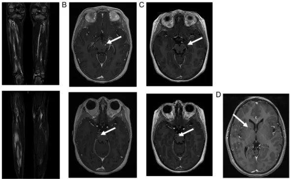
Patient 1: A 9-year-old girl had presented at the age of 3.5 years with fever, cutaneous vasculitis and myalgia. Biological analyses demonstrated inflammation on both C reactive protein (CRP) (100 mg/L) and erythrocyte sedimentation rate (ESR) (40 mm), elevated antinuclear antibodies (ANA 1/800), a transient lupus anticoagulant, low IgM and IgA level, mild CD19 B cells, CD8 T cells and natural killer (NK) lymphopenia. Nailfold capillaro-scopy showed no vessels due to a severe vasculitis. Muscle testing was abnormal with a predominant proximal muscle weakness. The creatine kinase level was normal. Muscle MRI and positron emission tomography (PET) scan revealed inflammatory lesions of the limbs (figure
1A). Muscle biopsy identified focal necrotising angiitis compatible with periarteritis nodosa.
At the age of 5 years, she had presented with two epi-sodes of severe vertigo, vomiting and encephalopathy. Cerebral MRI was normal, but a cerebrospinal fluid (CSF) pleocytosis was identified (16 lymphocytes/mm3). Clinical symptoms and vertigo improved on 2 mg/kg of prednisone. Following steroid treatment withdrawal, a cutaneous vasculitis appeared, necessitating the intro-duction of cyclosporine. The patient then developed an ileitis and cyclosporine was switched to azathioprine.
At the age of 7 years, a period of 2 months of persist-ent headache had been followed by an encephalopathy and, a few weeks later, despite the increase of steroids, by left internuclear ophthalmoplaegia. Cerebral MRI with diffusion was again reported as normal. Treatment was changed to cyclophosphamide, but the patient then developed ataxia with right body hemiparesis, left facial paresis and aphasia, 2 months later. MRI showed gadolinium-enhancing lesions with subtle abnormality diffusion sequences suggestive of a vascular stroke (figure 1B), which was later confirmed by their evolu-tion into lacunar lesions a few months later (figure 1C). During follow-up, the patient developed moderate hypertension. Despite immunosuppressive therapy, her pyramidal syndrome worsened and systemic in flamma-tion persisted. After switching to anti-tumour necrosis factor (anti-TNF) therapy (etanercept), the biological abnormalities resolved, with no further evidence of disease activity 1 year later. The recent MRI showed the occurrence of another asymptomatic lacunar lesion of the internal capsule (figure 1C).
Patient 2: The younger sister had been followed for uncharacterised microcytic anaemia since the age of 1 year, with a mild inflammatory syndrome of unknown
origin. IgM and IgA levels were low, with mild CD19 B lymphopenia. ANA was positive (1/200). The patient had neither cutaneous, visceral nor muscle signs, nor fever. At the age of 3 years, she presented with a transi-ent paralysis of the right extrinsic third cranial nerve, with persistent systemic inflammation. Cerebral MRI with diffusion sequences and CSF were normal. At the age of 5 years she presented again, with transient gait disturbance, unexplained fever and mild biological inflammation. A provisional diagnosis of viral vestibulitis was made. Cerebral MRI showed a 2 mm lacunar stroke of the internal capsule. CSF was again normal. Treatment with anti-TNF was started, associated with a rapid normalisation of the inflammation and no other symptoms over the following year.
CECR1 sequencing revealed a heterozygous c.1358A>G; p.Tyr453Cys mutation in both siblings (figure 2A). This variant is predicted to be pathogenic by in silico analysis, and has been described previously.1Furthermore, real-time quantitative PCR revealed a heterozygous deletion of exon 7 (c.973-?_1081+?del; p.Val325Thrfs*7), predicted to lead to a frameshift and truncated protein in both children (figure 2B). Parental analysis (father’s genotype: c.[973-? _1081+?del];[=]; mother’s genotype: c.[1358A>G];[=]) confirmed that the sisters were compound heterozygous for these two lesions.
ADA2 activity was measured on two occasions and was consistently reduced to 50% of wild-type levels in both children (figure 2C).
IFN and neutrophil signatures were evaluated in unstimulated patient cells from whole blood samples. The expression of a panel of IFN-stimulated genes was significantly elevated in both patients (figure 3A), though for the older sister the second sampling showed no elevation of IFN-stimulated genes.
Neutrophil-stimulated genes were not overexpressed (figure 3B). The expression of a panel of other cytokine transcripts was not significantly altered (see online supplementaryfigure S1).
DISCUSSION
We report the case of two sisters with ADA2 deficiency and unusual clinical features at disease onset with initial normal cerebral MRI. Both presented with a previously undescribed mutation in CECR1, low ADA2 activity and the association of a high IFN signature on gene expres-sion profiling, and a normal neutrophilic signature.
The clinical description of these two girls highlights the variable phenotype associated with ADA2 deficiency due to CECR1 mutations. ADA2 deficiency was first described in two majors articles, with fever, visceral and cutaneous lesions compatible with polyarteritis nodosa, and peripheral and central nervous system involve-ment.1 2 While patient 1 in this report eventually demonstrated a comparable spectrum of disease fea-tures, she initially presented with an inflammatory myo-sitis, which had not yet been described.2 Moreover,
on May 31, 2021 at Univ of Montpellier. Protected by copyright.
repeated cerebral MRIs, with diffusion sequences, were normal in the acute period, which is most often asso-ciated with encephalopathy, suggestive probably of tran-sient ischaemic strokes affecting medium and small arteries evolving later to a stroke with lacunar lesions. The clinical presentation of this patient’s sister was dif-ferent. While patients have been described with only
cutaneous involvement,6 7 this is the first report of a child with early stroke and no other features beyond a persistent mild inflammatory syndrome, including no cutaneous features. Hence, ADA2 deficiency should be considered as a differential diagnosis of atypical myositis or unexplained neurological events in the context of persistent inflammation, even in the absence of
Figure 1 Imaging of patient 1. (A) Muscle MRI of both legs performed at the age of 5 years: coronal gadolinium-enhanced
T1-weighted sequence at different levels, showing patchy inflammatory muscular lesions. (B) Cerebral MRI performed at the age of 7 years. Left: axial gadolinium-enhanced T1-weighted sequence, showing gadolinium-enhanced mesencephalic (up) and peduncular (down) lesions. Right: axial diffusion-weighted sequence showing mesencephalic (up) and peduncular (down) hyperintensities. (C) Cerebral MRI performed 4 months later: axial gadolinium-enhanced T1-weighted sequence showing evolution towards lacunar lesions (up: mesencephalic lesions/down: peduncular lesions). (D) Cerebral MRI performed 3 years later: gadolinium-enhanced T1-weighted sequences showing lacunar lesions located in the internal capsule.
Figure 2 Illustration of the mutation and ADA2 activity. (A) Sanger sequencing analysis illustrating the point c.1358A>G mutation which was identified in both sisters. (B) Reverse quantitative PCR (RQ-PCR, copy number calculated) illustrating the exon 7 deletion in the two sisters and in the father. CECR1-E7a and CECR1-E7b are two distinct amplicons encompassing exon 7 of CECR1 gene, to avoid a possible rare polymorphism in a primer hybridisation site. (C) CECR1 mutations result in a
decrease in ADA2 activity in patient plasma. The figure shows the ADA2 activity in the plasma of the two patients tested on two occasions, compared with ADA2 activity in the plasma of four controls (***p<0.001).
on May 31, 2021 at Univ of Montpellier. Protected by copyright.
cutaneous involvement.6 8Finally, our patients exhibited neither hypogammaglobulinaemia nor recurrent infec-tions, even though there were low IgM and IgA levels and mild B cell lymphopenia, in accordance with the hypothesis that ADA2 deficiency may lead to a defect in memory B cells.9 Interestingly, both patients also had mild biological markers of autoimmunity (ANA/lupus anticoagulant), as already reported.1 2
While the p.Tyr453Cys mutation has been reported previously, we provide the first description of an exonic deletion of CECR1. This possibility should therefore be borne in mind when testing forCECR1 mutations. Some phenotype–genotype correlations have been tentatively suggested, in particular for the G47R mutation (which might be associated less frequently with cerebrovascular involvement).10 However, this report and others under-line the fact that marked phenotypic variability can occur even in the same family.11
ADA2 enzymatic activity was reduced by almost 50% in both patients, although the clinical presentation was dif-ferent. This observation indicates that ADA2 activity does not reflect the severity of the disease.1 2We suggest that ADA2 activity dosage could be a complementary tool to diagnose ADA2 deficiency in cases where the clinical picture is suggestive and genetic testing is not conclusive (eg, in the presence of a missense variant of uncertain significance).
Gene expression analysis has already been performed in two patients with ADA2 deficiency. IFN-stimulated genes and neutrophil expressed gene overexpression have been suggested by Belot et al5to be relevant to the disease phenotype. Here, an upregulation of IFN-stimulated gene expression was demonstrated in both sisters, with the exception of the second sampling in the older sister. This might be partially explained by the fact that the patient did not have active disease at
Figure 3 IFN and neutrophil
gene expression studies. The RQ value for each transcript is equal to the normalised fold change relative to a control. (A)
Quantitative reverse transcription PCR of a panel of six
IFN-1-stimulated genes in CECR1 mutation-positive sisters on two occasions. IFN score was 4.82 before treatment and 1.86 after treatment in patient 1 and 11.79 and 9.47 before treatment in patient 2. IFN-stimulated genes are variably overexpressed in the two patients. (B) The expression of a panel of neutrophil-stimulated genes was measured in patients 1 and 2 on two occasions. Neutrophil-stimulated gene expression was essentially similar to that in controls. IFN-1,
interferon 1; NA, not applicable; RQ value, relative quantification value.
on May 31, 2021 at Univ of Montpellier. Protected by copyright.
the time of sampling and was under anti-TNF treatment. However, it is unclear to what extent the disease activity and the treatment affect cytokine transcription profiles in this disease, a point that should certainly be addressed further in future studies.
Of further note, neutrophil-derived gene expression was not elevated. It was previously hypothesised that a neutrophilic signature might explain part of the ADA2 deficiency phenotype, with a suggested role for ADA2 as plasmatic regulator of peripheral blood mononuclear cell activation.3 5 12 The patients described here make that possibility less likely. Meanwhile, inflammatory cyto-kine expression assessment did not reveal any consistent disease correlations, in accordance with previous data.1
Regarding ADA2 deficiency treatment, more than 20 patients have previously received systemic therapy includ-ing anti-interleukin 1 (IL-1), without efficacy in most cases. Moreover, treatment with fresh frozen plasma and bone marrow transplantation have been proposed in regard to the protein deficiency.13 14Furthermore, it was also reported that treatment with anti-TNF and IL-6 blockade could lead to clinical improvement (and, in the case of the former, almost normalise a previously high neutrophil signature).5 14 In our patients, short-term response to anti-TNF therapy was excellent.
To conclude, screening forCECR1 mutations should be considered in patients with an atypical presentation of inflammatory disease, including myositis. Efforts to collect new cases, in particular through the Eurofever registry, may allow us to improve our disease knowledge, straddling the borders of autoinflammation and immuno-logical deficiency.15 Importantly, although the patho-logical basis of this severe disease remains unclear, highly promising therapeutic strategies have already emerged. Author affiliations
1Pediatric Immunology-Hematology and Rheumatology Unit, Institut Imagine, Hôpital Necker-Enfants Malades, Assistance Publique Hôpitaux de Paris, Paris, France
2Transversal Unit of Allergology and Rheumatology, CHRU Tours, Tours, France
3Laboratory of Rare and Autoinflammatory Genetic Diseases, CHRU, Montpellier, France
4INSERM UMR 1163, Laboratory of Neurogenetics and Neuroinflammation, Institut Imagine, Hôpital Necker, Paris Descartes–Sorbonne Paris Cité University, Paris, France
5Manchester Centre for Genomic Medicine, Institute of Human Development Faculty of Medical and Human Sciences, Manchester Academic Health Sciences Centre, University of Manchester, Manchester, UK
6Inserm U930, CNRS 2448, Tours, France
7Service Neuropédiatrie et Handicaps, CHRU de Tours, Tours, France 8Pediatric Emergency Unit, CHRU Tours, Tours, France
9Pediatric Neurology Department, National Referral Center for Neuro-Inflammatory Diseases in Children, Hôpitaux Universitaires Paris Sud, Assistance Publique-Hôpitaux de Paris and UMR 1184, Center for Immunology of viral infections and autoimmune diseases, University Paris Sud, Le Kremlin-Bicêtre, France
Acknowledgements YJC acknowledges the European Research Council (GA 309449: Fellowship), and a state subsidy managed by the National Research Agency (France) under the’Investments for the Future’ program (reference ANR-10-IAHU-01).
Contributors FU wrote the manuscript and followed the two children with YM for the rheumatology and the help of two neurologists EL and KD for the analyses of cerebral MRI. GS and IT performed the genetics analyses. GIR and MPR performed the ADA2 activity dosage and the interferon and neutrophil signature under the supervision of YJC. YJC also mostly participated in the correction of the manuscript. PQ contributed to all the work and especially to the correction of the manuscript.
Competing interests None declared.
Patient consent Parental/guardian consent obtained.
Provenance and peer review Not commissioned; externally peer reviewed.
Data sharing statement No additional data are available.
Open Access This is an Open Access article distributed in accordance with the Creative Commons Attribution Non Commercial (CC BY-NC 4.0) license, which permits others to distribute, remix, adapt, build upon this work non-commercially, and license their derivative works on different terms, provided the original work is properly cited and the use is non-commercial. See: http:// creativecommons.org/licenses/by-nc/4.0/
REFERENCES
1. Zhou Q, Yang D, Ombrello AK, et al. Early-onset stroke and vasculopathy associated with mutations in ADA2.N Engl J Med
2014;370:911–20.
2. Navon Elkan P, Pierce SB, Segel R, et al. Mutant adenosine deaminase 2 in a polyarteritis nodosa vasculopathy.N Engl J Med
2014;370:921–31.
3. Zavialov AV, Engström A. Human ADA2 belongs to a new family of growth factors with adenosine deaminase activity.Biochem J
2005;391(Pt 1):51–7.
4. Poursharifi P, Saghiri R, Ebrahimi-Rad M, et al. Adenosine deaminase in patients with primary immunodeficiency syndromes: the analysis of serum ADA1 and ADA2 activities.Clin Biochem
2009;42:1438–43.
5. Belot A, Wassmer E, Twilt M, et al. Mutations in CECR1 associated with a neutrophil signature in peripheral blood.Pediatr Rheumatol Online J2014;12:44.
6. Westendorp WF, Nederkoorn PJ, Aksentijevich I, et al. Unexplained early-onset lacunar stroke and inflammatory skin lesions: consider ADA2 deficiency.Neurology2015;84:2092–3.
7. Gonzalez Santiago TM, Zavialov A, Saarela J, et al. Dermatologic Features of ADA2 deficiency in cutaneous polyarteritis nodosa.
JAMA Dermatol2015;151:1230–4.
8. Segel R, King MC, Levy-Lahad E. Mutant ADA2 in vasculopathies.
N Engl J Med2014;371:481–2.
9. Schena F, Volpi S, Pastorino C, et al. B cell characterization in ADA2 deficiency patients.Ann Rheum Dis2015;74(Suppl 2):1132. 10. Batu ED, Karadag O, Taskiran EZ, et al. A case series of adenosine
deaminase 2 deficient patients emphasizing genotype-phenotype correlations. Ann Rheum Dis 2015;74(Suppl 2):536.
11. Caorsi R, Grossi A, Insalaco A, et al. Prevalence of CECR1 mutations in pediatric patients with polyarteritis nodosa, livedo reticularis and/or stroke. Ann Rheum Dis 2015;74(Suppl 2):835. 12. Gakis C, Cappio-Borlino A, Pulina G. Enzymes (isoenzyme system)
as homeostatic mechanisms the isoenzyme (ADA2) of adenosine deaminase of human monocytes-macrophages as a regulator of the 20deoxyadenosine. Biochem Mol Biol Int 1998;46:487–94. 13. van Montfrans J, Zavialov A, Zhou Q. Mutant ADA2 in
vasculopathies.N Engl J Med2014;371:478.
14. Van Eyck L, Liston A, Wouters C. Mutant ADA2 in vasculopathies.
N Engl J Med2014;371:480.
15. Toplak N, Frenkel J, Ozen S, et al. An international registry on autoinflammatory diseases: the Eurofever experience.Ann Rheum Dis2012;71:1177–82.
on May 31, 2021 at Univ of Montpellier. Protected by copyright.