HAL Id: inserm-00851266
https://www.hal.inserm.fr/inserm-00851266
Submitted on 13 Aug 2013
HAL is a multi-disciplinary open access
archive for the deposit and dissemination of
sci-entific research documents, whether they are
pub-lished or not. The documents may come from
teaching and research institutions in France or
abroad, or from public or private research centers.
L’archive ouverte pluridisciplinaire HAL, est
destinée au dépôt et à la diffusion de documents
scientifiques de niveau recherche, publiés ou non,
émanant des établissements d’enseignement et de
recherche français ou étrangers, des laboratoires
publics ou privés.
Dopamine deficiency increases synchronized activity in
the rat subthalamic nucleus.
Alessandra Lintas, Isabella Silkis, Lavinia Albéri, Alessandro Villa
To cite this version:
Alessandra Lintas, Isabella Silkis, Lavinia Albéri, Alessandro Villa. Dopamine deficiency increases
synchronized activity in the rat subthalamic nucleus.. Brain Research, Elsevier, 2012, 1434,
pp.142-51. �10.1016/j.brainres.2011.09.005�. �inserm-00851266�
Dopamine Deficiency Increases Synchronized Activity in the Rat Subthalamic Nucleus
Alessandra Lintasa,b,c, Isabella Silkisd, Lavinia Alberia, Alessandro E.P. Villae
aDept. of Medicine/Unit of Anatomy, University of Fribourg, Switzerland bNeuroheuristic Res. Group, Information Science Inst., Univ. of Lausanne, Switzerland
cThe Schulich School of Medicine, University of Western Ontario, London, Canada
dInstitute of Higher Nervous Activity and Neurophysiology, Russian Academy of Sciences, Moscow, Russia eINSERM U836; Grenoble Inst. of Neuroscience; Universit´e Joseph Fourier, Eq7 Nanom´edecine et Cerveau, Grenoble, France
Abstract
Abnormal neuronal activity in the subthalamic nucleus (STN) plays a crucial role in the pathophysiology of Parkinson’s disease (PD). In this study we investigated changes in rat STN neuronal activity after 28 days following the injection of 6-OHDA in the substantia nigra pars compacta (SNc). This drug provoked a lesion of SNc that induced a dopamine (DA) depletion assessed by changes in rotating capacity in response to apomorphine injection and by histological analysis. By means of extracellular recordings and waveshape spike sorting it was possible to analyze simultaneous spike trains and compute the crosscorrelations. Based on the analysis of the autocorrelograms we classified four types of firing patterns: regular (Poissonian-like), oscillatory (in the range 4-12 Hz), bursty and cells characterized by a long refractoriness. The distribution of unit types in the control (n=61) and lesioned (n=83) groups was similar, as well as the firing rate. In 6-OHDA treated rats we observed a significant increase (from 26% to 48%) in the number of pairs with synchronous firing. These data suggest that the synchronous activity of STN cells, provoked by loss of DA cells in SNc, is likely to be among the most significant dysfunctions in the basal ganglia of Parkinsonian patients. We raise the hypothesis that in normal conditions, DA maintains a balance between funneling information via the hyperdirect cortico-subthalamic pathway and parallel processing through the parallel cortico-basal ganglia-cortico-subthalamic pathways, both of which are necessary for selected motor behaviors.
Keywords: Firing pattern, Basal Ganglia, 6-OHDA-lesioned rats, Parkinson’s disease
1. Introduction
The subthalamic nucleus (STN) is a part of the cortico-basal ganglia-thalamocortical circuit and abnormal activity of STN plays a crucial role in the pathophysiology of Parkinson’s dis-ease (PD) (DeLong and Wichmann, 2007), which is a progres-sive neurodegenerative disorder manifested by tremor, rigidity, akinesia and bradykinesia. Deep Brain Stimulation (DBS) of STN in Parkinsonian patients, which produces a reversible sup-pression of its activity, alleviates most of the neurological PD symptoms (Benabid et al., 1994; Limousin et al., 1998). Bilat-eral STN DBS improved behavioral performance in 6-OHDA lesioned rats (Li et al., 2010).
The STN receives topographically organized inputs from the cerebral cortex, and it provides the major glutamatergic exci-tation to the output nuclei of the basal ganglia (BG), which are the substantia nigra pars reticulata (SNr) (Villa and Loren-zana, 1997) and internal part of the globus pallidus (GPi) (en-topeduncular nucleus in rodents, EPN) (Parent and Hazrati, 1995a,b). Since cortico-basal ganglia circuits are organized in parallel channels, sensory information flow from functionally distinct cortical areas remains segregated within the striatum and through its direct projections to BG output structures (Utter and Basso, 2008). Experimental data indicate that such segre-gation is only partly maintained in the STN (Kolomiets et al., 2001). However, the STN is in a strategic position to exert a
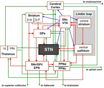
prominent control over the BG-related motor functions since it integrates the somatic motor information from various cor-tical/subcortical brain areas (including the motor cortex, tha-lamus and pedunculopontine nucleus) (Takada et al., 1988). Figure 1 illustrates the cortico-BG-thalamocortical circuit in-fluenced by dopamine (DA) neurons of substantia nigra pars compacta (SNc).
Understanding the mechanism of dopamine (DA) influence on the neural circuit involving the BG and STN is important for understanding disturbances in motor behavior and the de-velopment of possible therapies for PD. The functional role of DA is complex due to the heterogeneity of neurons within the STN and BG nuclei, presence of dopaminergic D1/D5 or D2/D3 receptors on diverse cell types and their terminals and opposite modulatory effects provoked by DA on different types of pre- and postsynaptic receptors (Sil’kis, 2005). Injection of DA or of D1 receptor agonists into the STN induced an increase in firing rate of the majority of STN neurons in both normal and 6-OHDA rats, but systemic administration of apomorphine (which is a non-selective DA agonist of both D1-like and D2-like receptors) provoked a decrease in the firing rate of STN neurons in rats with 6-OHDA lesions (Ni et al., 2001).
Investigation in rat and nonhuman primate models (Bergman et al., 1994; Vila et al., 2000; Tai et al., 2003; Breit et al., 2007) and human PD patients (Pogosyan et al., 2010) showed that the death of DA cells in the SNc may provoke changes of the
fir-to mofir-toneurons Cerebral Cortex GPe VL PfN Thalamus SNr/GPi/ EPN SNc VTA to brainstem to superior colliculus to habenula
ventral striatum ventral pallidum Limbic loop to spinal cord PPNd PPNc Striatum S-N S-P STN
Figure 1: GPe and GPi, external and internal segment of the globus pallidus; D1 and D2, dopaminergic receptors; S-N and S-P, striatonigral and striatopallidal cells; SNr and SNc, substantia nigra pars reticulata and compacta; STN, sub-thalamic nucleus; PfN, parafascicular sub-thalamic nucleus, PPNd and PPNc, pars dissipate and compacta of pedunculopontine nucleus; VL, ventral lateral nu-cleus; VTA, ventral tegmental area. Connections ending with an arrow (green) correspond to excitatory (Glu) projections; ending with a filled circle (red) correspond to inhibitory projections (GABAergic); ending with a filled dia-mond (blue) indicate the modulatory influence exerted by DA projections of SNc/VTA.
ing rate and firing pattern of STN neurons and characteristic changes in motor behavior. However, due to the complex or-ganization of BG and the variety of receptors it is difficult to determine the net effects produced by 6-OHDA or MPTP le-sion of DA cells in SNc. This is particularly true given the wide range of anesthetics used in acute studies cited in the literature. In this study, we investigated the changes in rodent STN neu-ronal activity using multielectrode recordings in the STN per-formed 28 days after 6-OHDA injection in the SNc. It is im-portant to mention that we performed our recordings under Eq-uithesin anesthesia. The choice of this drug is due to the fact that the other commonly used general anesthetics for rodents (mainly ketamine and urethane) have been reported to exert a strong influence upon the discharge pattern of extracellularly recorded units participating to the cortico-BG-thalamocortical circuit (Villa and Lorenzana, 1997; Ruskin et al., 2003; Hutchi-son et al., 2004; Chetrit et al., 2009).
We report that in our experimental condition the main effect of 6-OHDA lesion is a significant increase in the synchronous activity of STN cells, which we believe will provoke the main effect exerted by STN in PD models.
2. Materials and Methods
2.1. Subjects and surgical procedure
Experiments were performed in male Wistar rats weigh-ing 300 − 350 g. They were kept under controlled condi-tions of light, temperature and humidity, with food and
wa-ter available ad libitum. All animal experiments were car-ried out in accordance with the UK Animals (Scientific Pro-cedures) Act, 1986 and associated guidelines, the EEC Coun-cil Directives (86/609/EEC, OJ L 358, 1, 12 December, 1987) and the Guide for the Care and Use of Experimental Ani-mals (Canadian Council on Animal Care). Rats received at-ropine sulfate (0.4 mg/kg, ip) immediately prior to surgery as a prophylactic against respiratory distress. All surgical wounds were infiltrated with Scandicaine 0,5% (AstraZeneca) for lo-cal anesthesia. General anesthesia was induced by Equithesin (3ml/kg, i.p.) (chloral hydrate 4.24 mg, sodium pentobarbital 0.97 mg, and magnesium sulfate MgSO4 2.13 mg in 100 ml so-lution with 11% ethanol, 42% propylene glycole v/v) at a dose corresponding to 130 mg/kg chloral hydrate and 30 mg/kg pen-tobarbital (Preda et al., 2008). During the anesthesia the body temperature was monitored and maintained in the range 38– 39◦C by means of a heating pad.
2.2. 6-hydroxydopamine lesion of the SNc
The subjects (n=8) received one injection of 6µl of solution in the left substantia nigra pars compacta (SNc) aimed at target stereotaxic coordinates A 3.2; L +2.2; V 7.2 mm (from lambda and dura) (Paxinos and Watson, 1986). The injected solution contained 12µg 6-OHDA hydrobromide (Sigma, Paris, France) dissolved in 6µl of 0.9% NaCl with 0.1% ascorbic acid (Jouve et al., 2010). It is generally adviced to inject desipramine or pargyline prior to 6-OHDA administration to protect noradren-ergic neurons (Breese and Traylor, 1971) but this practice has been questioned (Debeir et al., 2005). There is a nonlinear dose dependent effect of 6-OHDA on noradrenergic neurons, and more recent work has shown that desipramine is not needed at the lower doses of 6-OHDA used in current study (Jouve et al., 2010). Notice that even in case of minor noradrenergic degener-ation following 6-OHDA injection, the animal model would be closer to human PD where same degeneration of catcholaminer-gic cells is likely to be associated to the patholocatcholaminer-gical state (Pil-lon et al., 1989; Mavridis et al., 1991; Delaville et al., 2011). For control subjects (n=8), an equal volume of vehicle (6µl saline with 0.1% ascorbic acid) was injected. The 6-OHDA solution were kept on ice (4◦C) and protected from light to
min-imize oxidation. The injection was made using a stainless steel cannula connected via a polyethylene catheter to a Hamilton microsyringe which was controlled by an infusion pump at a flow rate of 0.1µl/min. At the end of each injection, the syringe was kept in place for additional 5 minutes before being very slowly retracted from the brain, in no less than 5 more minutes.
2.3. Rotational behavior
Three weeks after the 6-OHDA-induced lesions were per-formed, the rats were screened for rotation response to sub-cutaneous injection of apomorphine (0.5 mg/kg, s.c., Sigma) (Casas et al., 1999). Five minutes after the apomorphine injec-tion DA-lesioned rats showed more than 20 rotainjec-tions per 5 min to the controlateral side. One subject did not match this cri-terion and was discarded from further study. It has previously 2
been demonstrated that rats satisfying this criterion are charac-terized by striatal DA depletion greater than 95% (Papa et al., 1994).
2.4. Electrophysiological recordings
General anesthesia was induced by Equithesin (3ml/kg, i.p.). The pedal withdrawal reflex was periodically checked and sup-plemental doses of Equithesin were provided during the whole recording session if necessary. Extracellular single unit record-ings in STN (3.6 - 4.2 mm posterior to bregma, L 2.3-3.7 mm, V 6.5-8.5 mm from the dura), (Paxinos and Watson, 1986) ipsi-lateral to the side of injection in SNc (in this study always the left hemisphere), were performed with glass-coated platinum-plated tungsten microelectrodes having an impedance in the range 0.5 − 2 MΩ measured at a frequency of 1 kHz (Fred-erick Haer & Co, Bowdoinham, Maine, USA). Signals from the microelectrodes were amplified, filtered (400 Hz − 20 kHz), viewed on an oscilloscope, and digitally recorded in WAV for-mat (44100 Hz sampling rate, 16 bit resolution) for computer-ized offline analysis. Spike sorting of the electrode signal files, based on a template matching algorithm, was able to separate up to three spikes recorded from the same channel (Asai et al., 2005). The time of spike discharges were digitally stored at a time resolution of 1 ms for later processing.
The first recording session started approximately 90 min af-ter the end of the surgical preparation. The data were gathered during spontaneous activity, i.e. in the absence of any operator-induced stimulation, for an interval of 5 − 10 min. All record-ings started at least 15 min after any supplementary dose of anesthetic and terminated at least 20 min before a new injec-tion, thus assuming the recording conditions corresponded to a steady level of anesthesia.
2.5. Histological and immunohistological examinations
Upon completion of the recording session (lasting in total 5 − 8 h), electrolytic lesions were placed at specific depths of the electrode track using 10 current pulses of 8 µA for 7 s at regular intervals of 10 seconds. Subjects were sacrificed by transcardial perfusion with 100 ml 0.9% NaCl immediately fol-lowed by 500 ml of fixative solution (4% paraformaldehyde in phosphate buffer 0.1M, pH 7.3). The brains were removed af-ter perfusion and cut into 40 µm sections using a microtome. Sections including SNc were examined for immunohistochem-ical staining of tyrosine hydroxylase (TH). In sections from 6-OHDA treated rats this analysis was aimed at determining the extent of the SNc lesion. Brain slices were immunoreacted with a primary, polyclonal antibody against rat tyrosine hydroxylase (TH, Abcam PLC, Cambridge, UK) and then with a biotiny-lated goat anti-rabbit IgG (Vector Labs, Burlingame, CA) sec-ondary antibody. The signal was amplified using avidin and biotinylated horseradish peroxidase using an Elite ABC Vectas-tain Kit (Vector, Burlingame, CA) (Park et al., 2007). Sections including STN and basal ganglia from all subjects were stained with cresyl violet for the reconstruction of the electrode tracks and localization of the electrolytic lesions.
2.6. Statistical analysis
Spike trains were analyzed by time series renewal density histograms (Abeles, 1982). Using this technique, all histograms were scaled in rate units (spikes/s) and smoothed after convo-lution with a moving Gaussian-shaped bin of 5 ms width. For each histogram, the 99% confidence limits were calculated, as-suming that spike occurrences followed a Poisson distribution. Firing rates of STN neurons of both groups of rats were com-pared by Mann-Whitney’s U-test because of the limited sample sizes we could not test properly the normality of the distribu-tions. A one-tailed Fisher’s exact t-test was used to compare the number of each cell type identified and the proportion of significant cross-correlations in both groups of rats.
3. Results
3.1. Effect of 6-OHDA-induced dopamine cell lesion
Consistent rotational response to the systemic injection of apomorphine (0.5 mg/kg) was observed in 7 out of 8 treated
a
*
SNc
b
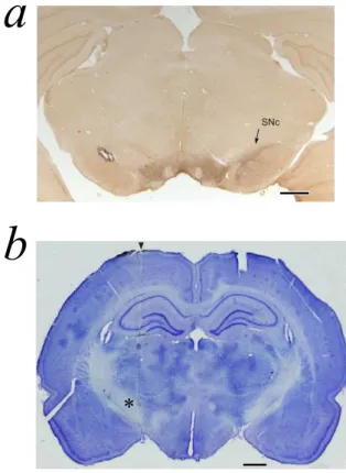
Figure 2: 6-OHDA lesion and reconstruction of the electrode track. Scale bars: 1 mm. (a) Coronal section stained for tyrosine hydroxylase (TH) showing the substantia nigra pars compacta (SNc) at interaural stereotaxic coordinate ap-proximately equal to Ia=2.9 (Paxinos and Watson, 1986). Note the lack of TH staining in the SNc of the lesioned hemisphere (left) compared to the non-lesioned hemisphere (right). (b) Coronal section, stained with cresyl violet, of the subthalamic nucleus of a control rat in the left hemisphere illustrating the microelectrode penetration at interaural stereotaxic coordinate approximately equal to Ia=5.6. The entrance of the electrode is indicated by arrow and an asterisk is placed to the left of the site of the electrolytic lesion made at the end of the session. A second lesion is visible dorsally in the lateral dorsal thalamic nucleus.
rats after 21 days from 6-OHDA injection. All rats that were characterized by a positive rotational behavior showed also a complete loss of TH-immunoreactive neurons restricted to the substantia nigra ipsilateral to the injected hemisphere, as exem-plified by the microphotographs in Figure 2a.
3.2. Electrophysiological recordings
Extracellular single units were recorded in 15 rats anes-thetized with Equithesin (7 lesioned and 8 control) . We per-formed a total of 25 electrode tracks (13 tracks in the control group and 12 tracks in the treated group). A total of 144 single units (n=61 in the control group and n=83 in the treated group), localized in STN, were characterized by stable firing rate and a stable autocorrelogram, i.e. no significant difference between the begin and the end of the recording session. An additional 94 single units were recorded in the same experiments, but ei-ther they did not match the above-mentioned criteria or their location was not in the STN after histological check. Figure 2b illustrates an example of an electrolytic lesion in STN (record-ing performed in the left hemisphere) and a second lesion per-formed in the lateral dorsal thalamic nucleus for measurement of tissue retraction necessary for an accurate reconstruction of the recording sites.
3.2.1. Properties of individual neurons
The analysis of the autocorrelograms allowed us to define 4 types of firing patterns of the spontaneous activity of STN cells. Units characterized by a significant refractory period were termed “initial prolonged through” (IPT) cells (Fig. 3a). Units that showed a tendency to fire in bursts (“bursting cells”, BC) were characterized by a hump in the autocorrelogram near time zero (Fig. 3b). Units that presented an autocorrelogram with dampened periodic oscillatory activity were termed “oscil-latory cells” (OSC) (Fig. 3c). Units with a flat autocorrelogram formed the “regular” (REG) type class (Fig. 3d).
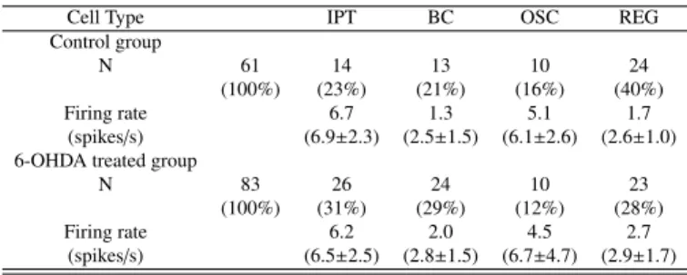
In the control group type REG units were the most abundant (40%, n=24) followed by the IPT (23%), BC (21%) and OSC (16%) which were almost equally represented. In 6-OHDA le-sioned rats the distribution of the four cell types (Table 1) was not statistically different from the controls (two sided Fischer’s exact test, not significant). However, we cannot rule out that
Table 1: Firing rates (median, mean±S.E.M.) of the STN units grouped by cell types in control and 6-OHDA treated rats recorded under general anesthesia induced by Equithesin.
Cell Type IPT BC OSC REG Control group
N 61 14 13 10 24
(100%) (23%) (21%) (16%) (40%) Firing rate 6.7 1.3 5.1 1.7
(spikes/s) (6.9±2.3) (2.5±1.5) (6.1±2.6) (2.6±1.0) 6-OHDA treated group
N 83 26 24 10 23 (100%) (31%) (29%) (12%) (28%) Firing rate 6.2 2.0 4.5 2.7 (spikes/s) (6.5±2.5) (2.8±1.5) (6.7±4.7) (2.9±1.7) 0 lag (ms) 400 F irin g ra te [sp ike s/ s] 0 40
a
b
0 lag (ms) 400 F irin g ra te [sp ike s/ s] 0 20 0 lag (ms) 400 F irin g ra te [sp ike s/ s] 0 20 IPT BCc
0 lag (ms) 400 F irin g ra te [sp ike s/ s] 0 20d
OSC REGFigure 3: Example of the four specific types of spontaneous firing pattern ob-served in STN. Autocorrelograms (auto renewal density histograms) with lag (ms) on the abscissa and rate (spike/s) on the ordinate. Abscissa is scaled to 400 ms for all plots. The histograms are smoothed by Gaussian shaped bin of 5 ms calculated according to Abeles (1982). The dashed lines correspond to 99% confidence limits. (a) Initial prolonged through cell (IPT), firing at 5.9 spikes/s; (b) Bursting cell (BC), firing at 1.9 spikes/s; (c) Oscillatory cell (OSC) with a period of 62 ms, firing at 7.0 spikes/s. (d) Regular cell (REG), firing at 1.7 spikes/s;
lack of statistical significance is due to the limited size of our samples; there may have been a trend for a decrease in the rel-ative proportion of REG cells and an increase of IPT and BC cells. The firing rate of IPT and OSC was significantly higher than that of BC and REG types, for both groups of rats (Mann Whitney’s U-test; U = 609, z = 2.07, p < 0.05 for the controls and U = 992.5, z = 3.02, p < 0.01 for the lesioned). However, the DA depletion did not alter the firing rate of either cell type (Mann-Whitney’s U-test, p > 0.05 for either cell type).
3.2.2. Interaction between pairs of cells
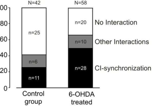
We could record overall 100 pairs of cells located in the STN, 42 in normal and 58 in 6-OHDA treated rats. All cell pairs were simultaneously recorded from the same electrode tip. The inter-action types were classified according to the shape of the cross-correlogram densities (CRD) calculated according to Abeles (1982). The independence of firing of a pair of units corre-sponded to a flat cross-correlogram with statistical fluctuations within the limits of significance (e.g., Fig. 4f). Correlograms with a symmetrical hump centered near zero delay, often re-ferred to as “common input” (CI), indicate that the pair of cells tended to discharge in synchrony (e.g., Fig. 4c).
The classes of correlograms showing significant deviations from flatness were more frequently (p < 0.05) observed in 6-OHDA treated rats (38/58, 66%) than in control subjects (17/42, 40%) (Fig. 5). Most of the significant interactions were of CI type, namely 11/42 (26%) in the control group and 28/58 (48%) in the 6-OHDA treated group. The other curves with significant deviations from flatness were grouped together in the “other” classes of interactions.
a
b
c
d
e
f
6-OHDA TREATED CONTROL GROUP autocorrelograms 0 400 lag (ms) F iri n g ra te [sp ike s/ s] 0 20 cell #1 -400 0 lag (ms) F iri n g ra te [sp ike s/ s] 0 25 400 crosscorrelogram cell #1→cell#2 0 400 lag (ms) F iri n g ra te [sp ike s/ s] 0 20 cell #2 autocorrelograms 0 400 lag (ms) F iri n g ra te [sp ike s/ s] 0 20 cell #1 -400 0 lag (ms) F iri n g ra te [sp ike s/ s] 0 20 400 crosscorrelogram cell #1→cell#2 0 400 lag (ms) F iri n g ra te [sp ike s/ s] 0 20 cell #2Figure 4: Autocorrelograms and crosscorrelogram of a cell pair recorded in a treated rat (panels a,b,c) and of a cell recorded in a control rat (panels d,e,f). The peak centered near time zero of panel (c) shows synchronous firing of the cells (a,b), referred to as “common input” (CI) type. The flat curve in panel (f) shows no-interaction between cells (d,e). The curves are smoothed by a Gaussian shaped bin of 5 ms. The firing rates of c#1 (a) and c#2 (b) are 1.6 and 1.9 spikes/s, respectively. The firing rates of c#1 (d) and c#2 (e) are 9.2 and 1.1 spikes/s, respectively. 0 100 Control group 6-OHDA treated No Interaction Other Interactions CI-synchronization n=25 n=20 n=6 n=10 n=11 n=28 80 60 40 20 N=58 N=42
Figure 5: Distribution of crosscorrelogram types (“No Interaction”, “Common Input-synchronization”, and “Other Interactions” referred to significant inter-actions other than synchronicity) between pairs of STN units recorded with the same electrode. The ordinate axis is in percentage. The raw numbers of correl-ograms are indicated by n and N.
4. Discussion
In the 6-OHDA treated rats of the present study we have demonstrated that the lack of DA does not significantly change the rate and type of firing patterns of STN neurons. We found that cells with initial through in their autocorrelograms (IPT) and cells with oscillatory activity (OSC) were characterized by a similar rate of firing (approximately equal to 6.6 spikes/s on average for control and treated rats) which was about twice higher than the rate of the other cell types (approximately equal to 2.7 spikes/s on average for control and treated rats). The cell types IPT and OSC represented together 39% and 43% of STN units in control and treated rats, respectively. Within this group, the lesion of SNc tended to increase the relative number of IPT firing pattern and to decrease the relative number of OSC. The observation about IPT and OSC might be associated with an increase in recurrent and possibly synchronous feedforward in-hibition provided by the loop STN-GPe-STN (Fig. 1) and by the facilitation of the inhibitory interneurons within the STN. The loop STN-GPi-STN can also support recurrent inhibition (Fig. 1).
The stronger STN firing the more effective would be the ex-citatory effect on the GPe and GPi and subsequent recurrent inhibition of STN neurons as suggested by administration of the D1/D2 agonist apomorphine (Ruskin et al., 2003; Chetrit et al., 2009). In this case the net effect on the firing rate would very small, if any, given an increase in the strength of inhibi-tion of STN neurons following a rise in their activity. In or-der to unor-derstand our results we must also consior-der the inter-dependency of the activity of STN and GPe given their strong interconnections. STN and GPe could constitute a kind of cen-tral pacemaker modulated by striatal inhibition of GPe neurons responsible for synchronized oscillatory activity in the normal and pathological BG (Plenz and Kital, 1999). DA denervation of the striatum and extrastriatal BG is likely to alter recurrent activity within this circuit thus leading to pathological activ-ity that resonates throughout the BG and motor system (Bevan et al., 2006). The activity of GPe is strongly dependent on
fir-ing of striatopallidal neurons which mainly express D2 recep-tors, and give rise to the indirect pathway through the BG. We assume that this pathway is the one that determines mainly the activity of STN neurons. In case of loss of DA in the striatum, the STN could be disinhibited via the indirect pathway due to stronger activity of striatopallidal cells and suppression of fir-ing of the GABAergic GPe cells projectfir-ing to the STN (Sil’kis, 2005). This hypothesis is supported by the fact that a high DA concentration is necessary for the activation of D2 receptors on striatopallidal cells (Williams and Millar, 1990). which in turn would affect STN activity according to the intensity of DA cell lesion.
Our results are partially in disagreement with previous stud-ies that reported an increase in the firing rate of STN neurons with more irregular and bursty-firing patterns of STN neurons as well as significant decrease in relative amount of tonic neu-rons (Breit et al., 2007). In addition, many existing studies show contrasting tendencies to increasing or to decreasing fir-ing rate in STN after lesion of the SNc (Hollerman and Grace, 1992; Hassani et al., 1996; Vila et al., 2000; Ni et al., 2001; Magill et al., 2001). We observed a decrease in the relative number of OSC cell types, in contrast with an increase in the incidence of slow oscillations (0.3-2.5 Hz) reported by Walters et al. (2007) after dopamine cell lesion in urethane-anesthetized rats. A possible source of discrepancy with previous studies may arise from the use of different anesthesia. We avoided the use of ketamine because we had previously observed that this non-competitive antagonist of NMDA receptors produces a massive change of firing patterns in the SNr of control animals (Villa and Lorenzana, 1997). We have also avoided the use of urethane because of urethane-induced slow wave cortical activ-ity that alters significantly the analysis of correlations between spike trains (Manns et al., 2000a,b; Jones, 2004; Kasanetz et al., 2008). Our finding that IPT and regular spiking neurons were the commonest types of firing under Equithesin anesthesia ap-pears in agreement with recordings performed in the STN of unanesthetized 6-OHDA treated rats that shown also a decrease in the number of oscillatory units (Kreiss et al., 1997; Allers et al., 2000). This indicates that the active metabolites of Eq-uithesin are unlikely to exert a strong influence upon the dis-charge patterns of STN units. Accordingly, Equithesin anesthe-sia could be viewed as a good control condition for studies in anesthetized rats.
The main finding of this study was that in the 6-OHDA treated rats pairs of simultaneously recorded adjacent STN units fired significantly more often in synchrony. A significant in-crease in cross-correlations between STN neurons has also been observed following a lesion of GPe without 6-OHDA treatment (Ryan et al., 1992). This result is comparable with ours because a lesion of GPe decreases STN inhibition, and DA deficiency leads to STN disinhibition via the indirect pathway through the BG (Fig. 1) . In the rat the neurons of STN are charac-terized by an important dendritic field around the cell bodies, thus suggesting that excitatory recurrent collaterals could rep-resent the element of circuitry subserving the synchronization of closely spatially related neurons (Hammond et al., 1983). Strong synchronization in STN cell firing could also modify
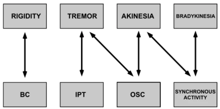
BRADYKINESIA AKINESIA TREMOR RIGIDITY SYNCHRONOUS ACTIVITY OSC IPT BC
Figure 6: Dependencies between human symptoms and firing pattern parame-ters. Adapted from Chibirova (2006).
propagation of signals from the neocortex to the output BG nu-clei. It is known that hyperdirect cortico-subthalamic pathway may funnel the information whereas the indirect cortico-striato-subthalamic pathway may process the information through par-allel loops (Parent et al., 2000). Indeed, excitatory responses to stimulation of diverse cortical areas were found in distinct stri-atal territories, and none of the cells responded to two cortical stimulation sites (Kolomiets et al., 2001). In contrast, the ex-istence of specific patterns of convergence of information flow from functionally distinct cortical areas in the STN allows in-teractions between parallel channels (Kolomiets et al., 2003; Bar-Gad et al., 2003).
It is widely believed that Parkinsonian symptoms are most likely caused by abnormal synchronous oscillating neuronal ac-tivity within the BG (Bergman and Deuschl, 2002; Avila et al., 2010). Akinesia and hypokinesia could be caused by abnor-mal hypoactivity of the GPe neurons and subsequent exces-sive inhibition of BG targets (Wichmann and DeLong, 1996). Recordings in human STN of PD patients during DBS neuro-surgery (Chibirova, 2006) showed that the same firing patterns observed in our study (i.e., IPT, BC, OSC and REG represented 33%, 19%, 15%, 33%, respectively) in a proportion similar to the one in 6-OHDA treated rats (Table1). Chibirova (2006) demonstrated that the clinical symptoms were associated to spe-cific features in the firing pattern observed in the STN cells of PD patients (Fig. 6). Motor impairments typically associated to clinical symptoms in human PD patients have been observed in rodents treated with MPTP or 6-OHDA. A complete lesion of SNc by 6-OHDA injection impaired dramatically (by 80%) the stepping of the contralateral paw (Barn´eoud et al., 2000). An-imals with more than 95% bilateral SNc lesion demonstrated bradykinesia symptoms (Sakai and Gash, 1994) and ≈75% DA cells loss produced forelimb akinesia manifested in stepping deficit (Tseng et al., 2005). It is important to note that akinesia induced by 6-OHDA could be restored to normal locomotion by DBS of the STN, which caused a decrease in firing rate and burst firing in the contralateral side of STN (Shi et al., 2006) and stepping performance in MPTP-treated mice was significantly improved by administration of L-DOPA (Blume et al., 2009). Animal models of PD can never reproduce the entire range of symptoms observed in human patients but they have proven to be valuable for the investigation of the subsequent mechanisms. 6
It is interesting to compare our results, which emphasize the increase in STN synchronous activity in 6-OHDA treated rats, with those of Chibirova (2006) who found a significant asso-ciation between synchronous firing in the STN of PD patients with akinesia and bradykinesia but not with tremor and rigid-ity (Figure 6). Given the complexrigid-ity of the physiopathologi-cal mechanisms which generate parkinsonian symptomatology the assumption that each class of clinical signs (tremor, akine-sia, rigidity, dyskineakine-sia, bradykinesia) is produced by a specific and separate mechanism was questioned (Gross et al., 1999). We support the hypothesis that synchronous activity of STN neurons is correlated with akinesia because it could promote increase in SNr cell firing due to simultaneous glutamate re-lease from numerous STN terminals in the SNr and its high concentration in synaptic cleft must promote increase in SNr cell firing. Increased glutamatergic innervation of the SNr is known to contribute to the motor deficits in PD, whereas sup-pression of glutamate release by injection of agonists of group III mGlu receptors into the SNr exerts functional protection against 6-OHDA lesion (Austin et al., 2010). The same proce-dure reversed reserpine-induced akinesia (Austin et al., 2010) indirectly suggesting the important role of glutamate concen-tration in the SNr for akinesia. It has been proposed that the normal dopaminergic system supports segregation of the func-tional subcircuits of the BG (Bergman et al., 1998) and that the BG-thalamocortical loop dynamically modulates the degree of correlation of neuronal activity in order to select the proper motor behavior (Yasoshima et al., 2005; Gale et al., 2009). We suggest that dopaminergic input is necessary to maintain a bal-ance between funneling and parallel processings, and changes in DA concentration exert modulatory role upon switching be-tween one to the other mode of transmission.
In conclusion, the finding of increased synchronicity between spatially adjacent STN neurons in 6-OHDA treated rats and the careful analysis of the cell types defined by their firing patterns offer a new insight to the mechanisms of motor disturbances in PD.
Acknowledgments
The Authors wish to thank Marguerite Kaczorowski for her technical assistance throughout the histological procedures and the reviewers whose comments have contributed to improve the clarity of the paper. We thank A.L. Benabid, S. Chabardes, T. Aksenova, O.K. Chibirova and the neurosurgery and neurology teams of the C.H.U. Grenoble for giving access to the results gathered in human PD patients.
References
Abeles, M., 1982. Quantification, smoothing, and confidence limits for single-units’ histograms. J Neurosci Methods 5, 317–325.
Allers, K.A., Kreiss, D.S., Walters, J.R., 2000. Multisecond oscillations in the subthalamic nucleus: effects of apomorphine and dopamine cell lesion. Synapse 38, 38–50.
Asai, Y., Aksenova, T., Villa, A.E.P., 2005. On-line real-time oriented applica-tion for neuronal spike sorting with unsupervised learning. Lecture Notes in Computer Science 3696, 109–114.
Austin, P.J., Betts, M.J., Broadstock, M., O’Neill, M.J., Mitchell, S.N., Duty, S., 2010. Symptomatic and neuroprotective effects following activation of nigral group iii metabotropic glutamate receptors in rodent models of parkin-son’s disease. Br J Pharmacol 160, 1741–1753.
Avila, I., Parr-Brownlie, L.C., Brazhnik, E., Casta˜neda, E., Bergstrom, D.A., Walters, J.R., 2010. Beta frequency synchronization in basal ganglia output during rest and walk in a hemiparkinsonian rat. Exp Neurol 221, 307–319. Bar-Gad, I., Heimer, G., Ritov, Y., Bergman, H., 2003. Functional correlations
between neighboring neurons in the primate globus pallidus are weak or nonexistent. J Neurosci 23, 4012–4016.
Barn´eoud, P., Descombris, E., Aubin, N., Abrous, D.N., 2000. Evaluation of simple and complex sensorimotor behaviours in rats with a partial lesion of the dopaminergic nigrostriatal system. Eur J Neurosci 12, 322–336. Benabid, A.L., Pollak, P., Gross, C., Hoffmann, D., Benazzouz, A., Gao, D.M.,
Laurent, A., Gentil, M., Perret, J., 1994. Acute and long-term effects of subthalamic nucleus stimulation in parkinson’s disease. Stereotact Funct Neurosurg 62, 76–84.
Bergman, H., Deuschl, G., 2002. Pathophysiology of parkinson’s disease: from clinical neurology to basic neuroscience and back. Mov Disord 17 Suppl 3, 28–40.
Bergman, H., Feingold, A., Nini, A., Raz, A., Slovin, H., Abeles, M., Vaa-dia, E., 1998. Physiological aspects of information processing in the basal ganglia of normal and parkinsonian primates. Trends Neurosci 21, 32–38. Bergman, H., Wichmann, T., Karmon, B., DeLong, M.R., 1994. The primate
subthalamic nucleus. ii. neuronal activity in the mptp model of parkinson-ism. J Neurophysiol 72, 507–520.
Bevan, M.D., Atherton, J.F., Baufreton, J., 2006. Cellular principles underly-ing normal and pathological activity in the subthalamic nucleus. Curr Opin Neurobiol 16, 621–628.
Blume, S.R., Cass, D.K., Tseng, K.Y., 2009. Stepping test in mice: a reliable approach in determining forelimb akinesia in mptp-induced parkinsonism. Exp Neurol 219, 208–211.
Breese, G.R., Traylor, T.D., 1971. Depletion of brain noradrenaline and dopamine by 6-hydroxydopamine. Br J Pharmacol 42, 88–99.
Breit, S., Bouali-Benazzouz, R., Popa, R.C., Gasser, T., Benabid, A.L., Be-nazzouz, A., 2007. Effects of 6-hydroxydopamine-induced severe or par-tial lesion of the nigrostriatal pathway on the neuronal activity of pallido-subthalamic network in the rat. Exp Neurol 205, 36–47.
Casas, M., Prat, G., Robledo, P., Jan´e, F., 1999. Rotational behavior in dopamine nigrostriatal denervated rats: effects of a wide range of time inter-vals between apomorphine administrations. Pharmacol Biochem Behav 62, 481–485.
Chetrit, J., Ballion, B., Laquitaine, S., Belujon, P., Morin, S., Taupignon, A., Bioulac, B., Gross, C.E., Benazzouz, A., 2009. Involvement of basal ganglia network in motor disabilities induced by typical antipsychotics. PLoS One 4, 1–14.
Chibirova, O., 2006. Interactions fonctionnelles dans les ganglions de la base ´etudi´ees par l’enregistrement simultan´e des activit´es unitaires discrimin´ees par un algorithme non supervis´e de tri de potentiels d’action. Ph.D. thesis. Universit´e Joseph-Fourier - Grenoble I. http://tel.archives-ouvertes.fr/tel-00115720/en/.
Debeir, T., Ginestet, L., Franc¸ois, C., Laurens, S., Martel, J.C., Chopin, P., Marien, M., Colpaert, F., Raisman-Vozari, R., 2005. Effect of intrastriatal 6-OHDA lesion on dopaminergic innervation of the rat cortex and globus pallidus. Exp Neurol 193, 444–454.
Delaville, C., Deurwaerd`ere, P.D., Benazzouz, A., 2011. Noradrenaline and parkinson’s disease. Front Syst Neurosci 5, 31–31.
DeLong, M.R., Wichmann, T., 2007. Circuits and circuit disorders of the basal ganglia. Arch Neurol 64, 20–24.
Gale, J.T., Shields, D.C., Jain, F.A., Amirnovin, R., Eskandar, E.N., 2009. Sub-thalamic nucleus discharge patterns during movement in the normal monkey and parkinsonian patient. Brain Res 1260, 15–23.
Gross, C.E., Boraud, T., Guehl, D., Bioulac, B., Bezard, E., 1999. From exper-imentation to the surgical treatment of parkinson’s disease: prelude or suite in basal ganglia research? Prog Neurobiol 59, 509–532.
Hammond, C., Rouzaire-Dubois, B., F´eger, J., Jackson, A., Crossman, A.R., 1983. Anatomical and electrophysiological studies on the reciprocal projec-tions between the subthalamic nucleus and nucleus tegmenti pedunculopon-tinus in the rat. Neuroscience 9, 41–52.
Hassani, O.K., Mouroux, M., F´eger, J., 1996. Increased subthalamic neuronal activity after nigral dopaminergic lesion independent of disinhibition via the
globus pallidus. Neuroscience 72, 105–115.
Hollerman, J.R., Grace, A.A., 1992. Subthalamic nucleus cell firing in the 6-ohda-treated rat: basal activity and response to haloperidol. Brain Res 590, 291–299.
Hutchison, W.D., Dostrovsky, J.O., Walters, J.R., Courtemanche, R., Boraud, T., Goldberg, J., Brown, P., 2004. Neuronal oscillations in the basal ganglia and movement disorders: evidence from whole animal and human record-ings. J Neurosci 24, 9240–9243.
Jones, B.E., 2004. Activity, modulation and role of basal forebrain cholinergic neurons innervating the cerebral cortex. Prog Brain Res 145, 157–169. Jouve, L., Salin, P., Melon, C., Kerkerian-Le Goff, L., 2010. Deep brain
stimu-lation of the center median-parafascicular complex of the thalamus has effi-cient anti-parkinsonian action associated with widespread cellular responses in the basal ganglia network in a rat model of parkinson’s disease. J Neurosci 30, 9919–9928.
Kasanetz, F., Riquelme, L.A., Della-Maggiore, V., O’Donnell, P., Murer, M.G., 2008. Functional integration across a gradient of corticostriatal channels controls up state transitions in the dorsal striatum. Proc Natl Acad Sci U S A 105, 8124–8129.
Kolomiets, B.P., Deniau, J.M., Glowinski, J., Thierry, A.M., 2003. Basal gan-glia and processing of cortical information: functional interactions between trans-striatal and trans-subthalamic circuits in the substantia nigra pars retic-ulata. Neuroscience 117, 931–938.
Kolomiets, B.P., Deniau, J.M., Mailly, P., M´en´etrey, A., Glowinski, J., Thierry, A.M., 2001. Segregation and convergence of information flow through the cortico-subthalamic pathways. J Neurosci 21, 5764–5772.
Kreiss, D.S., Mastropietro, C.W., Rawji, S.S., Walters, J.R., 1997. The response of subthalamic nucleus neurons to dopamine receptor stimulation in a rodent model of parkinson’s disease. J Neurosci 17, 6807–6819.
Li, X.H., Wang, J.Y., Gao, G., Chang, J.Y., Woodward, D.J., Luo, F., 2010. High-frequency stimulation of the subthalamic nucleus restores neural and behavioral functions during reaction time task in a rat model of parkinson’s disease. J Neurosci Res 88, 1510–1521.
Limousin, P., Krack, P., Pollak, P., Benazzouz, A., Ardouin, C., Hoffmann, D., Benabid, A.L., 1998. Electrical stimulation of the subthalamic nucleus in advanced parkinson’s disease. N Engl J Med 339, 1105–1111.
Magill, P.J., Bolam, J.P., Bevan, M.D., 2001. Dopamine regulates the impact of the cerebral cortex on the subthalamic nucleus-globus pallidus network. Neuroscience 106, 313–330.
Manns, I.D., Alonso, A., Jones, B.E., 2000a. Discharge profiles of juxtacellu-larly labeled and immunohistochemically identified gabaergic basal fore-brain neurons recorded in association with the electroencephalogram in anesthetized rats. J Neurosci 20, 9252–9263.
Manns, I.D., Alonso, A., Jones, B.E., 2000b. Discharge properties of juxta-cellularly labeled and immunohistochemically identified cholinergic basal forebrain neurons recorded in association with the electroencephalogram in anesthetized rats. J Neurosci 20, 1505–1518.
Mavridis, M., Colpaert, F.C., Millan, M.J., 1991. Differential modulation of (+)-amphetamine-induced rotation in unilateral substantia nigra-lesioned rats by alpha 1 as compared to alpha 2 agonists and antagonists. Brain Res 562, 216–224.
Ni, Z.G., Bouali-Benazzouz, R., Gao, D.M., Benabid, A.L., Benazzouz, A., 2001. Time-course of changes in firing rates and firing patterns of subtha-lamic nucleus neuronal activity after 6-ohda-induced dopamine depletion in rats. Brain Res 899, 142–147.
Papa, S.M., Engber, T.M., Kask, A.M., Chase, T.N., 1994. Motor fluctuations in levodopa treated parkinsonian rats: relation to lesion extent and treatment duration. Brain Res 662, 69–74.
Parent, A., Hazrati, L.N., 1995a. Functional anatomy of the basal ganglia. i. the cortico-basal ganglia-thalamo-cortical loop. Brain Res Brain Res Rev 20, 91–127.
Parent, A., Hazrati, L.N., 1995b. Functional anatomy of the basal ganglia. ii. the place of subthalamic nucleus and external pallidum in basal ganglia circuitry. Brain Res Brain Res Rev 20, 128–154.
Parent, A., Sato, F., Wu, Y., Gauthier, J., L´evesque, M., Parent, M., 2000. Or-ganization of the basal ganglia: the importance of axonal collateralization. Trends Neurosci 23, 20–27.
Park, Y.S., Jeon, M.F., Lee, B.H., Chang, J.W., 2007. Lesion of subthalamic nucleus in parkinsonian rats : effects of dopamine d(1) and d(2) receptor agonists on the neuronal activities of the substantia nigra pars reticulata. J Korean Neurosurg Soc 42, 455–461.
Paxinos, G., Watson, C., 1986. The Rat Brain in Stereotaxic Coordinates. Aca-demic Press, Sydney and Orlando. 2nd edition.
Pillon, B., Dubois, B., Bonnet, A.M., Esteguy, M., Guimaraes, J., Vigouret, J.M., Lhermitte, F., Agid, Y., 1989. Cognitive slowing in parkinson’s disease fails to respond to levodopa treatment: the 15-objects test. Neurology 39, 762–768.
Plenz, D., Kital, S.T., 1999. A basal ganglia pacemaker formed by the subtha-lamic nucleus and external globus pallidus. Nature 400, 677–682. Pogosyan, A., Yoshida, F., Chen, C.C., Martinez-Torres, I., Foltynie, T.,
Limousin, P., Zrinzo, L., Hariz, M.I., Brown, P., 2010. Parkinsonian im-pairment correlates with spatially extensive subthalamic oscillatory synchro-nization. Neuroscience 171, 245–257.
Preda, S., Govoni, S., Lanni, C., Racchi, M., Mura, E., Grilli, M., Marchi, M., 2008. Acute beta-amyloid administration disrupts the cholinergic control of dopamine release in the nucleus accumbens. Neuropsychopharmacology 33, 1062–1070.
Ruskin, D.N., Bergstrom, D.A., Tierney, P.L., Walters, J.R., 2003. Correlated multisecond oscillations in firing rate in the basal ganglia: modulation by dopamine and the subthalamic nucleus. Neuroscience 117, 427–438. Ryan, L.J., Sanders, D.J., Clark, K.B., 1992. Auto- and cross-correlation
analysis of subthalamic nucleus neuronal activity in neostriatal- and globus pallidal-lesioned rats. Brain Res 583, 253–261.
Sakai, K., Gash, D.M., 1994. Effect of bilateral 6-ohda lesions of the substantia nigra on locomotor activity in the rat. Brain Res 633, 144–150.
Shi, L.H., Luo, F., Woodward, D.J., Chang, J.Y., 2006. Basal ganglia neural responses during behaviorally effective deep brain stimulation of the sub-thalamic nucleus in rats performing a treadmill locomotion test. Synapse 59, 445–457.
Sil’kis, I.G., 2005. Possible mechanisms of involvement of the subthalamic nu-cleus and structures interconnected with this nunu-cleus in movement disorders evoked by dopamine depletion. Usp Fiziol Nauk 36, 66–83.
Tai, C.H., Boraud, T., Bezard, E., Bioulac, B., Gross, C., Benazzouz, A., 2003. Electrophysiological and metabolic evidence that high-frequency stimula-tion of the subthalamic nucleus bridles neuronal activity in the subthalamic nucleus and the substantia nigra reticulata. FASEB J 17, 1820–1830. Takada, M., Nishihama, M.S., Nishihama, C.C., Hattori, T., 1988. Two
sepa-rate neuronal populations of the rat subthalamic nucleus project to the basal ganglia and pedunculopontine tegmental region. Brain Res 442, 72–80. Tseng, K.Y., Kargieman, L., Gacio, S., Riquelme, L.A., Murer, M.G., 2005.
Consequences of partial and severe dopaminergic lesion on basal ganglia oscillatory activity and akinesia. Eur J Neurosci 22, 2579–2586.
Utter, A.A., Basso, M.A., 2008. The basal ganglia: an overview of circuits and function. Neurosci Biobehav Rev 32, 333–342.
Vila, M., P´erier, C., F´eger, J., Yelnik, J., Faucheux, B., Ruberg, M., Raisman-Vozari, R., Agid, Y., Hirsch, E.C., 2000. Evolution of changes in neuronal activity in the subthalamic nucleus of rats with unilateral lesion of the sub-stantia nigra assessed by metabolic and electrophysiological measurements. Eur J Neurosci 12, 337–344.
Villa, A.E.P., Lorenzana, V.M.B., 1997. Ketamine modulation of the temporal pattern of discharges and spike train interactions in the rat substantia nigra pars reticulata. Brain Research Bulletin 43, 525 – 535.
Walters, J.R., Hu, D., Itoga, C.A., Parr-Brownlie, L.C., Bergstrom, D.A., 2007. Phase relationships support a role for coordinated activity in the indirect pathway in organizing slow oscillations in basal ganglia output after loss of dopamine. Neuroscience 144, 762–776.
Wichmann, T., DeLong, M.R., 1996. Functional and pathophysiological mod-els of the basal ganglia. Curr Opin Neurobiol 6, 751–758.
Williams, G.V., Millar, J., 1990. Concentration-dependent actions of stimulated dopamine release on neuronal activity in rat striatum. Neuroscience 39, 1– 16.
Yasoshima, Y., Kai, N., Yoshida, S., Shiosaka, S., Koyama, Y., Kayama, Y., Kobayashi, K., 2005. Subthalamic neurons coordinate basal ganglia func-tion through differential neural pathways. J Neurosci 25, 7743–7753.