HAL Id: hal-02380944
https://hal.archives-ouvertes.fr/hal-02380944
Submitted on 6 Oct 2020
HAL is a multi-disciplinary open access
archive for the deposit and dissemination of
sci-entific research documents, whether they are
pub-lished or not. The documents may come from
teaching and research institutions in France or
abroad, or from public or private research centers.
L’archive ouverte pluridisciplinaire HAL, est
destinée au dépôt et à la diffusion de documents
scientifiques de niveau recherche, publiés ou non,
émanant des établissements d’enseignement et de
recherche français ou étrangers, des laboratoires
publics ou privés.
Distributed under a Creative Commons Attribution| 4.0 International License
system for bacterial competition
Helena Spiewak, Sravanthi Shastri, Lili Zhang, Stephan Schwager, Leo Eberl,
Annette Vergunst, Mark Thomas
To cite this version:
Helena Spiewak, Sravanthi Shastri, Lili Zhang, Stephan Schwager, Leo Eberl, et al.. Burkholderia
cenocepacia utilizes a type VI secretion system for bacterial competition. MicrobiologyOpen, Wiley,
2019, 8 (7), �10.1002/mbo3.774�. �hal-02380944�
MicrobiologyOpen. 2019;8:e774.
|
1 of 26 https://doi.org/10.1002/mbo3.774www.MicrobiologyOpen.com
Received: 21 August 2018
|
Revised: 7 November 2018|
Accepted: 7 November 2018DOI: 10.1002/mbo3.774 O R I G I N A L A R T I C L E
Burkholderia cenocepacia utilizes a type VI secretion system for
bacterial competition
Helena L. Spiewak
1| Sravanthi Shastri
1| Lili Zhang
2| Stephan Schwager
3|
Leo Eberl
3| Annette C. Vergunst
2| Mark S. Thomas
1This is an open access article under the terms of the Creative Commons Attribution License, which permits use, distribution and reproduction in any medium, provided the original work is properly cited.
© 2019 The Authors. MicrobiologyOpen published by John Wiley & Sons Ltd.
1Department of Infection, Immunity and
Cardiovascular Disease, The Medical School, The University of Sheffield, Sheffield, UK
2VBMI, INSERM, Université de Montpellier,
Nîmes, France
3Department of Plant and Microbial
Biology, University of Zurich, Zurich, Switzerland
Correspondence
Mark S. Thomas, Department of Infection, Immunity and Cardiovascular Disease, The Medical School, The University of Sheffield, Sheffield, UK. Email: m.s.thomas@sheffield.ac.uk Present Address Helena L. Spiewak, Northern Genetics Service, The Newcastle upon Tyne Hospitals NHS Foundation Trust, Institute of Genetic Medicine, International Centre for Life, Newcastle upon Tyne, UK
Lili Zhang, Section of Molecular Biology, Division of Biological Sciences, University of California, San Diego, La Jolla, California Stephan Schwager, Analytical
Chemistry, Synthes GmbH, Oberdorf BL, Switzerland Funding information Schweizerischer Nationalfonds zur Förderung der Wissenschaftlichen Forschung, Grant/Award Number: Project 31003A_169307; Biotechnology and Biological Sciences Research Council, Grant/ Award Number: BB/F016840/1 and BB/ L024209/1
Abstract
Burkholderia cenocepacia is an opportunistic bacterial pathogen that poses a signifi‐
cant threat to individuals with cystic fibrosis by provoking a strong inflammatory re‐ sponse within the lung. It possesses a type VI secretion system (T6SS), a secretory apparatus that can perforate the cellular membrane of other bacterial species and/or eukaryotic targets, to deliver an arsenal of effector proteins. The B. cenocepacia T6SS (T6SS‐1) has been shown to be implicated in virulence in rats and contributes toward actin rearrangements and inflammasome activation in B. cenocepacia‐infected mac‐ rophages. Here, we present bioinformatics evidence to suggest that T6SS‐1 is the archetype T6SS in the Burkholderia genus. We show that B. cenocepacia T6SS‐1 is active under normal laboratory growth conditions and displays antibacterial activity against other Gram‐negative bacterial species. Moreover, B. cenocepacia T6SS‐1 is not required for virulence in three eukaryotic infection models. Bioinformatics analy‐ sis identified several candidate T6SS‐dependent effectors that may play a role in the antibacterial activity of B. cenocepacia T6SS‐1. We conclude that B. cenocepacia T6SS‐1 plays an important role in bacterial competition for this organism, and prob‐ ably in all Burkholderia species that possess this system, thereby broadening the range of species that utilize the T6SS for this purpose.
K E Y W O R D S
antibacterial, bacterial competition, Burkholderia, protein secretion, T6SS, type VI secretion system
1 | INTRODUCTION
Bacteria utilize many systems to establish a niche, including mech‐ anisms to exploit eukaryotic organisms and/or to compete effec‐ tively with other bacterial species colonizing the same ecosystem. Many Gram‐negative bacteria possess a protein secretion sys‐ tem termed the type VI secretion system (T6SS) that participates in one or both processes, depending on the species (Ho, Dong, & Mekalanos, 2014). The T6SS is found in ~25% of Gram‐nega‐ tive species (Bingle, Bailey, & Pallen, 2008), including the human pathogens Pseudomonas aeruginosa (Mougous et al., 2006), Vibrio
cholerae (Pukatzki et al., 2006), Serratia marcescens (Murdoch et
al., 2011), and Burkholderia pseudomallei (Burtnick et al., 2011). The system is composed of multiple copies of at least thirteen different subunits (TssA‐TssM) and a single copy of the PAAR protein, which are organized into a dynamic protein injection machine containing two distinct interacting subassemblies (Basler, 2015). The first is a contractile structure that shares homology with components of the T4 bacteriophage tail and is comprised of multimers of TssD (also termed Hcp) that assemble into a tube that is sharpened at one end by a trimer of TssI (also known as VgrG) subunits capped by a monomer of the PAAR protein. The tube, in turn, is surrounded by a contractile sheath composed of polymerized TssBC subunits. The tube–sheath structure is assembled on a platform known as the baseplate that consists of the TssEFGK subunits (Brackmann, Nazarov, Wang, & Basler, 2017; Brunet, Zoued, Boyer, Douzi, & Cascales, 2015; Leiman et al., 2009; Nguyen et al., 2017). The sec‐ ond subassembly, composed of the TssJLM subunits, is a channel/ chamber complex that spans the inner membrane, periplasm, and outer membrane, and serves to anchor the contractile machinery to the bacterial cell envelope (Brunet et al., 2015; Durand et al., 2015; Nguyen et al., 2017). The role of the TssA subunit is less certain, but it has been proposed to play roles in priming and po‐ lymerization of the tube–sheath structure or act as a baseplate component (Planamente et al., 2016; Zoued et al., 2016).
Contraction of the sheath against the baseplate drives the sharpened inner tube through the chamber complex to the exterior where it punctures the cellular membrane of a neighboring target cell. Effector proteins, which may be noncovalently associated with the TssD, TssI, or PAAR subunits (“cargo” effectors) or occur as ad‐ ditional domains on these proteins (“specialized” effectors), are thus delivered into the target cell where they kill or subvert the recipient (Durand, Cambillau, Cascales, & Journet, 2014). In many T6SS‐con‐ taining bacteria, these targets are other competing species of bac‐ teria, and so the system plays a major role in bacterial competition (Diniz & Coulthurst, 2015; Hood et al., 2010; MacIntyre, Miyata, Kitaoka, & Pukatzki, 2010; Schwarz et al., 2010). Such T6SS‐depen‐ dent competition can occur in a variety of environments, including plant hosts (Ma, Hachani, Lin, Filloux, & Lai, 2014) or the mamma‐ lian gut (Chassaing & Cascales, 2018; Sana et al., 2016; Zhao, Caro, Robins, & Mekalanos, 2018). Some T6SSs also specifically target eu‐ karyotic cells and have more of a direct role in virulence, including the T6SS‐5 of B. pseudomallei and H2‐ and H3‐T6SS of P. aeruginosa
(Burtnick et al., 2011; Jiang, Waterfield, Yang, Yang, & Jin, 2014; Sana et al., 2012).
A variety of T6SS‐dependent effectors and cognate immunity proteins have now been described, including superfamilies of an‐ tibacterial effectors. These include effectors that target the pepti‐ doglycan layer, phospholipid membrane, or host DNA/RNA, such as the amidase effector–immunity pairs termed Tae‐Tai (for type VI amidase effector/immunity; Hood et al., 2010; Russell et al., 2011; Russell et al., 2012; Fritsch et al., 2013), the type VI lipase effec‐ tors (Tle) that possess phospholipase A1, A2, or D activity (Russell et al., 2013), or the type VI DNase effectors (Tde; Ma et al., 2014), respectively. A number of anti‐eukaryotic effectors have also been described, including a P. aeruginosa effector with phospholipase D activity that can target both bacterial and eukaryotic cells (Jiang et al., 2014), the catalase effector, KatN, responsible for intramacro‐ phage survival of enterohemorrhagic E. coli (Wan et al., 2017), and a VgrG subunit with a C‐terminal actin cross‐linking domain utilized by
V. cholerae (VgrG‐1) that impairs the phagocytic activity of eukary‐
otic host cells (Ma, McAuley, Pukatzki, & Mekalanos, 2009; Pukatzki, Ma, Revel, Sturtevant, & Mekalanos, 2007).
The genus Burkholderia constitutes a large and diverse group of Gram‐negative bacterial species, including primary and opportunis‐ tic human pathogens, plant pathogens, and plant‐associated species with biocontrol properties (Eberl & Vandamme, 2016). Recently, the classification of the Burkholderia has undergone a proposed re‐ vision, with all members of the Burkholderia cepacia complex (Bcc) and Pseudomallei groups, together with some phytopathogenic spe‐ cies, remaining as Burkholderia, while all the other species (typically nonpathogenic environmental strains) have been reassigned to the new genera Paraburkholderia (Sawana, Adeolu, & Gupta, 2014) and
Caballeronia (Dobritsa, Linardopoulou, & Samadpour, 2017). The Bcc
is a group of at least twenty closely related species that have gained notoriety as opportunistic respiratory pathogens in cystic fibrosis (CF) patients, as some strains are highly transmissible between indi‐ viduals and the resulting infections can be difficult to treat effectively and result in fatal pneumonia and septicemia (Depoorter et al., 2016; Drevinek & Mahenthiralingam, 2010). One of the most prevalent Bcc species in CF infections is B. cenocepacia. However, despite many studies investigating the virulence mechanisms of this bacterium, the molecular pathogenesis of B. cenocepacia infection is not fully understood. Numerous strategies have been proposed to account for its virulence, including its ability to invade and survive intracel‐ lularly within host cells (Burns et al., 1996; Cieri, Mayer‐Hamblett, Griffith, & Burns, 2002; Gavrilin et al., 2012; Martin & Mohr, 2000; McKeon, McClean, & Callaghan, 2010; Mesureur et al., 2017), induce pro‐inflammatory responses (Kotrange et al., 2011; Mesureur et al., 2017), scavenge iron (reviewed in Butt & Thomas, 2017), and secrete hydrolytic enzymes such as zinc metalloproteases (Corbett, Burtnick, Kooi, Woods, & Sokol, 2003; Sokol et al., 2003).
As many as eight different T6SSs have been identified across the redefined Burkholderia genus, with anywhere up to six of them being encoded in the genome of an individual species (Angus et al., 2014; Shalom, Shaw, & Thomas, 2007). The six T6SSs in B. pseudomallei
have been described using two numbering systems (Schell et al., 2007; Shalom et al., 2007), with a further two T6SSs identified in other Burkholderia species referred to as T6SSa and T6SSb (Angus et al., 2014). In the present investigation, we have adopted the nomen‐ clature of Shalom et al., 2007, and for consistency, we refer to T6SSa and T6SSb as T6SS‐7 and T6SS‐8, respectively. B. cenocepacia strains are generally considered to contain only a single T6SS that corre‐ sponds to T6SS‐1 of B. pseudomallei and B. thailandensis (Angus et al., 2014; Aubert, Flannagan, & Valvano, 2008; Aubert, Hu, & Valvano, 2015; Schwarz et al., 2010; Shalom et al., 2007).
The T6SS‐1 in the epidemic B. cenocepacia CF isolate K56‐2 was shown to contribute to bacterial survival within a rat model of chronic lung infection (Hunt, Kooi, Sokol, & Valvano, 2004). Subsequent work has suggested that T6SS‐1 is responsible for the ability of B. cenoce‐
pacia to subvert predatory eukaryotic cells, including the amoeba Dictyostelium discoideum and murine and human monocyte‐derived
macrophages, and this involves actin cytoskeletal rearrangement (Aubert et al., 2008; Xu et al., 2014). The T6SS‐1 has been shown to exert its effect on cytoskeletal rearrangement through Rho GTPase inactivation (Aubert et al., 2008; Flannagan et al., 2012; Keith, Hynes, Sholdice, & Valvano, 2009; Rosales‐Reyes, Skeldon, Aubert, & Valvano, 2012). More recent studies have suggested that the T6SS‐dependent interactions between B. cenocepacia and human‐derived phagocytic cells are important for triggering an innate immune response through pyrin inflammasome activation upon GTPase inactivation, which may promote bacterial clearance and protection from potentially lethal in‐ fections in a mouse model (Aubert et al., 2016; Gavrilin et al., 2012; Xu et al., 2014). Several observations which have been attributed to T6SS‐1 activity have been obtained using a B. cenocepacia strain in which atsR, a gene encoding a hybrid sensor kinase, has been deleted. This results in upregulation of the system and allows for detection of T6SS‐1 secre‐ tion activity in a B. cenocepacia strain (Aubert et al., 2008, 2015).
Here, we present a bioinformatics analysis of the T6SS‐1 in the genus Burkholderia and related species. We demonstrate sufficient T6SS‐1 secretion activity in B. cenocepacia isolates growing under stan‐ dard laboratory conditions to investigate the role of the T6SS in this Bcc species, without the need for upregulation of the system by atsR inactivation. From this, we provide evidence to support a functional role of the T6SS‐1 in B. cenocepacia in bacterial competition through a series of bacterial competition assays. The contribution of the T6SS‐1 to pathogenesis in three established eukaryotic models of B. cenoce‐
pacia infection was also investigated, but our results indicated that the
system does not contribute to pathogenesis in these models.
2 | MATERIALS AND METHODS
2.1 | Strains, plasmids, and growth conditions
The bacterial strains and plasmids used in this study are indicated in Table A1 (Appendix 1). For cultivation of bacteria, strains were routinely grown in LB medium (E. coli, P. putida) or M9 minimal salts agar containing 0.5% glucose (B. cenocepacia) at 37°C. M9 minimal salts contained 42 mM Na2HPO4, 22 mM KH2PO4, 19 mM NH4Cl,
9 mM NaCl, 1 mM MgSO4, and 0.1 mM CaCl2. Antibiotics were used, when appropriate, at the following concentrations: ampicillin (Ap), 100 μg/ml (E. coli); chloramphenicol (Cm), 25 μg/ml (E. coli, P. putida) and 50–100 μg/ml (B. cenocepacia); kanamycin (Km), 50 μg/ml (E. coli and B. cenocepacia); rifampicin (Rf), 100 μg/ml (E. coli and B. cenoce‐
pacia); and trimethoprim (Tp), 25 μg/mL (E. coli), 25 μg/ml (B. cenoce‐ pacia H111 and Pc715j), and 100 μg/ml (B. cenocepacia K56‐2). For
selection of trimethoprim resistance in E. coli, Iso‐Sensitest Agar (Oxoid) was employed, and for selection of kanamycin resistance in
B. cenocepacia, Lennox agar was utilized. Dialyzed brain‐heart infu‐
sion (D‐BHI) broth was prepared according to Sokol, Ohman, and Iglewski (1979) and used as the liquid growth medium for cultures of
B. cenocepacia undergoing secreted protein extraction.
2.2 | DNA preparation and manipulation
Recombinant DNA techniques were performed essentially as de‐ scribed in Sambrook et al. (1989). DNA amplification by PCR was performed with KOD DNA polymerase enzyme (Millipore) or GoTaq G2 Flexi DNA Polymerase (Promega) according to manufacturer's instructions using boiled cell lysate as template DNA. Primers used in this study are indicated in Table A2 (Appendix 1) and were pur‐ chased from Eurogentec, Belgium. PCR products were purified from solution or by agarose gel extraction using a QIAquick PCR Purification Kit (Qiagen). DNA restriction enzymes were purchased from Promega or New England Biolabs. DNA was ligated using T4 DNA ligase (Promega). Nucleotide sequence determination was per‐ formed by the Core Genomic Facility at The University of Sheffield, UK. Genome sequencing was provided by MicrobesNG (https:// www.microbesng.uk), Birmingham, UK. These sequence data have been submitted to the NCBI GenBank database under accession number MK051000. Details of data submission can be found at www.ncbi.nlm.nih.gov/genbank/.
2.3 | Construction of B. cenocepacia
strains and plasmids
Burkholderia cenocepacia chromosomal mutants with insertion‐
ally inactivated genes were generated by allelic replacement using the suicide vector pSHAFT2, as previously described (Shastri et al., 2017). Briefly, DNA fragments containing ~1,200 bp of the N‐terminal coding region of tssM (tssM’) and the entire tssK and
tagY genes were amplified from B. cenocepacia H111 using primer
pairs tssMfor and tssMrev, tssKfor and tssKrev, and tagYfor and tagYrev, respectively. Each gene/gene fragment was cloned into the vectors pBBR1MCS or pBluescriptII, where tssK was cloned between the restriction sites HindIII and BamHI, tssM’ between
XbaI and XhoI, and tagY between BamHI and XhoI, generating
pBBR1‐tssK, pBBR1‐tssM’, and pBluescript‐tagY. To disrupt each target gene, pBBR1‐tssK was restricted with EcoRI, pBBR1‐tssM’ with BamHI, and pBluescriptII‐tagY with ZraI, and ligated to the trimethoprim (dfrB2) resistance cassette that was excised from p34E‐Tp by EcoRI, BamHI, and SmaI, respectively. The disrupted
alleles, tssK::Tp, tssM::Tp, or tagY::Tp, were then transferred to pSHAFT2 as XhoI‐NotI (tssK and tssM) or XhoI‐XbaI (tagY) frag‐ ments. pSHAFT2‐derived constructs were conjugated into B. ceno‐
cepacia strains H111, K56‐2, and Pc715j using E. coli donor strain
S17‐1(λpir) according to Herrero, Lorenzo, and Timmis (1990) and de Lorenzo and Timmis (1994) and selected using M9 agar contain‐ ing trimethoprim. The previously constructed pSHAFT2‐tssA::Tp plasmid was similarly introduced into K56‐2 and Pc715j. Double crossover recombinants were identified by chloramphenicol sen‐ sitivity and verified by PCR using primers pairs that annealed to genomic regions of the target gene located just outside the ho‐ mologous region contained within the pSHAFT2 construct. See Appendix 2 for further details. Construction of the B. cenoce‐
pacia H111 tssM in‐frame deletion mutant has been described
previously (Dix et al., 2018). The tssM complementation plasmid, pBBR1‐tssM(+), was constructed by amplifying tssM from B. ceno‐
cepacia H111 with primers tssMforAcc65I and tssMrevXbaI, and
ligating the amplicon to the Acc65I and XbaI sites of pBBR1MCS, which places tssM under control of the vector lacZ promoter.
2.4 | Extraction and detection of
extracellular proteins
Culture supernatants were collected from 15 ml D‐BHI broth cul‐ tures of B. cenocepacia strains grown at 37°C until at OD600 of 0.6– 0.8 and filter sterilized using a 0.22‐μM syringe‐driven filter unit. Sodium deoxycholate was added to supernatants to a final concen‐ tration of 0.2 mg/ml, which were then incubated on ice for 30 min. To precipitate proteins, TCA was added at 10% (w/v) final concen‐ tration and incubated overnight at −20°C. Supernatants were cen‐ trifuged to collect the protein pellets, which were then washed with acetone, collected by centrifugation, and air‐dried. Protein pellets were resolubilized with 15 μl of 1x SDS‐loading buffer (125 mM Tris‐HCl, 5% (w/v) SDS, 10% (v/v) glycerol, 5% (v/v) 2‐mercaptoeth‐ anol, 0.005% (w/v) bromophenol blue, pH 6.8). For cell‐associated protein fractions, the whole‐cell pellet was concentrated 20‐fold in PBS and combined with an equal volume of 2x SDS‐sample buffer.
Protein samples were separated in a 15% SDS–polyacrylamide gel, transferred onto 0.45‐μM PVDF membrane (Millipore), and in‐ cubated for 1 hr in blocking solution (5% (w/v) milk, TBS, 0.05% (v/v) Tween‐20). TssD secretion was analyzed by Western blotting as standard protocol using a custom rat antibody raised against purified recombinant TssD (The University of Sheffield Biological Services, 1:2,000) and goat anti‐rat HRP secondary antibody (SouthernBiotech, 1:5,000). RNA polymerase β‐subunit was de‐ tected as a lysis control using a monoclonal mouse anti‐RNA poly‐ merase β‐subunit primary antibody (1:2,500, NeoClone) and rabbit anti‐mouse HRP secondary antibody (Thermo Scientific, 1:5,000).
2.5 | Bacterial competition assay
Attacker (B. cenocepacia) and prey (e.g., P. putida, E. coli CC118(λpir)) strains were grown overnight in LB at 37°C. Each culture was then
normalized to an OD600 of 0.5. Bacterial suspensions were combined in a 5:1 ratio of attacker:prey. Monoculture controls of target and attacker strains with LB were included using the same number of bacteria as in the attacker:prey sample, respectively. 25 μl of each coculture and control culture was spread over a 0.45‐μm nitrocellu‐ lose filter membrane on a prewarmed LB agar plate and incubated at 30°C for 4 hr. After incubation, bacteria from each filter membrane were harvested in 1 ml LB and 10−1 to 10−5 serial dilutions made.
10 μl of each dilution was spotted onto selection plates in triplicate using the surface viable count method (Miles, Misra, & Irwin, 1938).
B. cenocepacia was selected by Tc resistance, P. putida by Cm resist‐
ance, E. coli CC118(λpir) by Rf resistance, and E. coli SM10(λpir) by Km resistance. Plates were incubated at either 37°C or 30°C overnight, dependent on the strain. The number of viable CFU was counted and used to calculate the CFU/mL for each coculture or control culture tested. All experiments were carried out at least three times.
2.6 | Galleria mellonella larvae killing assay
Final‐instar Galleria mellonella larvae were purchased fresh from Livefood UK and maintained at 4°C before infection. For preparation of bacteria for injection, B. cenocepacia K56‐2 strains were cultured at 37°C in BHI broth until an OD600 of 0.6 was reached. The bacteria were centrifuged at 5,000 g for 2 min and resuspended in PBS to OD600 ~0.5 and serially diluted. For determination of the virulence of the strains, larvae (n = 30) were injected with 4 × 10⁴ and 4 × 10²
CFU/larvae (in 10 μl) into the hindmost left proleg semi‐automati‐ cally using a PB‐600‐1 Repeating Dispenser (Hamilton) affixed to a Gastight 500‐μL Hamilton syringe (Model 1750 RN (large hub) SYR with a 22‐gauge, large hub RN NDL, 2 inch, point style 2 needle). Three control groups (n = 20) were injected with 10 μl of sterile PBS, 10 μl heat‐killed bacteria (the lowest dilution of the bacterial culture used for infection boiled at 100°C for 10 min), or left untreated. Serial dilutions of the bacterial suspension were plated onto BHI agar and grown at 37°C overnight to estimate the bacterium inocu‐ lum. The heat‐killed bacterial suspension was also spotted onto BHI agar to check sterility. Larvae were incubated at 37°C for 26 hr in sterile plastic Petri dishes lined with filter paper discs. Larval survival was assessed from 16 to 26 hr postinfection at 2‐hr intervals. Dead larvae were classed as those that were stationary and no longer re‐ sponded to touch. All experiments were carried out at least three times.
2.7 | Caenorhabditis elegans killing assay
Analysis of the virulence of B. cenocepacia strains toward C. elegans N2 was performed as described in Uehlinger et al. (2009). Briefly, to form a bacterial lawn, overnight cultures of B. cenocepacia strains were adjusted to a density of approximately 1.3–1.5 × 10⁴ CFU/
ml, and 100 μl of the suspension was plated onto six‐well plates containing nematode growth medium (NGM II) and incubated at 37°C for 24 hr. Following this, approximately 20–40 hypochlo‐ rite‐synchronized L4 larvae of C. elegans Bristol N2 (obtained from
the Caenorhabditis Genetics Centre, University of Minnesota, Minneapolis) were used to inoculate the plates. Plates were then incubated at 20°C and the percentage of live worms scored after 48 and 72 hr. Nematodes were considered dead when they failed to respond to touch. E. coli OP50 was used as a negative control. All experiments were carried out at least three times.
2.8 | Zebrafish embryo infection assay
Infection of zebrafish (Danio rerio) embryos was performed as described in Vergunst, Meijer, Renshaw, and O’Callagha (2010), Mesureur and Vergunst (2014). Briefly, B. cenocepacia K56‐2 and the otherwise isogenic tssM::Tp and tssA:Tp mutants were grown overnight in LB containing the appropriate antibiotics. Thirty hours postfertilization, zebrafish embryos were dechorionated and an‐ esthetized in E3 medium with 0.02% buffered tricaine methane‐ sulfonate (MS222). Embryos (n = 20) were then microinjected with around 100 CFU of bacteria directly into the blood circulation and maintained in E3 medium at 28°C. Embryo survival was monitored at regular intervals from 40 hr postinfection (hpi). Dead embryos were scored as those without a heartbeat. The experiment was carried out twice.
From the same experiments, five infected embryos per treat‐ ment group were taken randomly at 0 and 24 hpi and subjected to bacterial enumeration as described in Mesureur & Vergunst, 2014. Statistical analysis was performed using Prism 6 (GraphPad). Survival assays are represented in Kaplan–Meier graphs and analyzed with a log‐rank (Mantel–Cox) test. In CFU count experiments, significance was determined using one‐way ANOVA, with Sidak's multiple com‐ parison test.
2.9 | Bioinformatic analysis
Relevant DNA and protein sequences were obtained from the NCBI GenBank database (Clark, Karsch‐Mizrachi, Lipman, Ostell, & Sayers, 2016). Unannotated GenBank entries were manually inter‐ rogated for coding regions and the respective protein sequences using SnapGene® software (from GSL Biotech; available at http://
www.snapgene.com). All protein homology analyses were per‐ formed using NCBI blastp and the nonredundant protein sequences (nr) database. T6SS‐1 clusters were identified in a two‐step process. First, the amino acid sequences of TssH (BCAL0347) and TagX pro‐ teins (BCAL0352) from B. cenocepacia J2315 were used as search queries to identify homologous proteins. Second, the loci encoding these proteins were interrogated for the presence of other T6SS‐re‐ lated genes. If homologues of the additional tag genes tagM, tagN, and tagY and the majority of core tss genes were present, the re‐ gion was defined as a T6SS‐1 cluster. To identify T6SS‐7 clusters, the protein sequence of the TssH homologue in the H111 T6SS‐7 cluster (I35_RS17330) was used as the query sequence to identify homologous proteins with a percentage sequence identity ≥70% in
Burkholderia and Paraburkholderia species. The surrounding loci were
then interrogated. If homologues of the core tss genes (tssA‐tssM)
were present in a similar genetic arrangement as that in the H111 T6SS‐7 cluster, the region was defined as a T6SS‐7 cluster.
Multiple sequence alignments were performed using Clustal W or Clustal Omega (Larkin et al., 2007; Sievers et al., 2011) and formatted for display using BoxShade (https://www.ch.embnet. org/software/BOX_form.html). The prediction of transmembrane helices within proteins was performed using TMHMM Server v.2.0 (Krogh, Larsson, Heijne, & Sonnhammer, 2001).
3 | RESULTS
3.1 | Comparative analysis of the T6SS‐1 in
Burkholderia and non‐Burkholderia species
In a previous study, six T6SSs were identified in B. pseudomallei (Shalom et al., 2007). The only one encoded on the large chromosome (T6SS‐1) has been identified in nine other Burkholderia species and three members of the Paraburkholderia (Angus et al., 2014). We have extended this analysis to include all Burkholderia and Paraburkholderia species, and members of other related proteobacteria for which ge‐ nome sequence information is available. Therefore, the amino acid sequence of protein products encoded by the T6SS‐1 gene cluster of B. cenocepacia J2315 was used in blastp searches to identify ho‐ mologous proteins in other Burkholderia, Paraburkholderia, and related species, and the respective T6SS‐1 gene clusters that encoded them were identified. All members of the genus Burkholderia (i.e., the Bcc and pseudomallei groups and the phytopathogenic strains B. gladioli,
B. plantarii, and B. glumae), with the exception of the recently described
species B. singularis, were found to harbor the T6SS‐1 gene cluster (Table A3 in Appendix 1). In species for which a complete genome assembly was available, the T6SS‐1 was located on chromosome 1 in every case. We also found homologous loci of the Burkholderia T6SS‐1 gene cluster in many species of the closely related Paraburkholderia genus, including P. acidipaludis, P. phytofirmans, and P. fungorum (see Table A4 in Appendix 1 for additional species), several of which were located on chromosome 2 or 3 instead of chromosome 1. The T6SS‐1 cluster of P. acidipaludis is shown in Figure 1. A T6SS‐1 cluster with a similar, but not identical, genetic organization was also found in other β‐proteobacteria, including Ralstonia solanacearum, Rubrivivax gelati‐
nosus, Achromobacter xylosoxidans, and the γ‐proteobacteria species Xanthomonas oryzae and Acinetobacter baumannii (Figure 1).
Most clusters were found to contain the core tss genes on three closely linked transcriptional units. However, in the majority of Bcc species, genes encoding the core T6SS subunits TssI and PAAR were not observed to be located in the T6SS‐1 gene cluster and are in‐ stead present in multiple copies at other loci distributed through‐ out the genome (as observed for B. cenocepacia by Aubert et al., 2015). Curiously, the T6SS‐1 gene cluster of members of the genus
Acinetobacter lacked a copy of the core tssJ gene, as previously noted
(Weber et al., 2013). It was also observed that several T6SS‐1 clus‐ ters contained insertions of one or more additional genes between the core genes or translocations of gene blocks, such as those in
Type VI‐associated genes (tag) are conserved in some T6SSs but not others and encode proteins related to T6SS function, such as reg‐ ulators or auxiliary subunits (Aschtgen, Thomas, & Cascales, 2010; Lossi et al., 2012; Shalom et al., 2007; Silverman et al., 2011). Five
tag genes were recognized as being conserved in almost all T6SS‐1
clusters. These are tagF, which encodes a post‐translational regulator and is also present in some unrelated T6SSs such as the H1‐T6SS of
P. aeruginosa (Lin et al., 2018; Silverman et al., 2011); tagM, encoding
a putative outer membrane‐anchored lipoprotein of unknown func‐ tion (Shalom et al., 2007); tagN, encoding a putative PG‐anchoring protein (Aschtgen et al., 2010); tagX, encoding a Sec‐dependent membrane‐anchored peptidoglycan hydrolase that facilitates T6SS sheath assembly through formation of holes in the peptidoglycan layer (Aubert et al., 2015; Ringel, Hu, & Basler, 2017; Weber et al., 2016); and a previously undescribed gene referred to here as tagY.
tagY corresponds to BCAL0353 in B. cenocepacia J2315 and is
located upstream from tagX, but in the reverse orientation in nearly
all T6SS‐1 gene clusters (Figure 1). It does not occur in unrelated T6SS gene clusters. In most Burkholderia species, tagY is likely to constitute a monocistronic operon due to the presence of a putative Rho‐independent transcription termination sequence located down‐ stream from the tagY coding sequence, but in some non‐Burkholderia species, it constitutes the first gene of a polycistronic operon that encodes additional T6SS‐related proteins such as TssI and putative Tle effectors (Figure 1). Therefore, TagY is likely to play a role in the activity of T6SS‐1. It should be noted that tagM and tagY are not present in the Acinetobacter T6SS‐1 gene cluster. As members of this genus also appear to lack a TssJ orthologue, they are devoid of three periplasmic proteins that are present in the Burkholderia‐type T6SS‐1 in other species.
Analysis of the predicted protein product of tagY orthologues identified a putative transmembrane domain (TMD) located approx‐ imately 55 residues from the N‐terminus. The region located N‐ter‐ minal to the TMD contains two short conserved motifs separated
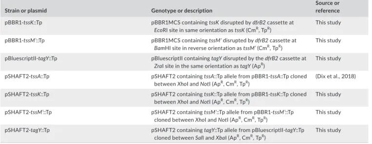
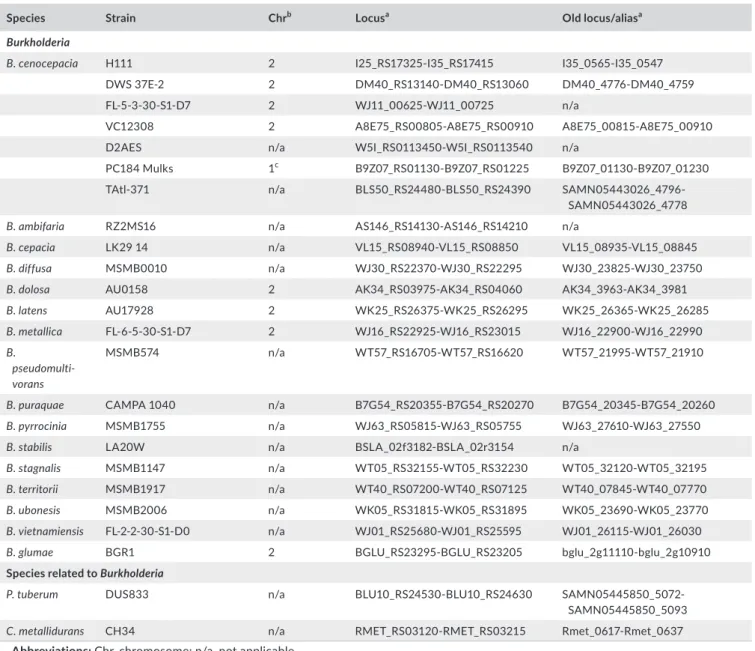
F I G U R E 1 Gene arrangement and distribution of the Burkholderia T6SS‐1 gene cluster. Schematic representation of the Burkholderia
T6SS‐1 gene cluster and related gene clusters in members of the Proteobacteriaceae. The box shows the genetic organization of the archetype Burkholderia T6SS‐1 gene cluster harbored by the indicated species, including B. cenocepacia (for reference, the B. cenocepacia T6SS‐1 gene cluster corresponds to BCAL0337‐BCAL0353 in strain J2315 and I35_RS01700‐I35_RS01780 in H111, as indicated).
Variations on the same basic theme found in other members of the Burkholderia, related genera within the β‐proteobacteria (Achromobacter,
by 10–13 amino acids (Appendix Figure A1). Based on the “positive inside rule” (Elofsson & von Heijne, 2007), the presence of amino acid residues with basic side chains immediately N‐terminal to the TMD suggests that the N‐terminal region constitutes a small cytoplasmically located domain. The TMD is followed by a long linker‐like region of low complexity, which in TagY orthologues in the Burkholderia spp. shares homology to the RnfC barrel sandwich hybrid domain (cl26195), a domain found at the N‐terminus of the RnfC electron transport complex subunit in Rhodobacter capsulatus (Biegel, Schmidt, González, & Müller, 2011; Schmehl et al.., 1993). A conserved C‐terminal region of ~40 amino acids that contain four cysteine residues was identified in most TagY orthologues (Appendix Figure A1). Due to the known role of cysteine thiols in various cel‐ lular activities, it is possible that this part of the protein, which is predicted to be located in the periplasmic space, constitutes a do‐ main which assembles an iron–sulfur cluster. Alternatively, it may be involved in binding other transition metal ions such as zinc or copper, or act as a redox sensor.
Two additional genes are conserved in the T6SS‐1 cluster of spe‐ cies that are not members of the Burkholderia and Paraburkholderia genera. They correspond to RSp0764 and RSp0765 of R. sola‐
nacearum GM1000, RGE_RS12595 and RGE_RS12600 of R. gelati‐ nosus IL144, XOO3320 and XOO3321 of X. oryzae MAFF 311018,
AT699_RS16195, and an unannotated gene of A. arsenitoxydans NCTC10807, and ABAYE2409 and ABAYE2405 of A. bauman‐
nii (which were previously annotated as asaB and asaC as they
were thought to be unique to the Acinetobacter T6SS; Carruthers, Nicholson, Tracy, & Munson, 2013). Bioinformatic analysis predicts that the first of each pair of genes encodes a protein possessing TMDs close to the N‐terminus (Appendix Figure A2), whereas the latter has been recognized as a putative PAAR domain‐containing protein in A. baylyi and named accordingly (Weber et al., 2016).
Homologues of the asaB gene (from herein referred to as tagZ) are also present in some, but not all Burkholderia and in a single
Paraburkholderia species (P. bannensis), while paar is present in all Burkholderia and Paraburkholderia species. However, both genes
reside outside the T6SS‐1 cluster in these two genera and in some cases are located within a conserved gene cluster on chromosome 1 that encodes three TssI subunits and one or more effector–im‐ munity protein pairs (Appendix Figure A3). The gene encoding the PAAR domain protein is located immediately upstream of tagZ in these T6SS‐related gene clusters, as is the case where these genes occur in the main T6SS‐1 gene cluster (Figure 1). As a number of the
Burkholderia and Paraburkholderia species possess only T6SS‐1, it can
be concluded that despite its location outside of the main T6SS‐1 gene cluster, the products of the paar‐tagZ gene pair play a role in the activity of T6SS‐1.
3.2 | Identification of an additional, isolate‐specific,
type VI secretion system in Burkholderia cenocepacia
During the bioinformatic analysis of the T6SS‐1 described above, an additional, complete T6SS gene cluster was identified in
B. cenocepacia strain H111, a cystic fibrosis isolate (Carlier et al.,
2014; Geisenberger et al., 2000). Further genome mining revealed that it was also present in B. cenocepacia strains FL‐5‐3‐30‐S1‐D7, VC12308, and DWS 37E‐2, and several additional B. cenocepacia isolates for which only contig or scaffold‐level genomic data are currently available, including D2AES, PC148, and TAtl‐371 (see Table A5 in Appendix 1 for loci). This second T6SS cluster is lo‐ cated on chromosome 2 in the completely sequenced strains and encodes orthologues of all the core T6SS subunits, including TssI and PAAR (Figure 2). The cluster shares a genetic arrangement that is similar to a T6SS cluster present in the plant pathogenic
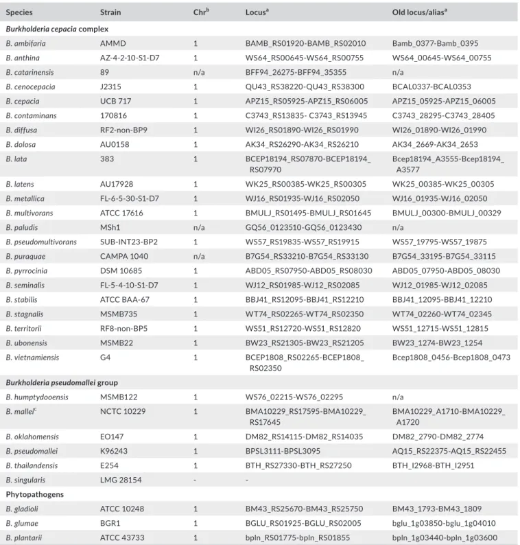
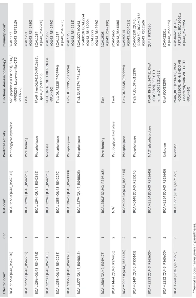
F I G U R E 2 Burkholderia cenocepacia H111 possesses an additional T6SS that is present in some plant‐associated and human pathogenic
bacteria. Schematic representation of a T6SS gene cluster identified in B. cenocepacia H111 (top) (I35_RS17325–I35_RS17415), which has a similar genetic organization to the T6SS‐7 cluster (also known as T6SS‐a) previously identified in B. glumae BGR1 and P. tuberum DUS833. A related T6SS cluster is also present in C. metallidurans CH34, EAEC 042 (the T6SS‐1 or sci‐1 cluster), and Y. pseudotuberculosis IP 32953 (T6SS‐2)
Burkholderia species, B. glumae, and to a T6SS gene cluster pre‐
sent in several Paraburkholderia species, including P. tuberum, which has been referred to as T6SSa (Angus et al., 2014), but for consistency with the established nomenclature is referred to here as the Burkholderia T6SS‐7. Our analysis also identified T6SS‐7 clusters in some but not all isolates of other Bcc species and in
Cupriavidus metallidurans, a species that is closely related to the Burkholderia/Paraburkholderia clade (Table A5 in Appendix 1 and
Figure 2).
Burkholderia T6SS‐7 is notable in possessing a TagL orthologue
which serves as an auxiliary subunit that anchors the T6SS to the peptidoglycan (Aschtgen et al., 2010). Accordingly, the genetic orga‐ nization of this T6SS gene cluster is also similar to those encoding TagL‐dependent T6SSs present in human pathogens such as T6SS‐2 of Yersinia pseudotuberculosis, the T6SS of the uropathogenic E. coli strain CFT073, and the T6SS‐1 (Sci‐1 T6SS) of enteroaggregative
E. coli (Figure 2).
Bioinformatic analysis of the T6SS‐7 gene cluster also suggests that it encodes a phospholipase D (PLD) effector and two corre‐ sponding Tli immunity proteins in members of the Burkholderiaceae. This particular PLD belongs to the Tle5 group of phospholipase effectors and is closely related to the PldB protein, PA5089, en‐ coded by the H3‐T6SS of P. aeruginosa that has been shown to serve as a transkingdom effector (Russell et al., 2013; Jiang et al., 2014; Appendix Figure A4).
3.3 | The Burkholderia cenocepacia T6SS‐1
is functional during growth under standard
laboratory conditions
The presence of the core T6SS subunit, TssD, in bacterial cul‐ ture supernatants is the hallmark of an active T6SS and can be used as a method to determine functionality of the T6SS. This assay was used to determine whether B. cenocepacia isolates possess an active T6SS‐1 during growth under standard labora‐ tory conditions and to validate T6SS‐1 mutants prior to their use
in bacterial competition and virulence assays described below. Therefore, mutants defective in the core tssA, tssK, and tssM components of T6SS‐1 were generated in strains H111, K56‐2, and Pc715j, and TssD secretion of the mutants was compared to that of the corresponding wild‐type parent strains grown in broth culture.
Western blotting showed that TssD was absent in the culture supernatant of the tssA, tssK, and tssM mutants but present in the respective wild‐type H111 and K56‐2 supernatants consistent with previous results obtained using a B. cenocepacia atsR mutant (Aubert et al., 2015; Figure 3a). The H111 and K56‐2 tssM mutants were sub‐ jected to a complementation analysis, whereby TssD secretion could be restored in both strains by introduction of a plasmid expressing
tssM (Figure 3b). Together, these results indicate that B. cenocepacia
isolates H111 and K56‐2 have an active T6SS‐1 under standard lab‐ oratory conditions.
The additional B. cenocepacia isolate analyzed, Pc715j (and its T6SS‐deficient derivatives), was unable to secrete TssD into the extracellular milieu, despite detection of this protein in whole‐cell extracts (Figure 3a), indicating that TssD was being expressed but that the T6SS‐1 was incapable of firing and/or assembly in this strain. Whole‐genome sequencing of our laboratory stock of Pc715j indicated that an IS element was inserted into the
tssM gene. The IS element exhibited homology to the ISUmu23
insertion sequence found in the Bcc‐specific phage KS5 (Lynch, Stothard, & Dennis, 2010), and its insertion into the tssM coding sequence was predicted to result in production of a nonfunctional truncated form of the TssM subunit that lacked the C‐terminal 447 amino acids.
The role of the candidate post‐translational regulatory protein, TagY, in T6SS‐1 activity was also explored by inactivating the tagY gene in strain H111. However, no significant difference in TssD se‐ cretion was observed between the wild‐type and the mutant strains (results not shown). These results could be explained if TagY acts to further upregulate the system in response to an unknown signal that is not present under the assay conditions.
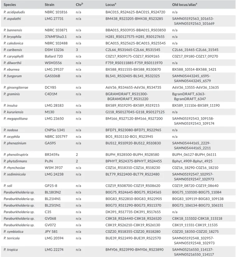
F I G U R E 3 Burkholderia cenocepacia T6SS‐1 is active under standard laboratory conditions. Secretion activity of B. cenocepacia T6SS‐1
in vitro. Anti‐TssD immunoblot was performed on proteins extracted from culture supernatants (SN) and cell‐associated proteins (CA) of
B. cenocepacia wild‐type (WT) strains H111, K56‐2, and Pc715j, and corresponding T6SS‐1 mutants (tssA::Tp, tssK::Tp, tssM::Tp, and/or ΔtssM)
(a) and the H111 and K56‐2 WT and ΔtssM or tssM::Tp strains carrying a complementation or empty control plasmid (pBBR1‐tssM(+) and pBBR1MCS (“pBBR1”), respectively) (b). Anti‐β‐RNAP antibody was used as an indicator of bacterial cell lysis in preparations. Scales and labels as indicated. The H111 tssA::Tp mutant was included as a control
3.4 | Burkholderia cenocepacia T6SS‐1 exhibits
antibacterial activity
It has been demonstrated that the T6SS can target effector proteins to other bacteria, thereby helping the organism to compete more ef‐ fectively against other bacterial species in its growth environment. However, to date, the antibacterial nature of the T6SS‐1 in any mem‐ ber of the Bcc has not been reported. Therefore, we addressed the role of the T6SS‐1 in the ability of B. cenocepacia to compete effec‐ tively with other bacterial species.
As basal‐level TssD secretion appeared to be greater in B. ceno‐
cepacia H111 than in strain K56‐2 (Figure 3a), the former was cho‐
sen to evaluate the role of the T6SS‐1 in competition in this species. Strains H111 and H111‐ΔtssM were used as “attackers” in a bacte‐ rial competition experiment against Gram‐negative “prey” species
Pseudomonas putida KT2440, Escherichia coli CC118(λpir), and E. coli
SM10(λpir). Following cocultivation for four hours on solid medium, viable prey bacteria were enumerated and the number that survived attack by the wild‐type and mutant attackers were compared.
For all three prey strains tested, the number of recovered sur‐ viving prey bacteria was significantly lower (by one to two orders of magnitude) when they were cocultured with the wild‐type attacker
strain in comparison with no attacker, demonstrating that B. ceno‐
cepacia can restrict the growth of E. coli and P. putida (Figure 4a).
Furthermore, following coculture with the ΔtssM attacker strain, the number of surviving prey bacteria was similar to those ob‐ served when no attacker was present (Figure 4a). The number of recoverable attacking H111 bacteria was similar for both the WT and T6SS mutant strains and was unaffected by coculture with all prey strains (Appendix Figure A5). To validate these results, a complementation experiment was performed using the E. coli SM10(λpir) strain as the prey. The antibacterial activity of the tssM mutant attacker toward the E. coli strain could be restored to wild‐ type levels by introduction of a plasmid expressing tssM into the mutant attacker strain (Figure 4b). These data strongly suggest that the T6SS‐1 in B. cenocepacia has antibacterial properties.
3.5 | Burkholderia cenocepacia T6SS‐1 is not required
for virulence in eukaryotic models of infection
Several eukaryotic infection models have been used to identify viru‐ lence factors of B. cenocepacia, including the nematode C. elegans, larvae of the waxmoth G. mellonella, and zebrafish embryos (Seed & Dennis, 2008; Uehlinger et al., 2009; Vergunst et al., 2010). To
F I G U R E 4 The Burkholderia cenocepacia T6SS‐1 plays a role in bacterial competition. (a) Recovery of viable P. putida, E. coli SM10(λpir)
and E. coli CC118(λpir) (in CFU/ml) “prey” strains following coculture with the indicated B. cenocepacia H111 “attacker” strains for 4 hr at 30°C. (b) Comparison of recovery of E. coli SM10(λpir) prey following coculture with B. cenocepacia H111 WT or ΔtssM mutant attacker strains carrying complementation or control plasmids pBBR1‐tssM(+) (“ptssM”) and pBBR1MCS (“pBBR1”), respectively. n ≥3 and error bars indicate SD
ascertain the contribution of T6SS‐1 to the virulence of B. cenoce‐
pacia, we utilized all three of these infection models. Comparison of
the survival of C. elegans infected with B. cenocepacia strain H111 and its tssA and tssK mutant derivatives for 48 and 72 hr showed that the wild‐type and mutant strains exhibited a similar killing ef‐ ficiency during both time periods (Figure 5a). tssA and tssM mutants of strain K56‐2 were used to explore the role of T6SS‐1 in virulence toward G. mellonella larvae and zebrafish embryos. Comparison of the survival of G. mellonella following infection with these mutants
demonstrated that they were able to kill the larvae as effectively as the wild‐type strain at 24 hr postinfection, whether high or low bacterial loads were employed (4 × 10⁴ and 4 × 10² CFU/larvae,
respectively), (Figure 5b). Wild‐type K56‐2 and its T6SS‐1 mutant derivatives were also found to be similarly virulent in the zebrafish model, both in terms of mortality and multiplication of the bacteria in the host (Figure 5c). Taken together, these results suggest that the T6SS‐1 in B. cenocepacia is primarily used to target other bac‐ terial species. Although T6SS‐1 does not significantly contribute to
F I G U R E 5 The Burkholderia cenocepacia T6SS‐1 is not required for virulence toward eukaryotes. (a) Percentage survival of C. elegans
following 48‐hr (white bars, left) and 72‐hr (black bars, right) infection with the indicated B. cenocepacia H111 strains at 20°C. Twenty to 40 worms were used per condition. E. coli OP50 was used as a negative control. Each point indicates mean (n = 3), and error bars indicate SD. (b) Percentage survival of wax moth larvae following 24‐hr infection with high (1 × 10⁴) (left) and low (1 × 10²) (right) doses of B. cenocepacia
K56‐2 (WT) and indicated mutant strains at 37°C. Thirty larvae were infected per condition. Uninfected (UI), heat‐killed B. cenocepacia WT (HK), and mock‐infected (PBS) controls were included. Each point indicates mean % survival (n = 3), and error bars indicate SD. (c) Zebrafish embryos were microinjected with ~100 CFU of indicated B. cenocepacia K56‐2 strains and kept at 28°C in individual wells containing E3 medium. About 20 embryos were used for determination of survival percentage over time (representative experiment shown on the left), and five embryos per indicated time point were used to determine recovery of viable B. cenocepacia K56‐2 counts (n = 5 per time point per experiment, geometric mean; right‐hand graph, showing summary of two independent experiments). ns: not significant
virulence in the eukaryotic models tested here, it is possible that it may have an impact in other systems.
3.6 | In silico identification of putative T6SS‐
dependent effectors in B. cenocepacia
The T6SS‐1 cluster in B. cenocepacia encodes no obvious T6SS‐de‐ pendent effectors. However, an earlier bioinformatics survey of the
B. cenocepacia K56‐2 genome identified ten TssI proteins encoded
at other locations within the genome, of which two (BCAL1359 and BCAS0667) contain C‐terminal effector domains (Aubert et al., 2015). Here, using what is known from previously characterized T6SS‐de‐ pendent effectors, coupled with bioinformatic tools, we have identi‐ fied additional putative T6SS‐dependent non‐TssI effectors and their cognate immunity proteins encoded by the B. cenocepacia genome. As T6SS‐dependent effector genes in other species are often located within close proximity to tssI genes (Lien & Lai, 2017), we used the predicted amino acid sequences of protein products encoded within close proximity to the ten intact tssI genes and one disrupted tssI gene (BCAL2503) present within the B. cenocepacia J2315 genome as que‐ ries in BLASTP searches to identify putative functional domains and homology to proteins belonging to established T6SS effector super‐ families (Appendix Figure A6). Twelve putative T6SS effectors were identified using this approach, with each of the tssI clusters in B. ceno‐
cepacia J2315 encoding at least one putative effector. Of the putative
effectors identified, six were predicted to be phospholipases (encoded by BCAL1296, BCAL1358, BCAL1366, BCAL2277, BCAM0046, and BCAM0149), five of which belong to the Tle antibacterial effector superfamily (Russell et al., 2013). Of the six remaining effectors, one is a predicted peptidoglycan hydrolase (BCAL1166), two were identi‐ fied as putative nuclease effectors (BCAL1298 and BCAS0663), of which the latter contains RHS repeats, an additional RHS repeat pro‐ tein (BCAM2253) containing a RES‐type NAD+ glycohydrolase CTD
(Skjerning, Senissar, Winther, Gerdes, & Brodersen, 2018), and two homologues of the antibacterial pore‐forming toxin Tse4 (BCAL1292 and BCAL2505; Whitney et al., 2014; LaCourse et al., 2018) were also identified. An additional putative T6SS effector was identified by using homologues of the Tae peptidoglycan hydrolase T6SS effec‐ tor superfamilies as queries to search the entire translated genome of B. cenocepacia, resulting in identification of a Tae4‐Tai4 effector immunity pair (BCAM1464‐BCAM1465) located away from a tssI gene cluster. Further details of the putative T6SS effectors identi‐ fied in these searches are included in Table A6 in Appendix 1, which includes the specific domains identified and putative immunity pro‐ teins. It should be noted that the previously described TecA effector (Aubert et al., 2016) is not encoded within a tssI gene cluster and was not independently identified in our analysis.
4 | DISCUSSION
Although some species of bacteria, such as S. marcescens and V. chol‐
erae V52, do exhibit high basal levels of T6SS activity during growth
in laboratory media (Gerc et al., 2015; MacIntyre et al., 2010), in other cases the T6SS is observed to be inactive (Burtnick et al., 2011; Mougous et al., 2006; Zheng, Shin, Cameron, & Mekalanos, 2010), necessitating the use of bacterial strains that have a constitutively active T6SS in order to investigate the functional role of the system and aid in the identification of T6SS‐dependent substrates (Hood et al., 2010; Russell et al., 2011). Here, we demonstrate that the T6SS‐1 of B. cenocepacia is active under standard laboratory conditions with sufficient basal activity to allow detection of TssD in concentrated culture supernatant by immunoblotting. This observation is consist‐ ent with a previous proteomic study in which a protein identified as “hemolysin‐coregulated protein” (i.e., Hcp or TssD) was detected in the extracellular fraction of strain H111 through 2‐DE coupled mass spectrometry which was not recognized as a T6SS subunit at the time (Riedel, Carranza, Gehrig, Potthast, & Eberl, 2006). These results are also consistent with an investigation that demonstrated TssD secre‐ tion in strain K56‐2 could be increased upon inactivation of a global virulence regulator, atsR (Aubert et al., 2008). This study showed the presence of very small amounts of a protein corresponding in size to TssD in wild‐type K56‐2 culture supernatants by SDS‐PAGE, which was confirmed by mass spectrometry rather than immunoblotting, as in our study. Moreover, the low abundance of this protein in the secreted fraction led the authors to consider the T6SS activity to be insufficient to use the wild‐type strain in further investigations into the role of the T6SS in B. cenocepacia. It is possible that our method of sample preparation and detection in wild‐type K56‐2 was more sensitive than that used in the Aubert and co‐workers study.
The role of the T6SS in interspecies and intraspecies bacterial competition has been recognized as a prominent feature of the sys‐ tem in a variety of T6SS‐containing bacteria, including P. aeruginosa,
V. cholerae, and S. marcescens (Hood et al., 2010; MacIntyre et al.,
2010; Murdoch et al., 2011). In this study, we provide evidence to support a role for the B. cenocepacia T6SS‐1 in competition against two bacterial species, P. putida and E. coli. We have also identified an arsenal of potential antibacterial cargo effectors that could be de‐ livered by T6SS‐1, notably including peptidoglycan hydrolases. The additional T6SS cluster we identified in B. cenocepacia H111 (T6SS‐7) is very unlikely to function in bacterial competition under the con‐ ditions tested, as the bacterial competition experiments performed in this study indicated that the level of prey survival was the same in the presence of a mutant attacker with an inactive T6SS‐1 as it was when there was no B. cenocepacia attacker strain present (Figure 4a). Our results are consistent with observations in other species that encode a Burkholderia T6SS‐1‐type secretion system. This includes the T6SS‐1 in B. thailandensis, which was found to be the sole T6SS cluster involved in bacterial competition (Schwarz et al., 2010), and the T6SS‐1 homologue in Acinetobacter spp. that was shown to con‐ tribute to interbacterial competition (Basler, Ho, & Mekalanos, 2013; Carruthers et al., 2013; Repizo et al., 2015; Weber et al., 2016). They are also consistent with recent observations in the related
Paraburkholderia species P. phymatum, where two non‐T6SS‐1‐type
secretion systems (T6SS‐3 and T6SS‐b (T6SS‐8)) were found to be responsible for interbacterial competition against β‐rhizobia strains
in vitro and as a consequence were less efficient in root nodulation (de Campos, Lardi, Gandolfi, Eberl, & Pessi, 2017).
The H1‐T6SS in P. aeruginosa PAO1 is thought to be triggered by attacks from the T6SS (or T4SS) of neighboring cells as a defensive strategy (Basler & Mekalanos, 2012; Basler et al., 2013; Ho, Basler, & Mekalanos, 2013). As a result, P. aeruginosa does not display a fit‐ ness advantage over T6SS‐deficient competing species (Basler et al., 2013). The T6SSs in S. marcescens Db10 and V. cholerae V52, on the other hand, fire indiscriminately and do not require activation from a neighboring attacking bacterium, and thereby confer a fit‐ ness advantage on the host bacterium against various Gram‐nega‐ tive competitor species, such as a E. coli, Salmonella typhimurium, and
Pseudomonas fluorescens (Gerc et al., 2015; MacIntyre et al., 2010).
Here, we demonstrate that the T6SS‐1 confers a fitness advantage on B. cenocepacia over both T6SS‐positive (P. putida KT2440) and T6SS‐negative (E. coli SM10(λpir)) bacterial species. This may sug‐ gest that, like S. marcescens and V. cholerae, the T6SS‐1 in B. cenoce‐
pacia is constitutively active and its activation is not stimulated by
external T6SS attacks, which is also supported by the evidence of T6SS activity in wild‐type strains of B. cenocepacia H111 and K56‐2 under standard laboratory conditions. This would provide additional support for the idea that the defensive regulatory strategy used by
P. aeruginosa is atypical among T6SSs (Gerc et al., 2015). In addition,
one of the T6SSs in P. putida KT2440 has been shown to be highly efficient at killing phytopathogens such as X. campestris and P. syrin‐
gae (Bernal, Allsopp, Filloux, & Llamas, 2017). However, our results
indicate that B. cenocepacia survival is unaffected by the presence of
P. putida. The constitutive activity of the T6SS‐1 we have observed
in B. cenocepacia may account for this, where B. cenocepacia may be able to subvert P. putida before P. putida can attack with its own T6SS. Alternatively, B. cenocepacia may be immune to the T6SS activ‐ ity of P. putida due to the presence of T6SS immunity proteins with interspecies reactivity, as seen for some Tae‐Tai and Tse‐Tsi effec‐ tors–immunity pairs in other species (Russell et al., 2012).
In comparison with the antibacterial T6SSs of other species, the fitness advantage of B. cenocepacia over the prey species tested is notably less than that observed in several other attacker species, in‐ cluding P. aeruginosa, V. cholerae, and S. marcescens. In these species, an active T6SS is responsible for 1,000‐ to 100,000‐fold reduction in the number of recovered prey bacteria in a bacterial competition assay (Hood et al., 2010; MacIntyre et al., 2010; Murdoch et al., 2011), whereas we only observed a modest 10‐ to 58‐fold reduction. This observation may be due to several reasons. For example, T6SS expression and activity may be lower in B. cenocepacia than these other T6SS‐positive strains, the prey strains used in our competition assay may produce their own antibacterial factors (such as bacterio‐ cins, siderophores, or effectors secreted by other systems), or there may be inherent differences in growth rates between B. cenocepacia and the prey species. However, upon enumerating the B. cenocepa‐
cia attacker species in our bacterial competition assays, we found
that B. cenocepacia survival was not affected by coculture with the prey species (Appendix Figure A5). It is also conceivable that the prey used here may have immunity toward specific T6SS effectors
due to cross‐reacting T6SS‐immunity proteins between species (Russell et al., 2012). It is possible that by screening a larger panel of bacterial species, a species may be identified that is more suscep‐ tible to the T6SS‐1‐dependent antibacterial activity of B. cenocepa‐
cia. Moreover, as the T6SS‐1 cluster harbors a number of genes that
potentially encode post‐translational regulators (i.e., tagF, tagM, and
tagY), this system may have the capacity to be further upregulated
under certain conditions.
The B. cenocepacia T6SS‐1 was first implicated in virulence to‐ ward eukaryotes in a signature‐tagged mutagenesis (STM) study carried out in a rat model of chronic lung infection in which trans‐ poson insertions within the T6SS‐1 gene cluster were associated with impaired survival of the bacterium (Hunt et al., 2004). In subse‐ quent studies, this group demonstrated that the T6SS‐1 contributes toward cytoskeletal rearrangements and inflammasome activation in B. cenocepacia‐infected macrophages through host Rho GTPase inactivation (Aubert et al., 2008; Flannagan et al., 2012; Keith et al., 2009; Rosales‐Reyes et al., 2012; Xu et al., 2014). In contrast to the reported impaired survival of T6SS mutants during rat lung infec‐ tion (Hunt et al., 2004), more recent evidence suggests that the T6SS may contribute to a pyrin inflammasome‐dependent innate immune response that promotes lung tissue inflammation and bacterial clear‐ ance in a mouse infection model (Aubert et al., 2016; Xu et al., 2014). The study by Aubert and co‐workers presented data to show that a putative T6SS effector was responsible for this mechanism.
We have tested several T6SS‐inactive strains of B. cenocepacia in three eukaryotic host–pathogen models, nematodes, larvae of the wax worm, and zebrafish larvae (Seed & Dennis, 2008; Uehlinger et al., 2009; Vergunst et al., 2010). We found no significant differ‐ ence in host survival rates in comparison with infection with the WT strain in any of these infection models, suggesting that the T6SS‐1 in B. cenocepacia does not have a functional role in pathogenicity. Of note, we have performed our assays in the presence of a functional
atsR, so the T6SS is not constitutively upregulated as occurs in the
absence of AtsR, and instead activation above basal levels would de‐ pend on the presence of the appropriate stimulus (as yet unknown) in any of the model systems. Therefore, the T6SS is either not ex‐ pressed in these models in the presence of AtsR, or does not con‐ tribute to a significant host‐induced protective immune response, as seen in mice (Aubert et al., 2016; Xu et al., 2014). We cannot exclude, however, that in the absence of atsR, a measurable effect on viru‐ lence could be detected.
To conclude, we have carried out a bioinformatic and functional analysis of the T6SS‐1 in the Bcc species B. cenocepacia. We have shown that it is encoded on the large chromosome in nearly all
Burkholderia species, unlike the other T6SSs associated with mem‐
bers of this genus, which are not conserved in all species and are usually specified by chromosome 2. Therefore, T6SS‐1 can be con‐ sidered as the ancestral Burkholderia T6SS and may serve as a marker for this genus. We also showed that T6SS‐1 was constitutively active in two representative clinical strains and could be used to compete against other bacterial species, including P. putida and E. coli. This is the first demonstration that T6SS‐1 in a Bcc member plays a role in
interbacterial competition and adds to the catalogue of Gram‐nega‐ tive bacteria that use the T6SS for this purpose. The natural reservoir of B. cenocepacia is within the environment, particularly in the soil around plant root systems where many other bacteria compete to establish themselves. It is therefore unsurprising that B. cenocepa‐
cia has evolved a mechanism for competitive fitness against other
bacteria, in a similar manner to other ubiquitous Burkholderia and
Paraburkholderia species (de Campos et al., 2017; Schwarz et al.,
2010). Future work will look to identify and characterize the se‐ creted components responsible for the T6SS‐dependent antibacte‐ rial activity of B. cenocepacia.
ACKNOWLEDGEMENT
This work was supported by a BBSRC Doctoral Training Grant (BB/ F016840/1) awarded to H.L.S. Genome sequencing was provided by MicrobesNG (http://www.microbesng.uk), which is supported by the BBSRC (grant number BB/L024209/1). VBMI is supported by l'Institut National de la Santé et de la Recherche Médicale INSERM and Université de Montpellier. L.Z. was a Marie‐Curie fellow in the Initial Training Network FishForPharma (PITN‐GA‐2011‐289209). L.E. is funded by the Swiss National Science Foundation (SNSF) (Project 31003A_169307).
CONFLIC T OF INTEREST
The authors declare no conflict of interest.
AUTHORS CONTRIBUTION
H.L.S., M.S.T., A.C.V., and L.E. conceived and designed experiments, and contributed to the writing of the manuscript. H.L.S., S.S., L.Z., and S.Sch. conducted experiments.
ETHIC S STATEMENT
Protocols and procedures employed in this investigation were re‐ viewed and approved by the appropriate institutional review com‐ mittees. Zebrafish (Danio rerio) were kept and handled in compliance with the guidelines of the European Union for handling laboratory animals (http://ec.europa.eu/environment/chemicals/lab_animals/ home_en.htm). Studies performed at VBMI are approved by the Direction Départementale de la Protection des Populations (DDPP) du Gard (ID 30–189–4). Infection experiments were terminated be‐ fore the larvae reached the free feeding stage and did not classify as animal experiments according to the 2010/63/EU Directive. Care and maintenance of zebrafish was as described previously (Vergunst et al., 2010).
DATA ACCESSIBILIT Y
All data are provided in full in the results section of this paper apart from the DNA sequence contig encompassing B. cenocepacia Pc715j
T6SS‐1 gene cluster which is available at www.ncbi.nlm.nih.gov/gen‐ bank/ under accession number MK051000.
ORCID
Helena L. Spiewak https://orcid.org/0000‐0003‐1039‐5520
Leo Eberl https://orcid.org/0000‐0002‐7241‐0864
Annette C. Vergunst https://orcid.org/0000‐0002‐4359‐1491
Mark S. Thomas https://orcid.org/0000‐0003‐0701‐2584
REFERENCES
Angus, A. A., Agapakis, C. M., Fong, S., Yerrapragada, S., Estrada‐de los Santos, P., Yang, P., … Hirsch, A. M. (2014). Plant‐associated symbiotic Burkholderia species lack hallmark strategies required in mammalian pathogenesis.
PLoS ONE, 9, e83779. https://doi.org/10.1371/journal.pone.0083779
Aschtgen, M.‐S., Thomas, M. S., & Cascales, E. (2010). Anchoring the type VI secretion system to the peptidoglycan: TssL, TagL, TagP…what else? Virulence, 1, 535–540. https://doi.org/10.4161/viru.1.6.13732 Aubert, D. F., Flannagan, R. S., & Valvano, M. A. (2008). A novel sensor
kinase‐response regulator hybrid controls biofilm formation and type VI secretion system activity in Burkholderia cenocepacia. Infection and
Immunity, 76, 1979–1991. https://doi.org/10.1128/IAI.01338‐07
Aubert, D. F., Hu, S., & Valvano, M. A. (2015). Quantification of Type VI secretion system activity in macrophages infected with Burkholderia
cenocepacia. Microbiology, 161, 2161–2173. https://doi.org/10.1099/
mic.0.000174
Aubert, D. F., Xu, H., Yang, J., Chen, S., Valvano, M. A., Shao, F., … Chen, S. (2016). A Burkholderia type VI effector deamidates Rho GTPases to activate the Pyrin inflammasome and trigger inflammation. Cell Host &
Microbe, 19, 664–674. https://doi.org/10.1016/j.chom.2016.04.004
Basler, M. (2015). Type VI secretion system: Secretion by a contrac‐ tile nanomachine. Philosophical Transactions of the Royal Society
B: Biological Sciences, 370, 20150021. https://doi.org/10.1098/
rstb.2015.0021
Basler, M., Ho, B. T., & Mekalanos, J. J. (2013). Tit‐for‐tat: Type VI secre‐ tion system counterattack during bacterial cell‐cell interactions. Cell,
152, 884–894. https://doi.org/10.1016/j.cell.2013.01.042
Basler, M., & Mekalanos, J. J. (2012). Type 6 secretion dynamics within and between bacterial cells. Science, 337, 815. https://doi. org/10.1126/science.1222901
Bernal, P., Allsopp, L. P., Filloux, A., & Llamas, M. A. (2017). The
Pseudomonas putida T6SS is a plant warden against phytopathogens. ISME Journal, 11, 972–987. https://doi.org/10.1038/ismej.2016.169
Biegel, E., Schmidt, S., González, J. M., & Müller, V. (2011). Biochemistry, evolution and physiological function of the Rnf complex, a novel ion‐motive electron transport complex in prokaryotes. Cellular
and Molecular Life Sciences, 68, 613–634. https://doi.org/10.1007/
s00018‐010‐0555‐8
Bingle, L. E., Bailey, C. M., & Pallen, M. J. (2008). Type VI secretion: A beginner’s guide. Current Opinion in Microbiology, 11, 3–8. https://doi. org/10.1016/j.mib.2008.01.006
Brackmann, M., Nazarov, S., Wang, J., & Basler, M. (2017). Using force to punch holes: Mechanics of contractile nanomachines. Trends in Cell
Biology, 27, 623–632. https://doi.org/10.1016/j.tcb.2017.05.003
Brunet, Y. R., Zoued, A., Boyer, F., Douzi, B., & Cascales, E. (2015). The type VI secretion TssEFGK‐VgrG phage‐like baseplate is re‐ cruited to the TssJLM membrane complex via multiple contacts and serves as assembly platform for tail tube/sheath polymeriza‐ tion. PLOS Genetics, 11, e1005545. https://doi.org/10.1371/jour‐ nal.pgen.1005545