HAL Id: hal-02937576
https://hal.archives-ouvertes.fr/hal-02937576
Submitted on 14 Sep 2020
HAL is a multi-disciplinary open access
archive for the deposit and dissemination of
sci-entific research documents, whether they are
pub-lished or not. The documents may come from
teaching and research institutions in France or
abroad, or from public or private research centers.
L’archive ouverte pluridisciplinaire HAL, est
destinée au dépôt et à la diffusion de documents
scientifiques de niveau recherche, publiés ou non,
émanant des établissements d’enseignement et de
recherche français ou étrangers, des laboratoires
publics ou privés.
disability and callosal agenesis inhibit cell migration
Kévin Duarte, Solveig Heide, Sandrine Poea-Guyon, Veronique Rousseau,
Christel Depienne, Agnès Rastetter, Caroline Nava, Tania Attié-Bitach,
Ferechté Razavi, Jelena Martinovic, et al.
To cite this version:
Kévin Duarte, Solveig Heide, Sandrine Poea-Guyon, Veronique Rousseau, Christel Depienne, et al..
PAK3 mutations responsible for severe intellectual disability and callosal agenesis inhibit cell
migra-tion. Neurobiology of Disease, Elsevier, 2020, 136, pp.104709. �10.1016/j.nbd.2019.104709�.
�hal-02937576�
Contents lists available atScienceDirect
Neurobiology of Disease
journal homepage:www.elsevier.com/locate/ynbdi
PAK3 mutations responsible for severe intellectual disability and callosal
agenesis inhibit cell migration
Kévin Duarte
a, Solveig Heide
b,1, Sandrine Poëa-Guyon
a,1, Véronique Rousseau
a,
Christel Depienne
b,c, Agnès Rastetter
b, Caroline Nava
b, Tania Attié-Bitach
d, Ferechté Razavi
d,
Jelena Martinovic
e, Marie Laure Moutard
f, Jacqueline Cherfils
g, Cyril Mignot
b,
Delphine Héron
b,⁎⁎, Jean-Vianney Barnier
a,⁎aDepartment of Cognition and Behavior, Paris-Saclay Institute of Neuroscience (Neuro-PSI CNRS, UMR 9197), Paris-Sud and Paris-Saclay Universities, Orsay, France bDepartment of genetics, Reference Center for Intellectual Disabilities of Rare Causes, APHP, GH Pitié Salpêtrière, Paris, France
cInstitute of Human Genetics, University Hospital Essen, University of Duisburg-Essen, Essen, Germany
dUnité d'Embryofoetopathologie, Service of Histology-Embryology-Cytogenetics, APHP Necker Enfants Malades & Imagine Institute, Inserm U1163, Paris, France eUnité de foetopathologie, APHP Antoine Béclère, Paris, France
fDepartment of Pediatrics Neurology, Reference Center for Intellectual Disabilities of Rare Causes APHP, Armand-Trousseau Hospital, Paris, France gLaboratoire de Biologie et Pharmacologie Appliquée, CNRS and Ecole normale supérieure Paris-Saclay, Cachan, France
A R T I C L E I N F O
Keywords:
Corpus callosum agenesis (CCA) Cell adhesion Cell migration Cell spreading Intellectual disability Kinase Neurodevelopmental disorder PAK3 αPIX/ARHGEF6 A B S T R A C T
Corpus callosum agenesis (CCA) is a brain malformation associated with a wide clinical spectrum including intellectual disability (ID) and an etiopathological complexity. We identified a novel missense G424R mutation in the X-linked p21-activated kinase 3 (PAK3) gene in a boy presenting with severe ID, microcephaly and CCA and his fetal sibling with CCA and severe hydrocephaly. PAK3 kinase is known to control synaptic plasticity and dendritic spine dynamics but its implication is less characterized in brain ontogenesis. In order to identify de-velopmental functions of PAK3 impacted by mutations responsible for CCA, we compared the biochemical and biological effects of three PAK3 mutations localized in the catalytic domain. These mutations include two “se-vere” G424R and K389N variants (responsible for severe ID and CCA) and the “mild” A365E variant (responsible for nonsyndromic mild ID). Whereas they suppressed kinase activity, only the two severe variants displayed normal protein stability. Furthermore, they increased interactions between PAK3 and the guanine exchange factor αPIX/ARHGEF6, disturbed adhesion point dynamics and cell spreading, and severely impacted cell mi-gration. Our findings highlight new molecular defects associated with mutations responsible for severe clinical phenotypes with developmental brain defects.
1. Introduction
The corpus callosum is the main myelinated fiber tract in the human brain commissures between homotopic and heterotopic regions of the cerebral hemispheres. Its development begins early during brain onto-genesis, from the 13th to 20th weeks after conception (Edwards et al.,
2014). Corpus callosum agenesis (CCA) is among the most common brain morphological anomalies affecting about one out of 4000 new-borns and its prevalence reaches 2–3% of individuals with intellectual disability (ID) (Grogono, 1968;Schaefer and Bodensteiner, 1992;Wang et al., 2004). CCA is extremely heterogeneous both clinically and etiologically. Genetic causes are usually identified in 30–45% of CCA,
https://doi.org/10.1016/j.nbd.2019.104709
Received 9 May 2019; Received in revised form 13 November 2019; Accepted 8 December 2019
⁎Correspondence to: J.-V. Barnier, Department of Cognition and Behavior, Paris-Saclay Institute of Neuroscience (Neuro-PSI CNRS, UMR 9197), Paris-Sud and
Paris-Saclay Universities, 91400 Orsay, France.
⁎⁎Correspondence to: D. Héron, Department of Genetics, Reference Center for Intellectual Disabilities of Rare Causes, APHP, GH Pitié Salpêtrière, 75013 Paris,
France.
E-mail addresses:[email protected](K. Duarte),[email protected](S. Heide),[email protected](S. Poëa-Guyon),
[email protected](V. Rousseau),[email protected](C. Depienne),[email protected](A. Rastetter),
[email protected](C. Nava),[email protected](T. Attié-Bitach),[email protected](M.L. Moutard),
[email protected](J. Cherfils),[email protected](C. Mignot),[email protected](D. Héron),
[email protected](J.-V. Barnier).
1Solveig Heide and Sandrine Poëa-Guyon contributed equally to this study.
Available online 14 December 2019
0969-9961/ © 2019 The Author(s). Published by Elsevier Inc. This is an open access article under the CC BY-NC-ND license (http://creativecommons.org/licenses/by-nc-nd/4.0/).
including chromosomal rearrangements (10–20%) and Mendelian conditions (30%) (Edwards et al., 2014; Palmer and Mowat, 2014). Several chromosomal alterations and > 300 different causative genes have been reported to date (Edwards et al., 2014;Heide et al., 2017). However, in a relatively large proportion of cases, the genetic defect remains unidentified. We report here a new likely causative mutation p.Gly424Arg (G424R) in the X-linked p21-activated kinase PAK3 gene in a patient with CCA and severe ID, and in his sibling fetus with hy-drocephaly and CCA.
More than ten PAK3 mutations have been reported to date in mild to severe ID with or without brain anomalies such as microcephaly, macrocephaly, and callosal agenesis (Allen et al., 1998;Bienvenu et al., 2000; Cartwright et al., 2017; Gedeon et al., 2003;Hertecant et al., 2017;Horvath et al., 2018;Magini et al., 2014;McMichael et al., 2015; Muthusamy et al., 2017;Peippo et al., 2007;Rejeb et al., 2008). PAK3 belongs to the family of the p21-activated kinases which are down-stream effectors of the RAC and CDC42 GTPases and control cytoske-leton dynamics, cell movement and migration, as well as cell pro-liferation (Bokoch, 2003). Even if PAK3 is already known as a key regulator of synaptic plasticity and dendritic spine dynamics, its precise role in neurodevelopmental processes remains elusive (Kreis and Barnier, 2009).
PAK3 is a multi-functional protein whose functions depend on its kinase activity and on its different substrates. It also possesses scaf-folding functions with partners involved in signalling pathways (Kreis and Barnier, 2009). Several biochemical defects related to pathogenic variants have been characterized including alteration of its kinase ac-tivity, protein stability, GTPase binding, dimer formation, and MAPK activation (Combeau et al., 2012;Kreis et al., 2007;Magini et al., 2014; Thévenot et al., 2011). In order to elucidate the genotype-phenotype relationship of affected patients, and more precisely to decipher the molecular bases of PAK3-mediated CCA, we performed a comparative analysis at the structural, biochemical and cellular levels between two mutation types: i) two missense mutations, K389N and the new G424R mutation, both responsible for severe ID and brain anomalies including microcephaly and CCA and ii) another missense mutation, A365E, re-sponsible for nonsyndromic mild ID (Gedeon et al., 2003;Magini et al., 2014). These three mutations are located in the C-terminal lobe of the kinase domain but outside the catalytic site. We analysed their phylo-genic conservation, impact on protein structure and their consequences on catalytic functions. In this study, we also demonstrate that although these three mutations totally suppress PAK3 catalytic activity, only the two mutations causing severe ID and CCA maintain a wild-type (WT)-like protein stability, increase αPIX/ARHGEF6 binding, impair adhesion point dynamics, and inhibit cell migration.
Our results therefore suggest a new molecular defect associated with PAK3 mutations leading to severe ID and CCA that may be partly re-sponsible for cell migration defects and brain morphological abnorm-alities.
2. Materials and methods
2.1. Editorial policies and ethical considerations
Written informed consent was obtained for all participants of this family. The research was approved by the local ethics committee.
2.2. Genomic analysis
DNA of the patient isolated from blood sample was sequenced in trio with healthy parents. The Roche/Nimblegen v3 exome, 64 Mb kit was used for library preparation with 12 samples multiplexing, according to manufacturer's protocol. This library was then sequenced on a NextSeq 500 (Illumina) with a 2x150bp high output flowcell. The bioinformatic analyses were conducted by Polyweb using BWA 0.7.12, picard-tools-1.121, GenomeAnalysisTK-2014.3- 17-g0583013, SNPEff-4.2. The
identified PAK3 variant was then confirmed by Sanger sequencing in the proband, the sibling fetus and their mother. We performed familial segregation by Sanger sequencing in a healthy maternal uncle. Probes are available upon request.
2.3. In silico analysis
In silico prediction analyses were assessed by using SIFT/Provean,
Polyphen-2, and mutation taster (Adzhubei et al., 2013;Kumar et al., 2009;Schwarz et al., 2014). NM_001128166 transcript sequence from NCBI and ENSP00000361077 protein sequence from Ensembl version 93 were used for this study (Zerbino et al., 2018). Common variants of this mutation were investigated by analyzing 1000 Genome Project, gnomAD (genome aggregation database), and Database of Single Nu-cleotide Polymorphism (dbSNP) databases (1000 Genomes Project Consortium et al., 2012;Sherry et al., 2001). Analysis of Φ and ψ angles was done using RAMPAGE server (Lovell et al., 2003). The PAK3 3D structure referred as PDB entry 6FD3 and the unphosphorylated acti-vation loop taken from the structure of unphosphorylated PAK1 (PDB entry 3Q4Z) were extracted from Protein Data Bank and modeled with PyMOL (Sorrell et al., 2019;Wang et al., 2011).
2.4. Mutagenesis and plasmids
The following plasmids were previously described: HA-PAK3-WT encodes the HA-tagged wild type PAK3 protein, the PAK3-K297L encodes K297L kinase dead mutant and pcDNA3-HA-PAK3-A365E encodes a ID-mutated PAK3 variant (Kreis et al., 2007; Rousseau et al., 2003). The two mutants K389N and PAK3-G424R were generated from pcDNA3-HA-PAK3-WT plasmid with Q5 high-fidelity DNA polymerase (NEB, #M0491S) by PCR and the primers
set (5’-CCAAGTGATCCACAGAGACATAAATAGCGACAACATTCTCC
TCG-3′ and 5’-CGAGGAGAATGTTGTCGCTATTTATGTCTCTGTGGATC ACTTGG-3′) and the primer set (5’-GTAAACGAAGCACTATGGTGAGA ACTCCCTATTGGATGGCAC-3′ and 5’-GTGCCATCCAATAGGGAGTTCT CACCATAGTGCTTCGTTTAC-3′), respectively using Quick change mu-tagenesis protocols. Mutated PAK3 gene sequences were verified by sequencing (Eurofins-GATC Company). The PIX interaction mutant (PIM) HA-PAK3-P194G-R195A was generated from pcDNA3-HA-PAK3-WT using a Phusion site-directed mutagenesis kit (Thermo-fischer #F541) with the oligonucleotide set (5’-GGAGCTCCAGAGCAT ACAAAATCAATCTATACTCGC-3′ and 5’-TGCAATGACAGGCGGGGG TTC-3′). Lentiviral vectors (pHAGE-HA-PAK3) used for COS7 cells in-fection were derived from pHAGE backbone using a “Gateway”® cloning protocol (Invitrogen) by introducing specific AttB sequences using the pcDNA3-HA-PAK3 plasmids as templates (WT, K297L, A365E, K389N and G424R) and the oligonucleotide set (5’-GGGGACAAGTTT GTACAAAAAAGCAGGCTTCACCATGGCTAGCTATCC-3′ and 5’-GGGGA CCACTTTGTACAAGAAAGCTGGGTGTCTAGACTAACGGCTACTG TTC-3′) (Murphy et al., 2006). Then a specific recombination (BP cloning step) between AttB-PCR products and the pDONR221 plasmid was performed using the BP clonase enzyme (Invitrogen, #12–535-029). The resulting pENTR-HA-PAK3 plasmids were used with LR clo-nase enzyme (Invitrogen, #11791-020) in addition to the pHAGE backbone (LR cloning step) to generate the lentiviral vectors (PAK3). The PAK3 coding sequences cloned in these pHAGE-HA-PAK3 were finally verified by sequencing.
2.5. Cell culture, transfection, virus production and infection
COS7 cells and HEK 293T cells were cultured in Dulbecco's modified Eagle's medium (Gibco, #31966-020) supplemented with 10% fetal bovine serum (FBS), 100 U/mL penicillin and 100 μg/mL streptomycin. HEK cells (used for kinase assay and protein interaction assay) were plated at 8.106 cells/100 mm dish and transfected with 30 μg of
combination with 10 μg of either pGFP-CDC42-V12, pGFP-CDC42-N17, pFLAG-αPIX, or pFLAG-βPIX) using a calcium-phosphate protocol. After 12 h of transfection, medium was changed with a fresh one and cells were then starved during 24 h with 0.5% FBS. Cell lines for each variant were established as follow: the different virus supernatants were pro-duced in HEK cells by transfection of the pHAGE-HA-PAK3 variant plasmids, with the PAX2 gag-pol and the VSVg envelop pMD2 plasmids using lipofectamine-2000 (Invitrogen, #11668030). After 2 days, the viral suspension was filtered with a 0.45 μm filter and was then added on COS7 cells. Seventy-two hour after infection, transduced cells were selected by addition of 0.5 mg/mL of Geneticin (Sigma, #G8168) for 8 days. In these established COS7-Lv-PAK3 cell lines, expression of PAK3 variants was induced by doxycycline treatment (2 μg/mL, Sigma). PAK3 expression reaches maximal value as soon as 24 h of doxycycline treatment (data not shown).
2.6. Biochemical assays
For kinase assay, HA-PAK3 mutant proteins were coexpressed in HEK cells either with a constitutively active GTP-loaded form of the CDC42 (CDC42-V12) or with a dominant-negative GDP-loaded mutant (CDC42-N17). Thirty-six hours later, cells were washed twice with ice-cold phosphate buffered saline (PBS) supplemented with 0.1 mM or-thovanadate and lysed in cold lysis buffer containing 50 mM Tris-HCl pH 7.5, 1% Triton X-100, 100 mM NaCl, 50 mM NaF, 10 mM Na4P2O7,
5 mM EDTA, 1% aprotinin supplemented with protease and phospha-tase inhibitors mixture. PAK3 proteins were HA-affinity purified, wa-shed as previously described and wawa-shed one more time with kinase buffer (25 mM HEPES pH 7.4, 25 mM MgCl2and 25 mM
β-glycer-ophosphate). Immunoprecipitates were incubated in kinase buffer containing 40 μM ATP and 5 μCi of [γ-32P] ATP for 20 min at 30 °C in
presence of 3 μg of myelin basic protein (MBP) as a substrate. Samples were then boiled in SDS-Laemmli buffer to stop the reaction and the products were resolved by 12% SDS-PAGE. Gels were dried and ex-posed to autoradiography. Immunoblot analysis were performed using anti-GFP (Millipore, #MAB3580), anti-βPAK (N-19, Santa Cruz, #SC-1871), anti-HA (Cell Signaling Technology, #2367). Membranes were then incubated using anti-mouse (Biorad, #172-1011), anti-goat (Pierce, PA1-86326) and/or anti-rabbit (Biorad, #75011) peroxidase-linked secondary antibodies. Membranes were then revealed with Luminata Crescendo (Millipore).
To assess the protein stability, PAK3 expression was induced by doxycycline in COS7-Lv-PAK3 cells during 48 h. PAK3 protein synthesis was stopped by doxycycline withdrawal, cells were lysed at the in-dicated time and decrease of PAK3 protein level was monitored by western blot on 10 μg of Total cell lysates (TCL) proteins as described above, using anti-HA antibodies and with anti-α-tubulin (Sigma, T6199) western blot as loading control. For MAP Kinase activation,
PAK3 expression in Lv-transduced COS7 cells was induced by
doxycy-cline for 2 days, or not; cells were cultured overnight in low serum conditions, and then treated, or not, with serum (10% final foetal bo-vine serum) for 10 min before to be washed (ice-cold PBS with 0.1 mM orthovanadate) and lysed with the same lysis buffer than for kinase assays and proteins samples were processed by the same method (see above). ERK activation was analysed by western blot with anti phospho-ERK antibodies (T202/Y204, Cell Signaling, #4370), and then sample loading assessed by anti-α tubulin revelation.
To test αPIX and βPIX binding, HEK cells were cotransfected with the plasmids of interest with Lipofectamine as described above and controls were done with a PAK3 variant bearing the double point mu-tation (P194G-R195A) in the proline-rich segment known to totally suppress PAK/PIX interaction and with a βPIX construct with a deletion of the SH3 domain (ΔSH3). Transfected cells were lysed 36 h later with a modified Robert's lysis buffer containing 0.1% Triton X-100, 10% glycerol, 20 mM Tris-HCl pH 8.0, 137 mM NaCl, 50 mM NaF supple-mented with protease and phosphatase inhibitors mixture. After 15 min
on ice, lysates were cleared by centrifugation at 10,000 ×g for 15 min. A Bradford protein assay (Biorad) was then performed to measure the protein concentration. TCL were immunoprecipitated overnight at 4 °C using monoclonal anti-HA agarose antibody (Sigma-Aldrich, #A2095). Anti-HA immunocomplexes were then washed four times with lysis buffer (previously described). Protein samples were separated by 10% SDS-PAGE. Immunoblot analysis were performed using anti-β-actin (Santa Cruz, #SC-69879), anti-HA (Cell Signaling Technology, #2367), anti-FLAG (Sigma, #F7425) primary antibodies. Membranes were then incubated using anti-mouse (Biorad, #172-1011) peroxidase-linked secondary antibodies. Membranes were then revealed with Luminata Crescendo (Millipore). Quantification was assessed using a CCD camera (Fusion FX imaging system, Vilber Lourmat) and analysis was per-formed with ImageJ software. Amounts of co-immunoprecipitated FLAG-PIX proteins, detected and quantified by western blot were ex-pressed relatively to HA-immunoprecipitated PAK3 proteins.
2.7. Cell imaging
COS7-Lv-PAK3 cells were plated at low density on glass coverslips and protein expression was induced with doxycycline during 24 h in half conditions. Cells were fixed with 4% p-formaldehyde for 15 min, washed with PBS and permeabilised with 0.1% Triton X-100 and 0.1% Tween 20 for 15 min at room temperature, then blocked with 0.3% bovine serum albumin (BSA) for 2 h at room temperature before an incubation in 0.1% Tween 20, 0.1% BSA with anti-paxillin (Millipore/ Upstate, #05–417) and anti-HA (Roche, #1867423) antibodies over-night at 4 °C. Cells were then washed and incubated for 2 h at room temperature in 0.1% Tween 20, 0.1% BSA with Alexa-568 anti-rat (Molecular Probes, #A11077) and Alexa-488 anti-mouse (Molecular Probews, #A21121) antibodies to label adhesion points and PAK3. Slips were mounted on slides with Fluoromount + Dapi (eBioscience, #00-4959-52), and cells were imaged using a Leica DMI6000B microscope and pictures were double blind analysed with ImageJ software (counting, Feret diameter measurement and colocalization). Paxillin-positive dots corresponding to adhesion points were classified as per-ipheral focal complexes and as central focal adhesions. The two classes of adhesion points were quantified for each mutant cell line with or without doxycycline induction. Each quantification was then normal-ized to its own non-induced control. Illustrating images were obtained with a confocal microscope LSM700 (Zeiss, Germany).
2.8. Cell circularity analysis
Protein expression of PAK3 variants was induced with doxycycline during 48 h in half conditions. Then, COS7-Lv-PAK3 cells were plated at low density on glass coverslips and fixed with 4% p-formaldehyde for 15 min after a 5 h period of spreading. Then coverslips were permea-bilised and blocked as above and incubated with anti-HA antibody (Roche, #1867423) overnight at 4 °C. Cells were then washed and in-cubated for 2 h at room temperature in 0.1% Tween 20, 0.1% BSA with 568 anti-rat antibody (Molecular Probes, #A11077) and Alexa-A488 phalloïdin (Molecular Probes, #A12379) to label PAK3 and F-actin. Slips were mounted on slides with Fluoromount + Dapi (eBioscience, #00–4959-52), and large tile-images at maximum mag-nification rate (25×) were made using an AxioZoom V16 (Zeiss, Germany). After stitching, pictures were analysed with ImageJ soft-ware. The Measure command calculates object circularity as follows: circularity = 4π(area/perimeter2). A circularity value of 1 indicates a
perfect circle. As the value approaches 0, it indicates an increasingly elongated polygon. This circularity index was quantified for each mu-tant cell line with and without doxycycline induction. Each quantifi-cation was then normalized to its own non-induced control.
2.9. Wound healing assay
COS7-Lv-PAK3 cells were plated at 7.105cells/mL as recommended
by Ibidi using a culture insert with two wells separated by a 500 μm gap (Ibidi GmbH, #81176). One day after, 50 μM of Cytosine Arabinoside (AraC) was added to each well and 2 μg/mL of doxycycline to half conditions. Twenty-four hour later, insert was removed, and wells were washed twice with PBS and a fresh complete growth medium (described before) with AraC and doxycycline (in induced wells) was added. Pictures were then taken using a Leica DMI6000B microscope at 0, 3 and 5 h after insert withdrawal. Migration analysis was performed with ImageJ software: each induced cell line migration was normalized and compared to its non-induced control.
To quantify cell apoptosis in these conditions, we performed anti-cleaved caspase 3 labelling using the following protocol: cells were fixed with 4% p-formaldehyde for 15 min, washed with PBS and per-meabilised/blocked with 5% goat normal serum and 0.3% Triton for 2 h at room temperature before an overnight incubation at 4 °C in PBS with 1% BSA and 0.3% Triton containing rabbit anti-cleaved caspase 3 antibody (Cell Signaling #9664). Cells were then washed and incubated for 2 h:30 in PBS with 1% BSA and 0.3% Triton containing goat anti-rabbit A568 antibody (Mol Probes #A11036). Slips were mounted on slides with Fluoromount + Dapi (eBioscience, #00-4959-52), and cells were imaged using a Leica DMI6000B microscope and pictures were analysed with ImageJ software.
2.10. Statistical analysis
Biochemical experiments (protein stability and protein interaction assay) and wound healing assay were conducted at least three times and data are presented as mean ± SEM. Normal distribution and variance equality were tested with a Shapiro test and Bartlett test. Then, a pairwise t-test with a «Benjamini-Hochberg» correction was performed. For cellular adhesion point analysis, experiments were realized at least three times and we performed a pairwise Wilcoxon test with a «Benjamini-Hochberg» correction as data were not following a normal distribution. A p-value of < 0.05 was considered to be significant.
3. Results
3.1. New PAK3 mutation in a family with X-linked CCA
The patient is the second son of healthy and unrelated parents from Maghreb (Fig. 1A). From a first consanguineous union, the mother had a healthy daughter. The mother had one healthy sister and two healthy brothers. The maternal grandmother had two brothers including one with ID who died at 11 years old and one who died at birth. The first pregnancy of the couple was terminated at 34 weeks of gestation after
prenatal ultrasound diagnosis of CCA associated with severe hydro-cephaly in a male fetus. Neuropathological examination showed that the brain weight was over the 95th centile and confirmed a subtotal CCA with presence of Probst bundles, a septal dysgenesis associated with triventricular dilatation, a massive gliosis of white matter and subependymal cystic lesions. The second pregnancy gave birth to a premature boy who died at birth. During the third pregnancy, CCA was diagnosed by ultrasound. The parents decided to continue the preg-nancy and the patient was born eutrophic at 39 weeks of gestation (weight 3595 g, length 47 cm, head circumference 33 cm). The first years of life were characterized by global development delay (the pa-tient walked at 3 years, said his first words at 5 years) and behavior disturbances (including frequent hand stereotypies). At age of 7 years, the patient's weight was 19 kg (− 1 SD), height at 108 cm (− 2 SD) and head circumference at 47 cm (− 3 SD). He had ID and spoke only a few words. At neurological examination, he had contractures of knees and hips with normal tendon reflexes. Brain MRI at the age of 6 years showed a short corpus callosum with global hypoplasia predominating in its posterior part (Fig. 1B). He had no other brain or extra-cerebral malformations. Clinical examinations of the patient over a long period did not reveal any ichthyosis or other cutaneous abnormalities. Kar-yotyping and microarray of the patient were normal. In search for the etiology, we performed trio-based whole exome sequencing and iden-tified a single nucleotide variation in the PAK3 gene (ChrX (GRCh37):g.110439731G > A, c.1270G > A in the NM_001128166 transcript, p.Gly424Arg) inherited from his mother. Familial segrega-tion showed that the sibling aborted fetus with hydrocephaly and CCA also carried the mutation at hemizygous state whereas the healthy maternal uncle did not. This mutation is absent from the control subject databases GnomAD, dbSNP, and 1000 Genome and was not found in the clinical database ClinVar (1000Genomes Project Consortium et al., 2012;Landrum et al., 2018;Sherry et al., 2001). Identified variant was deposited in the ClinVar Database where it is publicly available and can be found with accession number SCV000897773 for c.1270G > A at the following link: https://www.ncbi.nlm.nih.gov/clinvar/variation/ 626256/.
3.2. The conserved G424 residue forms the hinge of the activation loop of the catalytic domain
The c.1270G > A mutation leads to a non-synonymous change (p.Gly424Arg). This G424 residue is totally conserved among the human PAK paralogous (PAK1/2/4/5/6) and among the other PAK orthologous proteins in vertebrate and invertebrate organisms (Figs. 2A andS1A). This residue is conserved in around half of the human kinome (57%), especially in the AGC and STE kinase families (Fig. S1B). In silico prediction analyses indicated that the variant should be disease-causing since Polyphen-2 and SIFT/Provean predicts a damaging effect with any
Fig. 1. Genetical and clinical description. (A) Family pedigree of the identified PAK3 variant c.1270G > A. The variant is at hemizygous state in the patient (III-9)
and his sibling fetus (III-7). The mother (II-5) carries this variant at heterozygous state. Familial segregation showed the PAK variant was not carried by a healthy maternal uncle (II-3). (B) Brain MRI of the patient (III-9) at the age of 6 years old showing short corpus callosum with severe splenium hypoplasia.
substitution of this residue (Polyphen-2: 1.0; SIFT:0.00 scores) (Adzhubei et al., 2013;Kumar et al., 2009). The G424 residue is located in the kinase domain, like most of the previously described PAK3 mu-tations. It is located at the C-terminal hinge between the activation loop (which carries Threonine 421, the main phospho-site involved in PAK3 activation and controls substrate accessibility) and the P + 1 loop in the activation segment (Fig. 2B–D) (Nolen et al., 2004). More precisely, this residue supports the remodeling of the activation loop from its unphosphorylated, inactive conformation (shown in red) to its active, phosphorylated conformation (shown in green), (Fig. 2D). Φ and ψ angles analysis of the two hinge glycine residues of the activation loop shows that the N-ter Gly407 (Φ = −73, ψ = −11) is in the “general favoured” region of the Ramachandran plot while the C-ter Gly424 (Φ = 132, ψ = −160) is in a “Glycine favoured” region signifying that at the position 424 it can only be a glycine residue (Fig. S2) (Lovell et al., 2003). Thus, mutation to Arg424, a residue that cannot function as a hinge is predicted to impair the conversion of PAK3 to its active conformation without affecting its stability and likely affects the de-pendent processes from this segment, particularly the kinase activity. The two other mutations included in this study are also located in the kinase domain. Ala365 is buried inside the lobe, where its mutation into a bulkier Glu residue may result into structural misfolding. K389N is located at the tip of the loop that carries Arg386, a residue that is cri-tical for the stabilization of the activation loop by binding to phos-phorylated Thr421 in the activation loop. Its mutation to Asn, a residue known to support a wide range of main chain conformations, is likely to alter the stability of PAK3 and/or the conformation of the phosphory-lated, active form of the kinase.
3.3. The K389N and G424R mutations abrogate kinase activity without affecting protein stability
Since mutations in the activation segment may have opposite ef-fects, we analysed the effect of the G424R mutation on kinase activity (Dixit and Verkhivker, 2014). We compared the new G424R variant to the two previously described A365E and K389N mutations, which are both located in the kinase domain but lead to differential phenotypes,
i.e. nonsyndromic mild ID and severe ID associated with CCA,
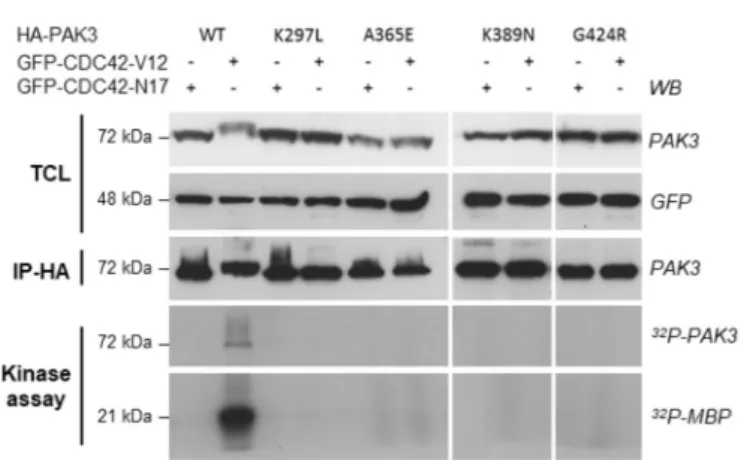
respec-tively. From this point on, the G424R and K389N variants will be coined as “severe PAK3” mutations (for “associated with severe phe-notypes”) and the A365E as “mild PAK3” mutation (for “associated with a mild phenotype”). These PAK3 mutants were transiently co-expressed in HEK cells with constitutively active or dominant negative forms of the CDC42 GTPase. Controls included the WT PAK3 protein and the K297L kinase-dead mutant, generated by disruption of the ATP-binding site of the catalytic domain and recurrently used as control for kinase activity. Upon co-expression with the constitutively active CDC42-V12 mutant, all three immunoprecipitated PAK3 mutant proteins displayed no kinase activity (like the negative control K297L mutant), whereas the WT protein displayed high activity (Fig. 3). The loss of kinase ac-tivity may contribute to the pathogenicity of the three mutants but would not explain the different clinical phenotypes associated with them.
As previously suggested by Magini and colleagues, the stability of
mutated PAK3 proteins may in part explain the severity of the disease (Magini et al., 2014). To explore this hypothesis, we tested the de-gradation time course of these proteins without affecting the bio-synthesis of other cellular proteins using a new approach based on the withdrawal of the antibiotic that positively controls the transcription of the gene of interest. We established COS7-Lv-PAK3 cell lines in which
PAK3 expression was induced by doxycycline thanks to a Tet-On
ex-pression system (Fig. 4A). HA-PAK3 mutated protein expression was high after 2 days of antibiotic treatment, except for the A365E mutant, and not detectable in absence of doxycycline (Fig. 4B). PAK3 protein stability was monitored by western blot after doxycycline withdrawal. Interestingly, the two K389N and G424R variants displayed a similar stability to those of the WT and the referent kinase-dead K297L proteins (Fig. 4C). Only the A365E mutation destabilized the protein, as pre-viously shown (Magini et al., 2014). This probably explains the low expression level of this mutant, upon doxycycline treatment. Thus, the two severe mutations maintain normal protein stability in contrast to the mild mutation, suggesting that catalytically inactive and stable proteins may induce cell defects by interfering with signalling pathways by protein/protein interaction in addition to the enzymatic loss of function.
3.4. PAK3 mutations associated with CCA affect αPIX binding and cell adhesion dynamics
An alteration of specific protein/protein interactions may char-acterize some mutation-associated phenotypes (Sahni et al., 2015). The Fig. 2. In silico analysis of the G424 residue. (A) Sequence alignment of PAK3, orthologous (Ort) and paralogous (Para). Accession numbers for Drosophila and
Caenorhabditis orthologous are respectively FBtr0083312 and C09B8.7a. The mutated residue is underlined. (B) Model representing PAK3 protein with previously described mutations and showing functional domains as p21-binding domain (PBD), autoinhibitory domain (AID) and kinase domain. The new G424R mutation is located at the kinase domain. (C) Sequence/structure corresponding of PAK3 protein. Yellow arrows are β-strands and red cylinders are α-helices. Red arrow shows the hinge point at the beginning of the p + 1 loop which correspond to the mutated residue (G424R). Purple boxes indicate the anchor points of the activation segment. Adapted from Nolen (Nolen et al., 2004). (D) 3D structure of PAK3 kinase domain depicting the localization of the three mutations in the C-terminal lobe of the catalytic domain (shown in pink). The unphosphorylated inactive conformation of the activation loop is shown in red and the phosphorylated active con-formation is shown in green. Ala365 is buried inside the lobe, K389N is located at the tip of the loop that carries Arg386, a residue that is critical for the stabilization of the activation loop, and G424 is located at the hinge of the activation loop. The unphosphorylated activation loop is taken from the structure of unphosphorylated PAK1 and overlaid to phosphorylated PAK3, with residue too flexible to be seen in the crystal structure schematized by a dotted line. fs: frameshift. (For inter-pretation of the references to colour in this figure legend, the reader is referred to the web version of this article.)
Fig. 3. Kinase activity suppression with the new mutant G424R and other PAK3
mutations. PAK3-WT or mutant proteins were immunoprecipitated after being coexpressed with either dominant-negative (N17) or constitutively active (V12) GFP-CDC42 proteins. Expression levels of transfected proteins were evaluated by western blot, using anti-PAK3 and anti-GFP antibodies (upper panel) on total cell lysate aliquots (TCL). Amount of immunoprecipitated proteins were ver-ified by HA-western blot (IP-HA). Note the electrophoretic shift observed for the WT PAK3 protein coexpressed with V12-CDC42, mainly due to autopho-sphorylation events. Radioactive phosphorylated MBP and PAK3 proteins were detected by autoradiography (lower panels: kinase assay). These results have been reproduced in three independent experiments. WB: western blot; IP: im-munoprecipitation.
main partners of PAK are the αPIX and βPIX Guanine exchange factors encoded by ARHGEF6 and ARHGEF7 genes, respectively (Bokoch, 2003). These GEF play a major role in neuronal signalling and some mutations leading to ID have been identified in the ARHGEF6 gene (Kutsche et al., 2000). PAK proteins form tight complexes with PIX through an interaction between a non-conventional proline-rich seg-ment of PAK and the PIX SH3 domain allowing their membrane re-cruitment (Bagrodia et al., 1998; Baird et al., 2005; Manser et al., 1998). We thus analysed the binding of PAK3 variants to αPIX and βPIX by co-immunoprecipitation assay in transiently transfected HEK cells and western blotting. The absence of co-immunoprecipitation using the P194G-R195A PIX-interacting mutant (PIM) of PAK3 (Fig. 5A and C) and a SH3 deleted βPIX construct (data not shown) demonstrated the specificity of this assay. We observed that the amount of PAK3-αPIX complexes formed with the K297L and A365E variants did not differ from those formed with the WT protein (WT mean = 0.41; K297L mean = 0.6, p = .37; A365E mean = 0.53, p = .44), whereas the K389N and G424R mutants tended to form more complexes with αPIX (WT mean = 0.41; K389N mean = 0.8, p = .056; G424R mean = 0.76,
p = .07;Fig. 5A and B). In the same experimental approach, PAK3 mutants and the WT protein formed equal amounts of PAK3/βPIX complexes (Fig. 5C and D).
Because the dynamics of adhesion points is regulated by the PAK/ PIX pathway, we then analysed the effect of PAK3 variants on the number of adhesion points (Nayal et al., 2006;Zhao et al., 2000). Cell adhesion points were detected by paxillin immunolabeling and quan-tified in dox-treated COS7-Lv-PAK3 cell lines relative to those observed in untreated cells. We first checked that doxycycline addition had no effect on the number of paxillin-positive adhesion points in non-trans-duced COS7 cells (Fig. S3B) and that the different expression levels of HA-PAK3-WT (low, medium or high) had no effect on the number of paxillin-positive points per cell (Fig. S3A). This result was also observed for the PAK3 mutants (data not shown). The expression of the WT protein did not change the number of paxillin-positive points compared to non-induced cells as indicated by a ratio around 1 (Fig. 6B). In cells expressing the A365E PAK3 mutation, the total number of paxillin-positive points was not significantly modified whereas the number of peripheral focal complexes was increased compared to WT-expressing cells (WT median = 1; A365E median = 1.2, p ≤ .001). In contrast, the expression of the severe K389N and G424R PAK3 variants significantly increased the total number of paxillin-positive points per cell (WT median = 0.98; K389N median = 1.27, p ≤ .001; G424R median = 1.17, p ≤ .01;Fig. 6B) with an increase of both peripheral focal complexes (WT median = 1; K389N median = 1.3, p ≤ .001; G424R median = 1.08, p ≤ .05) and central focal adhesions per cell (WT median = 1; K389N median = 1.8, p ≤ .001; G424R median = 1.4, p ≤ .01) compared to WT-expressing cells (Fig. 6C and D). After induction, we also observed an enrichment of the three kinase-dead PAK3 mutants in paxillin-positive points compared to WT condi-tion, as previously reported for the K297L kinase-dead protein (as il-lustrated onFig. 6A) (Sells et al., 2000). We conclude that both severe PAK3 mutants affect cell adhesion more severely than the mild A365E mutant.
3.5. Severe PAK3 mutations modify cell spreading and cell migration
While analyzing the effect of PAK3 variants on cell adhesion, we noticed that cells expressing kinase-dead mutants seemed to be more rounded after doxycycline induction in agreement with the role of PAK kinases in cell spreading (Price et al., 1998;Sells et al., 2000). Thus, we analysed in the first 5 h after seeding the cell circularity in non-induced cells and in PAK3 mutants-expressing cells. After fixation, COS7-Lv-PAK3 cells were labeled with fluorescent phalloidin in order to visualize the F-actin network and cell edges (Fig. 7A). We measured cell area, perimeter and circularity that is defined by the two previous para-meters. The expression of the three kinase-dead mutants triggered a significant increase in cell circularity (WT median = 0.89; A365E: median = 1.05; K389N: median = 1.28; G424R: median = 1.18;
p ≤ .001 for each mutant;Fig. 7B) and in cell area (WT median = 0.95; A365E: median = 1.08; K389N: median = 1.13; G424R: median = 1.28; p ≤ .001 for each mutant;Fig. 7C), compared to that of WT PAK3 expressing cells. No significant differences were observed for cell perimeter (data not shown). These results were all the more robust as doxycycline treatment of non-transduced COS7 cells induced a de-crease in cells area or circularity (Fig. S3C and S3D) and no effect on cell perimeter (Fig. S3E).
Because adhesion points and cell spreading play a critical role in cell mobility, we analysed the effect of the expression of PAK3 variants on the migration of COS7-Lv-PAK3 cells with a wound healing test. PAK3 expression was induced by addition of doxycycline and proliferation was blocked with antimitotic AraC treatment. The gap relative closure was quantified 3 and 5 h after inserts withdrawal. Expression of the WT PAK3 protein had no significant effect on the gap closure compared to the non-induced cells. In contrast, A365E-expressing cells displayed a 20% reduction of migration observed after 5 h compared to non-Fig. 4. Severe mutations do not affect PAK3 protein stability. (A) Schematic
representation of the Tet-On pHAGE vector containing the Tet-responsive ele-ment (TRE), mutant HA-tagged PAK3 coding sequences (HA-PAK3), a trans-activator (rtTA), and a gene of selection (Neo). (B) PAK3 variants are correctly expressed after a 48 h induction period with doxycycline. Anti-HA and anti-α-tubulin antibodies were used to reveal their expressions. Note however that the A365E variant was less detected than other PAK3 proteins. (C) Protein stability was assessed after doxycycline withdrawal and protein expression was analysed by western blot on TCL after 1–4 days of doxycycline withdrawal with anti-HA and anti-α-tubulin antibodies. Experiments were independently done three times and data shown are representative of a typical experiment. Ubq: ubiquitin promotor, CMVp: Cytomegalovirus promotor, IRES: Internal Ribosome Entry Site.
induced cells whereas the two severe kinase-dead variants K389N and G424R displayed a more drastic decrease of migration from 3 h of 40–60% compared to their own non-induced cells (Fig. 8). We verified that this decrease of migration is not due to an increase of apoptosis, by cleaved-caspase 3 labelling (Fig. S4). Together expression of severely pathogenic PAK3 variants impacted cell migration.
4. Discussion
CCA may be associated with neurodevelopmental disorders, mainly with ID. Hundreds of genetic anomalies (copy number variations and gene mutations) have been identified by chromosomal microarray analyses and next generation sequencing in patients with CCA asso-ciated with ID, thereby generating a great nosological and etiological complexity (Edwards et al., 2014;Heide et al., 2017). Moreover, dif-ferent mutations in a single gene may lead to distinct clinical pheno-types, including ID with and without CCA, which raises the issue of the genotype/phenotype correlation. In the present work, we described a new family with ID and CCA found in several males, which strongly suggested an X-linked disorder. Whole Exome Sequencing allowed us to identify a new mutation in the PAK3 gene located on the X chromo-some. Several PAK3 mutations have been previously identified in pa-tients with nonsyndromic ID highlighting the role of PAK3 in synaptic plasticity through a main signalling pathway involving Rho-GTPases of the RAC and CDC42 family and targeting actin dynamics and dendritic spine remodeling (Allen et al., 1998;Bienvenu et al., 2000;Dubos et al., 2012;Gedeon et al., 2003;Kreis and Barnier, 2009;Ramakers, 2002; Rejeb et al., 2008; Sala and Segal, 2014). This pathway also plays a crucial role in brain development since mutations in RAC1, CDC42 and
PAK1 genes were identified in patients with ID and microcephaly or
macrocephaly (Harms et al., 2018; Martinelli et al., 2018;Reijnders
et al., 2017). However the three PAK isoform seem to have specific and unique neuronal functions since their mutations lead to different symptoms, such as neurodevelopmental delay and microcephaly for PAK1, autism spectrum disorder for PAK2 and ID for PAK3 (Harms et al., 2018;Wang et al., 2018). Interestingly, several PAK3 mutations were identified in patients with ID and structural brain anomalies (Table 1). Microcephaly was reported in 13/29 individuals in 7 out 11 pedigrees and was associated with distinct mutations. Microcephaly did not correlate with the severity of ID, as illustrated by patients with the V294M variant and those with the R419X variant, both associated with microcephaly and mild ID (Allen et al., 1998;Muthusamy et al., 2017). Most mutations associated with microcephaly are missense muta-tions but two patients with the R419X nonsense mutation and two patients with a N-terminal frameshift also display this morphological defect. Concerning kinase activity, it is difficult to conclude that the loss of this function is sufficient to induce microcephaly. Indeed, patients harboring the A365E mutation which totally impairs the PAK3 enzy-matic activity do not display microcephaly (Gedeon et al., 2003). In-terestingly, one mutation probably responsible for a relative gain of function through a partial activation of its kinase activity is associated with macrocephaly (Hertecant et al., 2017). Concerning the severity of the pathogenicity of some missense mutations compared to nonsense or frame-shift mutations, we propose that the absence of protein has less pathogenic consequences than mutated variant proteins, because mis-sense variants can adversely interfere with signalling pathways. Such hypothesis has already been developed for PAK3 mutations that impact PAK dimer signalling and MAPK activation (Combeau et al., 2012; Magini et al., 2014).
Anomalies of the corpus callosum were reported in three pedigrees with different PAK3 missense variants, including our study (Table 1) (Horvath et al., 2018;Magini et al., 2014). However, all patients did not Fig. 5. αPIX binding increases with severe PAK3 K389N and G424R mutants. HEK cells were cotransfected with PAK3-WT or mutants, in presence of either αPIX or
βPIX, both FLAG-tagged. A PIX-interacting mutant (PIM) of PAK3 was also used to control specificity of assay. HA-tagged PAK3 proteins were immunoprecipitated
and western blots probed with anti-HA, anti-FLAG and anti-β-actin antibodies were performed on TCL and immunoprecipitated samples. Intensities were quantified with a chemiluminescence imager. Co-immunoprecipitated FLAG-PIX proteins were normalized to immunoprecipitated HA-PAK3 protein. Asterisks in western blot correspond to immunoglobulin bands. (A) Representative western blot of αPIX binding to PAK3. (B) Interaction quantification for αPIX. (C) Representative western blot of βPIX binding to PAK3. (D) Interaction quantification for βPIX. Results are presented as mean + SEM. N = 4.
undergo brain imaging, thus it is possible that some of the mutations listed inTable 1may also be associated with CCA. Moreover, the thin corpus callosum described in the patient with the S527G variant may be a secondary cerebral atrophy induced by self-injuries (Horvath et al., 2018). Thus, to date, only the K389N and G424R missense variants
located in the catalytic domain and co-segregating with CCA in all in-dividuals disrupt corpus callosum morphogenesis. Corpus callosum abnormalities are caused by developmental defects affecting neuronal proliferation and specification, cell migration and midline patterning, as well as by defects in axonal growth and guidance (Edwards et al.,
Fig. 6. Kinase dead mutants modify protein localization and severe PAK3 mutants increase cell adhesion point number. PAK3 variant expression was induced in
COS7-Lv-PAK3 cell lines during 24 h in presence of doxycycline. (A) Localization of PAK3 proteins. Cells were immuno-stained for HA-PAK3 and endogenous paxillin. Pictures were used to state on the localization of PAK3 which is enriched in cell adhesion points for all the kinase dead mutants. Scale bar = 20 μm. (B) Relative cell adhesion point number per cell. (C) Relative peripheral focal complexes number per cell. (D) Relative central focal adhesion number per cell. Pictures were analysed with ImageJ software in order to quantify cell adhesion point number. Each quantification was normalized to its own non-induced cell control. Three independent experiments have been realized and at least 120 cells were analysed. Data are represented as median and interquartile. Non-parametric tests were used for statistical analysis (cf. Material and methods;⁎p < 0.05;⁎⁎p < 0.01;⁎⁎⁎p < 0.001).
2014). It is well characterized that PAK kinases regulate several of these cellular processes, and more specifically that the PAK3 isoform controls neuroblast migration and axonal growth (Cobos et al., 2007;Kreis and Barnier, 2009; Liu et al., 2019). To decipher biological processes un-derlying the phenotypical variability of PAK3-associated neurodeve-lopmental disorders, we compared the effects of the two K389N and G424R mutations associated with severe ID and CCA with the A365E
PAK3 mutation previously reported in patients at the other edge of the
clinical spectrum, i.e. with mild nonsyndromic ID. We observed no correlation between the loss of kinase activity and the severity of the phenotype. This is in line with the severity of ID ranging from mild in patients with the A365E and W446S mutations that totally suppress the kinase activity to moderate or severe in those with the R67C mutation which retains it (Bienvenu et al., 2000;Gedeon et al., 2003; Peippo et al., 2007;Kreis et al., 2007;Magini et al., 2014).
The G424R mutation is the second identified disease-causing mu-tation of the PAK3 kinase located in the activation segment, with the
previously described Y427H mutation (Hertecant et al., 2017). A major determinant of the regulation of PAK and other kinases is the presence of an activation loop in the C-terminal lobe, which needs to be phos-phorylated so that the active site can bind the substrate protein in a productive way. Phosphorylation results in the stabilization of this loop through electrostatic interactions between the phosphorylated threo-nine and an argithreo-nine of the C-terminal domain, and this involves a lasso movement of this loop whose hinges are glycine located at its two ends, one of which is Gly424. As we demonstrated, mutation of this residue totally suppresses the kinase activity, which can be explained by the inability of the activation loop of this mutant to adopt its active con-formation. This mutation is thus anticipated to affect the structure and biochemistry of PAK3 in a different manner from other mutations of the catalytic domain involved in intellectual disabilities.
It was previously suggested that the stability of mutated PAK3 proteins may explain the severity of the associated phenotypes. We demonstrated that the A365E variant is unstable, a characteristic that Fig. 7. Kinase dead mutants lead to more rounded and spread cells. PAK3 variants expression was induced in COS7-Lv-PAK3 cell lines during 48 h in presence of
doxycycline. Cells were plated and then fixed 5 h later for phalloidin labeling. Cell circularity was then quantified with ImageJ software. (A) COS7 cells expressing either the WT or the different variants labeled with phalloidin. Scale bar = 20 μm. (B) Relative cell circularity quantification. (C) Relative cell area quantification. Data are represented as median and interquartile, and three independent experiments were realized. Non-parametric tests were used for statistical analysis; (cf. Material and methods;⁎p < 0.05;⁎⁎p < 0.01;⁎⁎⁎p < 0.001).
was previously reported by Magini et al. (Magini et al., 2014). This is readily explained by its buried location, where mutation to a bulkier residue should alter folding of the C-terminal lobe of the kinase domain. In contrast, the K389N and G424R variants display normal stability compared to the WT protein, which may exacerbate their functional consequences by forming inactive complexes with partners. Again, this
is consistent with their location at the surface of the kinase domain where they are not expected to impact significantly the stability of the protein (Sorrell et al., 2019). Another non-exclusive hypothesis is that the G424R mutation may modify protein/protein interactions affecting signalling pathways.
PIX proteins are the main partners of group I PAK kinases and form Fig. 8. Severe PAK3 variants K389N and G424R affect cell migration. Expression of PAK3 variants was induced in COS7-Lv-PAK3 cell lines during 24 h in presence of
doxycycline. Cell culture was performed using a migration insert and cells were then imaged at 0, 3 and 5 h after insert withdrawal. Gap closure for each mutant was normalized to its own control (non-induced cells). Three independent experiments were performed in duplicate and graphics represent the mean ± SEM. Parametric tests were used for statistical analysis after normality and variance equality verification; (⁎p < 0.05;⁎⁎p < 0.01;⁎⁎⁎p < 0.001).
Table 1
Summary of brain anomalies and intellectual defects of patients with PAK3 mutations.
Mutants ID Number of patients Microcephaly Macrocephaly Callosal anomalies Reference
R67C Moderate/severe 6 0/6 0/6 NR Bienvenu et al. (2000)
G92-fs Mild 4 2/2 0/2 NR Rejeb et al. (2008)
V294M Mild 3 3/3 0/3 NR Muthusamy et al. (2017)
A365E Mild 13 0/13 0/13 NR Gedeon et al. (2003)
K389N Severe 3 2/3 0/3 3/3 Magini et al. (2014)
R419X Mild 4 2/4 0/4 0/4 Allen et al. (1998)
G424R Severe 2 1/2 0/2 2/2 This study
Y427H Severe 2 0/2 2/2 0/2 Hertecant et al. (2017)
W446S Mild 5 2/5a 0/5 0/3; NR: 2/5 Peippo et al. (2007)
S527G Mild 1 1/1 0/1 1/1 Horvath et al. (2018)
Deletion Mild 1 0/1 0/1 NR Cartwright et al. (2017)
The number of affected patients relative to the total number of examined people was reported: Macrocephaly, microcephaly and corpus callosum (CC) abnormality were indicated. The symptoms of the patient harboring the R493C mutation were not described. NR: not reported.
supramolecular complexes with GIT proteins, and this complex plays a central role in neuronal signalling by regulating cell adhesion as well as migration, neurite extensions, and synaptic plasticity (Za et al., 2006). Mutations of the αPIX gene located on the X chromosome are also re-sponsible for ID (Kutsche et al., 2000). The regulation of PAK-PIX complexes involves the dimerization of PIXs, their phosphorylation and the combinatory of splice variants (Zhou et al., 2016). Thus, activity-dependent autophosphorylation regulates formation of the PAK/PIX complexes so that kinase-dead mutants form more complexes than WT proteins (Feng et al., 2004). By studying PAK3/PIXs interactions, we observed that the two PAK3 mutations inducing severe phenotypes tend to enhance PAK3 variant binding to αPIX and that their expression significantly increases the number of cell adhesion points suggesting abnormal turnover of adhesions. Adhesion point and integrin signalling are main regulators of cell migration which is often affected in CCA (Edwards et al., 2014;Zhou et al., 2016). Further experiments showed that the expression of both K389N and G424R mutants strongly mod-ified cell spreading and inhibited the cell migration tested in a wound healing assay. In PAK3 knock-out mice, it was observed that absence of PAK3 protein did not modify oligodendrocyte progenitor (OPC) mi-gration (Maglorius Renkilaraj et al., 2017). While this may reflect dif-ferential molecular mechanisms of migration in these cell types, it is likely that missense mutations affect signalling in a different manner than the absence of the protein, for example by exerting dominant-negative or up-regulated effects. Altogether, these results suggest that mutations that might affect PAK3-PIX signalling may affect cell mi-gration triggering brain morphological anomalies. Further studies with knock-in mouse model could bring a better comprehension of PIX function in pathophysiological mechanisms.
It is remarkable that although the missense K389N and G424R mutations are both expected to affect the kinase activity of PAK3, they display differences at both the clinical and cellular level. At the clinical level, ichthyosis is only diagnosed with the K389N variant whereas clinical traits such as ID, microcephaly, and ACC are observed with both
PAK3 mutations. Likewise, the K389N mutation was shown to impair
the MAP kinase pathway, which was proposed to occur through a ki-nase-independent pathway (Magini), while the G424R mutation has no effect on this pathway (Fig. S5). Links between PAK3 mutations and MAP kinase activation are complex and may involve both direct and indirect pathways, among which the PAK-PIX axis (Stockton et al., 2007), and that these pathways are differentially affected by PAK3 mutations with different biochemical defects. Notably, the two muta-tions affect different determinants of the catalytic machinery, with K389N being located in the catalytic site and G424R in the activation loop. We conclude that several pathways such as MAP kinase and/or PIX pathways may be involved in the wide clinical spectrum associated with PAK3 variants.
In conclusion, our data bring important insight into a cellular me-chanism possibly linking PAK3 mutations to brain dysgenesis. Remarkably, they highlight a remarkable correlation between struc-tural and biochemical defects induced by each mutation to the severity of the associated intellectual disorders. This study also depicts the complexity of the relationship between genotype and phenotype, and strongly suggests that the patient phenotype may be due to the addition of synaptic dysfunctions and developmental defects and may be gen-erated by the sum of biochemical defects such as kinase impairment, protein stability and partner-binding anomalies.
Funding
This work was supported by the french Fondation Jérôme Lejeune [grant number 1705-BJ2017B]; and the french Fondation pour la Recherche Médicale (grant number DEQ20150331694).
Declaration of competing interest
The authors declare that there is no conflict of interest that could be perceived as prejudicing the impartiality of the research reported.
Acknowledgements
We thank the family members for their participation in this study. We thank Emmanuel Thévenot, Nicolas Vitale, Kerstin Kutsche, and Célio Pouponnot for providing us with PAK3- PIM, β-PIX, α-Pix, and replicative lentiviral plasmids, respectively. pHAGE TRE dCas9-VP64 was a gift from Rene Maehr & Scot Wolfe (Addgene plasmid # 50916). We also want to thank the Fondation Jérôme Lejeune for supporting us (JVB) financially and the Fondation pour la Recherche Médicale for supporting JC.
Data availability statement
The data that support the findings of this study are openly available in ClinVar at https://www.ncbi.nlm.nih.gov/clinvar/variation/ 626256/, reference number SCV000897773.
References
Adzhubei, I., Jordan, D.M., Sunyaev, S.R., 2013. Predicting functional effect of human missense mutations using PolyPhen-2. Curr Protoc Hum Genet Chapter 7, Unit7.20. Curr. Protoc. Hum. Genet.https://doi.org/10.1002/0471142905.hg0720s76. Allen, K.M., Gleeson, J.G., Bagrodia, S., Partington, M.W., MacMillan, J.C., Cerione, R.A.,
Mulley, J.C., Walsh, C.A., 1998. PAK3 mutation in nonsyndromic X-linked mental retardation. Nat. Genet. 20, 25–30.https://doi.org/10.1038/1675.
Bagrodia, S., Jordon, K.A., Aelst, L.V., Cerione, R.A., 1998. A novel regulator of p21-activated kinases. J. Biol. Chem. 273, 23633–23636.https://doi.org/10.1074/jbc. 273.37.23633.
Baird, D., Feng, Q., Cerione, R.A., 2005. The Cool-2/α-pix protein mediates a Cdc42-Rac signaling cascade. Curr. Biol. 15, 1–10.https://doi.org/10.1016/j.cub.2004.12.040.
Bienvenu, T., des Portes, V., McDonell, N., Carrié, A., Zemni, R., Couvert, P., Ropers, H.H., Moraine, C., van Bokhoven, H., Fryns, J.P., Allen, K., Walsh, C.A., Boué, J., Kahn, A., Chelly, J., Beldjord, C., 2000. Missense mutation in PAK3, R67C, causes X-linked nonspecific mental retardation. Am. J. Med. Genet. 93, 294–298.
Bokoch, G.M., 2003. Biology of the p21-activated kinases. Annu. Rev. Biochem. 72, 743–781.https://doi.org/10.1146/annurev.biochem.72.121801.161742. Cartwright, A., Smith, K., Balasubramanian, M., 2017. Short case report: Xq23 deletion
involving PAK3 as a novel cause of developmental delay in a 6-year-old boy. Clin. Dysmorphol. 26, 38–40.https://doi.org/10.1097/MCD.0000000000000154. Cobos, I., Borello, U., Rubenstein, J.L.R., 2007. Dlx transcription factors promote
mi-gration through repression of axon and dendrite growth. Neuron 54, 873–888.
https://doi.org/10.1016/j.neuron.2007.05.024.
Combeau, G., Kreis, P., Domenichini, F., Amar, M., Fossier, P., Rousseau, V., Barnier, J.-V., 2012. The p21-activated kinase PAK3 forms heterodimers with PAK1 in brain implementing trans-regulation of PAK3 activity. J. Biol. Chem. 287, 30084–30096.
https://doi.org/10.1074/jbc.M112.355073.
Dixit, A., Verkhivker, G.M., 2014. Structure-functional prediction and analysis of cancer mutation effects in protein kinases. Comput. Math. Methods Med. 2014, 1–24.
https://doi.org/10.1155/2014/653487.
Dubos, A., Combeau, G., Bernardinelli, Y., Barnier, J.-V., Hartley, O., Gaertner, H., Boda, B., Muller, D., 2012. Alteration of synaptic network dynamics by the intellectual disability protein PAK3. J. Neurosci. 32, 519–527.https://doi.org/10.1523/ JNEUROSCI.3252-11.2012.
Edwards, T.J., Sherr, E.H., Barkovich, A.J., Richards, L.J., 2014. Clinical, genetic and imaging findings identify new causes for corpus callosum development syndromes. Brain 137, 1579–1613.https://doi.org/10.1093/brain/awt358.
Feng, Q., Baird, D., Cerione, R.A., 2004. Novel regulatory mechanisms for the Dbl family guanine nucleotide exchange factor Cool-2/α-Pix. EMBO J. 23, 3492–3504.https:// doi.org/10.1038/sj.emboj.7600331.
Gedeon, A.K., Nelson, J., Gécz, J., Mulley, J.C., 2003. X-linked mild non-syndromic mental retardation with neuropsychiatric problems and the missense mutation A365E in PAK3. Am. J. Med. Genet. A 120A, 509–517.https://doi.org/10.1002/ajmg.a. 20131.
Genomes Project Consortium, Abecasis, G.R., Auton, A., Brooks, L.D., DePristo, M.A., Durbin, R.M., Handsaker, R.E., Kang, H.M., Marth, G.T., McVean, G.A., 2012. An integrated map of genetic variation from 1,092 human genomes. Nature 491, 56–65.
https://doi.org/10.1038/nature11632.
Grogono, J.L., 1968. Children with agenesis of the corpus callosum. Dev. Med. Child Neurol. 10, 613–616.
Harms, F.L., Kloth, K., Bley, A., Denecke, J., Santer, R., Lessel, D., Hempel, M., Kutsche, K., 2018. Activating mutations in PAK1, encoding p21-activated kinase 1, cause a neurodevelopmental disorder. Am. J. Hum. Genet. 103, 579–591.https://doi.org/10. 1016/j.ajhg.2018.09.005.
Heide, S., Keren, B., Billette de Villemeur, T., Chantot-Bastaraud, S., Depienne, C., Nava, C., Mignot, C., Jacquette, A., Fonteneau, E., Lejeune, E., Mach, C., Marey, I., Whalen, S., Lacombe, D., Naudion, S., Rooryck, C., Toutain, A., Caignec, C.L., Haye, D.,