HAL Id: hal-02160945
https://hal.sorbonne-universite.fr/hal-02160945
Submitted on 20 Jun 2019
HAL is a multi-disciplinary open access archive for the deposit and dissemination of sci-entific research documents, whether they are pub-lished or not. The documents may come from teaching and research institutions in France or abroad, or from public or private research centers.
L’archive ouverte pluridisciplinaire HAL, est destinée au dépôt et à la diffusion de documents scientifiques de niveau recherche, publiés ou non, émanant des établissements d’enseignement et de recherche français ou étrangers, des laboratoires publics ou privés.
Fragments and dust after Holmium laser lithotripsy with
or without “Moses technology”: How are they different?
Etienne Keller, Vincent de Coninck, Marie Audouin, Steeve Doizi, Dominique
Bazin, Michel Daudon, Olivier Traxer
To cite this version:
Etienne Keller, Vincent de Coninck, Marie Audouin, Steeve Doizi, Dominique Bazin, et al.. Fragments and dust after Holmium laser lithotripsy with or without “Moses technology”: How are they different?. Journal of Biophotonics, Wiley, 2019, 12 (4), pp.e201800227, 1-9. �10.1002/jbio.201800227�. �hal-02160945�
F U L L A R T I C L E
Fragments and dust after Holmium laser lithotripsy with or
without
“Moses technology”: How are they different?
Etienne X. Keller
1,2,3| Vincent de Coninck
1,2,4| Marie Audouin
1,2| Steeve Doizi
1,2|
Dominique Bazin
5,6| Michel Daudon
7,8,9| Olivier Traxer
1,2*
1Service d'Urologie, Sorbonne Université, Service
d'Urologie, AP-HP, Hôpital Tenon, Paris, France
2Groupe de Recherche Clinique sur la Lithiase
Urinaire, Sorbonne Université, GRC no 20, Groupe de Recherche Clinique sur la Lithiase Urinaire, Hôpital Tenon, Paris, France
3Department of Urology, University Hospital
Zurich, University of Zurich, Zurich, Switzerland
4Department of Urology, AZ Klina, Brasschaat,
Belgium
5CNRS, Laboratoire de Chimie de la Matière
Condensée de Paris, UPMC, Collège de France, Paris, France
6Laboratoire de Physique des Solides, CNRS
UMR 8502, Université Paris Sud XI, Orsay, France
7CRISTAL Laboratory, Tenon Hospital, Paris,
France
8Laboratoire des Lithiases, Service des
Explorations Fonctionnelles Multidisciplinaires, AP-HP, Hôpital Tenon, Paris, France
9UMRS 1155 UPMC, INSERM, UMRS 1155
UPMC, Tenon Hospital, Paris, France *Correspondence
Olivier Traxer, Service d'Urologie, Hôpital Tenon, Assistance-Publique Hôpitaux de Paris, 4 rue de la Chine, 75020 Paris, France.
Email: olivier.traxer@aphp.fr Funding information
Belgische Vereniging voor Urologie (BVU); European Association of Urology; Kurt and Senta Herrmann Foundation; Travel Grant from the University Hospital Zurich
Urinary stones can be readily disinte-grated by Holmium:YAG laser (Holmium laser lithotripsy), resulting in a mixture of small stone dust parti-cles, which will spontaneously evacu-ate with urine and larger residual fragments (RF) requiring mechanical retrieval. Differences between frag-ments and dust have not been well
characterized. Also, it remains unknown how the recently introduced“Moses tech-nology” may alter stone disintegration products. Three complementary analytical techniques have been used in this study to offer an in-depth characterization of dis-integration products after in vitro Holmium laser lithotripsy: stereoscopic micros-copy, scanning electron microscopy and Fourier-transform infrared spectroscopy. Dust was separated from fragments based on its floating ability in saline irriga-tion. Depending on initial crystalline constituents, stone dust either conserved attributes found in larger RFs or showed changes in crystalline organization. These included conversion of calcium oxalate dihydrate towards calcium oxalate monohydrate, changes in carbapatite spectra towards an amorphous phase, changes of magnesium ammonium phosphate towards a differing amorphous and crystalline phase and the appearance of hydroxyapatite on brushite fragments. Com-paratively,“Moses technology” produced more pronounced changes. These findings provide new insights suggesting a photothermal effect occurring in Holmium laser lithotripsy. Figure: Appearance of hydroxyapatite hexagons on stone dust collected after Holmium laser lithotripsy of a brushite stone using“Moses technology.”
K E Y W O R D S
Fourier-transform infrared spectroscopy, holmium laser, lithotripsy, Moses effect, residual fragments, scanning electron microscopy, stone dust, urinary stone
1 | I N T R O D U C T I O N
Over the past two decades, Holmium:YAG laser (there-after referred as Holmium laser) has become a readily
available energy source for endourologic procedures allowing lithotripsy of all known urinary stone types [1]. Holmium laser operates at a wavelength of 2120 nm— near to the 1940 nm water absorption peak [2]—and transmits pulsed energy (pulse duration ≥250 μs)
Etienne X. Keller and Vincent de Coninck contributed equally to this study.
DOI: 10.1002/jbio.201800227
J. Biophotonics. 2019;12:e201800227. www.biophotonics-journal.org © 2018 WILEY-VCH Verlag GmbH & Co. KGaA, Weinheim 1 of 9 https://doi.org/10.1002/jbio.201800227
through thin silica fibers. The true effect of Holmium laser on stones has been the object of an ongoing debate. A photothermal effect is believed to be the pri-mary mechanism leading to fragmentation during Hol-mium laser lithotripsy [3, 4]. Contrarily, the formerly used pulsed dye laser (pulse duration ~1 μs) and Neo-dymium:YAG laser (pulse duration ~0.5 μs) had been shown to accomplish lithotripsy by a photoacoustical mechanism (plasma expansion and cavitation collapse), a characteristic that was not found for Holmium laser lith-otripsy [3, 5].
Delivery of Holmium laser energy on stones has been proposed to be most effective with the occurrence of the “Moses effect” in water. This phenomenon was first described in 1988 and relies on the vaporization of water by laser energy, such that the successive portion of the laser pulse transmits through vapor instead of water [6]. This allows for an enhanced transmission of the laser energy to the target stone. The recently developed“Moses technology” therefore uses pulse modulation to deliver a considerable fraction of total pulse energy through the vapor channel for ideal energy delivery [7].
Stone disintegration products from Holmium lithotripsy generally form a mixture of larger residual fragments (RF) and smaller stone dust particles [8, 9]. Growing enthusiasm for laser dusting techniques has emerged over the last years, owing to the observation that dust particles seem to have a propensity for spontaneous evacuation from kidney after ureteroscopic laser lithotripsy, omitting the need for time-consuming fragment extraction [10]. A previous study demonstrated the feasibility of stone dust aspiration through the working channel of flexible uretero-scopes (fURS) [11]. Nevertheless, a clear definition of stone dust has not been established yet. Also, it is not known whether alterations of the initial crystalline constitu-tion may be found after Holmium lithotripsy and whether “Moses technology” may produce differing stone disinte-gration products. It is thus necessary to further characterize RF and stone dust to help understand the true effect of Holmium laser on stones.
The present study offers an in-depth morpho-constitutional analysis of the disintegration products from Holmium lithotripsy for the seven most frequently encoun-tered crystalline constituents of urinary stones.
2 | M A T E R I A L S A N D M E T H O D S
Urinary stones were retrieved from a large stone biobank that was built over the last 33 years at our institution. Crys-talline constituents analyzed in this study were: calcium oxa-late monohydrate (COM), calcium oxaoxa-late dihydrate (COD), uric acid (UA), carbapatite (carbonated calcium phosphate; CA), struvite (magnesium ammonium phosphate; MAP), brushite (BR) and cystine (CYS). Criteria for stone selection
were size >1000 mm3 and >90% pure stone composition. The later was based on a detailed morpho-constitutional analysis of stones' surface, section and core, including Fourier-transform infrared (FTIR) spectroscopic analysis for each single stone available in the stone biobank, as previ-ously described [12]. Inappropriate stone storage (eg, wet storage conditions, missing or altered box labeling) was an exclusion criterion. The study was in accordance with ethical standards of the Helsinki declaration.
2.1 | Sample preparation
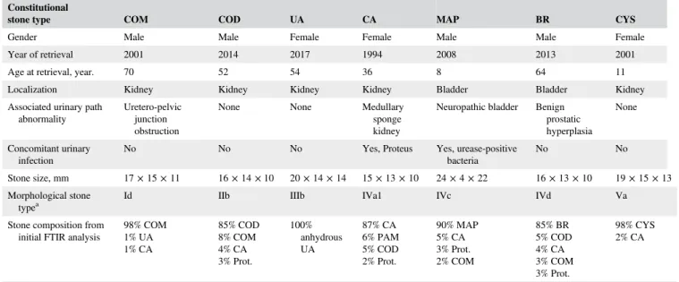
Each stone was cut with a surgical knife into pieces of ~300 mm3, which were immerged in sterile saline solution (0.9%) for 24 hours. Laser lithotripsy was then performed with a Lumenis P120H Holmium laser generator (Lumenis Ltd., Yokneam, Israel). A dusting setting (0.2 J, 40 Hz, long pulse, Lumenis SlimLine D/F/L 200μm fibers) was com-pared to the Moses mode (0.2 J, 40 Hz, Moses “contact” mode, MOSES SlimLine 200μm fibers), thereafter referred as Moses lithotripsy [13, 14]. Laser fibers were inserted into a Lithovue fURS (Boston Scientific, Maple Grove, Minne-sota) and laser lithotripsy was performed under direct visual control in a 5.8 mL glass container (inner diameter 10 mm, height 74 mm) using sterile saline irrigation at room temper-ature with an irrigation pressure of 40 cm H20 (Figure 1-A), until a total of 2400 J were delivered. Lithotripsy was per-formed free-hand with painting movements of laser fiber tip over stone samples. Laser fiber tips were cut with normal scissors before lithotripsy of each sample. The irrigation overflow was collected in a 100 mL plastic container.
After laser lithotripsy, larger RF were separated from stone dust using a method based on the floating ability of particles. For this, the content of the glass container compris-ing a mixture of RF and dust was poured into a 60 mL plas-tic container, which was perforated with a hole located 2 cm above the bottom of the container (Figure 1-B). This allowed for dust particles with floating ability to evacuate through the hole when a constant irrigation through the f-URS was applied (empty working channel, irrigation pressure of 40 cm H20). The size of the hole (5 mm) was chosen to allow sufficient irrigation outflow from the plastic container. The resulting irrigation overflow was collected in the same 100 mL plastic container that was used during lithotripsy (Figure 1-C). The RF without floating ability remained within the 60 mL plastic container and stone dust from the 100 mL was allowed to sediment under terrestrial gravity. 2.2 | Morphological analysis
For morphological analysis, each air-dried RF or dust sam-ple was separately transferred on carbon conductive double-faced adhesive tapes (Nisshin EM Co. Ltd., Tokyo, Japan). Images were obtained from an Olympus SZ61 stereoscopic microscope (Olympus Corporation, Tokyo, Japan), as well
as from a Zeiss Gemini Supra 55VP scanning electron microscope (SEM) (Carl Zeiss AG, Oberkochen, Germany) using low electron beam voltage (≤2 keV) [15]. For each sample, three different areas of interest were analyzed by SEM. No sample coating or fixation was needed for these analyses. Morphological stone types were classified accord-ing to the Daudon classification of stones [16, 17].
2.3 | Constitutional analysis
FTIR spectroscopy was used to identify chemical constitu-ents of each sample, as described previously [18]. Sample preparation included fine grinding of RF or dust, respec-tively, together with a purified salt (potassium bromide) and applying 10 tons to this mixture with a mechanical press to form a translucent pellet, which was placed into a Bruker Vector 22 spectrometer for analysis (Bruker Daltonik GmbH, Bremen, Germany). The density of a given RF or dust sample in its according pellet was limited to a level that generated spectral bands with a maximal intensity of 2.00 absorbance units in order to avoid scattering artifacts of intense bands. The FTIR spectra of the initial stones were compared to the spectra drawn from RF and dust samples.
3 | R E S U L T S
From >80 000 urinary stones listed in the biobank, one stone of each constitutional type was available for analysis. Table 1 summarizes patients' characteristics from whom stones were retrieved. Major constituent was >90% for each constitutional type, except for COD, CA and BR where a mixed stone of 85% COD, 87% CA and 85% BR had to be
selected, respectively, in order to align with the stone size requirements.
3.1 | Morphological analysis
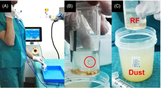
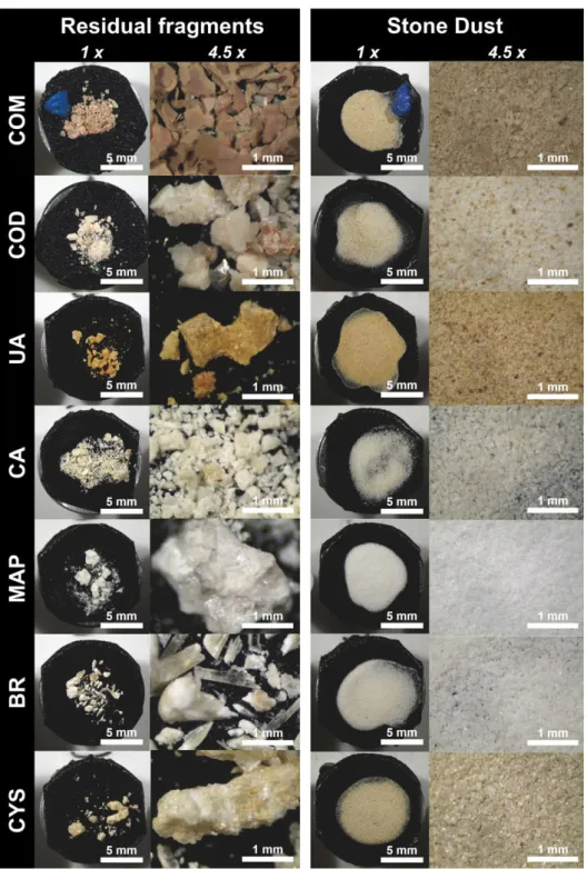
Observations from morphological analyses are summarized in Table 2 and illustrated in Figures 2, 3 and 4. While only a few stone characteristics could be retrieved from stereo-scopic microscopy (mainly color), the addition of SEM imagery allowed for an in-depth morphological analysis. For COM, the brown dust was characterized as small plates that kept a layered organization also found in larger RF. For COD, a bipyramidal organization was partially conserved in RF, whereas dust lost this attribute and merely conserved the initial beige-brown color within a heteroge-nous mixture of sharp particles. This was in contrast to UA, where both RF and dust revealed a partially conserved crys-talline organization with smoother edges and brown-orange color. The surface of CA was a mixture of smooth and bumpy surfaces both in RF and dust samples. The MAP samples revealed longitudinal white needles which seemed to be encased in another constituent, an observation found both in RF and dust. For BR, the typical longitudinal orga-nization was found in RF and was teared down to sharp and thin beige baguettes in dust samples. Finally, a layered organization of hexagons was found in RF and dust sam-ples of CYS.
Peculiar observations emerged from comparison of con-ventional Holmium with Moses lithotripsy. Moses litho-tripsy produced a more pronounced disruption of morphological characteristics of COD, MAP and CYS stones (Table 2). Also, areas with hexagonal plate-like sur-faces appeared on RF and dust from BR after Moses litho-tripsy (Figure 4). This plate-like morphology is
TABLE 1 Patient characteristics for each constitutional stone type Constitutional
stone type COM COD UA CA MAP BR CYS
Gender Male Male Female Female Male Male Female Year of retrieval 2001 2014 2017 1994 2008 2013 2001 Age at retrieval, year. 70 52 54 36 8 64 11 Localization Kidney Kidney Kidney Kidney Bladder Bladder Kidney Associated urinary path
abnormality
Uretero-pelvic junction obstruction
None None Medullary sponge kidney
Neuropathic bladder Benign prostatic hyperplasia
None
Concomitant urinary infection
No No No Yes, Proteus Yes, urease-positive bacteria
No No
Stone size, mm 17 × 15 × 11 16 × 14 × 10 20 × 14 × 14 15 × 13 × 10 24 × 4 × 22 16 × 13 × 10 19 × 15 × 13 Morphological stone
typea
Id IIb IIIb IVa1 IVc IVd Va
Stone composition from initial FTIR analysis
98% COM 1% UA 1% CA 85% COD 8% COM 4% CA 3% Prot. 100% anhydrous UA 87% CA 6% PAM 5% COD 2% Prot. 90% MAP 5% CA 3% Prot. 2% COM 85% BR 5% COD 4% CA 3% COM 3% Prot. 98% CYS 2% CA
Abbreviations: BR, brushite; CA, carbapatite; COD, calcium oxalate dihydrate; COM, calcium oxalate monohydrate; CYS, cystine; MAP, magnesium ammonium phos-phate; Prot., protein; UA, uric acid.
characteristic of hydroxyapatite formation and was not found in samples issued from conventional Holmium lithotripsy. 3.2 | Constitutional analysis
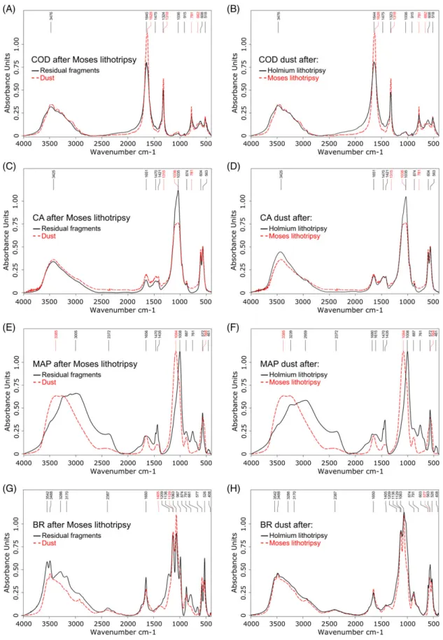
For COM, UA and CYS, the spectra of both RF and dust showed no changes when compared to their respective initial FTIR spectra. For COD, the characteristics bands at 1645 and 1324 cm−1remained unchanged in RF samples, whereas COD dust from conventional Holmium lithotripsy showed a tendency towards a conversion to COM. This was
characterized by displacement of major bands at 1644 and 1323 cm−1, as well as by the appearance of a prominent band at 781 cm−1, which is characteristic for COM. The conversion from COD to COM became even more apparent in COD dust from Moses lithotripsy, with displaced major bands at 1628 and 1318 cm−1, as well as a clearly identifi-able band at 781 cm−1 and the appearance of a discernable 662 cm−1 band (Figure 5-A-B). RF of CA conserved all characteristics from their initial spectrum with prominent bands at 1035, 604 and 563 cm−1, corresponding to phos-phate absorption peaks. In contrast, CA dust from Moses
TABLE 2 Morphological characteristics for each constitutional stone type Stone type Fragment type Morphological description Laser-related peculiarities Stereoscopic microscopy Scanning electron microscopy
COM RF Form: lamellar and radiating organization. Plates with matt smooth surfaces
Form: lamellar organization. Plates with smooth surfaces and few cracks, partially covered by smaller plates
— Color: homogenous, brown.
Dust Form: no visible organization Form: lamellar organization. Randomly shaped plates with <5 layers, sharp edges and smooth surfaces Color: homogenous, brown
COD RF Form: blocks with glossy rough surfaces and blunt translucid bipyramids
Form: blocks with partially conserved smooth bipyramidal organization and other more disorganized rough areas. Many cracks
Moses: more pronounced damages to the crystalline organization
Color: beige to yellow-brown.
Dust Form: no visible organization Form: randomly shaped particles with sharp edges and a mixture of smooth and rough surfaces. Loss of bipyramidal organization Color: heterogenous mixture of beige and
yellow-brown particles
UA RF Form: blocks with glossy rough surfaces Form: blocks with rough surfaces consisting of rather conserved smooth crystals
— Color: brown-orange
Dust Form: no visible organization Form: partially conserved crystalline organization with rather smooth edges and a mixture of smooth and rough surfaces. Few cracks
Color: heterogenous mixture of brown and orange particles
CA RF Form: blocks with matt bumpy surfaces on a background of small round particles.
Form: blocks with partly smooth and partly granular surfaces, many cracks. The granular surface organization is due to small spherical compounds
— Color: beige blocks, white round particles
Dust Form: no visible organization. Form: randomly shaped blocks with sharp edges and smooth to bumpy surfaces. Loss of spherical compounds Color: homogenous white-beige.
MAP RF Form: blocks with glossy smooth surfaces on a background of sharp baguettes
Form: blocks consisting of
mono-directional longitudinal needles encased in a constituent forming an amalgam with many cracks
Moses: greater tendency to separate needles from their encasement Color: white
Dust Form: no visible organization Form: randomly configured needles encased in an amalgam Color: homogenous white
BR RF Form: radial translucid glossy baguettes, Form: entangled longitudinal baguettes with smooth or needle-like surfaces. Deep cracks
Moses: areas of hexagonal plate-like surfaces at the tip of a larger baguette (Figure 4), as well as on dust particles. Color: translucid-white to yellow-brown
Dust Form: randomly overlapping baguettes Form: sharp and thin baguettes, few agglomerations of round particles Color: white-beige.
CYS RF Form: blocks with glossy rough surfaces, visible layers, few areas of discernable hexagonal organization
Form: blocks with a layered organization, smooth surfaces and recognizable hexagonal edges
Moses: many cracks and deep imprints.
Color: white-beige to yellow-brown Dust Form: glossy particles without visible
organization
Form: randomly shaped blocks with a layered organization, smooth surfaces and barely recognizable hexagonal edges
Color: homogenous, beige
Abbreviations: BR, brushite; CA, carbapatite; COD, calcium oxalate dihydrate; COM, calcium oxalate monohydrate; CYS, cystine; MAP, magnesium ammonium phos-phate; RF, residual fragments; UA, uric acid.
lithotripsy had a major band that moved towards 1038 cm−1 with a flattened shape signing a change towards an amor-phous phase (Figure 5C-D). Interestingly, this loss of crys-talline phase allowed for the appearance of clearly distinguishable bands at 1315 and 781 cm−1 in CA dust from Moses lithotripsy, thus, undercovering COM content in this sample. This COM arguably resulted from laser-induced conversion of the COD constituent that was present in the initial sample. MAP samples did not show any changes com-pared to their initial MAP spectrum, except for MAP dust from Moses lithotripsy where a profound loss of the ammo-nium bands at 1470 and 1435 cm−1, a displacement of 1008 cm−1 band towards 1084 cm−1 and a loss of water content was found (Figure 5E-F). Finally, for BR, conven-tional Holmium lithotripsy did not change the spectrum of RF, whereas there was a heightening of the indentation between the 1063 and 987 cm−1 bands in RF of BR after Moses lithotripsy, signing a higher proportion of apatite. The change from BR towards CA became even more evident on dust samples from both conventional Holmium and Moses lithotripsy, with the disappearance of a 987 and 661 cm−1 band, a profound disturbance of the 1136 and 1063 cm−1 bands, the appearance of 603 and 563 cm−1 bands and loss of water content (Figure 5-G-H).
4 | D I S C U S S I O N
To the best of our knowledge, the present study is the first of its kind providing a comprehensive morpho-constitutional analysis of disintegration products occurring during Hol-mium lithotripsy. The seven most frequently encountered stone types in our institution were analyzed [16]. Depending on major crystalline constituents, stone dust either conserved
all attributes found in larger RF or showed changes in the crystalline organization. Particularly, stone dust particles from COM, UA and CYS were found to have the same crys-talline organization as in RF. In contrast, stone dust from COD, CA, MAP and BR showed fundamental morpho-constitutional changes. Of interest, changes seemed to be more pronounced in samples issued from Moses lithotripsy.
Schafer et al. proposed that stone melting and recrystalli-sation occurs during Holmium lithotripsy [19]. This hypoth-esis was based on the observation of filamentous stone particles at microscopy after Holmium lithotripsy of pigmen-ted biliary stones. Such conclusions may not be valid for uri-nary stones based on the findings of the current study, because both RF and stone dust issued from COM, UA and CYS seemed to preserve all their morpho-constitutional characteristics. In line with these findings, a previous study involving SEM analysis of COM stones showed that lasered stone craters had a similar lamellar morphology found in non-ablated areas of stone fracture [20]. However, that study only analyzed stone craters, contrarily to the present report where stone fragments themselves were analyzed.
A previous matched-pair comparison of RF with stone dust showed 74% concordance in stone constituents, with intact FTIR spectra in all UA dust sample and complete loss of MAP spectra in all according to dust samples [11]. In line with those findings, UA dust indeed conserved morphological-constitutional characteristics of RF in the pre-sent study. As for MAP, a fundamentally novel characteristic was discovered by SEM analysis in both RF and dust sam-ples, which revealed an organization of longitudinal white needles encased in another constituent. An according change of FTIR spectrum was found in MAP dust from Moses litho-tripsy, which showed changes towards a differing amor-phous and crystalline phase.
FIGURE 1 Holmium laser lithotripsy and sample preparation. A, Laser lithotripsy performed under direct visual control with a Lumenis P120H Holmium laser generator, a 200μm laser fiber and a Lithovue fURS. B, Separation of stone dust from RF using a method based on the floating ability of particles which can escape through a 5 mm hole drilled 2 cm from the bottom of the container (red circle). C, Separate collection of irrigation overflow containing dust particles
Several further peculiar observations such as the con-version of COD towards COM, changes in CA spectra towards an amorphous phase as well as the appearance of hydroxyapatite on BR fragments suggest that stones were subject to a direct photothermal effect from Holmium laser in this study. Considering that—at equivalent pulse energy—Moses technology may deliver superior laser beam through vapor channel compared to conventional Holmium laser, it may have been that higher local tempera-tures occurred during Moses lithotripsy. This would explain
why more pronounced morpho-constitutional changes were observed in Moses samples compared to conventional Hol-mium lithotripsy. The direct photothermal effect has been supported by prior chemical analyses which revealed ther-mal decomposition products after Holmium lithotripsy, including the appearance of hydroxyapatite in laser impact craters on BR stone surface, in line with the findings of the present study [3]. Other crystalline phase changes, such as COM to calcium carbonate, UA to cyanide, BR to CA, hydroxyapatite to calcium pyrophosphate, MAP to
FIGURE 2 Morphological analysis by stereoscopic microscopy. Each stone constituent was analyzed by stereoscopic microscopy at magnifications of 1× and 4.5×. Dried RF and stone dust were put on carbon conductive double-faced adhesive tapes to allowed subsequent morphological analysis by SEM. A blue plastic marker was placed on the COM samples for orientation purposes during sample positioning in the SEM machine
ammonium carbonate and magnesium carbonate as well as CYS to free sulfur and cysteine suggest that a photothermal effect of Holmium laser causes urinary stones to assume their more stable crystalline form [3, 4].
A limitation of this study was the lack of a clear defini-tion of stone dust at the time the study was conducted. Var-ious definitions can be found in literature, mostly describing fragments <1 mm which cannot be retrieved by stone baskets [21, 22]. In this study, a genuine methodol-ogy relying on the floating ability of particles was proposed for separation of RF from stone dust. The rationale for this separation method was the observation of spontaneous evacuation of stone dust during retrograde surgery in our clinical practice, which seemed to be related to the floating ability of particles using an irrigation pressure of 40 cm
FIGURE 3 Morphological analysis by scanning electron microscopy. Each stone constituent was analyzed by SEM at magnifications of 100× and 500×. Low electron beam voltage (≤2 keV) was used and no sample coating or fixation was needed image acquisition
FIGURE 4 Plate-like hydroxyapatite. SEM analyzes areas of hexagonal plate-like surfaces at the tip of BR baguettes after Moses lithotripsy
FIGURE 5 Constitutional analysis by Fourier-transform infrared spectroscopy. Comparison between RF and stone dust revealed spectra changes for several constitutional stone types. A. Conversion from COD towards COM in dust after Moses lithotripsy. B, The conversion from COD towards COM in dust was more pronounced after Moses lithotripsy compared to conventional Holmium lithotripsy. C, Changes towards an amorphous phase in CA dust after Moses lithotripsy with flattening and displacement of the 1035 cm−1band. D, A spectral change of CA dust was only found after Moses lithotripsy, but not after conventional Holmium lithotripsy. E, Changes towards a differing and amorphous crystalline phase in MAP dust after Moses lithotripsy. F, A spectral change of MAP dust was only found after Moses lithotripsy, but not after conventional Holmium lithotripsy. G, Changes from BR towards CA after Moses lithotripsy. H, Spectral changes were found in CA dust both after Moses and conventional Holmium lithotripsy
H20 through the fURS working channel. We referred this phenomenon as the “snow globe effect”. Further studies are warranted to verify the validity of this new separation method of stone disintegration products. Also, further studies shall evaluate whether other experimental settings such as altered laser parameters or various morphological stone subtypes (eg, Ia instead of Id) would impact on the present findings.
5 | C O N C L U S I O N S
Morpho-constitutional analysis of stone disintegration prod-ucts reveals yet uncovered insight into the ablative effect of Holmium laser on urinary stones. Depending on major crys-talline constituents, stone dust either conserves all attributes found in larger RF or shows changes in crystalline organiza-tion. Moses technology seems to produce more pronounced changes. These findings suggest a photothermal effect of Hol-mium laser on stones and shall be considered for future studies on laser lithotripsy, as stone dust may not adequately reflect crystalline organization of the stones before laser lithotripsy.
A C K N O W L E D G M E N T S
Dr. E.X.K. is supported by a Travel Grant from the Univer-sity Hospital Zurich and by a grant from the Kurt and Senta Herrmann Foundation. Dr. V.D.C. is supported by a EUSP scholarship from the European Association of Urology and by a grant from the Belgische Vereniging voor Urologie (BVU). Prof. O.T. is a consultant for Coloplast, Rocamed, Olympus, EMS, Boston Scientific and IPG.
A U T H O R B I O G R A P H I E S
Please see Supporting Information online.
O R C I D
Etienne X. Keller https://orcid.org/0000-0003-1667-7609
Vincent de Coninck https://orcid.org/0000-0002-4983-5055
Olivier Traxer https://orcid.org/0000-0002-2459-3803
R E F E R E N C E S
[1] S. Pierre, G. M. Preminger, World J Urol 2007, 25, 235.
[2] E. D. Jansen, T. G. van Leeuwen, M. Motamedi, C. Borst, A. J. Welch, Lasers Surg Med 1994, 14, 258.
[3] K. F. Chan, G. J. Vassar, T. J. Pfefer, J. M. H. Teichman, R. D. Glickman, S. T. Weintraub, A. J. Welch, Lasers Surg Med 1999, 25, 22.
[4] G. J. Vassar, K. F. Chan, J. M. H. Teichman, R. D. Glickman, S. T. Weintraub, T. J. Pfefer, A. J. Welch, J Endourol 1999, 13, 181. [5] K. F. Chan, T. J. Pfefer, J. M. H. Teichman, A. J. Welch, J Endourol 2001,
15, 257.
[6] J. M. Isner, A. R. Lucas, Br J Hosp Med 1988, 40, 172.
[7] M. M. Elhilali, S. Badaan, A. Ibrahim, S. Andonian, J Endourol 2017, 31, 598. [8] J. M. H. Teichman, G. J. Vassar, J. T. Bishoff, G. C. Bellman, J Urol 1998,
159, 17.
[9] A. H. Aldoukhi, W. W. Roberts, T. L. Hall, K. R. Ghani, Front Surg 2017, 4, 57.
[10] C. A. Dauw, L. Simeon, A. F. Alruwaily, F. Sanguedolce, J. M. Hollingsworth, W. W. Roberts, G. J. Faerber, J. S. Wolf Jr., K. R. Ghani, J Endourol 2015, 29, 1221.
[11] E. R. Ray, G. Rumsby, R. D. Smith, BJU Int 2016, 118, 618.
[12] M. Daudon, A. Dessombz, V. Frochot, E. Letavernier, J.-P. Haymann, P. Jungers, D. Bazin, C R Chim 2016, 19, 1470.
[13] J. E. Santiago, A. B. Hollander, S. D. Soni, R. E. Link, W. A. Mayer, Curr Urol Rep 2017, 18, 32.
[14] B. R. Matlaga, B. Chew, B. Eisner, M. Humphreys, B. Knudsen, A. Krambeck, D. Lange, M. Lipkin, N. L. Miller, M. Monga, V. Pais, R. L. Sur, O. Shah, J Endourol 2018, 32, 1.
[15] D. Bazin, Ann Biol Clin 2015, 73, 517.
[16] M. Daudon, P. Jungers, in Urolithiasis: Basic Science and Clinical Prac-tice (Eds: T. H. Talati, A. D. M. JJ, Z. Ye), Springer, London, UK 2012, p. 113.
[17] J. Cloutier, L. Villa, O. Traxer, M. Daudon, World J Urol 2015, 33, 157. [18] L. Estepa, M. Daudon, Biospectroscopy 1997, 3, 347.
[19] S. A. Schafer, F. M. Durville, B. Jassemnejad, K. E. Bartels, R. C. Powell, IEEE Trans Biomed Eng 1994, 41, 276.
[20] G. J. Vassar, J. M. H. Teichman, R. D. Glickman, J Urol 1998, 160, 471. [21] J. Sea, L. M. Jonat, B. H. Chew, J. Qiu, B. Wang, J. Hoopman, T. Milner,
J. M. H. Teichman, J Urol 2012, 187, 914.
[22] M. Kang, H. Son, H. Jeong, M. C. Cho, S. Y. Cho, World J Urol 2016, 34, 1591.
How to cite this article: Keller EX, de Coninck V, Audouin M, et al. Fragments and dust after Holmium laser lithotripsy with or without “Moses technology”: How are they different? J. Biophotonics. 2019;12: e201800227.https://doi.org/10.1002/jbio.201800227