chemokine-blocking antibodies efficacious in arthritis
The MIT Faculty has made this article openly available.
Please share
how this access benefits you. Your story matters.
Citation
Angelini, Alessandro, et al. “Directed Evolution of Broadly
Crossreactive Chemokine-Blocking Antibodies Efficacious in
Arthritis.” Nature Communications, vol. 9, no. 1, Dec. 2018. © 2018
The Authors
As Published
http://dx.doi.org/10.1038/s41467-018-03687-x
Publisher
Nature Publishing Group
Version
Final published version
Citable link
http://hdl.handle.net/1721.1/118861
Terms of Use
Creative Commons Attribution 4.0 International License
Directed evolution of broadly crossreactive
chemokine-blocking antibodies ef
ficacious in
arthritis
Alessandro Angelini
1,2,3
, Yoshishige Miyabe
4
, Daniel Newsted
1
, Byron H. Kwan
1,5
, Chie Miyabe
4
,
Ryan L. Kelly
1,5
, Misha N. Jamy
1
, Andrew D. Luster
4
& K. Dane Wittrup
1,2,5
Chemokine receptors typically have multiple ligands. Consequently, treatment with a
blocking antibody against a single chemokine is expected to be insufficient for efficacy. Here
we show single-chain antibodies can be engineered for broad crossreactivity toward multiple
human and mouse proin
flammatory ELR
+CXC chemokines. The engineered molecules
recognize functional epitopes of ELR
+CXC chemokines and inhibit neutrophil activation
ex vivo. Furthermore, an albumin fusion of the most crossreactive single-chain antibody
prevents and reverses in
flammation in the K/BxN mouse model of arthritis. Thus, we report
an approach for the molecular evolution and selection of broadly crossreactive antibodies
towards a family of structurally related, yet sequence-diverse protein targets, with general
implications for the development of novel therapeutics.
DOI: 10.1038/s41467-018-03687-x
OPEN
1Koch Institute for Integrative Cancer Research, Massachusetts Institute of Technology, 500 Main Street, Cambridge, MA 02139, USA.2Department of Chemical Engineering, Massachusetts Institute of Technology, 77 Massachusetts Avenue, Cambridge, MA 02139, USA.3Department of Molecular Sciences and Nanosystems, Ca’ Foscari University of Venice, Via Torino 155, Venezia Mestre 30172, Italy.4Center for Immunology and Inflammatory Diseases, Division of Rheumatology, Allergy and Immunology, Massachusetts General Hospital, Harvard Medical School, 149 Thirteenth Street, Charlestown, MA 02129, USA.5Department of Biological Engineering, Massachusetts Institute of Technology, 77 Massachusetts Avenue, Cambridge, MA 02139, USA. Correspondence and requests for materials should be addressed to A.A. (email:[email protected]) or to K.D.W. (email:[email protected])
123456789
C
hronic inflammatory diseases usually involve multiple
ligands that act synergistically through promiscuous and
diverse receptors
1. This complexity is exemplified by the
ELR
+CXC chemokine system, a large family of secreted proteins
that have a prominent function in the development and
pro-gression of numerous inflammatory diseases, including
rheuma-toid arthritis (RA)
2. The ELR
+CXC chemokines are so named
because of the presence of an amino terminal Glu-Leu-Arg (ELR)
amino-acid motif followed by two invariant cysteines (C) that are
separated by a random residue (X)
3. The ELR
+CXC chemokine
system includes many small and structurally similar
chemoat-tractant ligands capable of binding to and activating the related
CXCR1 and CXCR2 G protein-coupled receptors (GCPR)
expressed on the surface of neutrophils
4. These ligands function
either by autocrine or paracrine mechanisms to induce signaling
networks that direct neutrophils to sites of inflammation.
Importantly, increased levels of ELR
+CXC chemokines have
been detected in the sera, synovial
fluid, and synovial tissue of
patients with RA
5–8. Studies in animal models have demonstrated
that genetic deletion of the most promiscuous ELR
+CXC
che-mokine receptor, CXCR2, can block the development of joint
inflammation in anti-type II collagen antibody-induced arthritis
9,
adjuvant-induced arthritis
10–12, and K/BxN serum
transfer-induced arthritis
13,14. This evidence indicates that the ELR
+CXC chemokine signaling network is an attractive therapeutic
target for the treatment of arthritic diseases
15.
Inhibition of ELR
+CXC chemokine-driven signaling has been
attempted with various antagonists against CXCR1 and CXCR2
receptors, including neutralizing antibodies, small molecules, and
peptide-derived inhibitors. Despite the broad variety of
approa-ches, these conventional receptor-based therapies have mostly
had limited success in the clinic
16,17. Failures have often been
attributed to (i) differences between the orthologous rodent
(pre-clinical) and human ((pre-clinical) systems and (ii) the extremely high
doses of antagonist required to guarantee continuous receptor
occupancy, such that all receptors in the body are
antag-onized
16,17. The latter phenomenon is particularly problematic, as
CXCR1 and CXCR2 undergo rapid internalization cycles (with a
half-life of 6‒8 h) and are expressed on neutrophils, which are the
most abundant (40‒75%) of the circulating leukocytes and have
short circulating half-life (6–8 h)
18. As a result, antagonists that
target these receptors are cleared quickly, reducing the amount of
drug available in circulation. Efforts to develop more effective
receptor-based therapies have led to the discovery of
non-a
b
c
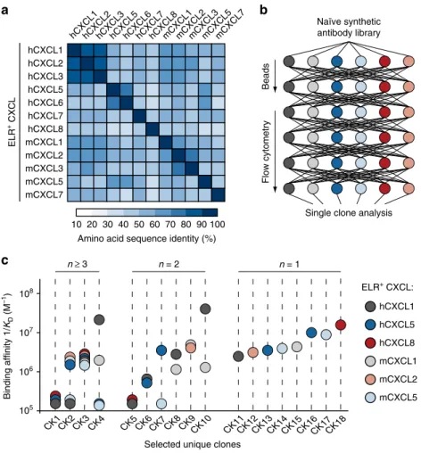
CK1CK2 CK3 CK4 CK5 CK6CK7 CK8
108
CK9CK10 CK11CK12CK13CK14CK15CK16
Selected unique clones
n ≥ 3 n = 2 n = 1 107 106 105 CK17CK18 mCXCL1 hCXCL1 mCXCL2 hCXCL5 mCXCL5 hCXCL8 hCXCL2 hCXCL3 hCXCL6
Amino acid sequence identity (%) mCXCL7 mCXCL3 hCXCL7 30 4050 60 70 80 90100 20 10 mCXCL1 hCXCL1hCXCL2hCXCL3 hCXCL6hCXCL5hCXCL7hCXCL8mCXCL2mCXCL3mCXCL5mCXCL7 Binding affinity 1/ KD (M –1 ) hCXCL1 hCXCL5 hCXCL8 mCXCL5 mCXCL1 mCXCL2 ELR+ CXCL: ELR + CXCL Naïve synthetic antibody library Beads Flow cytometry
Single clone analysis
Fig. 1 Isolation of crossreactive antibodies toward multiple ELR+CXC chemokines.a Heat map displaying the sequence identity among multiple human and murine ELR+CXC chemokines. The color of each element in the heat map indicates the sequence identity percentage, ranging from 10% (white) to 100% (dark blue).“h” and “m” indicates human and murine CXC chemokines, respectively. b Schematic representation of the iterative selection pathways applied to isolate crossreactive molecules from a naïve library of synthetic antibodies displayed on the surface of yeast. Two cycles of magnetic bead screening followed by four cycles offlow cytometry sorting were applied. c Plot of the binding affinities of 18 unique yeast-displayed synthetic antibody protein binders (CK) selected using six diverse human (hCXCL1, hCXCL5, and hCXCL8) and murine (mCXCL1, mCXCL2, and mCXCL5) ELR+CXC chemokine ligands. Each chemokine and its corresponding binding affinity values are reported as differently colored filled circles and indicate the means of at least three independent experiments. Data are presented as inverse equilibrium binding constants (1/KD; M−1)
competitive allosteric modulators of CXCR1 and CXCR2
19,20.
This class of inhibitors seems to provide unique advantages over
conventional drug formats and is being tested in advanced clinical
trials
21.
One
alternative
approach
to
inhibit
CXCR1
and
CXCR2 signaling is the blockade of the ELR
+CXC chemokine
ligands, which are often spatially confined to precise anatomical
locations and might enable improved drug accumulation and
specificity. However, generating synthetic compounds that
antagonize these ligands has proven difficult, owing to their small
size and lack of molecular pits or grooves. Many monoclonal
antibodies targeting single ELR
+CXC chemokines with high
affinity and specificity have been developed, but, despite their
potency and low toxicity, single neutralizing antibody-based
therapies have failed to block disease progression
22–27. This
limited therapeutic efficacy is often attributed to the multifactorial
and redundant nature of the ELR
+CXC chemokine system.
Consistent with this hypothesis, therapeutic intervention using a
cocktail of two or three monoclonal antibodies has resulted in
synergistic potency, suggesting that augmented efficacy might be
achieved by neutralizing multiple ligands at once
24,28.
In the present study, we use yeast-display technology to
engi-neer serum albumin (SA)–antibody fusions that can
simulta-neously block multiple orthologous human and mouse ligands,
thus providing the advantages of broad neutralization within a
single
molecule.
Importantly,
these
fusions
demonstrate
a
e
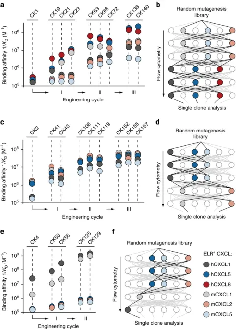
c
108 107 106 105 108 107 106 105 108 109 107 106 105 I II III CK1 CK19 CK21 CK23 CK63 CK66 CK72 CK138CK140 Engineering cycle Engineering cycle I II III CK2 CK41CK43 CK108CK111CK119 CK152CK155CK157 I II CK4 CK50 CK56 CK125CK129 Engineering cycle Random mutagenesis librarySingle clone analysis
Flow cytometry
Random mutagenesis library
Single clone analysis
Flow cytometry
Random mutagenesis library
Single clone analysis
Flow cytometry
d
b
f
Binding affinity 1/ KD (M –1) Binding affinity 1/ KD (M –1 ) Binding affinity 1/ KD (M –1 ) hCXCL1 hCXCL5 hCXCL8 mCXCL5 mCXCL1 mCXCL2 ELR+ CXCL:Fig. 2 Molecular co-evolution of antibody affinity and crossreactivity. Plots displaying binding affinities of engineered clones derived from a CK1, c CK2, and e CK4 lineages. Two independent processes of selection (I and II), each including the generation of random yeast-display antibody libraries and six cycles offlow cytometry sorting, followed by a third round of site-directed mutagenesis (III), were performed. ELR+CXC chemokines and their corresponding binding affinity values are reported as differently colored filled circles and indicate the means of at least three independent experiments. Data are presented as inverse equilibrium binding constants (1/KD; M−1). Selection pathways applied to isolate crossreactive molecules from a mutagenized yeast-display
synthetic antibody library that yieldedb CK1-, d CK2-, and f CK4-derived clones with improved binding affinity and crossreactivity. Each pathway comprisesfive to six cycles of flow cytometry sorting
promising prophylactic and therapeutic efficacy in vivo in the K/
BxN mouse model of inflammatory arthritis. Thus, we show that
directed evolution could be used to develop next-generation
therapeutics against multiple redundant pathological factors.
Results
Isolation of crossreactive antibodies. To evolve highly
cross-reactive protein binders toward multiple proinflammatory ELR
+CXC chemokines, we used synthetic single-chain variable
anti-body fragment libraries displayed on the surface of yeast
29,30. For
selection purposes, we chose to target three human (hCXCL1,
hCXCL5, and hCXCL8) and three murine (mCXCL1, mCXCL2,
and mCXCL5) chemokines based on their low sequence identity
(Fig.
1
a and Supplementary Table
1
) and proven therapeutic
relevance
8,14. We produced the selected chemokines and
con-firmed their purity as well as their biological activity based on
their ability to mobilize intracellular calcium in primary
Frequency (%) 100 10 CK1 lineage mutations CDR FWR
b
S98 Y100B Y100C N101 R6 H29 D30 A33 D6 A52 P55 A107 CDR FWR CK138: K103 100 10 F27 L29 K43 K44 S54 Y58 Frequency (%) E S P E E P Dc
E103 S27 P29 E43 E44 P54 D58 T100B T35 CK2 lineage mutations CDR FWR CK157: CDR FWRd
CK1 lineage CK2 lineage 100 10 1 0.1 0.01 Y100C Y100B S98 N101 CDR-H3 + + + + + – + + – + + + + + + – + – + – – + + – – – + – – – – – E103 E44 E43 100 10 1 FWR-VL FWR-VH + + + + + – – + + – + – – – – hCXCL1 hCXCL5 hCXCL8 mCXCL1 mCXCL2 mCXCL5a
0 4 6 8 2 10 0 5 1 2 3 4 Number m u tated residues F requency m utations (%)*
****
CK1 derived clones 0 2 4 6 8 0 1 2 3 4 F requency m utations (%) Number m u tated residues***
****
CK2 derived clonesInp Out Inp Out
0 2 4 6 8 0 1 2 3 4 F requency m utations (%) Number m u tated residues CK4 derived clones
Inp Out Inp Out
**
ns S32 G98 H100B H100C S100D D101 P S Y Y T N Binding / display (10 –3) ELR+ CXCL: I II n = 20/10 20/11Inp Out Inp Out
I II
n = 20/9 20/16
I II
neutrophils (Supplementary Fig.
1
a–d). To favor the selection of
crossreactive antibodies, we implemented a combinatorial
approach in which the output of each selection cycle was exposed
to a diverse array of ELR
+CXC chemokines in the next cycle
(Fig.
1
b). We took advantage of highly avid reagents preloaded
with ELR
+CXC chemokines to ensure that weak crossreactive
antibodies were also isolated. Subsequent DNA sequencing of
selected clones revealed the presence of 18 unique antibody
fragments with varying amino-acid compositions and loop
lengths within the complementarity-determining regions (CDRs).
Selected antibodies exhibited diverse affinities and specificities for
soluble ELR
+CXC chemokines (Fig.
1
c, Supplementary Fig.
2
,
and Supplementary Table
2
). When monomeric chemokines were
used to assess binding, only four of these clones (CK1–CK4)
recognized at least three different ELR
+CXC chemokines. The
most abundant and crossreactive of these antibodies (CK3)
recognized the biotinylation sequence located at the C-terminus
of each chemokine, thus explaining its exquisite promiscuity and
similar binding affinities for the various chemokines. However,
CK1, CK2, and CK4 remained as promising crossreactive
can-didates for further development. The remaining 14 antibody
clones were either bi-specific (CK5–CK10) or mono-specific
(CK11–CK18). Overall, this selection strategy yielded three
crossreactive antibodies that occurred at lower frequency within
the total selected variant pool and had weaker binding affinities
compared with the mono- and bi-specific antibodies.
Molecular co-evolution of af
finity and crossreactivity. In order
to further improve both the binding affinity and crossreactivity of
CK1, CK2, and CK4 clones, we deviated from our previous
selection strategy, which focused only on crossreactivity, and
implemented a novel selection approach to co-evolve affinity and
crossreactivity simultaneously. We
first applied error-prone PCR
amplification to introduce genetic diversity into our
antibody-encoding genes. We then promoted the selection of clones
dis-playing increased binding affinity by allowing the mutants to
evolve through sequential cycles of equilibrium-based selection
using decreasing concentrations of ELR
+CXC chemokines.
Concomitantly, we forced the development of crossreactivity by
exposing the outputs of each affinity selection cycle towards a
different ELR
+CXC chemokine in the following cycle. During
this iterative process, we solely collected variants whose affinity
and crossreactivity towards ELR
+CXC chemokines was higher
than that of their respective parental clones. After two iterative
evolutionary processes, each comprising
five to six consecutive
cycles of selection, we sequenced the isolated clones and assessed
their binding affinity and crossreactivity towards ELR
+CXC
chemokines (Supplementary Figs.
3
–
5
and Supplementary
Table
3
). Additionally, for cases in which we found distinct
affinity-conferring mutations scattered across different clones, we
combined mutations together to investigate the possibility of even
further promiscuity and higher affinity. A summary of the overall
co-evolutionary approach, including two iterative selection
pro-cesses for crossreactivity and affinity (I and II) and a third cycle of
combinatorial mutagenesis (III), is shown in Fig.
2
a, c, e.
This two-pressure selection strategy yielded antibodies with
improved affinity and, in most cases, increased crossreactivity
toward multiple ELR
+CXC chemokines. For example, the
engineered CK138 clone recognized double the number of
chemokines (from three to six) and achieved roughly a 30- to
340-fold improvement in affinity toward these chemokines (K
Dvalues ranging from 5.8 to 193 nM) relative to the parental CK1
clone. Similarly, the CK157 clone displayed crossreactivity toward
five targets and added a 20- to 55-fold improvement in affinity
(K
Dvalues ranging from 16.9 to 57.1 nM) as compared to the
initial CK2 clone. Finally, while CK129 retained only minimal
crossreactivity toward two targets, we observed a significant
increase in affinity of 50- and 800-fold, respectively, toward
human hCXCL1 (K
D= 0.79 nM) and its mouse homolog
mCXCL1 (K
D= 0.93 nM).
Importantly, the sequential order in which the ELR
+CXC
chemokine targets were exposed to the antibody mutant libraries
was critical to the success of the selection process. Among all the
possible selection pathways, we observed improvements in both
affinity and crossreactivity only when recombinant genetic
libraries were screened in order from lowest to highest affinity
chemokines (Fig.
2
b, d). Note that this was not applicable to the
development of CK129, as its parental clone (CK4) already
possessed high initial affinity toward hCXCL1 and mCXCL1, but
negligible affinity towards the other chemokines (Fig.
2
f). Overall,
our two-pressure selection approach promoted the evolution of
crossreactive molecules with improved affinity and revealed the
importance of the selection pathway for the achievement of
crossreactivity.
Crossreactive antibodies have a high level of mutations.
Although we applied reaction conditions that allowed, on average,
one to two amino-acid mutations per gene, selected clones from
each round of sorting showed higher mutation rates (Fig.
3
a).
While the crossreactive antibody CK138 predominantly gathered
mutations within the CDRs, CK157 collected numerous
muta-tions within the framework regions (FWRs) (Fig.
3
b, c and
Supplementary Fig.
6
). Both types of mutations were critical, as
reversion of either CDR or FWR mutations to the wild-type
amino acids resulted respectively in a loss of affinity of CK138
and CK157 toward ELR
+CXC chemokines (Fig.
3
d). Moreover,
Fig. 3 Frequency and distribution of mutations in crossreactive antibodies. a Box-and-whiskers graph comparing the total number (lefty-axis) and frequencies (righty-axis) of mutated residues detected in CK1-, CK2-, and CK4-derived clones before (light gray) and after (dark gray) the first (I) and second (II) process of selection, respectively.“Inp” indicates sequenced clones picked from the random yeast-display antibody library before selection (input).“Out” indicates sequenced clones after selection (output). “n” indicates samples size. The middle line within each box represents the median, and the lower and upper boundaries of the box indicate the 25th (Q1) and 75th (Q3) percentiles. Whiskers represent the 1.5× interquartile range (IQR= Q3–Q1) extending beyond box. Statistical comparisons were made between each group using one-way analysis of variance (ANOVA), followed by Tukey’s test to calculateP-values: *P < 0.05, **P < 0.01, ***P < 0.001; ****P < 0.0001. ns: non-significant. Homology model and frequencies of enriched mutations of engineeredb CK138 and c CK157 antibodies. Left, the VLand VHbackbones are represented as ribbons (light gray). Mutations acquired during the
selection process are depicted as spheres at the Cα positions. Mutated amino acids belonging to CDR loops of CK138 and CK157 are colored in dark blue and dark red, respectively. Diversified amino acids belonging to FWR regions of CK138 and CK157 are colored in light blue and light red, respectively. Right, columns graph reporting the mutation frequency in CDR (dark gray) and FWR (light gray) regions. Only amino-acid mutations of CK1 and CK2 lineages that showed at least 20% frequency and were enriched through two iterative processes of selection are reported. Wild type and mutated amino acids are listed at the top and bottom, respectively.d Fluorescence binding signal of CK1- (left) and CK2- (right) derived clones bearing highly frequent mutations within the CDR-H3 and FWRs, respectively, that were reverted to the wild-type amino acids. ELR+CXC chemokines and the corresponding binding/display values (y-axis) are indicated as differently colored filled circles and represent the means of at least three independent experiments
these mutations were found throughout different clones and
cycles of engineering, suggesting strong selection pressure for
these residues in conferring high crossreactivity and affinity
(Supplementary Figs.
3
a and
4
a).
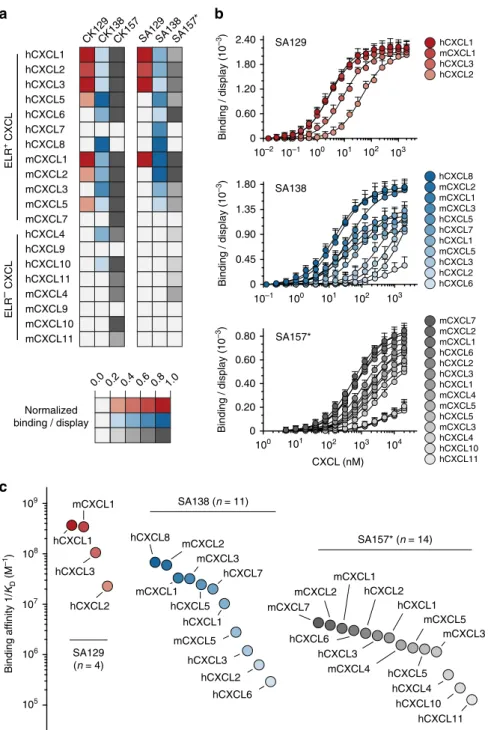
Engineered antibodies bind a large array of chemokines. To
assess the extent of crossreactivity of the engineered antibodies,
we characterized their binding affinity towards 20 human and
murine CXC chemokines. The chemokine panel included 12
human and mouse ELR
+CXC chemokines (which share 32–90%
sequence identity) and 8 human and mouse ELR
−CXC
che-mokines (which share 18–70% sequence identity). The ELR
+CXC chemokines share 20–51% sequence identity with the ELR
−CXC chemokines (Supplementary Fig.
7
a). In order to accurately
determine the K
Dvalues of these antibodies for the different
chemokines, we utilized two complementary configurations of
chemokines and antibodies in the context of yeast surface display
(Supplementary Fig.
7
b). Specifically, we performed titrations
using (i) soluble CXC chemokines with yeast-displayed antibodies
and (ii) soluble antibodies with yeast-displayed CXC chemokines
(Supplementary Fig.
7
c, d). Exploring both orientations was
necessary as some CXC chemokines are known to form oligomers
when present in high concentrations in solution, leading to
undesired multivalent binding phenomena. We produced the
CXC chemokines as fusions to the N-terminus of mouse SA
(Supplementary Fig.
1
e–h) and the engineered CK129, CK138,
and CK157 single-chain variable antibody fragments as fusion to
the C-terminus of SA, which are referred to as SA129, SA138, and
SA157* (Supplementary Fig.
8
and Supplementary Table
4
).
SA157* is denoted with an asterisk as instead of being produced
as a single chain with a linker, it was produced as separate V
Land
V
Hdomains and mixed in equimolar amounts. In both
orienta-tions, we observed similar crossreactivity of our engineered
antibodies towards CXC chemokines that were not included in
the selection cycles (Fig.
4
a). Yeast-displayed CK129, CK138, and
CK157 bind 7, 12, and 16 soluble CXC chemokines, respectively
(Fig.
4
a and Supplementary Fig.
7
f). Similarly, the soluble SA129,
SA138, and SA157* bind 4, 11, and 14 yeast-displayed CXC
chemokines, respectively (Fig.
4
a, b). With a few exceptions, the
K
Dvalues determined by using soluble SA129, SA138, and
SA157* antibody fusions with yeast-displayed CXC chemokines
were on average 2- to 5-fold higher than those measured in the
opposite arrangement (Supplementary Fig.
7
g and Supplementary
Table
5
). This discrepancy in measured K
Dvalues and extent of
crossreactivity between the two assay orientations was not
sur-prising and may reflect oligomeric CXC chemokines interacting
with multiple yeast-displayed antibodies, thus confounding
avidity effects with higher affinity. This phenomenon appears to
be pronounced for ELR
−CXC chemokines, such as hCXCL10
and hCXCL4, that are known to form highly avid oligomers in
solution
31.
We also observed that SA129, which only recognizes four
chemokines that share significant sequence identity, displays
relatively high affinity for those targets. In contrast, highly
crossreactive SA138 and SA157* had overall lower binding
affinities toward a larger array of targets, suggesting an inverse
correlation between affinity and extent of crossreactivity (Fig.
4
c
and Supplementary Fig.
7
h).
To assess that the crossreactivity was not merely due to
non-specific polyreactivity, we characterized the binding of our
engineered antibodies towards
five structural related CC
chemokines and eleven structurally unrelated proteins, including
the chemotactic factor C5a (Supplementary Fig.
7
e). No or weak
binding was detected in the case of CK129 and CK138,
confirming their specificity toward members of the CXC
chemokine family. Contrariwise, CK157 exhibits crossreactivity
toward C5a and some (CCL20, CCL22, CCL28) but not all CXC
and CC chemokines, suggesting a more promiscuous mode of
binding.
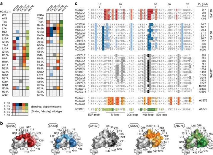
Crossreactive antibodies recognize functional epitopes. We
next performed
fine epitope mapping using alanine-scanning
mutagenesis to identify the residues that are directly involved in
the interactions
32. hCXCL1 was chosen as our model chemokine
over other ELR
+CXC chemokines because it is recognized by all
the engineered crossreactive antibodies and is well-characterized
biochemically
33,34. We
first combined three-dimensional
struc-tural analysis and literature data to identify hCXCL1 amino acids
suitable for mutagenesis. Structurally buried hydrophobic amino
acids and proline and cysteine residues were left unaltered, as
they are crucial for overall chemokine folding and stability
(Supplementary Fig.
9
a). Fifty-four predicted solvent-exposed
hCXCL1 residues were selected, individually mutated to alanine,
expressed on the surface of yeast (Supplementary Fig.
9
b), and
screened for decreased binding affinity to the soluble SA129,
SA138, and SA157*. Five mutants that exhibited an intense loss of
binding upon incubation with all three antibodies were excluded,
as this phenomenon was likely due to protein misfolding and
destabilization of the displayed variants (Supplementary Fig.
9
c).
We then assessed the binding of the remaining 49 hCXCL1
mutants to soluble SA129, SA138, and SA157*. Solvent-exposed
mutations that eliminated or significantly reduced binding
affi-nity were identified, which allowed us to pinpoint residues that
were likely critical for the interactions (Fig.
5
a and Supplementary
Table
6
). We further identified the epitopes of two commercially
available neutralizing antibodies: the highly specific Ab275 (binds
only hCXCL1) and the crossreactive Ab276 (binds hCXCL1,
hCXCL2, and hCXCL3). Next, we compared their epitope maps
with the maps assigned to our engineered antibodies (Fig.
5
a and
Supplementary Table
6
). Similarly to Ab275 and Ab276, SA129
and SA138 bind motifs along the functional N- and 40s-loops
that are known to be crucial for the binding of hCXCL1 to its
cognate receptor, CXCR2. In contrast, SA157* recognizes a
dis-tinctive epitope and engages binding with hCXCL1 residues that
are important for its interaction with glycosaminoglycans (GAGs;
Fig.
5
b). These epitope maps are also consistent with results from
a competitive assay (Supplementary Fig.
9
d). Notably, the
resi-dues recognized by the highly crossreactive SA138 and SA157*
are conserved among many different CXC chemokines, thus
explaining these antibodies’ broad promiscuity (Fig.
5
c).
We also observed that the total number of residues involved in
binding and the frequency of interaction strengths are different
across the characterized antibodies. The relatively more specific
Ab275, Ab276, and SA129 engage binding with a larger number
of hCXCL1 residues than the more crossreactive SA138 and
SA157*, and strong interactions are more frequent. In contrast,
the binding specificity of SA138 and SA157* appear to be
achieved mostly through weak interactions toward a few
conserved residues (Supplementary Fig.
9
e, f). These observations
are consistent with affinity measurements, which show that the
broadly crossreactive SA138 and SA157* display lower affinity
toward their targets compared to the more specific SA129. Taken
together, epitope mapping and competition assays revealed that
our engineered crossreactive molecules recognize distinct
con-served epitopes on CXC chemokines. Data also suggest that
crossreactivity towards a large array of chemokines is achieved
through weak recognition of a few highly conserved residues.
Crossreactive antibodies inhibit chemokine-binding in vitro.
As a measure of potential therapeutic efficacy, we then tested the
ability of crossreactive SA129, SA138, and SA157* antibody
fusions to inhibit binding of ELR
+CXC chemokines to their
cognate CXCR1 and CXCR2 receptors. To this end, we
used HEK293 cell lines expressing human CXCR1 and CXCR2
(Supplementary Fig.
10
a, b). We then incubated the cells
with various concentrations of hCXCL1 and hCXCL8 ligands to
determine the half-maximal effective concentrations (EC
50) of the
interactions (Supplementary Fig.
10
c and Supplementary
Table
7
). Next, we examined the ability of SA129, SA138,
and
SA157*
to
antagonize
the
interactions
between
hCXCL1 and hCXCL8 ligands and their cognate receptors.
Our engineered antibodies inhibited the ability of human
hCXCL1 and hCXCL8 chemokines to bind CXCR1 and CXCR2
receptors in a dose-dependent manner to various extents
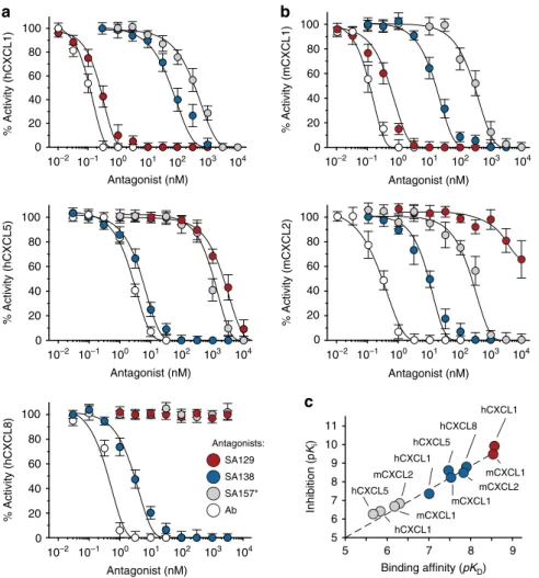
a
hCXCL1 hCXCL2 hCXCL3 hCXCL5 hCXCL6 hCXCL7 hCXCL8 mCXCL1 mCXCL2 mCXCL3 mCXCL5 mCXCL7 hCXCL4 hCXCL9 hCXCL10 hCXCL11 mCXCL4 mCXCL9 mCXCL10 mCXCL11 ELR + CXCL CK157CK129CK138 SA129SA138SA157*
b
hCXCL8 mCXCL2 mCXCL1 mCXCL3 hCXCL5 hCXCL7 hCXCL1 mCXCL5 hCXCL3 hCXCL2 hCXCL6 mCXCL7 mCXCL2 mCXCL1 hCXCL6 hCXCL2 hCXCL3 hCXCL1 mCXCL4 mCXCL5 hCXCL5 mCXCL3 hCXCL4 hCXCL10 hCXCL11 1.0 0.2 0.4 0.6 0.8 0.0 mCXCL1 hCXCL1 hCXCL3 hCXCL2 SA138 0 0.20 0.40 0.60 0.80 Binding / display (10 –3) SA157* CXCL (nM) SA129 Normalized binding / displayc
mCXCL7 mCXCL1 hCXCL6 hCXCL2 hCXCL3 hCXCL1 mCXCL4 mCXCL5 hCXCL5 mCXCL3 hCXCL4 hCXCL10 hCXCL11 hCXCL8 mCXCL2 mCXCL1 mCXCL3 hCXCL5 hCXCL7 hCXCL1 mCXCL5 hCXCL3 hCXCL2 hCXCL6 hCXCL1 mCXCL1 hCXCL3 hCXCL2 SA138 (n = 11) SA157* (n = 14) SA129 (n = 4) mCXCL2 ELR – CXCL 10–2 Binding / display (10 –3 ) Binding / display (10 –3 ) 103 102 101 100 10–1 103 102 101 100 103 104 105 106 107 108 109 102 101 100 10–1 0 0.45 0.90 1.35 1.80 0 0.60 1.20 1.80 2.40 Binding af finity 1/ KD (M –1)Fig. 4 Crossreactivity of engineered antibodies toward multiple CXC chemokines. a Heat map indicating the binding intensity of the engineered antibodies against 20 diverse human and murine CXC chemokines. Binding was assessed byflow cytometry using two cell-display arrangements: soluble CXC chemokine against yeast-displayed CK129 (red), CK138 (blue), and CK157 (gray) antibodies (on the left) and soluble serum albumin–antibody fusions SA129 (red), SA138 (blue), and SA157* (gray) against yeast-displayed CXC chemokines (on the right). Normalized binding/display signal intensities range from light to dark colors indicating low (0.0‒0.2) and high (0.8‒1.0) titers, respectively. b Binding isotherms of yeast-displayed CXC chemokines to soluble serum albumin–antibody fusions SA129, SA138, and SA157*. Equilibrium binding affinity (KD) values were determined only for chemokines exhibiting
signals at high concentrations of soluble antibody fusions. CXC chemokines are gradient colored ranging from dark (high affinity) to light (low affinity) red (SA129), blue (SA138), and gray (SA157*). Data are presented as mean (dots) ± s.e.m. (bars).c Plot showing binding affinities of yeast-displayed CXC chemokines to SA129 (red), SA138 (blue), and SA157* (gray) antibody fusions. The indicated values are displayed as differently coloredfilled circles and represent the means of at least three independent experiments presented as inverse equilibrium binding constants (1/KD; M−1)
(Supplementary Fig.
10
d and Supplementary Table
8
).
Remark-ably, the determined inhibitory constants (K
i) correlated well with
the previously reported K
Dvalues (Supplementary Fig.
10
e).
These results showed that crossreactive SA129, SA138, and
SA157* can interfere with the binding of ELR
+CXC chemokines
to both human CXCR1 and CXCR2 in vitro.
We further assessed the ability of the SA129, SA138, and
SA157* to antagonize the activation of ELR
+CXC chemokine
receptors. For this purpose, we utilized an intracellular calcium
mobilization assay in the presence of human- and mouse-derived
neutrophils activated respectively with human (hCXCL1,
hCXCL5, and hCXCL8) and murine (mCXCL1 and mCXCL2)
ELR
+CXC chemokines. To this end, we
first determined the
EC
50of these chemokines on the neutrophils (Supplementary
Fig.
10
f and Supplementary Table
9
). Then, we monitored
changes in intracellular calcium levels upon pre-incubation of
ELR
+CXC chemokines with varying concentrations of SA129,
SA138, and SA157* as antagonists. Commercial neutralizing
monoclonal antibodies were also used as a positive control. The
assays revealed that our engineered antibodies exhibited
encoura-ging inhibitory activity by preventing binding of the human and
murine ligands to their receptors in a dose-dependent manner
(Fig.
6
a, b and Supplementary Table
10
). Again, the calculated K
ivalues correlated well with the previously determined K
Daffinities
(Fig.
6
c). Taken together, these data indicate that engineered
crossreactive molecules are able to inhibit ELR
+CXC chemokine
signaling in vitro and ex vivo, and have the promise to suppress
CXCR1 and CXCR2 activation in vivo.
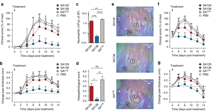
SA–antibody fusion reverses inflammation in vivo. Given the
promising results from our inhibitory assays, we then tested the
effect of our engineered antibody fusions in the K/BxN serum
transfer model of autoantibody-induced arthritis
35, which
Q13 T14 L15 I18 G47 R48 A50 N46 G17 N22 K45 R8 L12 Q13 T14 L15 I18 G47 R48 A50 N46 I18 G47 N46K45 I58 K65 K71 H19 K21 N22 T14 L15 I18 G47 R48 A50 N46 G17 Q16I58 H19 N22 K61 K71 K45 K65 K21 10 20 60 70 KD (nM) hCXCL1 hCXCL2 hCXCL3 hCXCL1 Ab276 Ab275 hCXCL8 mCXCL2 mCXCL1 mCXCL3 hCXCL5 hCXCL1 mCXCL5 hCXCL3 hCXCL2 hCXCL6 14.7 16.7 29.4 31.1 33.7 96.7 357 836 1590 SA138 5000 N.ELRCQCLQTLQG-IHLKN..PGP..ATLK-NGQKACLNP..KKIIEKMLKNGK.C N.ELRCQCIKTYSKPFHPKF..SGP..VKLS-DGRELCLDP..QRVVEKFLKRAE.C N.ELRCQCLQTMAG-IHLKN..SGP..ATLK-NGREACLDP..QKIVQKMLKGVP.C N.ELRCQCLNTLPR-VDFET..PGP..ATLK-DGQEVCLNP..QIIIKKILKSGK.C N.ELRCQCLKTLPR-VDFKN..PGP..ATLK-GGQKVCLDP..QKIIQKILNKGK.CN.ELRCVCLTVTPK-INPKL..AGP..AKLK-NQKEVCLDP..KKIIQKILGSDK.C N.ELRCQCLQTLQG-IHLKN..PGP..ATLK-NGKKACLNP..QKIIEKILNKGS.C N.ELRCQCLQTLQG-IHPKN..PGP..ATLK-NGRKACLNP..KKIIEKMLNSDK.C N.ELRCVCLQTTQG-VHPKM..IGP..ASLK-NGKEICLDP..KKVIQKILDGGN.C
N.ELRCTCLRVTLR-VNPKT..AGP..ASLK-NGKQVCLDP..KKVIQKILDSGN.C mCXCL7 mCXCL2 mCXCL1 hCXCL6 hCXCL2 hCXCL3 hCXCL1 mCXCL4 mCXCL5 hCXCL5 mCXCL3 hCXCL4 hCXCL10 hCXCL11 528 591 666 751 853 1034 1433 1770 2018 2125 2647 16033 32135 43890 SA157*
N.ELRCQCLQTMAG-IHLKN..SGP..ATLK-NGREACLDP..QKIVQKMLKGVP.C N.ELRCTCLRVTLR-VNPKT..AGP..ASLK-NGKQVCLDP..KKVIQKILDSGN.C N.ELRCQCLKTLPR-VDFKN..PGP..ATLK-GGQKVCLDP..QKIIQKILNKGK.C N.ELRCRCTNTISG-IPFNS..PGV..ATLK-NGQKTCLDP..KRIVMKILEGYC
N.ELRCQCLQTLQG-IHPKN..PGP..ATLK-NGRKACLNP..KKIIEKMLNSDK.C N.DLSCVCVKTISSGIHLKH..AGR..ATLK-NGRKICLDR..KKVIKKILESC N.ELRCQCLQTLQG-IHLKN..PGP..ATLK-NGKKACLNP..QKIIEKILNKGS.C N.ELRCQCLQTLQG-IHLKN..PGP..ATLK-NGQKACLNP..KKIIEKMLKNGK.C
N.ELRCVCLQTTQG-VHPKM..IGP..ASLK-NGKEICLDP..KKVIQKILDGGN.C N.ELRCVCLTVTPK-INPKL..AGP..AKLK-NQKEVCLDP..KKIIQKILGSDK.C N.TVRCTCISISNQPVNPRS..ASQ..ATMKKKGEKRCLNP..KNLLKAVSKERS.C N.RGRCLCIGPGVKAVKVAD..PSN..ITLKENKGQRCLNP..RLIIKKVERKNFC N.DLQCLCVKTTSQ-VRPRH..AGP..ATLK-NGRKICLDL..KKIIKKLLESC N.ELRCQCLNTLPR-VDFET..PGP..ATLK-DGQEVCLNP..QIIIKKILKSGK.C
N-loop 30s-loop 40s-loop 50s-loop ELR motif
50
N.ELRCQCLQTLQG-IHPKN..PGP..ATLK-NGRKACLNP..KKIIEKMLNSDK.C hCXCL1 mCXCL1 hCXCL3 hCXCL2 2.7 2.9 9.2 43.6 SA129
N.ELRCQCLQTLQG-IHLKN..PGP..ATLK-NGKKACLNP..QKIIEKILNKGS.C N.ELRCQCLQTLQG-IHLKN..PGP..ATLK-NGQKACLNP..KKIIEKMLKNGK.C N.ELRCQCLQTMAG-IHLKN..SGP..ATLK-NGREACLDP..QKIVQKMLKGVP.C N.ELRCQCLQTLQG-IHPKN..PGP..ATLK-NGRKACLNP..KKIIEKMLNSDK.C
N.ELRCQCLQTLQG-IHPKN..PGP..ATLK-NGRKACLNP..KKIIEKMLNSDK.C N.ELRCQCLQTLQG-IHLKN..PGP..ATLK-NGKKACLNP..QKIIEKILNKGS.C N.ELRCQCLQTLQG-IHLKN..PGP..ATLK-NGQKACLNP..KKIIEKMLKNGK.C
c
WT A4S T5A E6A L7A R8A Q10A L12A Q13A T14A L15A Q16A G17A I18A H19A K21A N22A Q24A S25A N27A K29A S30A G32A H34A A36S SA157* SA129SA138 hCXCL1: Ab276Ab275a
0 . 0 0 0 . 2 5 0 . 5 0 0 . 7 5 1 . 0 0(Binding / display) mutants (Binding / display) wild-type
Q37A T38A T43A K45A N46A R48A A50S A55S S56A K60A E64A M66A G47A K49A N53A I58A K61A K65A L67A N68A S69A K71A N73A D70A S72A SA157* SA129SA138 Ab276Ab275
b
SA129 SA138 SA157* Ab276L12 Q13 T14 L15 I18 G47 R48 A50 N46 Q10 Q16 G17 N22 K49 K45 N53 R8 Ab275
Fig. 5 Epitope mapping of crossreactive antibodies. a Binding of SA129 (red), SA138 (blue), SA157 (gray), Ab275 (green), and Ab276 (orange) to a defined panel of hCXCL1 alanine mutants was assessed byflow cytometry. Obtained median values were normalized to the display median fluorescence intensities of each single yeast surface displayed mutant (binding/display). Normalized values represent the means of at least three independent experiments. Mutations that do not significantly affect binding (0.75‒1.0) are shown in white, while mutations that weakly (0.5‒0.75), moderately (0.25‒0.5), or strongly (0.0‒0.25) disrupt binding are shown respectively in light, intermediate, and dark colors. b The identified contact residues of hCXCL1 (PDB ID: 1MGS) to each antibody as defined by epitope mapping are shown in red (SA129), blue (SA138), gray (SA157*), green (Ab275), and orange (Ab276). The color intensity correlates with the strength of the interaction, with weak and strong interactions shown as light and dark colors, respectively.c Sequence alignment of various CXC chemokine proteins. Positions of conserved solvent-exposed residues that appear to be involved in the interaction with SA129 (red), SA138 (blue), SA157* (gray), Ab275 (green), and Ab276 (orange) based on residues identified using hCXCL1 alanine mutants are shown. Amino-acid sequences have been listed based on binding affinity (KD), with the tightest CXC chemokine protein at the top and the weakest at the bottom. Upper
case N and C letters indicate the N- and C-terminus of the amino-acid sequence, respectively. Regions including residues that are not involved in binding are not reported for space reasons. The regions denoting the ELR-motif, N-loop, 30s-loop, 40s-loop, and 50s-loop that are known to be crucial for the binding of ELR+CXC chemokines to the cognate CXCR2 receptor are indicated at the bottom. Residues have been highlighted according to the strength of interaction determined using soluble antibodies against hCXCL1 alanine mutants, as shown in panela
displays clinical and histopathological similarities to human
RA
36. The levels of ELR
+CXC chemokines are elevated in the
joints of this arthritic mice and neutrophils, which have
upre-gulated CXCR2 in the joint, are the main effector cells, making
the K/BxN serum transfer-induced arthritis mice a valid model to
test the therapeutic efficacy of our engineered binders
13,14,37,38.
To antagonize circulating ELR
+CXC chemokines in vivo, we
generated SA–antibody fusions (Supplementary Fig.
8
). Fusion of
our engineered antibody fragments to SA improved solubility and
stability. In addition, SA is non-immunogenic, and the fusion
molecules could be produced in high yields conducive to high
drug doses
39. Furthermore, similarly to full-length antibody, SA
exploits the FcRn receptor to achieve a prolonged circulatory
half-life. Its plasma persistence is nonetheless still shorter than
that of full-length monoclonal antibodies, thus circumventing the
“buffering” effects that are experimentally observed with the use
of antibodies targeting small antigens
40–43. Unlike an antibody,
SA does not bind the FcγR receptors expressed on immune cells,
thus precluding additional immune activation and inflammation
mediated by antibody-dependent cell-mediated cytotoxicity
(ADCC)
44. Finally, SA has been shown to accumulate at high
levels in inflamed joints, making it a promising drug carrier for
RA
45. For this study, we used the SA129 and SA138 fusions
described earlier. We excluded SA157* because of its limited
solubility and crossreactivity towards non ELR
+CXC
chemo-kines (Supplementary Figs.
7
e and
8
d). As a negative control, we
used an irrelevant SA fusion (SA
CTR) encoding SA fused to an
antibody fragment that targets the human carcinoembryonic
antigen (CEA), a protein that does not exist in mice
46. To ensure
complete inhibition of all ELR
+CXC chemokines present in
circulation, we used fairly high doses of our engineered antibody
fusions (50 mg kg
−1). When injected into mice, SA129, SA138,
and SA
CTRdisplayed plasma half-lives between 42 and 47 h,
considerably longer than that of small synthetic compounds or
antibody fragments, but still shorter than that of full-length
monoclonal antibodies (Supplementary Fig.
11
a and
Supple-mentary Table
11
). Despite the high doses of SA129, SA138, and
SA
CTRused, the molecules were well tolerated. Treated mice
gained weight and exhibited good body condition
(Supplemen-tary Fig.
11
b). We initially assessed the ability of crossreactive
antibody fusions to prevent the manifestation of inflammatory
arthritis in the K/BxN serum transfer model. Mice were treated
on the same day that the arthritogenic serum was injected
(Supplementary Fig.
11
c), and disease progression was evaluated
by both blinded clinical scores and measurements of ankle
thickness. Mice treated with the more crossreactive SA138, which
binds all four murine ELR
+CXC chemokines (mCXCL1,
mCXCL2, mCXCL3, and mCXCL5), were protected from
devel-oping arthritis, with an approximately 80% reduction of clinical
score compared with negative controls at the peak of the disease
(day 8 after arthritogenic K/BxN serum transfer and disease
initiation). In contrast, the more specific SA129 that recognizes
a
b
0 20 40 60 80 100 % Activity (hCXCL1) Antagonist (nM) Antagonist (nM) Antagonist (nM) Antagonist (nM) SA129 Antagonists: SA138 SA157* Ab Antagonist (nM) % Activity (mCXCL1) % Activity (hCXCL5) % Activity (mCXCL2) % Activity (hCXCL8)c
5 6 7 8 9 5 6 7 8 9 10 11 hCXCL1 mCXCL1 hCXCL8 mCXCL2 hCXCL1 mCXCL1 hCXCL5 mCXCL2 mCXCL1 hCXCL1 hCXCL5 Binding affinity (pKD) Inhibition (p Ki ) 104 103 102 101 100 10–1 10–2 104 103 102 101 100 10–1 10–2 10–2 10–1 100 101 102 103 104 104 103 102 101 100 10–1 10–2 104 103 102 101 100 10–1 10–2 0 20 40 60 80 100 0 20 40 60 80 100 0 20 40 60 80 100 0 20 40 60 80 100Fig. 6 Crossreactive antibodies inhibit ELR+CXC chemokine signaling in vitro. Residual activity ofa human hCXCL1, hCXCL5, and hCXCL8 and b mouse mCXCL1 and mCXCL2 chemokines incubated with varying concentrations of SA129 (red), SA138 (blue), and SA157* (gray) fusions, and commercial neutralizing antibodies (Ab, white). The indicated values are the means of three independent experiments.c Plot displaying pKiversus the calculated pKDof
just one murine ELR
+CXC chemokine (mCXCL1) only
mod-erately reduced joint inflammation, with an approximately 30%
reduction of clinical score at day 8. Mice treated with SA
CTRshowed the typical clinical signs of untreated mice that received
arthritogenic serum and developed inflammatory arthritis with
pronounced joint swelling. There were no differences between
mice treated with SA
CTRor with vehicle (PBS) only (Fig.
7
a, b).
We next quantified the number of synovial fluid neutrophils
isolated from the arthritic joints of mice treated with SA129,
SA138, and SA
CTRantibody fusions. Synovial
fluids were
harvested at the peak of the disease (day 8 after disease
initiation). We observed that mice treated with the broadly
crossreactive SA138 had, respectively, 50- and 70-fold lower levels
of synovial
fluid neutrophils than mice treated with the more
specific SA129 or the irrelevant SA
CTR(Fig.
7
c). These data are
consistent with previous clinical score measurements and
resembled those observed for CXCR2-deficient mice (Cxcr2
−/−)
injected with arthritogenic serum
13,14. We also performed
histological analysis and scoring of inflamed ankle sections.
Inflammatory cell infiltration and pannus formation were absent
or minimally present in mice treated with the broadly
cross-reactive
SA138
(Fig.
7
d,
e).
Consistent
with
previous
clinical
findings, the joints of mice treated with arthritogenic
serum and control SA
CTRdisplayed abundant inflammatory cell
infiltration and pannus formation. These pathological changes
were present, though less pronounced, in mice treated with the
more specific SA129. In addition, the number of von Willebrand
factor (vWF)-positive endothelial cells (ECs) in the joint tissue of
mice treated with SA138 was lower than the number of
vWF-positive ECs in the joint tissue of mice treated with SA129 or
SA
CTR, suggesting that SA138 also prevented angiogenesis
(Supplementary Fig.
12
).
Finally, we tested the therapeutic efficacy of crossreactive
antibody fusions in mice with established arthritis. Arthritic mice
were treated 4 days after arthritogenic serum transfer, when joint
inflammation had developed (Supplementary Fig.
11
d). The
highly crossreactive SA138 rapidly reversed inflammation and
improved disease outcome with nearly 60% reduction of clinical
score and 0.3 mm of ankle thickness over control at the peak of
the disease (day 8 after disease initiation). The specific
SA129-treated mice exhibited only a modest reduction of both clinical
scores (~25%) and ankle thickness (0.1 mm) at day 8. The SA
CTRand vehicle-treated mice showed no difference in the rate of
disease development (Fig.
7
f, g). Taken together, these data show
that the broadly crossreactive SA138 molecule is capable of
blocking neutrophil infiltration in the synovial tissues, thus
preventing and even reversing inflammatory arthritis.
Discussion
Chronic inflammatory diseases usually involve multiple ligands
and receptors acting in concert. As a result, therapies targeting a
Time (days post treatment)
2 4 6 8 10 12 14
0
Change paw thickness (mm)
–0.2 0.0 0.2 0.4 0.6 0.8
a
c
b
10.0 Neutrophils (10 2/μ L of SF) 1.0 0.1 0.01 SA129 SA138 SA CTRf
SA129 SA138 SACTR PBS TreatmentChange paw thickness (mm)
g
SA129SA138 SACTR
PBS
Time (days post treatment)
4 6 8 10 12 14 –0.4 –0.2 0.0 0.2 0.4 Treatment **** ** *** 6.0 Histopathological score 0.0 SA129 SA138 SA CTR
d
** ns * Time (days post treatment)2 4 6 8 10 12 14 0 0 20 40 60 80 100
Clinical score (% of max)
SA129 SA138 SACTR PBS Treatment 120
Clinical score (% of max)
SA129 SA138 SACTR
PBS
Time (days post treatment) 0 20 40 60 80 100 4 6 8 10 12 14 Treatment 120
e
SA138 T N 200 μm SA129 200 μm T N SA CTR 200 μm T N 5.0 4.0 3.0 2.0 1.0Fig. 7 Crossreactive serum albumin–antibody fusion reverses inflammation in vivo. a Clinical score (% of max) and b change in ankle thickness (mm) of mice treated with serum albumin–antibody fusion proteins on day 0 (preventative regimen). Arthritogenic serum was injected into C57BL/6J on days 0 and 2. Mice were also treated daily with SA129, SA138, and SACTRfusions (1 mg per mouse in PBS i.p.) beginning on day 0. Paw thickness of ten mice per group (n = 10), pooled from two independent experiments, were measured every 2 days for a total of 14 days. Arrows indicate first day of treatment. Data are presented as mean (dots) ± s.e.m. (bars).c Columns graph reporting the number of infiltrating synovial fluid neutrophils (Ly6G+cells) from the ankles of serum-transferred arthritic mice measured at day 8 byflow cytometry (n = 3 per condition). Statistical comparisons were made between each group using one-way analysis of variance (ANOVA), followed by Tukey’s test to calculate P-values: *P < 0.05, **P < 0.01, ***P < 0.001; ****P < 0.0001. ns: non-significant. d Columns graph reporting the histopathological scoring and e representative H&E staining of ankle tissue sections of mice treated with SA129 (top), SA138 (middle), and control SACTR(bottom) on day 8. Scale bar represents 200μm. White arrows indicate joint-infiltrating inflammatory cells, and red arrows indicate pannus formation. T taulus, N navicular.f Clinical score (% of max) and g change in ankle thickness (mm) of K/BxN serum-induced arthritic mice treated beginning on day 4 with serum albumin–antibody fusion proteins (therapeutic regimen). Arthritogenic serum was injected into C57BL/6J on days 0 and 2, and mice were then treated daily i.p. with SA129, SA138, and SACTRfusions (1 mg per mouse in PBS i.p.) beginning on day 4 after inflammation had developed. Paw thickness of ten mice per group (n = 10), pooled from two independent experiments, was measured every 2 days for a total of 14 days. Arrows indicate the day treatment began. Data are presented as mean (dots) ± s.e.m. (bars)
single pathological factor are often insufficient to achieve desired
clinical outcomes, as has proven to be the case with
anti-chemokine therapies. To overcome this limitation, we engineered
highly crossreactive proteins capable of simultaneously blocking
multiple human and murine ELR
+CXC chemokines.
Highly crossreactive antibodies are challenging to obtain using
traditional methodologies involving animal immunization and
hybridoma development. The immune system tends to remove
self-reactive antibodies, making it difficult to generate in vivo
antibodies against sequence- and structurally related antigens
derived from different species. In contrast, in vitro antibody
libraries associated with display technologies are unaffected by
immune tolerance
47. Here we applied a multiple-pressure
itera-tive combinatorial approach for the isolation of
chemokine-blocking antibodies with broad crossreactivity. Even though none
of the engineered antibodies could recognize with high affinity all
members of ELR
+CXC chemokine family, this still represents, to
the best of our knowledge, the
first systematic study reporting the
selection strategy for the in vitro directed evolution of binders
with such extensive promiscuity towards a panel of structurally
related, yet sequence-diverse, protein targets. Nevertheless, our
in vitro engineering approach is not without limitations. Broadly
crossreactive antibodies were complex to develop, often had weak
binding affinities and greater number of mutations compared
with the more specific antibodies, which could lead to potential
instability and immunogenicity.
The biochemical characterization of crossreactive binders
iso-lated in this study allowed us to uncover some interesting
fea-tures. We observed that the binding mode of in vitro evolved
crossreactive CK138 and CK157 antibodies appear to resemble
those found in naturally existing crossreactive ELR
+CXC
chemokine-binding proteins. First, both the length and the
amino-acid composition and distribution of the CDR-H3 loop of
the crossreactive CK138 resembles that of the N-terminal
extra-cellular binding loop of the promiscuous CXCR2 receptor
48.
Moreover, CK138 binds ELR
+CXC chemokine residues that are
known to be recognized by that same N-terminal extracellular
loop, supporting the idea that both promiscuous molecules share
similar binding mode (Supplementary Fig.
13
a). Second, the
crossreactive CK157 accumulated numerous surface-exposed
FWR mutations at the dimerization interface between the V
Land V
Hchains. These mutations resulted in the replacement of
positive charges with negatively charged residues, allowing the
development of a defined structural patch that resembles the one
present in the naturally evolved highly crossreactive viral
chemokine-binding proteins (vCKBPs)
49,50. Epitope mapping
data revealed the ability of the highly crossreactive CK157 to
recognize the positively charged GAG-binding residues of ELR
+CXC chemokines. This supports the hypothesis that the binding
mode of CK157 could be similar to that of vCKBPs, where a large
negatively charged area engages conserved GAG-binding regions
on CC and CXC chemokine proteins, thus explaining their
extensive promiscuity (Supplementary Fig.
13
b).
In vitro evolved crossreactive antibodies also share some
fea-tures present in other crossreactive proteins of the immune
sys-tem
51. Indeed, similar to naturally occurring broadly neutralizing
antibodies, the in vitro evolved crossreactive antibodies
accu-mulated a high number of mutations during the selection process,
and in the case of CK157, mutations were preferentially located in
FWRs
52. Moreover, in vitro evolved crossreactive antibodies
appear to achieve binding to large array of diverse proteins by
engaging a small number of energetically favored hot-spots
(typically structurally and chemically conserved) surrounded by
weaker and more diverse peripheral interactions. This mechanism
of interaction resembles that of
αβ T-cell antigen receptors
(TCR), which bind multiple diverse short peptides presented by
major
histocompatibility
complex
(MHC)
molecules
53,54.
Nevertheless, other molecular mechanisms could also contribute
to high crossreactivity, such as (i) structural
flexibility, (ii)
molecular complementarity, (iii) entropic contributions, and (iv)
the chemical composition of both binding (paratope) and
recognition (epitope) sites (Supplementary Discussion)
55–59.
Using the engineered crossreactive molecules described in the
present study, we proposed a SA–antibody fusion-based strategy
to enable optimal pharmacokinetic profiles, thus overcoming
buffering effect phenomena that have limited previous
interven-tions. Importantly, when these fusions were tested in the murine
K/BxN serum transfer model of inflammatory arthritis, broadly
crossreactive SA138 demonstrated greater therapeutic efficacy
than the more specific molecule SA129, confirming that blocking
multiple neutrophil-active ELR
+CXC chemokines is required to
completely inhibit neutrophil infiltration into the joints and both
prevent and resolve inflammatory arthritis in vivo. Future
research efforts should be oriented toward testing their efficacy in
other models of arthritis in order to assess their therapeutic
potential for the treatment of RA. In addition to inflammatory
disease, we envisage that usage of such a molecule could augment
the efficacy of current cancer treatments. Synergistic therapeutic
antitumor immunity effects of CXCR2 blockade combined with
chemotherapy
60or anti-PD1 immunotherapy
61,62have recently
been shown.
In conclusion, we report here the in vitro engineering of
broadly crossreactive chemokine-blocking antibodies with
pro-mising inhibitory effects in vivo and provide a basis for the
development of next-generation therapeutics. We developed this
concept with crossreactive antibodies against ELR
+CXC
che-mokine ligands, but these studies also have value as a
proof-of-concept for a general approach that could be applied to other
therapeutic interventions targeting multiple soluble factors.
Although many challenges still remain, the ability to evolve
promiscuous and species-crossreactive proteins using directed
evolution techniques might provide not only superior therapeutic
efficacy, but also better assessment of treatment toxicity as well as
simpler and lower cost clinical trials, by facilitating the transition
from pre-clinical models to clinical studies in humans.
Methods
Cloning of chemokine fusion proteins. CXC chemokines were cloned as C-terminal fusion of the immunoglobulin fragment crystallizable (Fc) domain (N Fc-CXCLC) and as N-terminal fusion of the murine SA protein (NCXCL-SAC). All mammalian expression vectors are based on gWiz (Genlantis) containing an optimized human cytomegalovirus promoter and a kanamycin antibiotic resistance gene. Constructs for expression ofNFc-CXCLCfusion proteins were generated by using a modified Pfu DNA polymerase-mediated site-directed mutagenesis pro-tocol63. PfuUltra II Fusion HS DNA polymerase was obtained from Agilent Technologies, DpnI enzyme from New England BioLabs, and the oligonucleotide primers from Integrated DNA Technologies. The synthetic DNA coding for the active form of three highly diverse human (hCXCL1, hCXCL5 and hCXCL8) and murine (mCXCL1, mCXCL2 and mCXCL5) ELR+CXC chemokines were obtained from GeneArt Gene Synthesis (Thermo Fisher Scientific). Genes were codon-optimized for expression in mammalian cells. A sequence encoding for Gly–Gly dipeptide spacer (G2,NGGC) followed by a 15 amino-acid peptide sequence
(AviTag) containing a defined lysine for site-specific biotinylation (N GLNDI-FEAQKIEWHEC) were inserted at the C-terminus of the ELR+CXC chemokine to obtainNCXCL-G
2-AviTagCsynthetic genes. The AviTag sequence for enzymatic
biotinylation was placed at the well tolerated C-terminus of the ELR+CXC che-mokines to (i) preserve unaltered the functional N-terminus region, (ii) avoid loss of epitope recognition, and (iii) prevent additional structural heterogeneity that could be triggered by performing a chemistry-based amine-reactive succinimidyl esters based biotinylation. The de novo synthesizedNCXCL-G2-AviTagCsynthetic
sequences were subsequently inserted into a previously modified gWiz expression vector containing a DNA sequence encoding for a secretory leader peptide sequence (NMRVPAQLLGLLLLWLPGARCC), an Fc domain derived from a murine IgG2 heavy-chain constant regions CH2 and CH3, followed by a sequence
encoding a hexa-histidine tag (His6;NHHHHHHC), an eight amino-acidflexible
linker (NSSGVDLGTC) and a Tobacco Etch Virus proteolytic cleavage site (TEV; NENLYFQ∣A/VC) to obtain thefinalNFc-His