DOI 10.1007/s00345-008-0296-6
T O P I C P A P E R
Complications of retroperitoneoscopic living donor nephrectomy:
single center experience after 164 cases
Alexander Bachmann · Stephen Wyler · Thomas WolV · Lorenz Gürke · Jürg Steiger · Christoph Kettelhack · Thomas C. Gasser · Robin Ruszat
Received: 29 February 2008 / Accepted: 19 May 2008 / Published online: 27 June 2008 © Springer-Verlag 2008
Abstract
Objectives Retroperitoneoscopic living donor nephrec-tomy (RLDN) is used by only a few centers worldwide. Similar to laparoscopic living donor nephrectomy it oVers the donor rapid convalescence and excellent cosmetic results. However, concerns have been expressed over the safety of endoscopic living donor nephrectomy.
Methods We review the results of 164 consecutive RLDN from November 2001 to November 2007. Complications were classiWed into intra- and early postoperative.
Results Mean donor age was 53.4 § 10.7 years (27–79). Left kidneys were harvested in 76% of cases. Mean operation time was 146 § 44 min (55–270), and warm ischemia time 131 § 45 s (50–280). In two patients (1.2%) conversion to open nephrectomy was necessary. The intraoperative compli-cation rate was 3.0%. In the postoperative period we observed in 17.7% minor complications with no persisting impairments for the donor. The rate of major complications in the early postoperative period was 4.3%. Three patients (1.8%) necessitated revision, due to laceration of the external iliac artery in one patient and chyloretroperitoneum in two patients. Mean donor creatinine was 113.1 § 26.6 mg/dl (63–201) on the Wrst postoperative day, and 102.0 § 22.2 mg/dl (68–159) on the Wfth postoperative day.
Conclusion Retroperitoneoscopic living donor nephrec-tomy can be performed with acceptable intraoperative and early postoperative morbidity. Operation times and warm ischemia times are comparable to the open approach.
Keywords Retroperitoneoscopy · Living donor nephrectomy · Complications · Laparoscopy · Minimal-invasive
Introduction
During the past 15 years, the use of minimal invasive renal surgery has increased tremendously. In 1995, Ratner et al. [1] performed the Wrst laparoscopic living donor
nephrec-tomy. Since then, minimally invasive techniques have become more and more favored procedures. In 2000, Gill et al. [2] reported for the Wrst time of a retroperitoneoscopic
approach to donor nephrectomy and autotransplantation where successful allograft outcome was achieved without vascular complications. Extensive data have been published showing that conventional laparoscopic and retroperitoneo-scopic techniques for donor nephrectomy have a similar complication rate [3–5]. Retroperitoneoscopy provides two major advantages: Wrst, it oVers a quick access to the blood vessels comparable to the open approach and second, it has no interference with bowel, liver, or abdominal adhesions. The need to mobilize the colon like in transperitoneal kid-ney surgery is obviated.
Many studies demonstrated an improvement of donor outcome by means of minimally invasive techniques [4–7]. Furthermore, there is evidence that endoscopic minimal-invasive kidney donation can increase the number of kid-ney donations because of shorter hospital stay, earlier return to work, good cosmetic outcome and better overall A. Bachmann · S. Wyler · T. C. Gasser · R. Ruszat (&)
Department of Urology, University Hospital Basel, Spitalstr. 21, 4031 Basel, Switzerland
e-mail: [email protected]
T. WolV · L. Gürke · C. Kettelhack
Department of Surgery, University Hospital Basel, Basel, Switzerland
J. Steiger
Division of Transplantation Immunology and Nephrology, University Hospital Basel, Basel, Switzerland
patient satisfaction [8–11]. More than 60% of the centers in the USA currently perform minimal-invasive donor nephrectomy and numbers are rising in Europe as well.
As kidney donors are healthy individuals, it is important to ensure that the donor operation is safe and allows a quick return to normal activity. Large series of laparoscopic liv-ing donor nephrectomies (LLDN) have reported that a lengthy learning curve is required to make this operation feasible. Furthermore, the complication rate has varied from 6.4 to 16.9% [12].
We reviewed our 6-year single-institution experience of 164 retroperitoneoscopic living kidney nephrectomies (RLDN), evaluating the incidence and management of complications.
Methods
From November 2001 to November 2007, 164 consecutive RLDNs were performed at our institution. Preoperatively, the suitability of all potential donors and recipients was dis-cussed by an interdisciplinary transplantation team. The data obtained included donor age, sex, body mass index, operative time (OT), warm ischemia time (WIT), blood loss, and intra- and postoperative complications. After a complete history, physical examination, and laboratory investigation, contrast enhanced magnet resonance angiog-raphy was used for evaluation of vessel anatomy. If both kidneys were functionally and anatomically equivalent, the left kidney was preferentially selected to provide a longer length of the vein for the implantation process. The right kidney was taken if morphological Wndings such as multi-ple arteries, venous anomalies, or an early arterial branch-ing were present on the left side. Furthermore, the decision was based on the policy of selecting the kidney with the lowest risk of technical failure, but most importantly, leav-ing the donor with the “better” kidney [13, 14]. Our retro-peritoneoscopic technique was described recently in publication [3, 15].
Results
Preoperative donor characteristics and perioperative results Table1 shows detailed preoperative patient characteristics. The mean donor age was 53.4 § 10.7 years with a mean body mass index of 25.6 kg/m2. A total of 66% (n = 109) of the donors were females and 34% (n = 55) males. In 76% (n = 125) of the donors the left kidney was procured. Rea-sons for procurement of the right kidney were in most cases multiple arteries on the left side (n = 19, 49%) and an early left bifurcation (n = 7, 18%). In Wve patients (13%) we found
a right renal artery stenosis and in further Wve patients (13%) venous anomalies making right kidney harvesting necessary.
A total of 19 patients (11.6%) had a history of prior sig-niWcant abdominal surgery (e.g. colon surgery, open chole-cystectomy).
Mean OT was 146 § 44 min, mean WIT, deWned as the time from closure of the artery until clear outXow from the vein of the explanted and perfused kidney, was 131 § 45 s, and mean blood loss was 159 § 108 ml. All planned living donor nephrectomies were performed, and the kidneys were successfully transplanted. The average creatinine level on postoperative day Wve was 102.0 § 22.2 mg/dl compared to 67.5 § 13.1 mg/dl before the operation. The mean length of hospital stay was 6.8 days.
Intraoperative complications
Intraoperative complications are subdivided into minor and major complications (Table2). In the early period after
Table 1 Preoperative donor characteristics and perioperative data
a Including colon surgery, open cholecystectomy, etc
Mean § SD (range) No. (%)
Number of patients 164
Age 53.4 § 10.7 (27–79)
Body mass index (kg/m2) 25.6 § 4.0 (17–45)
BMI < 25 87 (53%) BMI 25–30 61 (37%) BMI > 30 16 (10%) Male 55 (34%) Female 109 (66%) Left kidney 125 (76%) Right kidney 39 (24%)
Reason for right procurement
Multiple arteries left 19 (49%)
Early bifurcation left 7 (18%)
Right renal artery stenosis 5 (13%)
Venous anomalis 5 (13%)
Double left sided pyelon 1 (3%)
Nephrolithiasis right 1 (3%)
Fibromuscular dysplasia 1 (3%)
Prior abdominal surgery in historya
19 (11.6%)
Baseline creatinine (mg/dl) 67.5 § 13.1 (40–140) Creatinine day 1 (mg/dl) 113.1 § 26.6 (63–201) Creatinine day 5 (mg/dl) 102.0 § 22.2 (68–159) Operation time (min) 146 § 44 (55–270) Warm ischemia time (s) 131 § 45 (50–280) Blood loss (ml) 159 § 108 (0–600) Hospitalization time
(post op days)
switching from laparoscopic hand-assisted donor nephrec-tomy to RLDN two patients (cases 11 and 18) needed con-version. In a 71-year-old man a disruption of a fragile atherosclerotic left renal artery occurred, necessitating immediate open conversion and subsequent transfusion. Despite this event, the kidney was harvested safely and implanted successfully. Another patient with two very short veins on the right side was converted to open left kidney donation. The conversion had no persistent impairments for the donor or the recipient.
In a healthy slim 42-year-old female, a totally left hemidi-aphragm rupture during left-sided retroperitoneoscopic donor nephrectomy occurred. This was most likely caused when creating the retroperitoneal working space by blunt balloon dilation. Because the cardiopulmonary situation of the patient remained stable, retroperitoneoscopic donor nephrectomy was performed with our standard technique. Afterwards, the diaphragm was repaired with three 2-0 polyglactin running sutures. Two large 2-mm absorbable clips were applied at the end of each suture to guarantee tightness. The postoperative course of the donor was uneventful.
Furthermore, two kidneys (1.2%) from the right side had renal veins shorter than 2 cm necessitating lengthening by means of v. saphena patch before implantation could be performed. Longer functional follow-up of both grafts was uneventful, although early glomerular function was delayed in these grafts.
In a female donor with a history of repeated pyelonephri-tis, ureteral injury in the region of the ureteropelvic junc-tion occurred. The kidney was harvested, irrigated with cold UW Belzer solution, and an ex vivo pyeloplasty was performed. The kidney was implanted successfully without persisting impairments for the donor or the recipient. Postoperative complications
Postoperative complications are shown in Table2. Most of the complications were minor without persisting disadvan-tages for the donor or the recipient.
After an initial uncomplicated course, three left-sided kidney donors developed chyloretroperitoneum. After 6, 9, and 28 days the patients were readmitted to hospital due to increasing pain in the left lower abdomen. A major retro-peritoneal Xuid accumulation could be demonstrated sono-graphically. The abdominal CT scan revealed a retroperitoneal accumulation of Xuid. In all three cases drainage was initially inserted and the patient’s diet was switched to short- and medium-chain fatty acids. As a sup-porting measure, 0.1 mg somatostatin was administered subcutaneously three times a day. Despite this regime, two donors needed operative revision due to persisting chylus production and could be discharged after 18 and 30 days, respectively. The patient treated conservatively was dis-charged after a period of 45 days.
In a 48-year-old woman with left-sided kidney donation signiWcant hemoglobin decrease was noticed immediately after operation. CT scan revealed bleeding from a lacera-tion of the left external iliac artery. The patient was reoper-ated and the laceration was sutured. The further course was uneventful.
In another donor, bleeding at a trocar incision occurred 2 days after operation, necessitating blood transfusion. The bleeding was controlled with a deep skin suture.
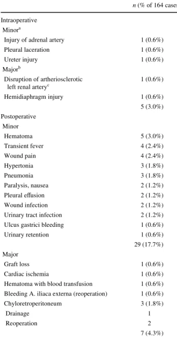
Table 2 Intra- and postoperative complications after
retroperitoneo-scopic donor nephrectomy
a
Minor complications related to surgery, no reoperation required, no eVects on graft function
b
Major complications related to severe disadvantages for the donor, including conversion reoperation, transfusion or eVects on graft function
c Leading to conversion and blood transfusion
n (% of 164 cases) Intraoperative
Minora
Injury of adrenal artery 1 (0.6%)
Pleural laceration 1 (0.6%)
Ureter injury 1 (0.6%)
Majorb
Disruption of artheriosclerotic left renal arteryc
1 (0.6%) Hemidiaphragm injury 1 (0.6%) 5 (3.0%) Postoperative Minor Hematoma 5 (3.0%) Transient fever 4 (2.4%) Wound pain 4 (2.4%) Hypertonia 3 (1.8%) Pneumonia 3 (1.8%) Paralysis, nausea 2 (1.2%) Pleural eVusion 2 (1.2%) Wound infection 2 (1.2%)
Urinary tract infection 2 (1.2%)
Ulcus gastrici bleeding 1 (0.6%)
Urinary retention 1 (0.6%)
29 (17.7%) Major
Graft loss 1 (0.6%)
Cardiac ischemia 1 (0.6%)
Hematoma with blood transfusion 1 (0.6%) Bleeding A. iliaca externa (reoperation) 1 (0.6%)
Chyloretroperitoneum 3 (1.8%)
Drainage 1
Reoperation 2
We observed one postoperative myocardial infarction in a 79-year-old female donor with subsequent intensive care unit observation and uneventful further follow up. In our series one graft loss due to renal artery kinking after left-sided RLDN occurred. Although the diagnosis of impaired arterial blood Xow by doppler ultrasonography led to immediate reoperation, the graft was lost.
Discussion
Living donor transplantation is unique in that it aVects not just a patient but also a healthy donor. There is evidence that endoscopic minimal-invasive kidney donation can increase the number of kidney donations because of shorter hospital stay, earlier return to work, good cosmetic outcomes and bet-ter overall patient satisfaction [9–11, 16, 17]. Furthermore, it has been shown that LLDN and RLDN donors experience less postoperative pain than after open living donor nephrec-tomy over the early postoperative days [4, 18].
Today, most surgeons use the transperitoneal approach, except few clinics who established primarily the retroperi-toneoscopic approach [19–22]. Although the limited work-ing space makes the retroperitoneoscopic approach more demanding compared to the transperitoneal approach, it oVers two major advantages: Wrst, it is possible to operate without interfering with intraperitoneal organs or adhesions in case of prior abdominal surgery. Second, the approach provides quick access to the renal vessels, there is no need for mobilizing the ascending or descending colon which saves operation time. When compared to fully laparoscopic and hand-assisted laparoscopic living donor nephrectomy performed in other centers, we experienced lower OTs and WITs with RLDN [17, 23–26]. Furthermore, we found sim-ilar OTs and WITs compared to our own open series [27].
In 2005, we analyzed the outcome of 65 transplanted kidneys after RLDN and compared the results to 69 kidneys harvested with open nephrectomy [15]. Early functional follow-up showed signiWcant lower 24-h urine output after RLDN. However, the creatinine reduction within 24 h was comparable between the groups and from the seventh post-operative day there was no statistically signiWcant diVer-ence regarding serum creatinine levels. Overall recipient complication rate and total early rejection rate within 30 days were similar within the two groups.
While advantages of decreased pain, faster recovery, and shorter hospital stay are appealing to potential donors, overall safety and graft survival remain paramount in living donor nephrectomy. The relatively long hospitalization time in our series is related to the diVerent health insurance arrangements of patients in European and US American hospitals. Since these are fundamentally diVerent, the personal Wnan-cial pressure to leave the hospital after the operation is low.
With increased attention to donor safety, we examined our results of 164 consecutive RLDNs performed by three surgeons.
We separated donor morbidity into intra- and postopera-tive complications, with rates of 3.0 and 22.0%, respec-tively. One of the largest series in the literature is from the University of Maryland, which reported a 6.8% intraopera-tive and 17.1% postoperaintraopera-tive complication rate with 738 laparoscopic donor nephrectomies [24]. The rate of major complications in our series (5.5%) was in the range of pub-lished data for open (1–6%) or laparoscopic techniques (1– 6.3%) [3, 4, 28].
Our conversion rate was 1.2%, which is comparable to the 1.6–2.8% reported in the literature [24–26]. The two conversions occurred within the Wrst 20 operations (case numbers 11 and 18) and could be attributed to our learning curve.
Recently, we could also show that right kidney harvest-ing is feasible and safe with the retroperitoneoscopic approach [29]. At the beginning of right kidney procure-ment we had two patients with very short renal veins, necessitating vein lengthening using a venous saphena patch. After these initial problems we never experienced diYculties with right kidney donation. We prefer to use a TA-30-2.5 (AutoSuture®) disposable stapler on both artery and vein. It provides an additional 2–3 mm length of the right graft vein, compared to the commonly used Endo-GIA stapler. It is placed parallel and Xushed to the vena cava, obtaining maximal renal vein length by raising the kidney with hand-assistance, thereby extending the renal vein.
In a 42-year-old slim female we observed a completely bisected hemidiaphragm after blunt creation of the retro-peritoneal space with the dilation balloon. Our retrospective complication analysis revealed that the typical bulge of the lateral abdominal wall was less pronounced in this case. Additionally, the inXated volume of approximately 1,200 ml water was probably too much for the slim (BMI 19.8 kg/m2) female donor. The diaphragmatic injury must be seen as a complication of blunt balloon dissection of the retroperitoneum, with the dilatation device placed too close to the diaphragm and inadequate inXated volume, conse-quently leading to its rupture. Pleural injury is an uncom-mon, but potentially serious complication. In a multi-institutional study reviewing 1,765 laparoscopic procedures pleural injury was detected in ten patients (0.6%) [30]. In most cases the diaphragm was injured during kidney dis-section and rarely during inadvertent trocar placement into the pleural cavity. We recommend a careful and body-weight adjusted inXation of volume when water is used for blunt dilatation of the retroperitoneal space. If an air pump is used, endoscopic camera control should be used. Addi-tionally, a correlating bulge of the lateral abdominal wall between chest and iliac crest must be identiWed.
In our series, we observed three patients with chyloretro-peritoneum after a primary uncomplicated left-sided donor nephrectomy. One patient could be treated conservatively; the other two patients necessitated reoperation.
The left renal hilus is surrounded by well-developed lymphatics that often open directly into the cisterna chyli or variably into a lymphatic plexus. It is absolutely necessary to free the renal vessels as much as possible from the sur-rounding tissue. Perihilar and perivascular lymph nodes and lymphatics must be severed and coagulated in the course of this process. During the subsequent detachment of the kid-ney from the perirenal fatty tissue, iatrogenic injury to large lymphatic vessels, which are easy to overlook, may occur, in particular medial to the vascular pedicle. Especially in the case of these vessels, it is rarely possible to attain suY-cient bipolar coagulation of the injury due to the dissection. In addition, minor damage may be overlooked as a result of the suppression of lymph Xow by the pneumretroperito-neum of 10–15 mmHg. Consequently, the lymph/chylous Wstula will only develop postoperatively. Iatrogenic injury should be treated directly with small clips, bipolar cautery, or, if possible, direct suturing with 4-0 monoWlament sutures. As a prophylactic measure to prevent injuring the large lymphatics, care must be taken to achieve deWnitive coagulation of the lymphatics, especially medial to the renal vessels and anterior to the aorta.
Conclusion
The results of our series support the safety of RLDN. The observed complication rate is similar to other published data. We found that pure retroperitoneoscopy has a short learning curve. This is reXected by short operation times, which are comparable to the open approach. The main advantage of RLDN is the quick access to the renal vessels, without interfering with intraperitoneal organs or abdomi-nal adhesions.
ConXict of interest statement There is no conXict of interest.
References
1. Ratner LE, Ciseck LJ, Moore RG, Cigarroa FG, Kaufman HS, Ka-voussi LR (1995) Laparoscopic live donor nephrectomy. Trans-plantation 60(9):1047–1049
2. Gill IS, Uzzo RG, Hobart MG, Streem SB, Goldfarb DA, Noble MJ (2000) Laparoscopic retroperitoneal live donor right nephrec-tomy for purposes of allotransplantation and autotransplantation. J Urol 164(5):1500–1504
3. Bachmann A, Dickenmann M, Gurke L, Giannini O, Langer I, Gasser TC, Steiger J, Sulser T (2004) Retroperitoneoscopic living donor nephrectomy: a retrospective comparison to the open ap-proach. Transplantation 78(1):168–171
4. Ratner LE, Kavoussi LR, Sroka M, Hiller J, Weber R, Schulam PG, Montgomery R (1997) Laparoscopic assisted live donor nephrectomy—a comparison with the open approach. Transplan-tation 63(2):229–233
5. Wolf JS Jr, Marcovich R, Merion RM, Konnak JW (2000) Prospec-tive, case matched comparison of hand assisted laparoscopic and open surgical live donor nephrectomy. J Urol 163(6):1650–1653 6. Stifelman MD, Hull D, Sosa RE, Su LM, Hyman M, Stubenbord W,
Shichman S (2001) Hand assisted laparoscopic donor nephrectomy: a comparison with the open approach. J Urol 166(2):444–448 7. Velidedeoglu E, Williams N, Brayman KL, Desai NM, Campos L,
Palanjian M, Wocjik M, Bloom R, Grossman RA, Mange K, Bark-er CF, Naji A, Markmann JF (2002) Comparison of open, laparo-scopic, and hand-assisted approaches to live-donor nephrectomy. Transplantation 74(2):169–172
8. Chung E, Grant AB, Hibberd AD, Sprott P (2007) Why potential live renal donors prefer laparoscopic nephrectomy: a survey of live donor attitudes. BJU Int 100(6):1344–1346
9. Giessing M, Reuter S, Deger S, Tullmann M, Hirte I, Budde K, Fritsche L, Slowinski T, Dragun D, Neumayer HH, Loening SA, Schonberger B (2005) Laparoscopic versus open donor nephrec-tomy in Germany: impact on donor health-related quality of life and willingness to donate. Transplant Proc 37(5):2011–2015 10. Kuo PC, Johnson LB (2000) Laparoscopic donor nephrectomy
in-creases the supply of living donor kidneys: a center-speciWc micro-economic analysis. Transplantation 69(10):2211–2213
11. Schweitzer EJ, Wilson J, Jacobs S, Machan CH, Philosophe B, Farney A, Colonna J, Jarrell BE, Bartlett ST (2000) Increased rates of donation with laparoscopic donor nephrectomy. Ann Surg 232(3):392–400
12. Nanidis TG, AntcliVe D, Kokkinos C, Borysiewicz CA, Darzi AW, Tekkis PP, Papalois VE (2008) Laparoscopic versus open live donor nephrectomy in renal transplantation: a meta-analysis. Ann Surg 247(1):58–70
13. Goldfarb DA (2001) Are concerns over right laparoscopic donor nephrectomy unwarranted? J Urol 166(6):2558–2559
14. Murray JE, Harrison JH (1963) Surgical management of Wfty pa-tients with kidney transplants including eighteen pairs of twins. Am J Surg 105:205–218
15. Bachmann A, WolV T, Ruszat R, Giannini O, Dickenmann M, Gurke L, Steiger J, Gasser TC, Stief CG, Sulser T (2005) Retro-peritoneoscopic donor nephrectomy: a retrospective, non-random-ized comparison of early complications, donor and recipient outcome with the standard open approach. Eur Urol 48(1):90–96 16. Oyen O, Andersen M, Mathisen L, Kvarstein G, Edwin B, Line
PD, Scholz T, PfeVer PF (2005) Laparoscopic versus open living-donor nephrectomy: experiences from a prospective, randomized, single-center study focusing on donor safety. Transplantation 79(9):1236–1240
17. Simforoosh N, Basiri A, Tabibi A, Shakhssalim N, Hosseini Mog-haddam SM (2005) Comparison of laparoscopic and open donor nephrectomy: a randomized controlled trial. BJU Int 95(6):851–855 18. Bachmann A, Giannini O, WolV T, Dickenmann M, Ruszat R, Langer I, Gurke L, Gasser TC, Reich O, Stief CG, Steiger J, Sulser T (2005) Retroperitoneoscopic living donor nephrectomy: a com-parison with the open approach in respect of early postoperative pain management. Transplant Proc 37(2):609–612
19. Ng CS, Abreu SC, bou El-Fettouh HI, Kaouk JH, Desai MM, Goldfarb DA, Gill IS (2004) Right retroperitoneal versus left transperitoneal laparoscopic live donor nephrectomy. Urology 63(5):857–861
20. Tanabe K, Miyamoto N, Ishida H, Tokumoto T, Shirakawa H, Ya-mamoto H, Kondo T, Okuda H, Shimmura H, Ishikawa N, Nozaki T, Toma H (2005) Retroperitoneoscopic live donor nephrectomy (RPLDN): establishment and initial experience of RPLDN at a sin-gle center. Am J Transplant 5(4 Pt 1):739–745
21. Wadstrom J (2005) Hand-assisted retroperitoneoscopic live donor nephrectomy: experience from the Wrst 75 consecutive cases. Transplantation 80(8):1060–1066
22. Yang SC, Rha KH, Kim YS, Kim SI, Park K (2001) Retroperiton-eoscopy-assisted living donor nephrectomy: 109 cases. Transplant Proc 33(1–2):1104–1105
23. Buell JF, Abreu SC, Hanaway MJ, Ng CS, Kaouk JH, Clippard M, Zaki S, Goldfarb DA, Woodle ES, Gill IS (2004) Right donor nephrectomy: a comparison of hand-assisted transperitoneal and retroperitoneal laparoscopic approaches. Transplantation 77(4):521–525
24. Jacobs SC, Cho E, Foster C, Liao P, Bartlett ST (2004) Laparo-scopic donor nephrectomy: the University of Maryland 6-year experience. J Urol 171(1):47–51
25. Leventhal JR, Kocak B, Salvalaggio PR, KoVron AJ, Baker TB, Kaufman DB, Fryer JP, Abecassis MM, Stuart FP (2004) Laparo-scopic donor nephrectomy 1997 to 2003: lessons learned with 500 cases at a single institution. Surgery 136(4):881–890
26. Su LM, Ratner LE, Montgomery RA, Jarrett TW, Trock BJ, Sin-kov V, Bluebond-Langner R, Kavoussi LR (2004) Laparoscopic
live donor nephrectomy: trends in donor and recipient morbidity following 381 consecutive cases. Ann Surg 240(2):358–363 27. Ruszat R, Sulser T, Dickenmann M, WolV T, Gurke L, Eugster T,
Langer I, Vogelbach P, Steiger J, Gasser TC, Stief CG, Bachmann A (2006) Retroperitoneoscopic donor nephrectomy: donor out-come and complication rate in comparison with three diVerent techniques. World J Urol 24(1):113–117
28. Buell JF, Hanaway MJ, Potter SR, Cronin DC, Yoshida A, Munda R, Alexander JW, Newell KA, Bruce DS, Woodle ES (2002) Hand-assisted laparoscopic living-donor nephrectomy as an alter-native to traditional laparoscopic living-donor nephrectomy. Am J Transplant 2(10):983–988
29. Ruszat R, Wyler SF, WolV T, Forster T, Lenggenhager C, Dicken-mann M, Eugster T, Gurke L, Steiger J, Gasser TC, Sulser T, Bachmann A (2007) Reluctance over right-sided retroperitoneo-scopic living donor nephrectomy: justiWed or not? Transplant Proc 39(5):1381–1385
30. Del Pizzo JJ, Jacobs SC, BishoV JT, Kavoussi LR, Jarrett TW (2003) Pleural injury during laparoscopic renal surgery: early rec-ognition and management. J Urol 169(1):41–44