Guidelines for Diabetic Foot Infections • CID 2004:39 (1 October) • 885
I D S A G U I D E L I N E S
Diagnosis and Treatment of Diabetic Foot Infections
Benjamin A. Lipsky,
1,aAnthony R. Berendt,
2,aH. Gunner Deery,
3John M. Embil,
4Warren S. Joseph,
5Adolf W. Karchmer,
6Jack L. LeFrock,
7Daniel P. Lew,
8Jon T. Mader,
9,bCarl Norden,
10and James S. Tan
11 1Medical Service, Veterans Affairs Puget Sound Health Care System, and Division of General Internal Medicine, Department of Medicine, University of Washington School of Medicine, Seattle, Washington;2Bone Infection Unit, Nuffield Orthopaedic Centre, Oxford, United Kingdom; 3Northern Michigan Infectious Diseases, Petoskey, Michigan;4Section of Infectious Diseases, Department of Medicine, University of Manitoba, Winnipeg, Manitoba;5Section of Podiatry, Department of Primary Care, Veterans Affairs Medical Center, Coatesville, Pennsylvania;6Division of Infectious Diseases, Department of Medicine, Harvard Medical School, and Beth Israel Deaconess Medical Center, Boston, Massachusetts; 7Dimensional Dosing Systems, Sarasota, Florida;8Department of Medicine, Service of Infectious Diseases, University of Geneva Hospitals, Geneva, Switzerland;9Department of Internal Medicine, The Marine Biomedical Institute, and Department of Orthopaedics and Rehabilitation, University of Texas Medical Branch, Galveston, Texas;10Department of Medicine, New Jersey School of Medicine and Dentistry, and Cooper Hospital, Camden, New Jersey; and11Department of Internal Medicine, Summa Health System, and Northeastern Ohio Universities College of Medicine, Akron, OhioEXECUTIVE SUMMARY
1.
Foot infections in patients with diabetes cause
substantial morbidity and frequent visits to health care
professionals and may lead to amputation of a lower
extremity.
2.
Diabetic foot infections require attention to local
(foot) and systemic (metabolic) issues and coordinated
management, preferably by a multidisciplinary
foot-care team (A-II) (table 1). The team managing these
infections should include, or have ready access to, an
infectious diseases specialist or a medical microbiologist
(B-II).
3.
The major predisposing factor to these infections
is foot ulceration, which is usually related to peripheral
neuropathy. Peripheral vascular disease and various
im-munological disturbances play a secondary role.
4.
Aerobic gram-positive cocci (especially
Staphy-lococcus aureus) are the predominant pathogens in
diabetic foot infections. Patients who have chronic
Received 2 July 2004; accepted 2 July 2004; electronically published 10 September 2004.
These guidelines were developed and issued on behalf of the Infectious Diseases Society of America.
aB.A.L. served as the chairman and A.R.B. served as the vice chairman of the Infectious Diseases Society of America Guidelines Committee on Diabetic Foot Infections.
b Deceased.
Reprints or correspondence: Dr. Benjamin A. Lipsky, Veterans Affairs Puget Sound Health Care System, S-111-GIMC, 1660 S. Columbian Way, Seattle, WA 98108-9804 (Benjamin.Lipsky@med.va.gov).
Clinical Infectious Diseases 2004; 39:885–910
This article is in the public domain, and no copyright is claimed. 1058-4838/2004/3907-0001
wounds or who have recently received antibiotic
ther-apy may also be infected with gram-negative rods, and
those with foot ischemia or gangrene may have obligate
anaerobic pathogens.
5.
Wound infections must be diagnosed clinically
on the basis of local (and occasionally systemic) signs
and symptoms of inflammation. Laboratory (including
microbiological) investigations are of limited use for
diagnosing infection, except in cases of osteomyelitis
(B-II).
6.
Send appropriately obtained specimens for
cul-ture prior to starting empirical antibiotic therapy in all
cases of infection, except perhaps those that are mild
and previously untreated (B-III). Tissue specimens
ob-tained by biopsy, ulcer curettage, or aspiration are
pref-erable to wound swab specimens (A-I).
7.
Imaging studies may help diagnose or better
de-fine deep, soft-tissue purulent collections and are
usu-ally needed to detect pathological findings in bone.
Plain radiography may be adequate in many cases, but
MRI (in preference to isotope scanning) is more
sen-sitive and specific, especially for detection of soft-tissue
lesions (A-I).
8.
Infections should be categorized by their severity
on the basis of readily assessable clinical and laboratory
features (B-II). Most important among these are the
specific tissues involved, the adequacy of arterial
per-fusion, and the presence of systemic toxicity or
meta-bolic instability. Categorization helps determine the
de-gree of risk to the patient and the limb and, thus, the
urgency and venue of management.
treat-Table 1.
Infectious Diseases Society of America–United States Public Health Service Grading System for ranking
rec-ommendations in clinical guidelines.
Category, grade Definition
Strength of recommendation
A Good evidence to support a recommendation for use; should always be offered B Moderate evidence to support a recommendation for use; should generally be offered
C Poor evidence to support a recommendation; optional
D Moderate evidence to support a recommendation against use; should generally not be offered E Good evidence to support a recommendation against use; should never be offered
Quality of evidence
I Evidence from⭓1 properly randomized, controlled trial
II Evidence from⭓1 well-designed clinical trial, without randomization; from cohort or case-controlled analytic studies (preferably from11 center); from multiple time-series; or from dramatic results from uncontrolled experiments
III Evidence from opinions of respected authorities, based on clinical experience, descriptive studies, or reports of expert committees
ing clinically uninfected ulcers with antibiotic therapy (D-III).
Antibiotic therapy is necessary for virtually all infected wounds,
but it is often insufficient without appropriate wound care.
10.
Select an empirical antibiotic regimen on the basis of
the severity of the infection and the likely etiologic agent(s)
(B-II). Therapy aimed solely at aerobic gram-positive cocci may
be sufficient for mild-to-moderate infections in patients who
have not recently received antibiotic therapy (A-II).
Broad-spectrum empirical therapy is not routinely required but is
indicated for severe infections, pending culture results and
an-tibiotic susceptibility data (B-III). Take into consideration any
recent antibiotic therapy and local antibiotic susceptibility data,
especially the prevalence of methicillin-resistant S. aureus
(MRSA) or other resistant organisms. Definitive therapy should
be based on both the culture results and susceptibility data and
the clinical response to the empirical regimen (C-III).
11.
There is only limited evidence with which to make
informed choices among the various topical, oral, and
paren-teral antibiotic agents. Virtually all severe and some moderate
infections require parenteral therapy, at least initially (C-III).
Highly bioavailable oral antibiotics can be used in most mild
and in many moderate infections, including some cases of
os-teomyelitis (A-II). Topical therapy may be used for some mild
superficial infections (B-I).
12.
Continue antibiotic therapy until there is evidence that
the infection has resolved but not necessarily until a wound
has healed. Suggestions for the duration of antibiotic therapy
are as follows: for mild infections, 1–2 weeks usually suffices,
but some require an additional 1–2 weeks; for moderate and
severe infections, usually 2–4 weeks is sufficient, depending on
the structures involved, the adequacy of debridement, the type
of soft-tissue wound cover, and wound vascularity (A-II); and
for osteomyelitis, generally at least 4–6 weeks is required, but
a shorter duration is sufficient if the entire infected bone is
removed, and probably a longer duration is needed if infected
bone remains (B-II).
13.
If an infection in a clinically stable patient fails to
re-spond to
⭓1 antibiotic courses, consider discontinuing all
an-timicrobials and, after a few days, obtaining optimal culture
specimens (C-III).
14.
Seek surgical consultation and, when needed,
interven-tion for infecinterven-tions accompanied by a deep abscess, extensive
bone or joint involvement, crepitus, substantial necrosis or
gan-grene, or necrotizing fasciitis (A-II). Evaluating the limb’s
ar-terial supply and revascularizing when indicated are particularly
important. Surgeons with experience and interest in the field
should be recruited by the foot-care team, if possible.
15.
Providing optimal wound care, in addition to
appro-priate antibiotic treatment of the infection, is crucial for healing
(A-I). This includes proper wound cleansing, debridement of
any callus and necrotic tissue, and, especially, off-loading of
pressure. There is insufficient evidence to recommend use of
a specific wound dressing or any type of wound healing agents
or products for infected foot wounds.
16.
Patients with infected wounds require early and careful
follow-up observation to ensure that the selected medical and
surgical treatment regimens have been appropriate and effective
(B-III).
17.
Studies have not adequately defined the role of most
adjunctive therapies for diabetic foot infections, but systematic
reviews suggest that granulocyte colony-stimulating factors and
systemic hyperbaric oxygen therapy may help prevent
ampu-tations (B-I). These treatments may be useful for severe
infec-tions or for those that have not adequately responded to
ther-apy, despite correcting for all amenable local and systemic
adverse factors.
18.
Spread of infection to bone (osteitis or osteomyelitis)
may be difficult to distinguish from noninfectious
osteoar-Guidelines for Diabetic Foot Infections • CID 2004:39 (1 October) • 887
Table 2.
Risk factors for foot ulceration and infection.
Risk factor Mechanism of injury or impairment
Peripheral motor neuropathy Abnormal foot anatomy and biomechanics, with clawing of toes, high arch, and subluxed metatarsophalangeal joints, leading to excess pressure, callus formation, and ulcers Peripheral sensory neuropathy Lack of protective sensation, leading to unattended minor injuries caused by excess pressure or mechanical or ther-mal injury
Peripheral autonomic neuropathy Deficient sweating leading to dry, cracking skin Neuro-osteoarthropathic deformities (i.e., Charcot disease)
or limited joint mobility
Abnormal anatomy and biomechanics, leading to excess pressure, especially in the midplantar area
Vascular (arterial) insufficiency Impaired tissue viability, wound healing, and delivery of neutrophils
Hyperglycemia and other metabolic derangements Impaired immunological (especially neutrophil) function and wound healing and excess collagen cross-linking
Patient disabilities Reduced vision, limited mobility, and previous amputation(s)
Maladaptive patient behaviors Inadequate adherence to precautionary measures and foot inspection and hygiene procedures, poor compliance with medical care, inappropriate activities, excessive weight-bearing, and poor footwear
Health care system failures Inadequate patient education and monitoring of glycemic control and foot care
thropathy. Clinical examination and imaging tests may suffice,
but bone biopsy is valuable for establishing the diagnosis of
osteomyelitis, for defining the pathogenic organism(s), and for
determining the antibiotic susceptibilities of such organisms
(B-II).
19.
Although this field has matured, further research is
much needed. The committee especially recommends that
ad-equately powered prospective studies be undertaken to
eluci-date and valieluci-date systems for classifying infection, diagnosing
osteomyelitis, defining optimal antibiotic regimens in various
situations, and clarifying the role of surgery in treating
oste-omyelitis (A-III).
INTRODUCTION
Purpose of the guideline.
Foot infections in persons with
di-abetes are a common, complex, and costly problem [1–4]. In
addition to causing severe morbidities, they now account for
the largest number of diabetes-related hospital bed–days [5]
and are the most common proximate, nontraumatic cause of
amputations [6, 7]. Diabetic foot infections require careful
at-tention and coordinated management, preferably by a
multi-disciplinary foot-care team (A-II) [8–13]. The team managing
these infections should preferably include, or have ready access
to, an infectious diseases specialist or a medical microbiologist
(B-III) [1]. Optimal management of diabetic foot infections
can potentially reduce the incidence of infection-related
mor-bidities, the need for and duration of hospitalization, and the
incidence of major limb amputation [14, 15]. Unfortunately,
these infections are frequently inadequately managed [16]. This
may result from a lack of understanding of current diagnostic
and therapeutic approaches, insufficient resources devoted to
the problem, or a lack of effective multidisciplinary
collabo-ration. The primary purpose of this guideline is to help reduce
the medical morbidity, psychological distress, and financial
costs associated with diabetic foot infections.
The focus of this guideline is primarily on managing the
diabetic patient with suspected or evident foot infection,
be-cause other published guidelines cover the general management
of the diabetic foot and diabetic foot ulceration [17–19]. The
committee members realize that the realities of primary care
practice and the scarcity of resources in some clinical situations
will restrict the implementation of some of the recommended
procedures and treatments. We believe, however, that in almost
all settings, high-quality care is usually no more difficult to
achieve or expensive than poor care and its consequences [20,
21].
This guideline should provide a framework for treating all
diabetic patients who have a suspected foot infection. Some
health care centers will be able to implement it immediately,
whereas others will need increased resources, better staff
train-ing, and intensified coordination of available expertise. Use of
this guideline may reduce the burdens (medical, financial, and
ecological) associated with inappropriate practices, including
those related to antibiotic prescribing, wound care,
hospitali-zation decisions, diagnostic testing, surgical procedures, and
adjunctive treatments. We hope it will contribute to reducing
the rates of lower extremity amputation, in line with the
in-ternational St. Vincent declaration [22]. Cost savings may
en-Table 3.
Pathogens associated with various clinical foot-infection syndromes.
Foot-infection syndrome Pathogens
Cellulitis without an open skin wound b-Hemolytic streptococcusaand Staphylococcus aureus Infected ulcer and antibiotic naiveb S. aureus and b-hemolytic streptococcusa
Infected ulcer that is chronic or was previously treated with antibiotic therapyc
S. aureus, b-hemolytic streptococcus, and Enterobacteriaceae
Ulcer that is macerated because of soakingc Pseudomonas aeruginosa (often in combination with other organisms)
Long duration nonhealing wounds with prolonged, broad-spectrum antibiotic therapyc,d
Aerobic gram-positive cocci (S. aureus , coagulase-negative staphylococci, and enterococci), diphtheroids, Enterobac-teriaceae, Pseudomonas species, nonfermentative gram-negative rods, and, possibly, fungi
“Fetid foot”: extensive necrosis or gangrene, malodorousc Mixed aerobic gram-positive cocci, including enterococci, Enterobacteriaceae, nonfermentative gram-negative rods, and obligate anaerobes
a Groups A, B, C, and G. b Often monomicrobial. c Usually polymicrobial. d
Antibiotic-resistant species (e.g., methicillin-resistant S. aureus, vancomycin-resistant enterococci, or extended-spectrum b-lactamase producing gram-negative rods) are common.
sue, although this may be offset by an increased demand for
preventive foot care, diagnostic testing (especially MRI), and
vascular interventions [12].
Methodology.
This guideline committee is comprised of
Infectious Diseases Society of America members with
experi-ence and interest in diabetic foot infections, many of whom
also have experience in writing guidelines. Committee members
are from several US states and other countries; their
back-grounds represent academia, bench and clinical research,
in-fectious diseases clinical practice, podiatry, and industry. Three
of the members are also members of the International Working
Group on the Diabetic Foot, which published its International
Consensus Guidelines on Diagnosing and Treating Diabetic
Foot Infections in 2003 [23]. After an extensive literature search
(which included the MEDLINE database, the EBSCO database,
the Cochrane Library, diabetic foot Web sites and
bibliogra-phies, and hand-searching of bibliographies of published
ar-ticles), committee members reviewed and discussed all available
evidence in a series of meetings and established consensus
through discussion and debate over a period of 3 years. Three
subcommittees drafted subsections that were modified and
ex-changed; these served as a basis for the final document, which
underwent numerous revisions that were based on both internal
and external reviews. Because of the relative paucity of
ran-domized controlled trials or other high-quality evidence in this
field, most of our recommendations are based on discussion
and consensus (B-II) (table 1) [24]. Thus, we elected to offer
a relatively brief summary and to provide an extensive
bibli-ography for those who would like to review the data themselves.
PATHOPHYSIOLOGY OF INFECTION
A diabetic foot infection is most simply defined as any
infra-malleolar infection in a person with diabetes mellitus. These
include paronychia, cellulitis, myositis, abscesses, necrotizing
fasciitis, septic arthritis, tendonitis, and osteomyelitis. The most
common and classical lesion, however, is the infected diabetic
“mal perforans” foot ulcer. This wound results from a complex
amalgam of risk factors [25, 26], which are summarized in
table 2. Neuropathy plays the central role, with disturbances
of sensory, motor, and autonomic functions leading to
ulcer-ation due to trauma or excessive pressure on a deformed foot
that lacks protective sensation. Once the protective layer of skin
is breached, underlying tissues are exposed to bacterial
colo-nization. This wound may progress to become actively infected,
and, by contiguous extension, the infection can involve deeper
tissues. This sequence of events can be rapid (occurring over
days or even hours), especially in an ischemic limb. Various
poorly characterized immunologic disturbances, especially
those that involve polymorphonuclear leukocytes, may affect
some diabetic patients, and these likely increase the risk and
severity of foot infections [27–30].
MICROBIOLOGY
Aerobic gram-positive cocci are the predominant
microorgan-isms that colonize and acutely infect breaks in the skin. S. aureus
and the b-hemolytic streptococci (groups A, C, and G, but
especially group B) are the most commonly isolated pathogens
[31–38]. Chronic wounds develop a more complex colonizing
flora, including enterococci, various Enterobacteriaceae,
obli-gate anaerobes, Pseudomonas aeruginosa, and, sometimes, other
nonfermentative gram-negative rods [39–43]. Hospitalization,
surgical procedures, and, especially, prolonged or
broad-spec-trum antibiotic therapy may predispose patients to colonization
and/or infection with antibiotic-resistant organisms (e.g.,
MRSA or vancomycin-resistant enterococci [VRE]) [44].
Al-Guidelines for Diabetic Foot Infections • CID 2004:39 (1 October) • 889
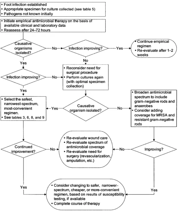
Figure 1.
Algorithm 1, part 1: approach to treating a diabetic patient with a foot wound
though MRSA strains have previously been isolated mainly
from hospitalized patients, community-associated cases are
now becoming common [45] and are associated with worse
outcomes in patients with diabetic foot infections [46–48].
Van-comycin (or glycopeptide)–intermediate S. aureus has been
iso-lated in several countries. Of note, the first 2 reported cases of
vancomycin-resistant S. aureus each involved a diabetic patient
with a foot infection [49].
The impaired host defenses around necrotic soft tissue or
bone may allow low-virulence colonizers, such as
coagulase-negative staphylococci and Corynebacterium species
(“diph-theroids”), to assume a pathogenic role [43, 50]. Acute
infec-tions in patients who have not recently received antimicrobials
are often monomicrobial (almost always with an aerobic
gram-positive coccus), whereas chronic infections are often
polym-icrobial [31, 36, 43, 51]. Cultures of specimens obtained from
patients with such mixed infections generally yield 3–5 isolates,
including gram-positive and gram-negative aerobes and
an-aerobes [14, 34, 37, 38, 40, 41, 52–58]. The pathogenic role of
each isolate in a polymicrobial infection is often unclear. Table
Table
4.
Evaluating
the
diabetic
patient
who
has
an
infected
foot.
Level
of
evaluation,
by
area(s)
to
be
assessed
Relevant
problems
and
obser
vations
Investigations
Patient
Systemic
response
to
infection
Fever
,
chills,
sweats,
vomiting,
hypotension,
and
tachycardia
Histor
y
and
physical
examination
Metabolic
state
V
olume
depletion,
azotemia,
hyperglycemia,
tachypnea
hyperosmolality
,
acidosis
Ser
um
chemistr
y
analyses
and
hematological
testing
Psychological/cognitive
state
Delirium,
dementia,
depression,
impaired
cognition,
and
stupor
Assessment
of
mental
and
psychological
status
Social
situation
Self
neglect,
potential
noncompliance,
and
lack
of
home
support
Inter
views
with
family
,
friends,
and
health
care
professionals
Limb
or
foot
Biomechanics
Deformities,
including
Charcot
arthropathy
,
claw/hammer
toes,
and
callosities
Clinical
foot
examination
and
radiography
(⭓
2
images)
V
ascular
status
Arterial
aIschemia,
necrosis,
or
gangrene
Foot
pulses,
blood
pressures
(ABI),
TcpO
2,
duplex
ultrasonography
,
and
angiograms
V
enous
Edema,
stasis,
or
thrombosis
Skin
and
soft-tissue
examination
and
duplex
ultrasonography
Neuropathy
Loss
of
protective
sensation
bLight
touch,
monofilament
pressure,
or
vibration
perception
W
ound
Size
and
depth
(tissues
involved)
Necrosis,
gangrene,
foreign
body
,
and
involvement
of
muscle,
tendon,
bone,
or
joint
Inspect,
debride,
cand
probe
dthe
wound;
and
radio-graphy
(⭓
2
images)
Presence,
extent,
and
cause
of
infection
Pur
ulence,
warmth,
tenderness,
pain,
induration,
cellulitis,
bullae,
crepitus,
abscess,
fasciitis,
and
osteomyelitis
Gram
staining
and
culture,
eultrasonography
or
CT
for
detection
of
deep
abscesses,
and
radiography
(⭓
2
images)
and/or
MRI
for
detection
of
osteomyelitis
f891
NOTE.
ABI,
ankle-brachial
index
(ratio
of
systolic
blood
pressures
from
arm
and
foot);
TcpO
2,
transcutaneous
partial
pressure
of
oxygen.
aAssess
the
foot’s
arterial
supply
in
ever
y
patient
with
a
diabetic
foot
infection
(A-II)
[59].
If
dorsalis
pedis
and
posterior
tibial
pulses
are
palpa
ble,
arterial
supply
is
generally
adequate
[60–62].
If
in
doubt,
other
diagnostic
tests
are
indicated
[63].
Any
trained
individual
can
perform
these
tests,
but
a
va
scular
specialist
(medical
or
surgical)
may
need
to
interpret
the
results.
Tests
are
available
to
measure
the
following:
(1
)
Doppler
arterial
pressure,
with
waveform
analysis;
(2
)
ABI.
V
alues
must
be
interpreted
cautiously
when
there
is
arterial
calcification
[59,
64–69],
which
is
suggested
by
ABIs
of
11
[68].
An
ABI
of
0.50–0.90
identifies
mild-to-moderate
peripheral
vascular
disease,
and
an
ABI
of
!0.50
suggests
ischemia
that
will
likely
impair
wound
healing
[66];
(3
)
Ankle
blood
pressure
(should
be
150
mm
Hg)
and
toe
pressure
(should
be
130
mm
Hg),
with
specially
designed
cuf
fs;
(4
)
TcpO
2[70],
which
may
provide
the
best
guidance
to
determine
the
anatomic
level
at
which
the
limb
is
adequately
perfused
(TcpO
2,
130
mm
Hg)
tissue
[71–74]
but
can
lack
reproducibility
,
and
they
require
a
skilled
technician
and
relatively
expensive
equipment
[75,
76].
bPeripheral
neuropathy
is
most
easily
detected
by
loss
of
protective
sensation,
defined
by
the
inability
to
detect
a
10-g
nylon
monofilament
(Semmes-W
einstein)
when
pressed
against
any
2
of
3
sites
(chosen
from
among
5)
on
the
foot
(plantar
surface
of
heel,
metatarsal
heads
and
arch,
and
tips
of
toes)
[25,
77,
78].
cMost
wounds
need
debridement,
which
involves
removing
both
the
hyperkeratosis
(callus)
sur
rounding
a
wound
and
the
necrotic
tissue
and
slough
from
its
base.
Debridement
reduces
pressure
at
callused
sites,
removes
colonizing
bacteria,
facilitates
the
collection
of
appropriate
specimens
for
culture,
and
permits
examination
for
the
presence
of
deep-tissue
involvement
[17,
79–82].
The
patient
should
be
forewarned
that
bleeding
is
likely
and
that
th
e
wound
will
be
larger
after
the
procedure.
Following
debridement,
measure
and
record
the
wound
size,
the
extent
of
sur
rounding
cellulitis,
and
the
qua
lity
and
quantity
of
any
drainage
(including
color
and
odor)—this
aids
any
other
clinicians
who
are
treating
the
patient
in
their
assessment
of
the
healing
progress
(B-III).
dUse
a
sterile,
blunt,
metal
probe
to
measure
the
depth
and
extent
of
the
wound,
including
noting
any
foreign
bodies,
soft-tissue
abscesses,
com-munications
with
joint
cavities
or
tendon
sheaths,
or
palpable
bone
(A-II).
Bone
touched
with
a
probe
has
a
characteristic
stony
feel
[83].
eKnowledge
of
the
etiologic
agent(s)
that
caused
the
wound
infection
is
generally
helpful
in
selecting
definitive
antibiotic
therapy
.
Obtain
specime
ns
for
culture
before
initiating
antibiotic
therapy
(if
possible)
or
after
discontinuing
therapy
(in
a
stable
patient
who
has
not
responded
to
therapy)
for
a
few
days
(B-III).
To
avoid
contaminants,
obtain
and
process
specimens
by
means
of
appropriate
methods.
Attempt
to
obtain
tissue
samples,
because
these
generally
provide
more
accurate
culture
results
than
do
superficial
swab
specimens
(A-I).
Most
studies
[41,
42,
84,
85]
indicate
that
the
latter
yield
a
greater
range
of
organisms
than
do
deeper
-tissue
material
and
yet
may
still
fail
to
identify
some
of
the
deep
flora.
Standard
swab
specimens
yield
fewer
anaerobes
and
are
often
minimally
processed
by
the
microbiology
laborator
y,
but
properly
collected
and
transported
anaerobic
swab
specimens
may
be
adequate
[86].
Skin
aspiration
may
yield
a
pathogen
in
cases
of
cellulitis
[87],
but
this
method
is
insensitive,
and
the
pathogens
obtained
from
cases
of
cellulitis
are
predictably
aerobic
gram-positive
cocci.
See
table
5
for
details
of
sampling
methods.
fUltrasonography
(especially
high
resolution)
[88]
and
CT
scanning
[89]
may
be
helpful
for
detecting
deep
soft-tissue
abscesses
or
sinus
tracts.
Pla
in
radiographs
and
MRIs
are
best
for
detecting
bone
involvement
(A-I).
MRI
may
also
provide
anatomic
information
about
the
presence
of
a
sinus
tract,
abscess,
or
muscle
involvement
[90–93].
Nuclear
medicine
scans
(especially
those
with
labeled
leukocytes
and,
sometimes,
of
bone)
are
highly
sensi
tive
and
may
be
useful
in
some
cases
[94–97]
but
are
generally
less
specific
than
MRI.
Table 5.
Collection of soft-tissue specimens from an infected diabetic foot for culture.
When7 Culturing clinically uninfected lesions is unnecessary, unless done as part of an infection-control surveillance protocol (C-III). 7 Cultures of infected wounds are valuable for directing antibiotic choices, but may be unnecessary in cases of acute mild
infection in an antibiotic-naive patient (B-III).
7 Blood cultures should be performed for a patient with a severe infection, especially if the patient is systemically ill (C-III). How
7 Cleanse and debride the lesion before obtaining specimens for culture.
7 In cases involving an open wound, obtain tissue specimens from the debrided base (whenever possible) by means of curet-tage (scraping with a sterile dermal curette or scalpel blade) or biopsy (bedside or operative) (A-I).
7 Avoid swabbing undebrided ulcers or wound drainage. If swabbing the debrided wound base is the only available culture option, use a swab designed for culturing aerobic and anaerobic organisms and rapidly transport it to the laboratory (B-I). 7 Needle aspiration may be useful for obtaining purulent collections or, perhaps, a specimen from an area of cellulitis. 7 Clearly identify samples (specimen type and anatomic location), and promptly send them to the laboratory in an appropriate
sterile container or transport media for aerobic and anaerobic culture.
3 lists common clinical infection syndromes and the pathogens
most likely isolated in conjunction with them.
EVALUATING THE PATIENT, THE WOUND,
AND THE INFECTION
Diabetic patients may develop many types of foot wounds, any
of which can become infected. Infection should be diagnosed
clinically on the basis of the presence of purulent secretions
(pus) or at least 2 of the cardinal manifestations of
inflam-mation (redness, warmth, swelling or induration, and pain or
tenderness); not all ulcers are infected (figure 1) (B-II) [23].
Curing an infection often contributes to, but is not defined by,
healing of an ulcer. Management of diabetic foot infections
involves evaluating and determining the severity of infection
as the basis for selecting the appropriate approach to treatment
[15, 23, 40] (B-II). The issue of osteomyelitis is particularly
complex and problematic and is thus dealt with separately.
Evaluation of the infection should occur at 3 levels, as
out-lined in tables 4 and 5 (B-III): the patient as a whole, the
affected limb or foot, and the infected wound. The goal is to
determine the clinical extent (table 4) and the microbial etiology
(table 5) of the infection, the biology or pathogenesis of the
wound, any contribution of altered foot biomechanics to the
cause of the wound (and, thus, its ability to heal), any
contri-bution of vascular (especially arterial) disease, and the presence
of any systemic consequences of infection. Clinicians lacking
the skills or experience to conduct any of these assessments
should seek appropriate consultation.
DETERMINING THE SEVERITY OF INFECTION
The results of the evaluations described in table 4 can be used
to determine the overall severity of the infection and to
for-mulate a management plan (figure 2) (B-II). Unfortunately, the
lack of consensus on wound definitions and infection
classi-fication systems hampers comparison of published studies. The
Wagner system [15, 40, 98, 99] has been widely used for 25
years but was developed for the “dysvascular” foot, is skewed
toward severe disease, and contains all infections within a single
grade [100–105]. Consensus is developing that the key issues
in classifying a diabetic foot wound are its depth (in particular,
which tissues are involved) and whether the wound is
com-plicated by either ischemia or infection [23, 101, 106–108]
(B-II). The International Consensus on the Diabetic Foot recently
published a preliminary progress report on a diabetic foot ulcer
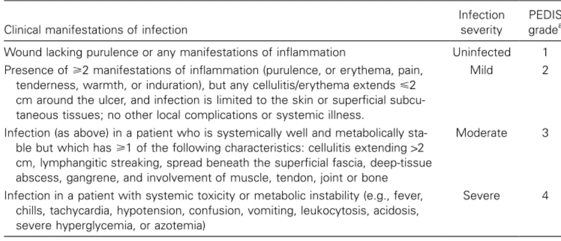
classification system for research purposes [23]. The key
ele-ments are summarized by the acronym PEDIS (perfusion,
ex-tent/size, depth/tissue loss, infection, and sensation). The
infection category includes grades 1 (no infection), 2
(involve-ment of skin and subcutaneous tissue only), 3 (extensive
cel-lulitis or deeper infection), and 4 (presence of a systemic
in-flammatory response syndrome). Because this research-based
system is designed to be applicable to all ulcers, it includes a
category of grade 1 for uninfected lesions; grades 2–4 are similar
to those we describe in table 6.
For infected wounds (figure 2), the most important initial
task is to recognize patients who require immediate
hospital-ization, parenteral and broad-spectrum empirical antibiotic
therapy, and urgent consideration of diagnostic testing and
surgical consultation. We have defined these potentially
life-threatening infections as “severe.” Infections defined as “mild”
must be distinguished from clinically uninfected lesions but are
otherwise relatively easy to recognize. Defining infections as
“moderate” poses the greatest difficulty, because this term
covers a broad spectrum of wounds, some of which can be
quite complicated and even limb threatening. Other
classifi-cation schemes have used the terms “uncomplicated” and
“complicated” synonymously with mild and moderate, but we
wish to avoid confusion with the various complications that
can beset a wound. The distinction between moderate and
severe infections has less to do with the status of the foot than
Guidelines for Diabetic Foot Infections • CID 2004:39 (1 October) • 893
Figure 2.
Algorithm 1, part 2: approach to treating a diabetic patient with a foot infection.
1Consider hospitalization if any of the following criteria
are present: systemic toxicity (e.g., fever and leukocytosis); metabolic instability (e.g., severe hypoglycemia or acidosis); rapidly progressive or
deep-tissue infection, substantial necrosis or gangrene, or presence of critical ischemia; requirement of urgent diagnostic or therapeutic interventions; and
inability to care for self or inadequate home support.
with the patient to whom it is attached. This distinction is
complicated by the fact that
⭓50% of patients with a
limb-threatening infection do not manifest systemic signs or
symp-toms. After debating several classification schemes, we propose
the one presented in table 6 as a basis for subsequent discussions
in and beyond this guideline (B-II).
TREATMENT OF INFECTION
Avoid prescribing antibiotics for uninfected ulcerations.
Some argue that many apparently uninfected diabetic foot
ul-cers are actually subclinically infected—that is, they contain a
high “bioburden” of bacteria (usually defined as
110
5organisms
per gram of tissue) that results in “critical colonization” levels
and impairs wound healing [54, 109–114]. Available published
evidence does not support the use of antibiotics for the
man-agement of clinically uninfected ulcerations, either to enhance
wound healing or as prophylaxis against infection [115, 116].
Because antibiotic use encourages antimicrobial resistance,
in-curs financial cost, and may cause drug-related adverse effects,
we discourage therapy of uninfected ulcers. In some
circum-stances, it is difficult to decide whether a chronic wound is
infected, such as when the foot is ischemic, has abnormal
col-Table 6.
Clinical classification of a diabetic foot infection.
Clinical manifestations of infection
Infection severity
PEDIS gradea Wound lacking purulence or any manifestations of inflammation Uninfected 1 Presence of⭓2 manifestations of inflammation (purulence, or erythema, pain,
tenderness, warmth, or induration), but any cellulitis/erythema extends⭐2 cm around the ulcer, and infection is limited to the skin or superficial subcu-taneous tissues; no other local complications or systemic illness.
Mild 2
Infection (as above) in a patient who is systemically well and metabolically sta-ble but which has⭓1 of the following characteristics: cellulitis extending12 cm, lymphangitic streaking, spread beneath the superficial fascia, deep-tissue abscess, gangrene, and involvement of muscle, tendon, joint or bone
Moderate 3
Infection in a patient with systemic toxicity or metabolic instability (e.g., fever, chills, tachycardia, hypotension, confusion, vomiting, leukocytosis, acidosis, severe hyperglycemia, or azotemia)
Severe 4
NOTE. Definitions of terms can be found in footnotes to table 4. Foot ischemia may increase the severity of any infection, and the presence of critical ischemia often makes the infection severe. PEDIS, perfusion, extent/size, depth/ tissue loss, infection, and sensation.
a
International Consensus on the Diabetic Foot [23].
oration or a fetid odor, has friable granulation tissue, is
asso-ciated with unexpected pain or tenderness, or when an
oth-erwise properly treated ulcer fails to show healing progress [117,
118]. In these unusual cases, a brief, culture-directed course of
antibiotic therapy may be appropriate (C-III).
Determine the need for hospitalization.
Hospitalization is
the most expensive part of treating a diabetic foot infection,
and deciding on its necessity requires consideration of both
medical and social issues. Patients with infections that are either
severe or complicated by critical limb ischemia should generally
be hospitalized (C-III) [119, 120]. Some patients with
appar-ently mild infections and more patients with moderate
infec-tions may also need hospitalization; this may be for observation,
urgent diagnostic testing, or because complicating factors are
likely to affect their wound care or adherence to antibiotic
treatment. In the absence of these complicating features, most
patients with mild or moderate infections can be treated as
outpatients (A-II) [84, 121].
Stabilize the patient.
Attending to the general metabolic
state of the patient is essential [25, 122]. This may involve
restoration of the fluid and electrolyte balances; correction of
hyperglycemia, hyperosmolality, acidosis, and azotemia; and
treatment of other exacerbating disorders. Critically ill patients
who require surgery should usually be stabilized before transfer
to the operating room, although surgery should usually not be
delayed for
148 h after presentation to the hospital (B-III)
[123]. The improvement of glycemic control may aid in both
eradicating the infection and healing the wound [124]. As the
infection improves, hyperglycemia may be easier to control.
Choose an antibiotic regimen.
Selection of the antibiotic
regimen initially involves decisions about the route of therapy,
the spectrum of microorganisms to be covered, and the specific
drugs to administer and later involves choosing the definitive
regimen and the duration of treatment. Initial therapy is usually
empirical and should be based on the severity of the infection
and on any available microbiological data, such as recent culture
results or current Gram-stained smear findings. For severe
in-fections and for more-extensive, chronic moderate inin-fections,
it is safest to commence therapy with broad-spectrum agents.
These should have activity against gram-positive cocci
(includ-ing MRSA in locations where this pathogen is common), as
well as gram-negative and obligate anaerobic organisms (B-III).
To ensure adequate and prompt tissue concentrations, therapy
should be given parenterally, at least initially (C-III). Although
some suggest broad-spectrum empirical therapy for most
in-fections [125–127], the majority of mild—and many
moder-ate—infections can be treated with agents with a relatively
narrow spectrum, such as those covering only aerobic
gram-positive cocci (A-II) [84]. Although anaerobic organisms are
isolated from many severe infections [42, 128], they are
infre-quent in mild-to-moderate infections [14, 84, 129], and there
is little evidence to support the need for antianaerobic therapy
in most infections (B-III). For mild-to-moderate infections in
patients without gastrointestinal absorption problems and for
whom an oral agent with the appropriate spectrum is available,
oral therapy is often appropriate, especially with highly
bio-available agents (A-II). For mildly infected open wounds with
minimal cellulitis, limited data support the use of topical
an-timicrobial therapy (B-I) [130].
Antibiotics vary in how well they achieve effective
concen-trations in infected diabetic foot lesions [131–137]; this is
as-sociated with the pharmacodynamic properties of the specific
agent and, especially, the arterial supply to the foot, rather than
with diabetes [138]. There are surprisingly few published
clin-ical trials of antibiotic therapy for diabetic foot infection.
Sev-eral antibiotic trials involving patients with various complicated
Guidelines for Diabetic Foot Infections • CID 2004:39 (1 October) • 895
skin and soft-tissue infections have included some patients with
diabetic foot infections. Table 7 provides a list of published
clinical trials that focused on therapy of diabetic foot infections,
either exclusively or as an identified subset of a larger study.
The lack of standardization among these trials makes the
com-parison of outcomes of different regimens inappropriate. The
differing definitions of infection severity and clinical end points
that were used in these publications highlight the need to
de-velop a consensus classification system for future studies. On
the basis of the available studies, no single drug or combination
of agents appears to be superior to others [129].
Table 8 summarizes some potential empirical antibiotic
reg-imens according to the clinical severity of the infection,
al-though the available data do not allow us to recommend any
specific antibiotic regimen for diabetic foot infections (B-II).
These suggested agents are derived from available published
clinical trials and our collective experience and are not meant
to be inclusive of all potentially reasonable regimens. Similar
agents could be used, depending on various clinical,
micro-biological, epidemiological, and financial considerations.
Con-sider modifying antibiotic therapy when culture and
suscep-tibility results are available (C-III). Empirical choices for
patients who are not responding to antibiotic therapy should
include agents that cover a different or more-extended
spec-trum of organisms (B-III) (figure 3). The regimens in table 8
are listed in approximate order of increasing broad-spectrum
coverage; the order does not indicate preferences by the
com-mittee. Dosages of antibiotic agents should be selected
accord-ing to suggestions of the US Food and Drug Administration,
the drug’s manufacturers, and the experience of the prescriber
and should be modified on the basis of any relevant organ
(especially renal) dysfunction and other clinical factors.
Determine the need for surgery.
Many infections require
surgical procedures that range from drainage and excision of
infected and necrotic tissues to revascularization of the lower
extremity and reconstruction of soft-tissue defects or
mechan-ical misalignments [164–168]. Unfortunately, surgmechan-ical
treat-ment of diabetic foot infections is based on even less-structured
evidence than that for antibiotic therapy [169]. Seek urgent
surgical consultation for life- or limb-threatening infections,
such as those presenting with necrotizing fasciitis, gas gangrene,
extensive soft-tissue loss, or evidence of compartment
syn-drome, or those in limbs with critical ischemia (A-II) [170,
171]. A surgical specialist should also evaluate patients who
have unexplained persistent foot pain or tenderness and/or
evidence of a deep-space infection, deep abscesses, or
progres-sive infection in the face of apparently appropriate medical care
(figure 3). Timely and aggressive surgical debridement,
includ-ing limited resections or amputations, may reduce the need for
more-extensive amputation (B-II) [172, 173]. Pus under
pres-sure, especially in an ischemic foot, can cause rapid and
irrep-arable damage. For patients with less-serious infections, it may
be appropriate to delay surgery to carefully observe the
effec-tiveness of medical therapy or to determine the demarcation
line between necrotic and viable tissue [174].
The surgeon must determine the adequacy of the blood
sup-ply to the remaining viable tissues, consider common operative
pitfalls (e.g., infection spreading among foot compartments, to
the deep plantar space, or along the tendon sheaths), and
for-mulate a strategy for eventual soft-tissue cover (e.g., primary
closure, delayed primary closure, secondary intention, or tissue
transfer) [175–177]. The surgical approach should optimize the
likelihood for healing and should attempt to preserve the
in-tegrity of the walking surface of the foot (B-II) [178]. In
ad-dition to manual dexterity, the surgeon must have sufficient
knowledge and experience to judge when and how to intervene.
The surgeon’s training specialty is less important than his or
her knowledge of the anatomy of the foot, the pathophysiology
of ulceration and infection, and experience with and
enthusi-asm for the field [8]. In most instances, the surgeon should
continue to observe the patient until the infection is under
control and the wound is healing (B-III).
In some cases, amputation is the best or only option [170,
179]. Urgent amputation is usually required only when there
is extensive necrosis or life-threatening infection [180]. Elective
amputation may be considered for the patient who has
recur-rent ulceration (despite maximal preventive measures), has
ir-reversible loss of foot function, or would require unacceptably
prolonged or intensive hospital care [181, 182]. Selection of
the level of amputation must take into consideration vascular,
reconstructive, and rehabilitation issues [183, 184]. Generally,
the surgeon should attempt to save as much of the limb as
possible. However, a higher-level amputation that results in a
more functional residual stump (even if a prosthesis is required)
may be a better choice than preserving a foot that is
mechan-ically unsound, unlikely to heal, or prone to future ulceration.
When all or part of a foot has dry gangrene, it may be preferable
(especially for a patient for whom surgery is a poor option) to
let the necrotic portions autoamputate. It may also be best to
leave adherent eschars in place, especially on the heel, until
they soften enough to be more easily removed, provided there
does not appear to be an underlying focus of infection [80,
81].
If the infected limb appears to be ischemic, the patient should
be referred to a surgeon with vascular expertise [185]. In most
cases, ischemia is due to larger-vessel atherosclerosis, rather
than to “small-vessel disease” [68]. Because vessels above the
knee and below the ankle tend to be relatively spared,
lower-extremity atherosclerosis may be amenable to angioplasty or
vascular bypass [186]. Patients with noncritical ischemia (e.g.,
those with an ankle to brachial artery blood pressure index of
0.5–0.9) can usually be successfully treated without a vascular
Table 7.
Antibiotic agents used in published clinical studies of diabetic foot infections.
Antibiotic (route)
No. of treated
patients Study design Patient group
Type/severity of
infection(s) Reference Cephalosporins
Cefoxitin (iv) 8 Prospective, noncomparative
Hospitalized Presumptive anaerobic [139]
Cefoxitin (iv) 23 RDBCT Hospitalized Moderate-to-severe [140] Cefoxitin (iv) 60 Prospective,
noncomparative
Hospitalized Failing to respond to therapy
[141]
Cefoxitin (iv) 18 RDBCT Hospitalized Mild-to-severe [142] Cefoxitin (iv) alone 12 RCT Hospitalized Mixed [143] Cefoxitin (iv) and
amdino-cillin (iv)
13 RCT Hospitalized Mixed [143]
Ceftizoxime (iv) 20 Prospective, uncontrolled Hospitalized PVD, moderate-to-severe [144] Ceftizoxime (iv) 23 RDBCT Hospitalized Moderate-to-severe [140] Cephalexin (po) 29 RDBCT Outpatient Mild-to-moderate [84] Ceftriaxone (iv) 90 Prospective, observational Hospitalized Severe limb-threatening [145] Penicillins
Ampicillin/sulbactam (iv) (then amoxicillin/clavu-lanate [po])
53 RCT Hospitalized initially Moderate [146]
Ampicillin/sulbactam (iv) 48 RDBCT Hospitalized Limb-threatening [147] Ampicillin/sulbactam (iv) 74 Prospective,
noncomparative
Hospitalized Moderate-to-severe [148]
Ampicillin/sulbactam (iv) 18 RDBCT Hospitalized Mild-to-severe [142] Ampicillin/sulbactam (iv)
and/or amoxicillin/clavu-lanate (po)
120 RCT Outpatient or hospitalized All types [121]
Amoxicillin/clavulanate (iv/po) 191 Observational, noncomparative
Mostly hospitalized Moderate [149]
Ticarcillin/clavulanate (iv) 28 RCT subgroupa Inpatient or outpatient Complicated soft-tissue [150] Ticarcillin/clavulanate (iv) 17 RCT subgroupa Hospitalized Complicated soft-tissue [151] Piperacillin/tazobactam (iv) 29 Prospective,
noncomparative Hospitalized Moderate-to-severe [152] Piperacillin/tazobactam (iv/im) 38 Prospective noncomparative
Outpatient Parenteral, mostly moderate
[153]
Piperacillin/tazobactam (iv) 34 RDBCT subgroupa Hospitalized Severe [154] Fluoroquinolones
Ciprofloxacin (po) 46 Prospective, randomized doses
Hospitalized PVD [155]
Ciprofloxacin (iv, then po) 43 Prospective, noncomparative
Hospitalized Soft-tissue or bone [156]
Ciprofloxacin (po) and clindamycin (po)
120 Uncontrolled, quasi-prospective
Hospitalized initially, re-ceived other iv agents, and was then dis-charged home
Moderate-to-severe [157]
Ofloxacin (iv, then po) 55 RCT Hospitalized initially Moderate [146] Ofloxacin (po) 420 RDBCT Outpatients Mild-to-moderately
infected ulcers
[130]
Levofloxacin (iv or po) 26 RCT subgroupa Outpatients or inpatients Complicated soft-tissue [150] Trovafloxacin (po) 214 Prospective,
noncomparative
… Soft-tissue [158]
Clinafloxacin (iv, then po) 42 RDBCT subgroupa Hospitalized Severe [154] Ofloxacin, levofloxacin, or
ciprofloxacin (iv and/or po)
90 Prospective, observational Hospitalized Severe limb-threatening [145]
Carbapenems
Imipenem/cilastatin (iv) 48 RDBCT Hospitalized Limb-threatening [147] Imipenem/cilastatin (iv) 94 Uncontrolled,
noncomparative
Hospitalized Moderate-to-severe [159]
Imipenem/cilastatin (iv) 22 Randomized, open, comparative
Hospitalized Wagner grade 2–4 wounds
[160]
Ertapenem (iv) 33 RDBCT Hospitalized Complicated soft-tissue [161]
Guidelines for Diabetic Foot Infections • CID 2004:39 (1 October) • 897
Table 7.
(Continued.)
Antibiotic (route)
No. of treated
patients Study design Patient group
Type/severity of
infection(s) Reference Miscellaneous agents
Aztreonam (iv) 20 Prospective subgroupa Hospitalized Acute, severe soft-tissue [162] Clindamycin (po) 29 RDBCT Outpatient Mild-to-moderate [84] Piperacillin/clindamycin (iv) 24 Randomized, open,
comparative
Hospitalized Wagner grade 2–4 wounds
[160]
Pexiganan (topical) 415 RDBCT Outpatient Mild-to-moderate infected ulcers
[130]
Linezolid (iv or po) 241 RCT Outpatient or hospitalized All types [121] Daptomycin (iv) 50 RCT subgroupa Hospitalized Complicated skin [163]
NOTE. Trials are those in which the purpose of the study was to examine the efficacy of antibiotic therapy, and the subjects were exclusively, predominantly, or separately identifiable as diabetic patients with foot infections. The clinical and microbiological outcomes were not consistently defined or routinely provided. PVD, peripheral vascular disease; RCT, randomized controlled trial; RDBCT, randomized, double-blinded, controlled trial (each arm is listed separately in the table).
a
Involved patients with diabetic foot infections who constituted an identified subgroup of a larger trial of skin and skin structure infections.
procedure. For more-severe vascular disease of the foot, many
centers have reported successful use of femoral-distal bypass
procedures in diabetic patients [186–189]. For a patient with
a severely infected ischemic foot, it is usually preferable to
perform any needed revascularization early after recognizing
the infection (i.e., within 1–2 days), rather than to delay this
procedure in favor of prolonged (and potentially ineffective)
antibiotic therapy (B-II) [123, 190]. On the other hand, careful
debridement of necrotic infected material should not be delayed
while awaiting revascularization. Optimal surgical management
may require multiple, staged procedures [191].
Formulate a wound-care plan.
The wound may require
additional attention after the debridement performed during
the initial assessment (table 4). The goal is to physically excise
dead and unhealthy tissue, thereby enabling wound healing and
removing a reservoir of potential pathogens [82, 192–194]. Any
experienced clinician may perform limited debridement. This
can usually be undertaken as a clinic or bedside procedure and
without anesthesia, especially for a neuropathic foot. Sharp
debridement with scalpel, scissors, or tissue nippers is generally
preferable to hydrotherapy or topical debriding agents, which
are less definitive and controllable and may require prolonged
and repeated applications (B-III) [194, 195]. There are many
wound-care products that are touted as being able to improve
healing in various ways [17, 23, 196–199], but a discussion of
these is beyond our scope. The infected wound should be
dressed in a manner that allows daily inspection and encourages
a moist wound-healing environment (B-III). No evidence
fa-vors any particular type of dressing; convenience and cost are
important considerations. Removal of pressure from a foot
wound (i.e., off-loading) is crucial to the healing process
(A-I) [200, 201]. Many types of devices can off-load the infected
wound, but it is important to choose one that permits easy
inspection [202].
Adjunctive treatments.
Investigators and industry
repre-sentatives have advocated many types of wound-care
treat-ments, including wound vacuum-drainage systems [203–206],
recombinant growth factors [207–212], skin substitutes [203,
213–216], antimicrobial dressings [217–219], and maggot
(ster-ile larvae) therapy [220–222]. Although each treatment likely
has some appropriate indications, for infected wounds,
avail-able evidence is insufficient to recommend routine use of any
of these modalities for treatment or prophylaxis.
Two adjunctive modalities do deserve brief comments. First,
granulocyte colony-stimulating factors (G-CSFs) have now
been investigated in 5 randomized trials involving diabetic foot
infections [223–227]. A preliminary meta-analysis of these trials
suggests that G-CSF does not accelerate resolution of infection
but may significantly reduce the need for operative procedures
(B-I) [228]. Second, several anecdotal and retrospective reports
suggest that hyperbaric oxygen therapy may be of value for
treatment of diabetic foot wounds, and a few recent prospective
studies have shown promising results [229–232]. A recent
Cochrane review concluded that hyperbaric oxygen therapy
significantly reduced the risk of major amputation related to a
diabetic foot ulcer [233] (B-I). Only additional randomized
clinical trials can establish when, for whom, and with what
protocols these expensive and limited resources might be used
in the treatment of diabetic foot infections. Neither should be
used as a substitute for proper surgical debridement and
con-ventional therapy.
FOLLOW-UP
Careful observation of the patient’s response to therapy (figure
4) is essential and should be performed daily for inpatients and
perhaps every 2–5 days initially for outpatients (B-III). The
primary indicators of improvement are resolution of local and
systemic symptoms and clinical signs of inflammation. Blood
test findings, including WBC counts [234, 235] and
inflam-Table 8.
Suggested empirical antibiotic regimens, based on clinical severity, for diabetic foot infections.
Route and agent(s) Mild Moderate Severe
Advised route Oral for most Oral or parenteral, based
on clinical situation and agent(s) selected Intravenous, at least initially Dicloxacillin Yes … … Clindamycin Yes … … Cephalexin Yes … …
Trimethoprim-sulfamethoxazole Yes Yes …
Amoxicillin/clavulanate Yes Yes …
Levofloxacin Yes Yes …
Cefoxitin … Yes …
Ceftriaxone … Yes …
Ampicillin/sulbactam … Yes …
Linezolida(with or without aztreonam) … Yes …
Daptomycina(with or without aztreonam) … Yes …
Ertapenem … Yes …
Cefuroxime with or without metronidazole … Yes …
Ticarcillin/clavulanate … Yes …
Piperacillin/tazobactam … Yes Yes
Levofloxacin or ciprofloxacin with clindamycin … Yes Yes
Imipenem-cilastatin … … Yes
Vancomycinaand ceftazidime (with or without metronidazole)
… … Yes
NOTE. Definitive regimens should consider results of culture and susceptibility tests, as well as the clinical response to the empirical regimen. Similar agents of the same drug class may be substituted. Some of these regimens may not have US Food and Drug Administration approval for complicated skin and skin-structure infections, and only linezolid is currently specifically approved for diabetic foot infections.
a
For patients in whom methicillin-resistant S. aureus infection is proven or likely.