18 JOURNAL OF PALEONTOLOGY, V. 65, NO. 1, 1991 . 1977. Calcareous Algae. Developments in Palaeontology and
Stratigraphy, 4. Elsevier, New York, 185 p.
., N.P.JAMES, AND R.N.GINSBURG. 1974. The puzzling Paleozoic phylloid algae—Holocene answer in squamariacean calcareous red algae. American Association of Petroleum Geologists, Annual Meet ing, 2:82-83.
J. Paleont., 65(1), 1991, pp. 18-33 Copyright © 1991, The Paleontological Society 0022-3360/91/0065-0018503.00
INTRODUCTION
C
ULCAJUNA CALCAR d'Orbigny, 1826, was repeatedly dis cussed in the literature with regard to its architecture and systematic position. It was placed in the genus Rotalia Lamarck, 1804, by Brady (1884) and by Hofker (1927), included in Para-rotalia Le Calvez, 1949, by Reiss and Merling (1958), Reiss (1963), Hofker (1970), and Hansen and Reiss (1971), and lately assigned again to Calcarina d'Orbigny, 1826, by Le Calvez (1977), Hottinger and Leutenegger (1980), and Reiss and Hottinger (1984).Differences of opinion stem from conflicting, in many cases incomplete or incorrect, descriptions of the architecture of C. calcar and of other species with which it has been compared or associated. Confusion has also arisen as a result of differing interpretations ofcertain test elements (and terminology applied to them) by various authors. Rigorously defined test elements are, however, besides their taxonomic value, of considerable importance in the understanding of functional morphology of the test and in relating test architecture to cytoplasmic differ entiation (Hottinger and Dreher, 1974; Hottinger, 1978; Hot
ZHANG YUN. 1989. Multicellular thallophytes with differentiated tis sues from late Proterozoic phosphate rocks of South China. Lethaia, 22:113-132.
ACCEPTED 26 JUNE 1990
Harvard University provided $500 in support of this article.
Sea, the architectural details of Calcarina calcar were reexam-ined by means of scanning electron microscopy. In order to solve the problems involved, the necessity arose to study, in addition, the following species with regard to certain morpho logical characters: 1) Rotalina inermis Terquem, 1882 (topo-types), the type species of Pararotalia, and 2) Pararotalia spi-nigera (Le Calvez, 1949), both from the Lutetien of Grignon, Paris Basin; 3) Rotalia mexicana Nuttall 1928, emend. Poag, 1966, the type-species of the genus Neorotalia Bermudez, 1952, placed generally in the synonymy of Pararotalia (see Hofker, 1957; Reiss and Merling, 1958; Reiss, 1963; Loeblich and Tap-pan, 1964, 1987), from the Lower Miocene of Victoria, Aus tralia; 4) Rotalia viennoti Greig, 1935 (topotypes from the Oligo-cene of Ramla, Israel), placed by Bermudez (1952) in Neorotalia and by Reiss and Merling (1958) in Pararotalia; as well as 5) Nautilus spengleri Gmelin, 1788, the type species of the genus Calcarina, from Keij Island, Indonesia, and from Sybai, Phil ippines.
The present paper summarizes observations on certain ar chitectural characters of the taxa mentioned above and the re
THE FORAMINIFERAL GENERA PARAROTALIA, NEOROTALIA,
AND CALCARINA: TAXONOMIC REVISION
LUKAS HOTTINGER,1 ELWIRA HALICZ,2 AND ZEEV REISS2
'Geologisch-Palaeontologisches Institut der Universitaet Basel, Bernoullistrasse 32, CH-4056 Basel, Switzerland and
2Department of Geology, Institute of Earth-Sciences, The Hebrew University of Jerusalem,
91904 Giv'at Ram, Jerusalem, Israel
ABSTRACT—Scanning electron microscopy of the architecture of Rotalina inermis Terquem, 1882, the type-species of the genus Pararotalia Le Calvez, 1949, and of Rotalia mexicana Nuttall, 1928, the type-species of the genus Neorotalia Bermudez, 1952, reveals that both taxa have in common: 1) an umbilical bowl closed by either a single or a compound umbilical plug; 2) an interiomarginal extraumbilical aperture, restricted by a toothplate that protrudes with a free edge into the aperture and forms an umbilical spiral canal; and 3) a septal flap and intraseptal interlocular spaces. Rotalia mexicana, however, also possesses an enveloping canal-system, similar to that found in the Calcarinidae. For this reason Neorotalia must be regarded as a valid distinct genus, not synonymous with Pararotalia, as proposed by some authors. Calcarina calcar d'Orbigny, 1839, variously placed by authors in Rotalia Lamarck, 1804, in Pararotalia Le Calvez, 1949, and lately again in Calcarina d'Orbigny, 1826, is shown to possess the same basic architecture as Neorotalia and is placed, consequently, into this latter genus. Irregular supplementary apertures occurring in N. calcar are not considered of generic value and neither are they regarded as indicating a relationship with Nautilus spengleri Gmelin, 1788, the type-species of Calcarina. The latter, although characterized by a complex enveloping canal-system, possesses primary multiple interioareal main apertures, surrounded by thick rims, as well as a small umbilical plate, but lacks a toothplate with a free edge. For comparison, Pararotalia spinigera (Le Calvez, 1949) and Neorotalia viennoti (Greig, 1935) were also studied. The subfamily Pararotaliinae Reiss, 1963, is emended to include the canal-system as a characteristic feature. A glossary of selected terms is appended.
unified and for this reason a glossary of some important terms, as used or redefined lately by the present authors, is appended at the end of this article.
SYSTEMATIC PALEONTOLOGY
Suborder ROTALHNA
Delage and Herouard, 1896
Superfamily ROTALIACEA
Ehrenberg, 1839
Family ROTALIIDAE Ehrenberg, 1839
Subfamily PARAROTALIINAE
Reiss, 1963, emend, herein
Diagnosis. — Trochospiral test with single interiomarginal main aperture into which a toothplate protrudes with a free edge; irregular supplementary apertures may be present; canal-system composed of intraseptal interlocular spaces, of a spiral umbilical canal formed by interconnected toothplates, and, in some gen era, of enveloping canals produced by secondary lamination.
Genus PARAROTALIA Le Calvez, 1949 PARAROTALIA INERMIS (Terquem, 1882)
Figure 1.1, 1.3, 1.4 Rotalina inermis TERQUEM, 1882, p. 68, PL 6, fig. 1.
Pararotalia inermis (Terquem). LE CALVEZ, 1949, p. 32, PI. 3, figs. 54-56; LOEBLICH AND TAPPAN, 1957 (emend.), p. 14-15, PI. 1, figs. 2, 3,
text-figs. 4, 5; REISS AND MERLING, 1958, p. 1-17, PI. 2, figs. 4-12,
PL 5, figs. 16-18; LOEBLICH AND TAPPAN, 1964, p. C612-C613, PL
486, figs. 1-3; LE CALVEZ,, 1970, p. 163, PL 34, figs. 6, 7; HANSEN
AND REISS, 1971, p. 335, PI. 9, fig. 6, PI. 10, fig. 3; LOEBLICH AND TAPPAN, 1987, p. 659, PI. 755, figs. 18-21.
Review.—Le Calvez (1949) described deeply incised sutures on the ventral side, a single umbilical plug, and an interioareal, elliptical aperture surrounded by a lip. Loeblich and Tappan (1957) described deeply depressed sutures on the ventral side, prominent umbilical shoulders, and the aperture as originally interiomarginal and extraumbilical—umbilical with a lip, mod ified, before the development of the next chamber, by a delicate umbilical plate into an areal ovate or comma-shaped opening, an internal septum being formed at the junction of umbilical plate and chamber wall. Reiss and Merling (1958) regarded the "umbilical plate" reported by Loeblich and Tappan (1957) an actual chamber wall and the "internal septum" as a primary toothplate. Reiss and Merling used, however, the term "tooth-plate" in a broad sense (Hofker, 1951), hence including foram-enal plate, umbilical plate, and cover plate (see Muller-Merz,
1980, and appended Glossary); they compared the toothplate of Pararotalia to the plate of Cuvillierina, Debourle, 1955, prov en by Muller-Merz (1980) to be a typical umbilical plate (see Glossary). The aperture was described and figured by Reiss and Merling (1958) as interiomarginal, extraumbilical and the in tercameral foramen as areal, modified by the attachment (in adaxial position) of the toothplate. The presence of a septal flap was demonstrated and the deeply incised ventral sutures were shown by Reiss and Merling (1958) to correspond to intraseptal interlocular spaces ("intraseptal passages"), now regarded as part of the canal-system (Hottinger, 1978). Hansen and Reiss (1971), in disagreement with Reiss and Merling (1958), de scribed in all Pararotalia a slightly contorted "foramenal plate," protruding into the chamber, to which an "umbilical cover plate" is attached in each preceding chamber, similar to the situation in Ammonia Briinnich, 1772. Lately, Loeblich and Tappan (1987), in their new description of the genus Pararotalia, re ported the presence of a septal flap doubling the septa and de scribed the aperture as interiomarginal, extending obliquely into the apertural face, and the intercameral foramen as areal, due
to the attachment of an imperforate toothplate that extends to the distal margin of the aperture.
Observations and remarks. —The accompanying Figures 1.1, 1.3, 1.4, shows the slightly inflated ventral (umbilical) chamber walls, which are smooth, except near the peripheral carina where they are finely pustulate, the prominent umbilical shoulder, the septal flap, and the deep and open intraseptal interlocular spaces on the ventral side communicating with a deep furrow between inner ventral walls, producing the umbilical bowl, and the single umbilical plug. In dissected chambers (Figure 1.3,1.4), the areal intercameral foramen with a thick lip or rim and the toothplate are shown. Originating from the septal flap and connected to the inner ventral chamber wall, an imperforate toothplate ex tends to the distal chamber wall, attached to the dorsal corner of the primarily interiomarginal, extraumbilical aperture, and protruding with a free, serrated edge into the latter. It is folded both parallel and normal to the axis of coiling and produces with the adjacent preceding coil a space. As shown by Reiss and Merling (1958, PL 2, figs. 5, 9) and by Hansen and Reiss (1971, PL 10, fig. 2; see also Pararotalia sp. on PL 10, fig. 1), each toothplate is attached to the preceding one in adaxial position. This produces a narrow continuous spiral space, here regarded as a canal. It communicates behind the fold of the toothplate with the main chamber lumen, as well as with the furrow around the umbilical plug. These findings are in general agreement with Reiss and Merling (1958) and with Loeblich and Tappan (1987), but differ from the descriptions by Loeblich and Tappan (1957,
1964) and by Hansen and Reiss (1971).
The presence in Pararotalia of a toothplate (s. str.) with a free edge, and the absence of such features as an umbilical plate (like e.g., in Rotalia or in Cuvillierina), of a foramenal plate connected to a cover plate (like e.g., in Ammonia), or of folia with foliar apertures (in the sense of Levy et al., 1980, like in Rotalia or in Ammonia) (see Glossary) set the Pararotaliinae well apart from the Rotaliinae Ehrenberg, 1839, Cuvillierininae Loeblich and Tappan, 1964, and Ammoniinae Saidova, 1981, in the classification of Loeblich and Tappan (1987). The description of the Pararotaliinae is emended here to include the canal-sys tem (compare, however, Loeblich and Tappan, 1987).
PARAROTALIA SPINIGERA (Le Calvez, 1949) Figure 1.2, 1.5-1.8
Globorotalia spinigera (Terquem). LE CALVEZ, 1949, p. 39, PL 6, figs.
97-98; LE CALVEZ, 1952, p. 48.
Pararotalia spinigera (Le Calvez). LOEBLICH AND TAPPAN, 1957 (emend.),
p. 18, PL 4, figs. 1-3; LE CALVEZ, 1970, p. 161, PL 39, fig. 6.
Review.—Loeblich and Tappan (1957) described the cham bers as gently convex on the ventral side with a pronounced umbilical shoulder and deeply depressed intercameral sutures, a single umbilical plug, and the short blunt, peripheral spines arising from each chamber. The aperture is described as interi omarginal and extraumbilical-umbilical with a lip, restricted to an areal, comma-shaped opening by a secondary umbilical plate which forms also a protruding "lower lip" roughly parallel with the lower margin of the chamber. Le Calvez (1970) regarded this species as a kind of Rotalia with an internal toothplate. This agrees with the latest redescription of the genus Pararotalia by Loeblich and Tappan (1987).
Observations and remarks. —Figure 1.2, 1.8, shows the slight ly inflated chambers on the ventral side and the deeply depressed sutures forming the intraseptal interlocular spaces, a deep furrow between the umbilical bowl and the single umbilical plug, the pustules, sometimes aligned or forming riblets, on the ventral-peripheral margin and on the carina, as well as (see also Figure
1.7) the spinose peripheral projections covered by pustules and radial ridges. These projections are solid and imperforate (Figure 1.5) and are thus pseudospines (see Glossary). Figure 1.2 shows the thick lip or rim along the aperture and Figure 1.8 shows the septal flap and the toothplate. Figure 1.6 shows an intercameral foramen, bordered by a thick pustulate lip and the distal, slightly serrated free edge ("inner lip") of the toothplate (belonging to the same chamber). The plate is in continuation of the inner ventral chamber wall (adaxial to the distinct umbilical shoulder), attached to the dorsal end of the interiomarginal, extraumbilical aperture (now an areal foramen), and forms a space (canal) with the preceding adjacent coil. Part of the succeeding chamber's septal flap and broken-off toothplate is also shown. At the ven tral end of the foramen the apertural face is bent inward. The architecture of P. spinigera is, therefore, the same as that of P.
inermis.
Genus NEOROTALIA
Bermudez, 1952, emend. NEOROTALIA MEXICANA (Nuttall, 1928) emend. Poag, 1966
Figures 2.1-2.9, 3
Rotalia mexicana NUTTALL, 1928, p. 374, PL 50, figs. 6-8.
Rotalia mexicana var. mecatepecensis NUTTALL, 1932, p. 26.
Neorotalia mexicana (Nuttall). BERMUDEZ, 1952, p. 75, PI. 12, fig. 4.
Pararotalia mexicana mecatepecensis (Nuttall). REISS AND MERLING,
1958, PI. 2, figs. 18-24, PI. 5, figs. 19-20; HANSEN AND REISS, 1971, p. 335, PI. 10, fig. 2.
Pararotalia mexicana (Nuttall). LOEBLICH AND TAPPAN, 1964, p. C612-C613, PI. 486, fig. 5 (lectotype); (emend.) POAG 1966, p. 414-415, PI. 6, figs. 11-19; LOEBLICH AND TAPPAN, 1987, p. 659, PI. 755, figs. 15-17.
/tev/evv.—NuttalFs (1928) description is supplemented by Bermudez (1952) who emphasized, in his description of the genus Neorotalia, the presence of strong ornamentation on both sides of the test, of an umbilical "knob," which may be sub divided, and especially of intraseptal canals. The aperture is described as a simple slit between the plug and the periphery. Reiss and Merling (1958), who regarded Neorotalia as a syn onym of Pararotalia, described and illustrated by micropho-tographs of oriented thin sections and by camera lucida drawings of dissected chambers the architecture of Pararotalia mexicana
mecatepecensis, placed by Poag (1966) in Pararotalia mexicana
as a forma (an invalid infrasubspecific category, according to the ICZN). The aperture is described by Reiss and Merling (1958) as interiomarginal extraumbilical with a thick, pustulate rim and the intercameral foramen as areal, due to the attachment of a toothplate, extending from the septal flap to the distal wall and cameral aperture. The toothplates, which appear s-shaped in horizontal section, are shown to be connected to each other in adaxial position and, although not mentioned in the text, to actually protrude into the aperture. A space between toothplate and adjacent coil is discernible (PI. 2, figs. 21, 23, 24, PI. 5, figs. 19, 20). Noteworthy is the inward bend of the apertural face near the ventral end of the aperture (PI. 2, figs. 19, 21). The
prominent shoulder, leading to the formation of a deep umbil ical bowl closed by a large single or by a compound umbilical plug, is clearly shown (PI. 2, figs. 22, 24, PI. 5, figs. 19, 20). Of particular interest is Reiss and Merling's (1958) figure 24 on Plate 2, which shows that the deep furrow between umbilical plug and inner umbilical walls of the surrounding chambers (forming the umbilical bowl) is covered by secondary lamination with spaces in between ("umbilical cavities" in Reiss and Mer ling, 1958). The septal flap is shown (PI. 5, fig. 19) to be rather narrow, covering only partly the preceding, ornamented by ridg es, septal face. The thick peripheral carina is shown to be dis sected into large granules by thin irregular spaces that com municate in places with the interlocular spaces (PL 2, figs. 19-23). Poag (1966) in his emended description of Pararotalia mex icana, described and illustrated the single or sometimes sub divided umbilical plug, the "granular" keel, as well as depres sions, perpendicular to the sutures on the ventral side and bordered, as shown on his Plate 6, figure 19, by prominent thickenings. The aperture is described by Poag (1966, p. 414) as "beginning as an elongate opening at the base of the final chamber, with a lip, later becoming partially closed by a sub-apertural plate, leaving an elongate narrow opening completely surrounded by a thickened lip." Hansen and Reiss (1971) il lustrated a horizontal section showing the internal architecture, but interpreted (like in Pararotalia) the plates in the chambers as foramenal plates connected to cover-plates (see Glossary); this is in spite of the fact that the plates appear to be each a single element protruding with a free edge into the aperture.
Observations and remarks. —The dorsal (spiral) side is strong ly ornamented by very large, rounded or elongated, imperforate pustules (Figure 2.1, 2.2). The chamber periphery is covered by thick, crowded, imperforate polygonal pustules ("granules"), with thin spaces (canals) inbetween, communicating in places with the deep interlocular intraseptal spaces (Figure 2.3-2.8).
On the ventral side, the chambers possess a prominent um bilical shoulder, the inner ventral walls producing a wide and deep umbilical bowl (Figure 2.5, 2.7), closed by a cluster of lamellar piles forming a compound plug. The latter is penetrated by canals and separated from the umbilical bowl by a deep furrow (Figure 2.3-2.5,2.7). The ventral chamber wall possesses a distinct radial shoulder, thickened by an imperforate ridge, from which ribs, normal to the suture lines, extend on both sides of the shoulder, bordering depressions or grooves communi cating with the intraseptal interlocular spaces. In some speci mens the chambers may be radially extended and pointed, pro ducing a stellate equatorial outline; in these cases the ribs on the chamber surfaces, paralleling the chamber shape, produce a chevron-shaped pattern (Figure 2.4, 2.7).
Blunt, spine-shaped projections occur on the peripheral mar gin, generally in extension of the apertural face; these projections are imperforate, inflational features enclosing irregular radial spaces (canals) communicating with intraseptal interlocular spaces, and are thus properly described as canaliculate spines (Figure 2.5-2.9). A conspicuous feature of the test is the external closing-off by secondary lamination of the interlocular intra septal spaces. This secondary lamination produces over the out er margins of the intraseptal spaces so-called "flying covers"
FIGURE 1 — 1, 3, 4, Pararotalia inermis (Terquem), Lutetien, Grignon, Paris Basin. 1. 898145, xl20, ventral (umbilical) view with two last chambers broken off; 3, 898150, xi20, profile view of specimen with last chamber removed; 4, x240, enlargement of part of 3. 2, 5-8, Pararotalia spinigera (Le Calvez), Lutetien, Grignon, Paris Basin. 2, 898140, x 120, ventral view; 5, 7, dorsal (spiral) view; 7, 898146, x 120; 5, enlargement of part of 7 (chamber periphery); 6, 898148, x240, oblique ventral view of penultimate chamber; 8, 898149, x 120, ventral view of specimen with ultimate chamber broken off. a, aperture; c, canal; f, intercameral foramen; fu, furrow; is, intraseptal interlocular space; li, apertural lip; p, plug; pu, pustules; r, retral bend of apertural margin at ventral corner of aperture; sf, septal flap; sp, pseudospine; tp, toothplate.
(Hottinger and Leutenegger, 1980), which leave at their margins
irregularly alternating rows of canal-openings ("residual open
ings") into the interlocular space (Figure 2.4,2.7,2.8). This kind
of test structure has been described by Hottinger and Leute
negger (1980) as an "enveloping canal-system" (see Glossary).
As part of this system, the furrow between umbilical bowl and
central umbilical plug is also gradually covered by secondary
lamellae with spiral spaces or cavities between them (see Reiss
and Merling, 1958, PL 2, fig. 24) and with canal openings to the
exterior at the margins of the compound plug (Figure 2.3, 2.4,
2.7).
The primary aperture is an arched extraumbilical and
interi-omarginal opening, bordered by a thick, pustulate lip. The
in-tercameral foramen is areal, modified by the attachment of a
toothplate (s. str.), which extends from the septal flap into the
chamber, connects with the inner ventral chamber wall, and
protrudes with its free edge into the aperture (Figure 2.5, 2.7).
Consecutive toothplates are interconnected in adaxial position
and produce a primary spiral canal. Figure 3 is a drawing of an
SEM photo of a horizontal section published by Hansen and
Reiss (1971) and here reinterpreted according to our observa
tions; it shows the septal flap, the toothplate extending from it
and protruding into the aperture, whereby a canal is formed
with the adjacent coil, the interlocular intraseptal spaces, and
part of the enveloping canal system.
Our present findings support largely those of Reiss and Mer
ling (1958) and of Poag (1966), but differ from those of Hansen
and Reiss (1971). Neorotalia, while sharing the basic architec
ture, including apertural features and toothplates, with typical
Pararotalia, differs from the latter by its enveloping canal-sys
tem and deserves, therefore, independent generic status.
NEOROTALIA CALCAR
(d'Orbigny, 1839)
Figures 4.1-4.6, 5.1-5.4, 6.1-6.6, 7.1, 7.2
Calcarina calcar D'ORBIGNY,
1839, p. 81, PL 5, figs. 22-24;
LECALVEZ,1977, p. 15, PL 2, figs. 1-5 (5, lectotype); HOTTINGER AND LEUTENEG
GER,
1980, p. 123-124, PL 1, figs. 1-17;
REISS AND HOTTINGER,p.
246-247, fig. G.28k-m.
Rotalia calcar (d'Orbigny).
BRADY,1884, p. 709, PL 108, figs. 3, 4?;
HOFKER,1927, p. 37, PL 17, figs. 1-13.
Pararotalia calcar (d'Orbigny).
REISS AND MERLING,1958, p. 2-4, 8-9,
11, 13, 15-16, PL 3, figs. 6-14;
REISS,1963, p. 85-86, PL 6, fig. 12;
HOFKER,1970, p. 55-56, PL 41, figs. 1-5,
HANSEN AND REISS,1971,
p. 335, PL 9, figs. 1-5, PL 10, figs. 4-6.
Rotalia defrancei (d'Orbigny). MdBius, 1880, p. 104-105, PL 14, figs.
1-7.
Review. —The canal-system of this species was already partly
described and illustrated by Mobius (1880) and by Hoflker (1927).
Neither these authors nor Brady (1884), who all regarded Cal
carina calcar as a Rotalia, described in detail the apertural and
inner architectural features. These features were described and
illustrated by microphotographs of oriented thin sections by
Reiss and Merling (1958): a septal flap and intraseptal interlo
cular spaces (PL 3, fig. 11), peripheral "tubulated spines" (PL
3, figs. 11, 13, 14), as well as "umbilical cavities interrupted
by pillars" and due to nonadherence of consecutive outer la
mellae on the ventral side (PL 3, figs. 6, 8-10) were reported.
An umbilical shoulder forming an umbilical bowl was shown,
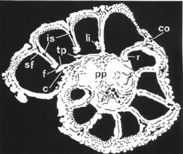
FIGURE
3—Neorotalia mexicana (Nuttall), Lower Miocene, Victoria,
Australia. Reinterpreted drawings of an SEM photo of a horizontal
section (from Hansen and Reiss, 1971), x94. c, canal (formed by
toothplate); co, cover of interlocular space (enveloping system); f,
intercameral foramen; is, intraseptal interlocular space; li, apertural
lip; pp, compound plug; r, retral bend; sf, septal flap; tp, toothplate.
as well as the furrow between the inner umbilical walls and the
multiple lamellar piles closing the bowl being secondarily cov
ered by outer lamellae (PL 3, fig. 6). The aperture was described
as similar to that of Pararotalia mexicana mecatepecensis, hence
interiomarginal extraumbilical, extending slightly towards the
periphery and bordered by a thick lip. The presence of a tooth
plate (s.l) was described and the latter actually illustrated (though
not described) as protruding into the aperture and forming a
space between it and the adjacent coil (PL 3, figs. 6, 11). In view
of their findings, Reiss and Merling (1958) transferred C. calcar
to the genus Pararotalia.
Hofker (1970) followed this usage, but drew attention to the
presence of two kinds of apertures, i.e., a large areal one, bor
dered by a thick lip, typical of Pararotalia, and some irregular,
small ones without lips in interiomarginal position, resembling,
in a "primitive" manner, apertures of Calcarina. Hofker (1970)
believed, therefore, P. calcar to be a link between Pararotalia
and Calcarina.
Hansen and Reiss (1971) described the internal plates as
fo-ramenal plates connected with cover-plates (see Glossary), al
though their SEM photos of sections show clearly that each plate
is a single element extending from the septal flap, connected to
the inner ventral chamber wall, and protruding into the aperture,
a space (canal) being formed between the adaxially intercon
nected plates and the adjacent coil. The chevron-shaped pattern
of ridges thickened by secondary lamination was described and
illustrated. It was emphasized that the latter covers the test in
such a manner that "the interlocular spaces are bridged over
FIGURE 2—1-9, Neorotalia mexicana (Nuttall), Lower Miocene, Victoria, Australia. 1, 2, dorsal (spiral) views; 7, 898156, x60, 2, 898155, x60;
3, 4, ventral views; 3, 898160, x60; 4, 898151, x60; 5, 6, 7, oblique ventral views; 5, 898159, xl20, ultimate chamber broken off; 6, 898126,
x240, part of peripheral margin; 7, 898154, x 120, part of specimen with ultimate chamber broken off; 8, 898161, x240, ventral view, part
of peripheral margin; 9, 898159, x600, canaliculate peripheral spine, c, canal, canal openings; co, cover of interlocular space (enveloping
system); f, intercameral foramen; fu, furrow; is, intraseptal interlocular space; li, apertural lip; pp, compound plug with canals; sf, septal flap;
tp, toothplate. Note radial chamber shoulders and chevron-shaped ornamentational pattern in 3, 4, 7.
and cavities are trapped," communicating with the outside
"through narrow canals leading into the peripheral spines." This
is, in fact, the "enveloping canal-system" with "flying covers"
described and illustrated in great detail by Hottinger and
Leu-tenegger (1980). The "flying covers" are formed by outer la
mellae that cover externally the interlocular spaces and are fold
ed into the latter in a manner that produces funnel-like
imaginations, alternating in position opposite each other at the
margins of the cover and putting in communication the inter
locular spaces with the exterior ("residual openings").
Hottinger and Leutenegger (1980) denied the presence of a
"hooked umbilical chamber extension" (i.e., a toothplate), which
can be seen, however, in their Plate 1, figures 16, 17. On the
other hand, Hottinger and Leutenegger (1980) regarded the inner
ventral chamber wall (adaxial from the umbilical shoulder) as
an "umbilical plate," forming an "interlocular space" subse
quently transformed into a spiral canal. Multiple intercameral
foramina were described (PI. 1, fig. 16), although a single, large
elongated and extraumbilical opening with a serrated lip was
shown as the intercameral foramen (PI. 1, figs. 11, 12). Mainly
on the basis of the enveloping canal-system, as well as of the
reported multiple intercameral foramina, Hottinger and Leu
tenegger (1980) attributed the species calcar again to Calcarina,
a usage followed by Reiss and Hottinger (1984).
Observations and remarks. —The chambers are radially elon
gated and peripherally pointed, with spinose projections giving
the test its typical stellate equatorial outline (Figure 4.1, 4.2).
The dorsal (spiral) side is covered with large inflational pustules,
as well as, in later chambers, by irregularly elongate, sometimes
vermiculate pustules (Figure 4.1). On the ventral (umbilical)
side each chamber shows a prominent radial shoulder. An um
bilical shoulder leads to the formation of a deep umbilical bowl
by the inner ventral chamber walls (Figure 4.2,4.3). The sutures
are deeply sunken, forming the interlocular intraseptal spaces,
which are covered externally by secondary lamination forming
the flying covers with their bordering openings of the enveloping
canal-system (Figure 4.2, 4.3). The openings of the covers cor
respond closely in position to the grooves running along prom
inent ribs from the radial imperforate chamber shoulder, and
nearly normal to it, into the intraseptal spaces (compare also
Hottinger and Leutenegger, 1980). Grooves and adjoining ribs
parallel roughly the peripheral chamber shape and form thus a
chevron-shaped pattern (Figure 4.2). Secondary lamination blurs
the ornamentation and covers part of the canal openings along
the flying covers, but produces new openings in irregular sutural
and radial position (Figure 4.2, 4.3).
The umbilical bowl is closed by a cluster of prominent infla
tional piles externally expressed as thick pustules, with distinct
canals between them (Figure 4.2,4.3). The furrow ("interlocular
space" in Hottinger and Leutenegger, 1980) produced by the
inner ventral chamber walls forming the umbilical bowl and
connected with the intraseptal interlocular spaces (Figure 4.2)
is gradually covered by partly nonadhering secondary lamellae
(Figure 4.3) and converted into a spiral canal. Spaces between
the ventral piles are covered in the same manner, producing the
"umbilical cavities" reported by Reiss and Merling (1958, PL
3, fig. 10). The distal chamber face is roughly triangular in shape,
densely covered by minute rounded to elongate pustules and
riblets, aligned more or less in the direction of the periphery
and of the radial chamber shoulder (Figure 4.4-4.6). Secondary
lamination enhances and modifies this ornamentation in later
instars and produces, together with ribs and adjoining grooves
running towards the intraseptal spaces, the characteristic chev
ron-shaped ornamentational pattern. The spinose projections
of the shell are built up stepwise by radial folds of secondary
lamellae glued at first against the peripheral radial ridges on the
apertural face of the penultimate chamber and later against the
previous folds of secondary outer lamellae. Thus, long infla
tional canaliculate spines are produced housing radially diverg
ing and anastomosing canals with bordering minute spikes, ex
tending every intraseptal interlocular space in radial direction
(Figure 4.1-4.3). Chambers are built against or around the can
aliculate spines of the preceding coil, which puts the spine canal
system in communication with both the exterior on the dorsal
side and the chamber lumina of the succeeding coil (Figures 4.1,
6.1). All inflational ornamentational features are covered by
minute ridges (Figure 4.1).
The primary cameral aperture is a slightly arched
interio-marginal, extraumbilical slit, slightly extended towards the pe
riphery and bordered by a strongly pustulate lip (Figure
4.4-4.6). The distinctly serrated free edge of a toothplate, connected
to the apertural lip at its distal corner, protrudes into the aperture
and produces a space (canal) between plate and adjacent coil
(Figure 4.4-4.6). The apertural face is bent backwards at the
proximal end of the apertural lip (Figure 4.5, 4.6). Between the
distal end of the main chamber aperture and the periphery,
irregular openings in interiomarginal position, bordered by pus
tules and spinules, can be observed, especially in later chambers
of well-preserved specimens (Figure 4.3,4.6). They are regarded
here as supplementary apertures.
Dissected specimens (Figures 5, 6) show the test architecture
and structure in detail. The imperforate toothplate is seen to
extend from the narrow septal flap through the chamber to the
interiomarginal aperture, to protrude into the latter, thus re
stricting it, and to produce with the adjacent coil a primary
spiral umbilical canal, communicating with the chamber lumen,
as well as with the furrow (Figure 5.1-5.4). Attachment of the
toothplate to the septal flap leads to the intercameral foramen,
which preserves the pustulate lip of the aperture, to become
truly areal. The retral bend of the apertural face at the ventral
corner of the aperture, mentioned above in Pararotalia inermis
and in Neorotalia mexicana, is shown in N. calcar to extend
finger-like into the chamber lumen (Figure 5.2-5.4). The covers
of the intraseptal interlocular spaces with their openings to the
exterior are shown in Figure 5.3, 5.4. The canaliculate spines
are incorporated into the succeeding coil on the spiral side as
the result of chambers being built against or around the spine
(Figure 6.1-6.3). The relationship of toothplate to septal flap
and to the spiral canal-system, the intercameral foramina and
apertural lips, as well as the intraseptal interlocular spaces and
their covers of the enveloping canal system are also shown in
Figure 6.3-6.6. The SEM photo of a sectioned araldite cast
(Figure 6.6) shows the main chamber lumina and intercameral
foramina, as well as, adjoining the latter, the toothplates, which
FIGURE 4—1-6, Neorotalia calcar (d'Orbigny), Recent, Gulf of Aqaba, Red Sea, 1.5-6.0 m depth. 1, 7604-G, x 75, dorsal view, note ornamentation;
2, 7645-G, x no, ventral view, note ornamentation; 3-6, oblique ventral views; 3, 875222, x240, apertural face and part of adjacent coil,
note canaliculate spines; 4, 7641-G, x400, apertural face of ultimate chamber; 5, 7613-G, x 370, apertural face of ultimate chamber; 6, 875227,
x 240, apertural face of ultimate chamber and part of adjacent coil, a, aperture; af, apertural (distal) face; c, canal openings; co, cover of
intraseptal interlocular space (enveloping system); fu, furrow; li, apertural lip; pp, multiple piles with canals in between; r, retral bend; sa,
supplementary apertures; sp, canaliculate spine; tp, toothplate.
26 JOURNAL OF PALEONTOLOGY, V. 65, NO. 1, 1991
FIGURE 5—1-4, Neorotalia calcar (d'Orbigny), Recent, Gulf of Aqaba, Red Sea, 1.5 m. Internal views of ventrally dissected specimens. 1, 898129, x240; 2, 875225, x600; 3, 898127, x240; 4, 898128, x240; 3, 4, same chambers seen from different angles, c, canal; co, cover of intraseptal interlocular space (enveloping system); df, distal chamber wall; f, intercameral foramen; is, intraseptal interlocular space; li, apertural lip; r, retral bend at ventral end of aperture; sa, supplementary apertures; sf, septal flap; tp, toothplate.
are adaxially connected in successive chambers, producing a
spiral umbilical canal. The latter communicates through narrow
openings with the furrow that borders the umbilical bowl and
is converted into a spiral canal continuous with the intraseptal
spaces. What appear to be multiple supplementary foramina are
also shown at the lower margin of Figure 6.6.
A reinterpreted drawing of an SEM photo of a horizontal
section of N. calcar, published by Hansen and Reiss (1971), is
shown in Figure 7.1, 7.2. The toothplates of consecutive cham
bers are seen to protrude into the main apertures and to be
interconnected adaxially, thus producing the spiral umbilical
canal. It is noteworthy that the toothplates are seen to be in
continuation of the wedging out, inner ventral, perforate and
laminated chamber wall where the spiral toothplate canal passes
into the spiral furrow around the multiple plugs (Figure 7.2 and
compare Figure 6.6). The retral bend at the ventral end of the
aperture is clearly seen. It is of interest to note that in this spot
the thick, pustulate lip does not border the aperture like it does
along most of the slightly forward-bent apertural margin. The
enveloping canal-system is shown at the outer margins of the
interlocular spaces, and so is the structure of the canaliculate
spines, communicating with the remainder of the canal system.
The present description supplements and modifies to some
extent that of Hottinger and Leutenegger (1980) and indicates
that the general architecture and test structure of Calcarina cal
car d'Orbigny is identical with that of Neorotalia mexicana. An
exception appears to be the supplementary apertures of Neo
rotalia calcar. However, these apertures are small and irregular,
lack penstomal thickenings, and are observed only in well-pre
served adult chambers. Presence or absence of such supple
mentary apertures is not definitely proven in the fossil tests of
Neorotalia mexicana. Moreover, presence or absence of sup
plementary apertures in the distal face in addition to a main
aperture in other rotaliacean genera is not regarded by the pres
ent authors as being of generic value (Hottinger et al., in press).
It is doubtful whether the interiomarginal supplementary ap
ertures ofN. calcar are related to the multiple, interioareal pri
mary apertures of the genus Calcarina, as assumed by Hofker
(1970) (see below).
NEOROTALIA VIENNOTI (Greig, 1935)
Figure 8.1, 8.2
Rotalia viennoti GREIG,
1935, p. 524, PI. 58, figs. 1-14.
Neorotalia viennoti (Greig). BERMUDEZ,
1952, p. 19.
Pararotalia viennoti (Greig).
REISS AND MERLING,1958, p. 1, PI. 3, figs.
1-5.
Review. —Rotalia viennoti has been compared with Rotalia
calcar by Greig (1935), who suggested that both of them should
be included perhaps in a new genus, different from Rotalia. It
was included by Bermudez (1952) in his genus Neorotalia. Reiss
and Merling (1958) included R. viennoti in Pararotalia. For
these reasons we have reexamined topotypes of this species.
In his original description, Greig (1935) emphasized the heavy
ornamentation by pustules of both sides of the test and especially
those forming "vertical pillars" in the center of the test, possibly
having a function in supporting and strengthening of the test.
Greig also reported a "typical canal-system." The aperture was
described as "peripheral and slightly ventral, near the base of
the apertural face . . . consisting of a plain slit... in the adult
of irregular openings... their configuration being controlled...
by the pustular arrangement of the previous coil." Greig's Plate
58, figure 1 strongly suggests an extraumbilical aperture restrict
ed by a plate. The umbilical bowl is shown on Plate 58, figures
1, 14; in the latter (a vertical section) an internal plate forming
a space with the adjacent coil is well apparent.
Reiss and Merling (1958) examined specimens identified as
Rotalia viennoti from the Middle Eocene, which seem closely
related to, if not conspecific with, the types from the Oligocene.
They described and illustrated in thin sections the extraumbil
ical interiomarginal aperture, a toothplate, intraseptal interlo
cular spaces, the cluster of thick "pillars" in the axial region, as
well as the heavy pustules on both sides of the test. By com
parison with Rotalia mexicana mecatepecensis, included in
Pararotalia, Reiss and Merling (1958) attributed R. viennoti to
this latter genus.
Observations and remarks. —The test is heavily pustulate on
both sides and on the periphery and especially in the central
ventral region (Figure 8.1). These pustules represent the ter
minations of the massive inflational piles forming the com
pound axial plug (see also Greig, 1935; Reiss and Merling, 1958).
The ornamentation is enhanced by thick secondary lamination,
which blurs the radial-chamber shoulders visible in the last few
chambers (Figure 8.1), as well as the deeply sunken sutures
forming the intraseptal interlocular spaces (Figure 8.1, 8.2). An
enveloping canal system due to secondary lamination is present,
the covers at the margin of the interlocular spaces with their
openings to the exterior visible only in ontogenetically late
chambers, where heavy ornamentation does not hide them (Fig
ure 8.1).
The rather narrow septal flap extends into a toothplate; the
latter is connected to the inner ventral chamber wall (forming
the largely hidden umbilical bowl) and protrudes with a dis
tinctly serrated free edge into the interiomarginal extraumbilical
aperture (bordered by a heavily pustulate lip), thus producing
a spiral umbilical canal (Figure 8.2). Thick pustules present on
the previous coil opposite the aperture (Figure 8.1) may lead
the latter to appear in the light-microscope as multiple (see
Greig, 1935). The architecture of Rotalia viennoti is thus iden
tical with that of Neorotalia mexicana and Neorotalia calcar
and is, therefore, included in Neorotalia, in agreement with
Bermudez (1952).
Family
CALCARINIDAESchwager, 1876
Genus CALCARINA d'Orbigny, 1826
CALCARINA SPENGLERI(Gmelin, 1791)
Figure 8.3, 8.7
Nautilus spengleri GMELIN,
1788-1793, Vol. 1, No. 6.
Calcarina spengleri (Gmelin). CUSHMAN, 1927, p. 72, PI. 31, fig. 2;
HOFKER,1927, p. 45, PI. 2, figs. 3-6, 8-10;
HANSEN AND REISS,1971,
p. 336, PI. 11, figs. 1-6, PI. 12, figs. 1-5; HOTTINGER AND LEUTENEG
GER, 1980, p. 124-125, PI. 6, figs. 1-13, PI. 7, figs. 1-3, text-fig. 2;
HANSEN,1981, PI. 4, figs. 1, 2, (neotype), PI. 5, figs. 1-6.
Calcarina mayori Cushman. REISS AND MERLING,
1958, PI. 3, figs. 17,
18, PI. 4, figs. 1-4.
Review.— The type-species of the genus Calcarina has been
extensively discussed in the literature (see references above with
bibliography) and only a few points should be raised here, per
taining to differences from Neorotalia.
Nearly all authors have described the aperture as multiple
rounded openings, surrounded by thick rims, interioareal in
position. Only Reiss and Merling (1958) have described the
aperture as a "strongly indented interiomarginal one, part of the
indentations coalescing and forming 'pores'." Scanning electron
microscopy (Hansen and Reiss, 1971; Hottinger and Leuteneg
ger, 1980) disproved this latter description and showed the mul
tiple apertures surrounded by thick rims. A toothplate (s.l.) was
recorded by Reiss and Merling (1958) "very near the adjacent
coil" and Hansen and Reiss (1971) reported a foramenal plate
and cover plate in early stages only. No internal plate was ob
served by Hottinger and Leutenegger (1980). Reiss and Merling
(1958) snowed in thin sections the presence of intraseptal
terlocular spaces, as well as of "umbilical and intramural cav
ities" interrupted by numerous "pillars," produced by second
ary lamination in the ventral chamber walls of the test. This
corresponds to the enveloping canal system described in detail
by Hottinger and Leutenegger (1980). The structure of the can
aliculate spines was shown by these latter authors (see also
Han-sen and Reiss, 1971).
Observations and remarks.—The accompanying Figure 8.4
and 8.7 demonstrates clearly the multiple, irregularly rounded
apertures with thick peristomal lips or rims, preserved as such
when converted into intercameral foramina. There is no
foram-enal or cover plate present and neither is there a toothplate with
free edge. On the other hand, a forward extension of the septal
flap, very near the adjacent coil, can be observed (Figure 8.7)
and is best described as an umbilical plate. It produces part of
the spiral umbilical canal described by Hottinger and Leute
negger (1980). The canal openings of the enveloping system
around the heavy pustules on the ventral side are shown in
Figure 8.5 and the club-shaped canaliculate spines in Figure
8.3-8.6. Particularly well developed spikes at the margins of
canal openings of the chamber wall and in the canaliculate spines
are shown in figure 8.6.
Calcarina differs, therefore, considerably from Neorotalia
mainly by the presence of multiple, interioareal main apertures
surrounded by thick rims, by the absence of a toothplate (s. str.)
with a free edge, as well as by the presence of an umbilical plate.
DISCUSSION AND CONCLUSIONS
Reexamination of the type-species of the genera Pararotalia
Le Calvez, 1949, and Neorotalia Bermudez, 1952, shows them
to possess the same basic architecture, including interconnected
toothplates with a free edge, producing a spiral canal, and a
single interiomarginal main aperture. However, Neorotalia dif
fers from Pararotalia in possessing in addition an enveloping
canal system, produced by secondary lamination, and must,
therefore, be regarded as a valid independent genus. Calcarina
calcar d'Orbigny, 1826, placed by some authors in Pararotalia,
has the same canal system, toothplates, and apertural features
as Neorotalia and must be placed in this latter genus. This de
spite the fact that N. calcar possesses irregular supplementary
apertures. The type-species of Calcarina d'Orbigny, 1826, also
characterized by an enveloping canal system, possesses, how
ever, multiple primary interioareal chamber apertures sur
rounded by thick rims and an umbilical plate, but lacks the
toothplate with free edge, typical of Pararotalia and Neorotalia.
FIGURE
7—1, 2, Neorotalia calcar (d'Orbigny), Recent, Gulf of Aqaba,
Red Sea. Reinterpreted drawing of an SEM photo of a horizontal
section (from Hansen and Reiss, 1971), x 185. 2, partial enlargement
of 1, x 500. c, canal; co, cover on intraseptal interlocular space (en
veloping system); df, distal chamber wall; f, intercameral foramen;
is, intraseptal interlocular space; li, apertural lip; mc, main chamber
lumen; pp, multiple piles; r, retral bend; sf, septal flap; spc, canaliculate
spines and canals; tp, toothplate, w, inner ventral, perforate chamber
wall coating toothplate at their junction.
Y'.'ii-.M^X
FIGURE
6—1-6, Neorotalia calcar (d'Orbigny). 1-5, Recent, Gulf of Aqaba, Red Sea, 1.5-6 m; 6, Recent, Keij Island, Indonesia. 1, 7605-G,
x 220, part of dorsal view showing incorporation of canaliculate spines from preceding coil; 2-5, internal views of ventrally dissected chambers;
2, 7640-G, x400, note incorporated canaliculate spine; 3, 898135, x240; 4, 875224, x240; 5, 898133, x240; 6, horizontal section of araldite
cast (shell material dissolved), note transition from toothplate canal to furrow canal in upper part of picture, c, canal; co, cover of intraseptal
interlocular space; es, enveloping canal-system; f, intercameral foramen; is, intraseptal interlocular space; li, apertural lip; mc, main chamber
lumen; o, opening between main chamber lumen and canal-system; sa, supplementary aperture; sf, septal flap; sp, spine; tp, toothplate.
30
JOURNAL OF PALEONTOLOGY, V. 65, NO. 1, 1991
es
The earliest, toothplate-bearing pararotaliids were observed in thin sections of hard rocks at the base of the Late Cretaceous limestone sequence of Terradets Gorge, Montsech, Lerida Prov ince, northern Spain (Hottinger and Rosell, 1973), level MS27, of Coniacian or Early Santonian age. They co-occur with true Rotalia and other rotaliid genera, like Kathina and Orbitoka-thina.
The derivation of all rotaliids from Pararotalia advocated by some authors (see also Reiss and Merling, 1958) is, thus, ques tionable. The differences between the Rotaliinae and the Para-rotaliinae with regard to toothplates, umbilical plates, folia, and canal-systems are so great that an independent development of these groups in the Turonian or earlier must be assumed. For the same reasons, it would be probably appropriate to consider the Pararotaliinae Reiss, 1963, and the Cuvillierininae Loeblich and Tappan, 1964 (as well as the Ammoniinae Saidova, 1981, with foramenal and cover plates) as separate families, and not as subfamilies of the Rotaliidae Ehrenberg, 1839 (see Loeblich and Tappan, 1987).
The functional significance of differentiation into dense en-doplasm in the main chamber lumina and rhizopodial ecto plasm (with different organelles and inclusions) in canal-systems has been discussed by Hottinger and Dreher (1974), Hottinger (1978), Hottinger and Leutenegger (1980), and Reiss and Hot tinger (1984). Basically, canal-systems serve as bypasses of en-doplasm-containing main chamber-lumina and intercameral fo ramina, thus connecting functional ectoplasm deep inside the test directly with the exterior. This is particularly advantageous to the individual (e.g., with regard to motility) under conditions of external stress.
Although test geometry is quite different, toothplates in the Neorotaliinae resemble those of certain Buliminidae, where they form a kind of axial canal, regarded to contain ectoplasm active in oxygen uptake under low-oxygen conditions (Veerhallen,
1986). Cytoplasmic differentiation of canal-cytoplasm in Neo rotalia calcar has been shown by Hottinger and Leutenegger (1980). With regard to the enveloping canal-system, it seems that it plays an important role in both Neorotalia and Calcarina in facilitating pseudopodial hold-fast to solid substrate or plants in the high-energy, shallow-water environments of the photic zone, to which these symbiontic diatom-bearing genera are con fined (Reiss and Hottinger, 1984). Geological evidence suggests that the Late Cretaceous-Miocene Pararotalia was characteristic of calmer waters. Analogies, homologies, and functional signif icance of foraminiferal test structures remain challenging topics for further studies on living individuals.
ACKNOWLEDGMENTS
The authors are indebted to M. Langer and M. Duggelin (University of Basel) and to M. Dvoraczek (Geological Survey of Israel, Jerusalem) for assistance in scanning electron micros copy. A. R. Loeblich, Jr. and B. K. Sen Gupta offered construc tive criticism of the manuscript. The material examined origi nates from collections at the University of Basel and The Hebrew University of Jerusalem. All figured specimens are deposited in the Natural History Museum of Basel.
REFERENCES
BERMUDEZ, P. J. 1952. Estudio sistematico de los foraminiferos ro-taliformes. Boletin de Geologia, Venezuela, 2(4): 1-230.
BRADY, H. B. 1884. Report on the foraminifera dredged by H.M.S. Challenger during the years 1873-1876. Reports of the Scientific Re sults of the Voyage of H.M.S. Challenger (Zoology), 9, 814 p. BRUNNICH, M. T. 1772. Brunnich Zoologiae Fundamenta. Hafhiae et
Lipsiae, Grunde i Dyeloeren, 253 p.
CUSHMAN, J. A. 1927. An outline of a reclassification of the fora minifera. Contributions from the Cushman Laboratory for Forami niferal Research, 3:1-105.
DEBOURLE, A. 1955. Cuvillierina eocenica, nouveau genre et nouvelle espece de foraminifere de TYpresien d'Aquitaine. Compte Rendu des Seances de la Societe Geologique de France, 1955:19.
DELAGE, Y. AND E. HEROUARD. 1896. Traite de Zoologie Concrete. 1. La Cellule et les Protozoaires. Schleicher Freres, Paris, 584 p. EHRENBERG, C. G. 1839. Uber die Bildung der Kreidefelsen und des
Kreidemergels durch unsichtbare Organismen. Physikalische Ab-handlungen der Koniglichen Akademie der Wissenschaften zu Berlin,
1838 [1840: separate 1839]:59-147.
GMELIN, J. F. 1788-1793. Systema naturae Linnaei, 1(6):3021-3909, Vermes (13th ed.). G. E. Beer, Lipsiae, Germania.
GREIG, D. A. 1935. Rotalia viennoti, an important foraminiferal spe cies from Asia Minor and western Asia. Journal of Paleontology, 9: 524.
HANSEN, H. J. 1981. On Lorentz Spengler and a neotype for the for-aminifer Calcarina spengleri. Bulletin of the Geological Society of Denmark, 29:191-201.
, AND Z. REISS. 1971. Electron microscopy of rotaliacean wall structures. Bulletin of the Geological Society of Denmark, 10:329-346.
HOFKER, J. 1927. The foraminifera of the Siboga Expedition, Pt. 1, Tinoporidae, Rotaliidae, Nummulitidae, Amphisteginidae. E. J. Brill, Leiden, 74 p.
. 1951. The toothplate Foraminifera. Archives Neerlandaises de Zoologie, 8:353-372.
. 1957. Foraminifera from the Cretaceous of Southern Limburg, Netherlands, XXIV. The development of Pararotalia tuberculifera (Reuss). Natuurhistorisch Maandblad, 46(3-4):31-39.
. 1970. Studies of foraminifera. Part II, Systematic problems. Publicaties van het Natuurhistorisch Genootschap in Limburg, 20: 1-98.
HOTTINGER, L. 1978. Comparative anatomy of elementary shell struc tures in selected large foraminifera, p. 203-266. In R. H. Hedley and C. G. Adams (eds.), Foraminifera, Vol. 3. Academic Press, Inc., Lon don.
, AND D. DREHER. 1974. Differentiation of protoplasm in Num mulitidae (Foraminifera) from Elat, Red Sea. Marine Biology, 25:41-61.
, E. HAUCZ, AND Z. REISS. In press. Architecture of Eponides and Poroeponides reexamined. Micropaleontology.
, AND S. LEUTENEGGER. 1980. The structure of calcarinid fora minifera. Schweizerische Palaontologische Abhandlungen, 101:115-151.
, AND J. ROSELL. 1973. El Cretacico superior del Montsec. XIII. Coloquio europeo de micropaleontologia, p. 73-85. Empresa Na tional Adaro Investigaciones Mineras S.A., Madrid.
LAMARCK, J. B. 1804. Suite des memoires sur les fossiles des environs de Paris. Annales Museum National d'Historie Naturelle, 5:237-245. LE CALVEZ, Y. 1949. Revision des foraminiferes Lutetiens du Bassin de Paris. II. Rotaliidae et families affines. Memoires du Service de la Carte Geologique Detaillee de la France, p. 1-54.
. 1952. Revision des Foraminiferes Lutetiens du Bassin de Paris,
FIGURE 8—1, 2, Neorotalia viennoti (Greig), Oligocene, Ramla, Israel. 1, 876512, x60, ventral view; 2, 876506, xl20, partial, oblique ventral view of specimen with last chamber dissected. 3-7, Calcarina spengleri (Gmelin). 3, 4, 6, 7, Recent, Keij Island, Indonesia; 5, Recent, Sybai, Mindoro Island, Philippines. 3, 764013, x60, ventral view; 4, 764086, x60, oblique profile view; 5, 898137, x220, young specimen; 6, partial enlargement of 3, showing canaliculate spines with spikes; 7, 764014, x 240, apertural view of penultimate chamber. Note multiple intercameral foramina surrounded by thick rims in 4 and 7. c, canal; co, cover of intraseptal interlocular space (enveloping system); es, enveloping canal system with exterior openings; f, intercameral foramen; is, intraseptal interlocular space; li, apertural lip; sf, septal flap; sk, spikes in canaliculate spines; tp, toothplate; up, umbilical plate.
32 JOURNAL OF PALEONTOLOGY, V. 65, NO. 1, 1991 IV. Valvulinidae, Peneroplidae, Ophthalmidiidae, Lagenidae.
Me-moires du Service de la Carte Geologique Detaillee de la France, p. 1-64.
. 1970. Contribution a Fetude des foraminiferes Paleogenes du Bassin de Paris. Cahiers de Paleontologie, Paris, Editions du Centre National de la Recherche Scientifique, p. 1-326.
. 1977. Revision des foraminiferes de la collection d'Orbigny. II—Foraminiferes de Tile de Cuba. Cahiers de Micropaleontologie,
1:1-127.
LEVY, A., R. MATHIEU, A. POIGNANT, M. ROSSET-MOULINIER, AND A. ROUVILLOIS. 1980. Revision de quelques genres de la famille Dis-corbidae (Foraminiferida) fondee sur Fobservation de leur architec ture interne. Revue de Micropaleontologie (1979), 22:66-88.
LOEBLICH, A. R., JR., AND H. TAPPAN. 1957. Morphology and tax
onomy of the foraminiferal genus Pararotalia Le Calvez, 1949. Smith sonian Miscellaneous Collections, 135(2): 1-24.
, AND . 1964. Sarcodina chiefly "thecamoebians" and For aminiferida, p. C1-C900. In R. C. Moore (ed.), Treatise on Inver tebrate Paleontology, Pt. C, Protista 2. Geological Society of America and University of Kansas Press, Lawrence.
, AND . 1987. Foraminiferal Genera and Their Classification. Van Nostrand Reinhold Company, New York, Vol. 1, 970 p., Vol. 2, 212 p.
MOBIUS, K. 1880. Foraminiferen von Mauritius, p. 65-112. In K. Mobius, F. Richter, and E. Martens (eds), Beitrage zur Meeresfauna der Insel Mauritius und der Seychelles Gutman, Berlin.
MULLER-MERZ, E. 1980. Strukturanalyse ausgewahlter rotaloider Fo raminiferen [Structural analysis of selected rotaliid Foraminifera]. Schweizerische Palaontologische Abhandlungen, 101:5-70. NUTTALL, W. L. F. 1928. Notes on the Tertiary foraminifera of south
ern Mexico. Journal of Paleontology, 2:372-376.
. 1932. Lower Oligocene foraminifera from Mexico. Journal of Paleontology, 6:32.
ORBIONY, A. D \ 1826. Tableau metodique de la classe des Cephalo-podes. Annales des Sciences Naturelles, 7(1):245-314.
. 1839. Foraminiferes, p. 1-224. In A. Bertrand (ed.), Ramon de la Sagra, Histoire Physique et Naturelle de Tile de Cuba. Paris. POAG, C. W. 1966. Paynes Hammock (Lower Miocene?) foraminifera
of Alabama and Mississippi. Micropaleontology, 12:393-440. REISS, Z. 1963. Reclassification of perforate foraminifera. Bulletin of
the Geological Survey of Israel, 35:1-111.
, AND L. HOTTINGER. 1984. The Gulf of Aqaba—Ecological Mi cropaleontology. Ecological Studies 50, Springer-Verlag, Berlin, Hei delberg, 354 p.
, AND P. MERLING. 1958. Structure of some Rotaliidea. Bulletin of the Geological Survey of Israel, 21:1-19.
SAIDOVA, K. H. 1981. O sovremennom sostoyanii sistemy nadvido-vykh taksonov Kaynozoyskikh bentosnykh foraminifer [On an up-to-date system of supraspecific taxonomy of Cenozoic benthonic fo raminifera]. Institut Okeanologii P. P. Shirshova, Akademyia Nauk SSSR, Moscow, 73 p.
SCHWAGER, C. 1876. Saggio di una classificazione dei foraminiferi avuto riguardo alle loro famiglie naturali. Bolletino R. Comitato Geo-logico dTtalia, 7:475-485.
TERQUEM, O. 1882. Les foraminiferes de FEocene des environs de Paris. Memoires de la Societe Geologique de France, ser. 3, 2(3): 1-193.
VEERHALLEN, P. J. 1986. Morphology and function of the internal structures of non-costate Bulimina. Proceedings of the Koninklijke Nederlandse Akademie van Wetenschappen, 89:367-385.
ACCEPTED 27 JUNE 1990
APPENDIX
GLOSSARY OF SELECTED TERMS
Synonyms or terms applied by some authors in a generalized manner, pertaining also to different test elements, are in brackets.
Canaliculate spine.— Spine- or club-shaped to arborescent radial structure composed of consecutive outer lamellae enclosing canals.
rated from the main chamber lumina, but with which they may com municate in the same or in successive whorls by openings other than intercameral foramina. Canal-systems contain functional microtubular ectoplasm and represent bypasses of main chamber lumina in the whorls, directly connecting ectoplasm deep inside the early parts of the test with extrathalamous rhizopodial endoplasm. The spaces forming canal-sys tems are delimited by different elements of the test: umbilical plate, cover-plate joined to a foramenal plate, toothplate, septal flap, folia, previous coil, as well as consecutive outer lamellae.
Cover plate.—\ more or less folded, imperforate extension of the septal flap into the preceding chamber through the intercameral fora men, cutting off, in the preceding chamber, the main chamber lumen from a foliar chamberlet. Usually attached to a preceding foramenal plate. It is a secondary feature, never present in the ultimate chamber and thus not homologous with a primary foramenal plate or umbilical plate. [Umbilical cover plate; retroparies, pars auct]
Enveloping canals.—More or less tubular spaces parallel with the test surface formed within lateral chamber walls and communicating with intraseptal interlocular spaces. The enveloping canals are produced by (nonadhering) imperforate portions of outer lamellae, also covering part ly the intraseptal spaces and folded into these spaces, leaving on both sides of this "flying cover" alternating rows of openings for ectoplasmic flow over grooves in the perforate wall, situated between imperforate inflational ridges. [Intramural cavities auct.]
Folium.—In spiral lamellar foraminifera the mostly triangular in out line and often texturally differentiated (porosity) axial-umbilical portion of the lateral chamber wall. The border between main lateral chamber wall and folium may be indicated by a short posterior indentation or "notch" or by an umbilical plate-suture. An opening (foliar aperture) is always present between the anterior margin of a folium and the adjacent previous coil. In addition, umbilical and/or posterior openings may be present, depending upon whether the folia are free or attached by their tips or along their posterior margin. In some genera folia may extend onto the preceding chamber and be attached to it, covering partly in traseptal interlocular spaces, wherever present. A folium is composed of the same layers as those forming the main chamber wall. [Lip; tenon; umbilical flap; astral lobe, pars auct.]
Foramenal plate.—Basically a primary infold in the direction of growth of the posterio-lateral chamber wall at a sutural notch, and attached to an intercameral foramen. A foramenal plate may be a short finger- or lobe-like closed structure, a short straight plate, a marginally open plate, folded at an angle to the axis of coiling and attached to the adjacent previous coil, or tunnel-like and reaching a spiral interlocular space. A foramenal plate partly separates a main chamber lumen from a foliar chamberlet and is commonly in continuation of a septal flap. It may or may not be connected with a cover plate, which, in the preceding cham ber, completely separates the main chamber lumen from a foliar cham berlet. (Compare umbilical plate; cover plate.) [Toothplate; paries prox-imus, pars auct.]
Main chamber lumen. —Complete or segmented chamber cavity pro duced by a single growth step and communicating with the preceding and succeeding main chamber lumina through (primary or secondary) intercameral foramina. In certain lamellar foraminifera separated partly or completely from a foliar or stellar chamberlet, as well as from canal systems by a foramenal plate, toothplate, umbilical plate, cover plate, or sealing-plate, or by infolded lateral wall producing septula.
Plug [umbilical plug/.—Expanding pile of thickened lamellae in axial position in an umbilicus or in an umbilical bowl. May be canaliculate and/or compound.
Primary spiral-umbilical canal.—More or less tubular or flattened space located between umbilical plates and the wall of the preceding adjacent coil or between plates, folia, and preceding coil; or between toothplates and preceding coil; present in the ultimate chamber.
Pseudospine. —A conical, elongated, normal to wall-surface, solid in flational ornament feature, becoming spine-like through superposition of subsequent outer lamellae. (Compare canaliculate spine.) [Murica; spine; pars auct.]
Septal flap. —That part of the inner lamella that covers the preceding septal face. A septal flap may be adhering to the preceding septal face producing in this case a trilamellar septum in primarily bilamellar fo