CLINICAL ARTICLE
Intracranial dermoid cysts: variations of radiological
and clinical features
B. Orakcioglu&M.-E. Halatsch&M. Fortunati&
A. Unterberg&Y. Yonekawa
Received: 10 October 2007 / Accepted: 3 June 2008 / Published online: 20 November 2008
# Springer-Verlag 2008
Abstract
Background Intracranial dermoid cysts are uncommon, and their clinical features as well as surgical management differ from patient to patient. Dermoids are generally benign lesions, but may cause spontaneous complications such as meningitis and/or hydrocephalus due to rupture and epileptic seizures depending on their location. Little has been reported about characteristic imaging findings with resulting therapeutic considerations, and only a few reports exist about associated hydrocephalus. Imaging modalities have changed and can facilitate differential diagnosis and follow-up if applied correctly. In this paper, we attempt to contribute our clinical experience with the management of dermoid cysts.
Patients and methods The charts of five men and two women with intracranial dermoid cysts were retrospectively reviewed. The patients were treated between September 1993 and September 2006. Selected patients are presented in detail.
Results Tumour location, size and radiographic character-istics varied in each patient. Clinical presentations com-prised focal neurological deficits as well as epileptic seizures, persistent headache, mental changes and psycho-organic syndromes. One patient underwent delayed ven-triculo-peritoneal shunting after ruptured fatty particles caused obstructive hydrocephalus. Despite dermoid rupture
into the subarachnoid space, three patients never developed hydrocephalus. Diffuse vascular supra-tentorial lesions were seen in one patient as a result of aseptic meningitis. Diffusion-weighted imaging (DWI) hyperintensity in der-moids is related to decrease of water proton diffusion and should be used for both the diagnosis and follow-up of this lesion.
Conclusion Although dermoid cysts are known to be benign entities per se, their rupture can cause a wide range of symptoms including aseptic meningitis and/or hydro-cephalus. This may be due to intraventricular obstruction and/or paraventricular compression. While rupture does not necessarily bring about hydrocephalus, radical removal of the tumour and close monitoring of ventricular size is required. Although not widely recognised as such, DWI is considered to be a useful imaging modality in the diagnosis and follow-up of dermoids.
Keywords Dermoid . Hydrocephalus . Olfactory disturbance . CT . MRI
Introduction
Intracranial dermoids are rare congenital conditions that
account for 0.04–0.6% of all intracranial tumours [18,33].
Derived from ectopic cell remnants that are incorporated into the neural tube, they are usually located along the midline. Various clinical aspects such as presentation with epileptic seizures, meningitis, focal neurological deficits, hydrocephalus, sudden death and incidental diagnosis have been reported. These acute symptoms associated with the presence of dermoids, their localisation and their rupture
are well documented [1,2,5,14,15,17,18,20,23,26,33,
40–42]. Diagnostic findings were evaluated in the past but
B. Orakcioglu
:
M. Fortunati:
Y. YonekawaDepartment of Neurosurgery, Universitätsspital Zürich, Zürich, Switzerland
B. Orakcioglu (*)
:
M.-E. Halatsch:
A. UnterbergDepartment of Neurosurgery, University Hospital of Heidelberg, Im Neuenheimer Feld 400,
Heidelberg, Germany
T able 1 Clinical details of patients with intracranial dermoids Patient number Age, sex Presentation Location CT MRI Rupture HCP Therapy Morbidity Follow-up Recurrence 1 19, m Recurrent seizures, headache for 10 years, hyposmia Left fronto-basal, intraventricular Hypodense, some solid structures N/a Y es N o Resection after 10 years Hyposmia (preexisting) Seizure 9 years after surg ery No 2 33, m Seizure Intrasellar , roof of orbit, right petrous bone, intraventricular Hypodense with calcification
T1-hyperintensity (nonhomogenous), T2-hyperintensity
, fatty particles T1-hyperintense Y es Y es Subtotal resection, ventriculo-peritoneal shunt No 18 months No 3 16, f Persistent posttraumatic headache Parasellar , right suprasellar , intraventricular , basal cisterns Hypodense
T1-hypointensity (nonhomogenous), T2-hyperintensity
, Gd enhancement, fatty particles T1-hyperintense Y es N o Subtotal resection T emporarily impaired vision 24 months No 4 60, m Headache, dizziness, ataxia, n/v , dysarthria, right facial paresis, dysmetria Right posterior fossa with compression of 4th ventricle Hypodense with calcification, leucencephalopathy T1-hypointensity (nonhomogenous), T2-hyperintensity
, Gd enhancement, DWI hyperintensity , leptomeningeal Gd enhancement, (convexity , Sylvian fissure, perimedullary) No No T otal resection No 3 months No 5 56, m Acute paranoid-psychotic episode, anterograde amnesia, neuropsychological deficits Suprasellar with compression of optic chiasm Hypodense T1-hypointensity , T2-hyperintensity , Gd enhancement, DWI hyperintensity , (inhomogeneous) Y es N o T otal resection No 6 months No 6 19, f Headache, complex partial epilepsy , blurred vision, n/v , dizzyness Meckel ’s cave, Foramen ovale, (extradural) Hypodense T1-hyperintensity , mild enhancement, T2-hyperintensity , fat suppression, DWI hyperintensity , extradural No No T otal resection No 24 months No 7 52, m 25-year history of epilepsy (generalised), dizziness, diplopia, EEG with pathological spikes right temporal Right temporo-mesial N/a
T1-hypointensity (nonhomogenous), T2-hyperintensity
, no Gd enhancement No No T otal resection No 6 months No Gd Gadolineum, N/a not available, HCP hydrocephalus, n/v nausea/vomiting, DWI dif fusion weighted imaging
show strong individual variation [4,8,9,13,22,24,27,31,
32,36,38,44,45]. The aim of this report is to contribute to
the understanding of this complex disease by presenting a series of patients with distinct characteristics and to provide a brief literature overview.
Patients and methods
We retrospectively reviewed 7 patients with intracranial dermoids who were treated in the Department of Neurosurgery at the University Hospital in Zurich, Switzerland, between 1993 and 2006. We excluded patients with the diagnosis of epidermoid or teratoma. Patients were analysed considering clinical profiles, radiological studies, intra-operative findings and devel-opment of hydrocephalus.
Results
The patients predominantly presented with headache and/or
seizures (Table1). Their ages ranged from 16–60 years and
the median age was 33 years. There were 5 males and 2 females. All diagnoses of dermoid were verified by microscopy. Four of our patients had a history of epilepsy, and subsequent investigations revealed an intracranial tumour. Co-existing symptoms were tumour location-dependent and included hyposmia, cerebellar symptoms, blurred vision and dizziness. Three patients who did not present with signs of epilepsy suffered from headache and one patient had acute paranoid-psychotic episodes. One patient was operated on for a large frontal dermoid 10 years after initial presentation with epileptic seizures and head-ache. Right-sided anosmia was the leading pre-operative symptom in this patient. Radiological investigations showed intracranial masses at different locations. As the period of inclusion of patients is rather long (1993–2006), imaging protocols were not identical. Cranial computed
tomography (CT) (Fig.1) and magnetic resonance imaging
(MRI) was obtained in all patients. Formerly, pre- and post-operative MRI contained standard protocols including
T2-WI (Fig. 2), T1-WI and post-contrast-enhanced T1-WI.
Meanwhile, these protocols were extended to also include DWI as these sequences were found useful in characterising these lesions. On MRI, the tumours typically presented with inhomogeneous T1-hypointensity, T2-hyperintensity and
moderate contrast enhancement (Figs.3,4,5and 6). DWI
may demonstrate a hyperintense lesion (Fig. 7). Due to
tumour rupture, fatty components were also observed intraventricularly (lateral ventricles) or in the subarachnoid space in four patients. In patient 4, leptomeningeal enhancement as a sign of aseptic meningitis was detected
in the basal cisterns, both Sylvian fissures, perimedullary and at the convexity. Multiple supratentorial vascular atherosclerotic lesions were visible.
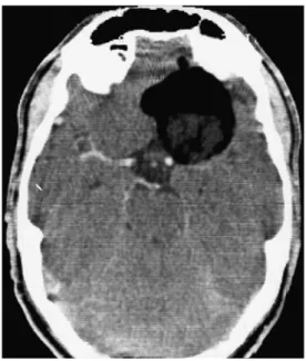
Gross total resection could be achieved in five of the patients. In two patients, subtotal removal was performed in order to reduce peri-operative neurological morbidity. In patient 2, the solid tumour components around the sella were subtotally resected, and parts of the calcified Fig. 1 Axial CT with left fronto-basal hypodense tumour adherent to the middle and anterior cerebral arteries (Patient 1)
Fig. 2 One-year follow-up MRI (T2-WI) of Patient 1 with CSF-filled resection site and left subdural hygroma; no tumour rest or spilled components
tumour adherent to the internal carotid artery (ICA) and middle cerebral artery (MCA) were left in situ. Due to pre-operative rupture of the dermoid cyst, fatty particles obstructed intraventricular pathways at the level of the Foraminae of Monro and subsequently led to post-operative hydrocephalus which was treated with a ven-triculo-peritoneal (VP) shunt. The intraventricular fatty
particles (Fig. 4) in patient 3 were not removed. The
patient never developed hydrocephalus and subtotal removal of the solid tumour components was accom-plished. The patient has been clinically followed regarding subsequent obstruction of CSF pathways and development of hydrocephalus but is free of specific symptoms up to now. Within the tumour, dermoid-specific findings such as hair, sebaceous glands and cholesterol crystals were
present in all cases and confirmed by histopathological examination. The radical resection of the dermoid includ-ed the capsule of the tumour.
Post-operative morbidity was low. In patient 1 the pre-operative hyposmia did not improve and patient 3 temporarily experienced right-sided blurred vision which recovered fully to the operative level. Due to pre-operative dermoid rupture, patient 2 developed an obstructive hydrocephalus that required a VP shunt. Fig. 3 Sagittal MRI (T1-WI) of a suprasellar dermoid with
fronto-temporal extension and subarachnoid spreading of fatty particles (Patient 2)
Fig. 4 Sagittal MRI (T1-WI) of an intra-, supra- and parasellar dermoid (Patient 2); spilling of fatty particles into the basal cisterns, ambient cistern, intraventricular and subarachnoid space with adhesion to the basilar artery
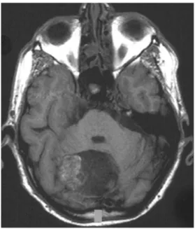
Fig. 5 Axial MRI (T2-WI) with hyperintensity of infratentorial dermoid with severe mass effect and compression of the 4th ventricle (Patient 4)
Fig. 6 Axial MRI (T1-WI) with mainly hypointense and laterally non-homogeneously iso- to hyperintense signal of dermoid (Patient 4)
Follow-up periods varied from 3 months to 9 years after surgery. Clinically, all patients remained neurologically intact without any new neurological deficits or residual epilepsy that required medical treatment.
Discussion
Intracranial dermoids are reported to account for 0.04% to
0.6% [28, 33] of all intracranial tumours. As congenital
dermoid and epidermoid cysts are formed by inclusion of ectodermal elements during neural tube closure, they may be diagnosed at a young age. Dermoids are composed of a thick fibrous capsule with a lining of stratified squamous epithelium, enclosing a thick viscous greenish-brown fluid. This fluid contains lipid metabolites, cholesterol crystals, whorls of hair, calcified areas and decomposed epithelial cells. Sebaceous or sweat glands may also be
present [36]. Mesodermal origin is sometimes ascribed to
dermoids, but this is a misconception, as dermoids derive from ectodermal cell lineages only. Other than true neoplasms, which grow by progressive cell division, they enlarge by desquamation of normal cells and secretion of dermal elements into a cystic cavity well circumscribed by a thick wall of connective tissue. Mainly capsular calcification is common, representing dystrophic changes or dental enamel, another ectodermal derivate. In contrast to epidermoids, dermoids are mostly located in or close to the midline, which is due to their earlier formation. A dermal sinus may be present with spinal, anterior and
posterior fossa lesions [6].
Ruptured dermoids present with various manifestations. Patients with dermoid cysts may either remain asymptom-atic or present with headaches, epileptic seizures, signs of compression of neighbouring tissues, aseptic meningitis, transient cerebral ischemia as a result of vasospasm and
even olfactory halucinations [11,14,18–20,25,33,40,45–
47]. After rupture of these cysts, their fatty content
disperses into the subarachnoid space and may be present
in any of the cisterns and ventricles [17,23,30,36,38,39,
43]. Most often the rupture occurs spontaneously although
trauma, such as a closed head injury, has been implicated in
certain instances (see patient 2) [25]. In patients with
generalised involvement of the meninges and/or subarach-noid distribution of dermoid particles, reactive vasospasm may occur. Vasospasm can also be a reaction to direct
mechanical compression by the space-occupying lesions [1,
11, 14]. Rarely, intraventricular fat leads to rapidly
developing occlusive hydrocephalus [17,23,24].
Certain radiographic characteristics have been
de-scribed in the literature [22, 24, 36, 38, 45]. Osborn et
al. concluded that dermoid contents usually resemble fat
on CT and MRI whereas epidermoids resemble CSF [29].
The literature refers to dermoids mostly associated with T1-WI hyperintensity and T2-WI hypointensity due to a higher density of cholesterol within the tumour. Less commonly, dermoids may display signal characteristics like epidermoids, if the fatty contents are less dense. A CSF-like signal with T2-WI hyperintensity and T1-WI hypointensity may be present. Chen et al. described epidermoids as tumours with a strong DWI hyperintensity as was seen in patient 4 due to an effect of the keratinoid
contents [7]. It was recommended to use fluid attenuated
inversion recovery (FLAIR) sequences and DWI to dis-tinguish epidermoids from cystic lesions, but as demon-strated by our patient 4, histopathologically confirmed dermoids can resemble epidermoids due to their bright
signal on DWI [7, 10]. Only few studies on the DWI
appearance of intracranial dermoids exist [3,35]. Aksoy et
al. reported a hyperintense DWI signal in a posterior fossa dermoid that pre-operatively mimicked an epidermoid. The solid components of the dermoid led to decreased diffusion within the tumour compared to purely cystic
lesions [3]. Other authors also reported a few examples in
which the CT appearance was hyperdense, and even partial
rim enhancement has been documented [8, 9,13,21,22,
27, 33]. Haemorrhage-like imaging patterns are also
described on CT and MRI [34]. Messori et al. reported a
dermoid that showed signal changes in T1-WI during follow-up changing from hypointensity to hyperintensity
related to alterations of the dermoid cyst contents [24].
Intra-operatively, the differentiation between both tumours (dermoid vs. epidermoid) is straightforward once remnants of dermal appendages such as hair and sebaceous glands Fig. 7 Axial MRI (DWI) hyperintensity of some tumour areas,
indicating decreased water proton diffusion as seen in keratin-rich lesions (Patient 4)
are found. In our patients 2, 3 and 4, the pre-operative imaging showed signal characteristics better compatible with epidermoids than with dermoids with T2-WI hyper-intensity and T1-WI non-homogeneous hypohyper-intensity in the solid tumour parts. The ruptured particles had a homogeneous, fatty aspect on MRI with strong T1-WI hyperintensity (patient 2 and 3) and intraventricular fat/ fluid levels on CT (patients 1, 2 and 3). According to the
literature, this is not a surprising finding per se [45], but
the high percentage of patients with atypical MRI signals (71%) in our series justifies critical re-evaluation of the diagnostic radiological criteria.
Patient 1 stands out because the long follow-up period before and after tumour resection is quite unusual. Yaşargil et al. discussed two patients that had a clinical history of 10–20 years before presentation, and Rubin et al. studied one patient with a long history of 15 years before diagnosis
and 9 years of follow-up [33,47]. Epileptic seizures are a
common presentation as mentioned previously. Our patient developed left-sided anosmia over the years. To the best of our knowledge, olfactory dysfunction due to the compres-sive effect of an enlarging dermoid has not been described in the literature. After 10 years of observation, the tumour was radically resected but hyposmia persisted. Nine years after the successful operation, the patient presented with a new onset of seizures. At that point, tumour re-growth was excluded and reactive parenchymal gliosis may have led to his late symptoms. This underlines the fact that despite radical tumour removal, epileptic seizures may recur within some years which has been reported to take place in 5% of all craniotomies for any type of pathology. In any event, complete excision should only be attempted when preser-vation of neurovascular structures is possible. Dermoids adhesive to critical surrounding structures should be decompressed as far as reasonably achievable, and its capsule should be left behind. If total resection cannot be achieved, gentle and copious irrigation of the tumour cavity was recommended by Ecker et al. in order to prevent
delayed vasospastic events [11].
Patient 4 presented with a variety of cerebellar symp-toms which were clearly related to the right-sided intra-axial solid dermoid and which resolved after resection of the mass. Infra-tentorial dermoids are known to present in the first two decades of life rather than at 60 years of age
[18]. This patient also presented with leptomeningeal
enhancement which was interpreted as a diffuse manifesta-tion of aseptic meningitis. Whether or not the meningitis was related to the dermoid remains a matter of debate. However, in the literature various ischaemic events are described in the setting of dermoids and their clinical
presentations [11, 14,26]. In these patients, asymptomatic
aseptic meningitis could have initiated reactive vasospasm manifesting as small infarcts in unusual locations.
There is a very large variety of clinical features and imaging aspects of intracranial dermoids. Comparing the findings of previous studies with our results, we would like to emphasise the diagnostic quandaries with MRI and CT
[22,24,36,38,45]. Mixed signals were observed according
to most published data. Although very recently some reports
have presented data with “non-classical” MRI and CT
findings in dermoids, we aim to emphasize the importance of using an MRI protocol involving T2-WI, T1-WI, T1 fat saturated-WI, magnet resonance angiography (MRA) and DWI. Diffusion-weighted imaging facilitates detecting tumour remnants or re-growth after resection. It may also help to identify solid tumour components that cause a decrease in water proton diffusion, resulting in DWI hyper-intensity as seen in other keratin-rich lesions. Therefore, this protocol should be used for pre-operative diagnostics and during follow-up to monitor potential tumour re-growth.
A variety of clinical presentations can occur due to
rupture of dermoids [1,12,14,16,17,20,23,24,26,30–
32, 36–40, 42,43,45]. In patient 1, dermoid rupture into
the ventricles never led to hydrocephalus. In contrast, ventricle size progressed post-operatively in case 2. Right-sided dilatation of the lateral ventricle was pre-existing, but intra-operative distribution of fatty particles probably caused full-blown occlusive hydrocephalus. The fatty intra-ventricular components may obstruct the Foraminae of Monro, the Foraminae of Luschka and Magendie or the aqueduct; furthermore, intra-operative retraction is a possi-ble explanation for further displacement of fatty droplets
and consecutive hydrocephalus [17,20,23]. If feasible, an
en bloc resection of the dermoid is therefore desirable. The subarachnoid dissemination of fatty particles has probably led to symptoms in patients 2 and 3, with the dermoids being silent until their rupture. Yaşargil et al. have emphasised that communicating hydrocephalus can emerge
as a consequence of aseptic meningitis/arachnoiditis [47].
Therefore, close monitoring of ventricle size is essential with this diagnosis.
Conclusion
This retrospective review of 7 histopathologically verified dermoids demonstrates the wide range of clinical presenta-tions. Reactive gliosis can lead to very late post-operative symptoms such as epileptic seizures. Hydrocephalus is a prominent complication, demanding close monitoring of ventricle size. Diffusion-weighted imaging provides essential information about the existence of solid tumour components
pre- and post-operatively. Complete removal of dermoids without dissemination of the contents should be achieved without injury to surrounding neurovascular structures in order to prevent re-growth and associated morbidity.
References
1. Ahmad I, Tominaga T, Ogawa A, Yoshimoto T (1992) Ruptured suprasellar dermoid associated with middle cerebral artery aneurysm: case report. Surg Neurol 38:341–346
2. Akdemir G, Daglioglu E, Ergungor MF (2004) Dermoid lesion of the cavernous sinus: case report and review of the literature.
Neurosurg Rev 27:294–298
3. Aksoy FG, Aksoy OG, Gomori JM (2001) Klippel-Feil syndrome in association with posterior fossa suboccipital dermoid cyst. Eur
Radiol 11:142–144
4. Brown JY, Morokoff AP, Mitchell PJ, Gonzales MF (2001) Unusual imaging appearance of an intracranial dermoid cyst.
AJNR Am J Neuroradiol 22:1970–1972
5. Bucciero A, Del Basso De Caro ML, Carraturo S, Vizioli L, Cerillo A, Tedeschi G (1995) Supratentorial dermoid cysts. Presentation
and management of five cases. J Neurosurg Sci 39:7–11
6. Caldarelli M, Massimi L, Kondageski C, Di Rocco C (2004) Intracranial midline dermoid and epidermoid cysts in children. J
Neurosurg Spine 100:473–480
7. Chen S, Ikawa F, Kurisu K, Arita K, Takaba J, Kanou Y (2001) Quantitative MR evaluation of intracranial epidermoid tumours by fast fluid-attenuated inversion recovery imaging and echo-planar diffusion-weighted imaging. AJNR Am J Neuroradiol 22:1089– 1096
8. Danaila L, Carp N (1989) Dermoid tumour of the fourth ventricle with hyperdense aspect demonstrated on CT scan. Case report.
Neurol Psychiatr (Bucur) 27:231–236
9. Drolshagen LF, Standefer M (1991) Dense dermoid cyst of the posterior fossa. AJNR Am J Neuroradiol 12:317
10. Dutt SN, Mirza S, Chavda SV, Irving RM (2002) Radiologic differentiation of intracranial epidermoids from arachnoid cysts.
Otol Neurotol 23:84–92
11. Ecker RD, Atkinson JL, Nichols DA (2003) Delayed ischaemic deficit after resection of a large intracranial dermoid: case report
and review of the literature. Neurosurgery 52:706–710 discussion
709–710
12. El-Bahy K, Kotb A, Galal A, El-Hakim A (2006) Ruptured intracranial dermoid cysts. Acta Neurochir (Wien) 148:457–462 13. Erdem G, Topcu M, Topaloglu H, Bertan V, Arikan U (1994)
Dermoid tumour with persistently low CSF glucose and unusual CT and MRI findings. Pediatr Neurol 10:75–77
14. Ford K, Drayer B, Osborne D, Dubois P (1981) Case report. Transient cerebral ischaemia as a manifestation of ruptured
intracranial dermoid cyst. J Comput Assist Tomogr 5:895–897
15. Gormley WB, Tomecek FJ, Qureshi N, Malik GM (1994) Craniocerebral epidermoid and dermoid tumours: a review of 32
cases. Acta Neurochir (Wien) 128:115–121
16. Hash CJ, Ritchie DJ (1978) Ruptured intraventricular dermoid cyst without clinical inflammation. Arch Neurol 35:61
17. Karabulut N, Oguzkurt L (2000) Tetraventricular hydrocephalus due
to ruptured intracranial dermoid cyst. Eur Radiol 10:1810–1811
18. Lunardi P, Missori P (1991) Supratentorial dermoid cysts. J
Neurosurg 75:262–266
19. Lunardi P, Missori P, Gagliardi FM, Fortuna A (1992) Dermoid and epidermoid cysts of the midline in the posterior cranial fossa.
Neurosurg Rev 15:171–175
20. Lunardi P, Missori P, Rizzo A, Gagliardi FM (1989) Chemical meningitis in ruptured intracranial dermoid. Case report and
review of the literature. Surg Neurol 32:449–452
21. Mamata H, Matsumae M, Yanagimachi N, Matsuyama S, Takamiya Y, Tsugane R (1998) Parasellar dermoid tumour with
intra-tumoural haemorrhage. Eur Radiol 8:1594–1597
22. Markus H, Kendall BE (1993) MRI of a dermoid cyst containing
hair. Neuroradiology 35:256–257
23. Martin R, Knone A, Schuknecht B, Kuhn W (1989) Rapid development of occlusion hydrocephalus by intraventricular fat possibly derived from a ruptured dermoid cyst. J Neurol Neurosurg Psychiatry 52:134–135
24. Messori A, Polonara G, Serio A, Gambelli E, Salvolini U (2002) Expanding experience with spontaneous dermoid rup-ture in the MRI era: diagnosis and follow-up. Eur J Radiol
43:19–27
25. Miller NR, Epstein MH (1975) Giant intracranial dermoid cyst: Case report and review of the literature on intracranial dermoids
and epidermoids. Can J Neurol Sci 2:127–134
26. Nakamura M, Mizuguchi M, Momoi MY, Chou H, Masuzawa T (2001) Transient cheiro-oral syndrome due to a ruptured
intracra-nial dermoid cyst. Brain Dev 23:261–263
27. Neugroschl C, David P, Sadeghi N, Soebert A, Pirotte B, Rorive S, Baleriaux D (2002) Unusual CT features of dermoid cyst in the
posterior fossa. Eur Radiol 12:2726–2729
28. Osborn A (1994) Dermoids. Osborn AG Diagnostic
Neuroradiol-ogy Mosby, Year Book inc:632–636
29. Osborn AG, Preece MT (2006) Intracranial cysts:
radiologic-pathologic correlation and imaging approach. Radiology 239:650–
664
30. Oursin C, Wetzel SG, Lyrer P, Bachli H, Stock KW (1999)
Ruptured intracranial dermoid cyst. J Neurosurg Sci 43:217–220
discussion 220–211
31. Patkar D, Krishnan A, Patankar T, Prasad S, Shah J, Limdi J (1999) Ruptured intracranial dermoids: magnetic resonance imaging. J Postgrad Med 45:49–52
32. Phillips WE 2nd, Martinez CR, Cahill DW (1994) Ruptured intracranial dermoid tumour secondary to closed head trauma. Computed tomography and magnetic resonance imaging. J
Neuroimaging 4:169–170
33. Rubin G, Scienza R, Pasqualin A, Rosta L, Da Pian R (1989) Craniocerebral epidermoids and dermoids. A review of 44 cases. Acta Neurochir (Wien) 97:1–16
34. Sanchez-Mejia RO, Limbo M, Tihan T, Galvez MG, Woodward MV, Gupta N (2006) Intracranial dermoid cyst mimicking haemorrhage. Case report and review of the literature. J Neurosurg
105:311–314
35. Shinoyama M, Kajiwara K, Harada K, Ideguchi M, Akimura T, Nishizaki T, Suzuki M (2002) [A case of a ruptured dermoid cyst in the sylvian fissure]. No Shinkei Geka
30:1197–1201
36. Smith AS, Benson JE, Blaser SI, Mizushima A, Tarr RW, Bellon EM (1991) Diagnosis of ruptured intracranial dermoid cyst: value
MR over CT. AJNR Am J Neuroradiol 12:175–180
37. Stendel R, Pietila TA, Lehmann K, Kurth R, Suess O, Brock M
(2002) Ruptured intracranial dermoid cysts. Surg Neurol 57:391–
398 discussion 398
38. Stephenson TF, Spitzer RM (1987) MR and CT appearance of ruptured intracranial dermoid tumours. Comput Radiol 11:249– 251
39. Sumida M, Taguchi H, Kuroki K (1999) [A case of
recurrent-rupture dermoid cyst]. No Shinkei Geka 27:261–266
40. Takeuchi H, Kubota T, Kabuto M, Izaki K (1993) Ruptured suprasellar dermoid cyst presenting olfactory delusion (Eigengeruchs erlebnis). Neurosurgery 33:97–99
41. Tekkok IH, Ayberk G, Kansu T, Saglam S (1989) Bilateral intranuclear ophthalmoplegia associated with fourth ventricular dermoid tumour. J Clin Neuroophthalmol 9:254–257
42. Venkatesh SK, Phadke RV, Trivedi P, Bannerji D (2002) Asymptomatic spontaneous rupture of suprasellar dermoid cyst:
a case report. Neurol India 50:480–483
43. Wallis SW, Van Roy WJ, Wijnalda D, Van Dijl R (1998) Ruptured intracranial dermoid cyst. J Belge Radiol 81:257
44. Warakaulle DR, Anslow P (2003) Differential diagnosis of intracranial lesions with high signal on T1 or low signal on
T2-weighted MRI. Clin Radiol 58:922–933
45. Wilms G, Casselman J, Demaerel P, Plets C, De Haene I, Baert AL (1991) CT and MRI of ruptured intracranial dermoids.
Neuroradiology 33:149–151
46. Wilms G, Plets C, Marchal G, Demaerel P (1990) Simultaneous occurrence of epidermoid and dermoid cysts in the posterior fossa: CT and MR findings. AJNR Am J Neuroradiol 11:1257–1258
47. Yasargil MG, Abernathey CD, Sarioglu AC (1989) Microneur-osurgical treatment of intracranial dermoid and epidermoid
tumours. Neurosurgery 24:561–567
Comment
Orakcioglu et al. submit an interesting report on their experience with intracranial dermoid cysts and give an nice overview on the current literature.
As intracranial dermoid cysts account for less than approx. 0.25% of all intracranial neoplasms, they are up 10 times less common than e.g. intracranial epidermoids. That is why a collection of 7 histologically proven cases seems worth publishing to me. The authors summarize all important information on the different clinical signs and symptoms of ruptured dermoid cysts. They conclude, that long-term follow-up is necessary because of the likelihood of post-op hydrocephalus. I
completely support the author’s opinion, that DWI scans are the most
useful imaging modality for diagnosis and follow-up exams.
Dr Suess Charite - Universitatsmedizin Berlin