Abstract. An RNA hairpin is an essential secondary
structure of RNA. It can guide RNA folding, determine in-teractions in a ribozyme, protect messenger RNA (mRNA) from degradation, serve as a recognition motif for RNA binding proteins or act as a substrate for enzymatic reac-tions. In this review, we have focused on cis-acting RNA hairpins in metazoa, which regulate histone gene expres-sion, mRNA localization and translation. We also review evolution, mechanism of action and experimental use of
DOI 10.1007/s00018-005-5558-5 © Birkhäuser Verlag, Basel, 2006
trans-acting microRNAs, which are coded by short RNA
hairpins. Finally, we discuss the existence and effects of long RNA hairpin in animals. We show that several pro-teins previously recognized to play a role in a specific RNA stem-loop function in cis were also linked to RNA silenc-ing pathways where a different type of hairpin acts in trans. Such overlaps indicate that the relationship between cer-tain mechanisms that recognize different types of RNA hairpins is closer than previously thought.
Keywords. dsRNA, miRNA, RNAi, stem-loop, hairpin, localization, translation.
Introduction
An RNA hairpin consists of a double-stranded RNA (dsRNA) stem, often containing mismatches and bulges (i.e. unpaired sequences within the stem), and a terminal loop. It is the most common secondary structure found in almost every RNA folding prediction. RNA hairpins originate by two mechanisms: (i) transcription by DNA-dependent RNA polymerase of an inverted repeat DNA resulting in the RNA folding into a stem-loop structure, and (ii) an RNA molecule formed as a folded-back tem-plate for RNA-dependent RNA polymerase, which syn-thesizes the second strand of the stem. This review will focus exclusively on the first case – RNA hairpins folded within RNA transcripts. The second mechanism, which produces perfect long dsRNA hairpins, is not widespread in nature and is probably restricted to a ‘copy-back’ mechanism of replication in certain viruses [1]. It is dif-ficult to classify RNA hairpins into distinct categories
be-cause these structures easily arise and differ in many as-pects. Structurally, RNA hairpins can occur in different positions within different types of RNAs; they differ in the length of the stem, the size of the loop, the number and size of bulges, and in the actual nucleotide sequence (Fig. 1a). These parameters provide an extreme variabil-ity allowing specific interactions with proteins (discussed in detail in [2]). Functionally, RNA hairpins can regulate gene expression in cis or trans, i.e. an RNA hairpin within an RNA molecule can regulate just that molecule (cis) or it can induce effects on other RNAs or pathways (trans). Hairpins serve as binding sites for a variety of proteins, act as substrates for enzymatic reactions as well as dis-play intrinsic enzymatic activities.
Many of the pathways utilizing and/or responding to RNA hairpins have evolved independently and are not linked to others. It is not possible to provide a systematic and complete review of such a broad topic as the biology of hairpin RNAs in a single article. We have therefore de-cided to provide an insight into the specific roles of RNA hairpins in animals. The rationale for this combination is that we want to highlight some common factors for both
Review
Hairpin RNA: a secondary structure of primary importance
P. Svoboda* and A. Di Cara
Friedrich Miescher Institute for Biomedical Research, Maulbeerstrasse 66, 4002 Basel (Switzerland), Fax: +41 61 6973976, e-mail: svoboda@fmi.ch
Received 21 November 2005; received after revision 3 January 2006; accepted 11 January 2006 Online First 29 March 2006
cis- and trans-acting RNA hairpins among the wide
spec-trum of possible biological effects.
This review is organized into three main sections dis-cussing (i) short RNA hairpins acting in cis, (ii) short RNA hairpins acting in trans (microRNA pathway) and (iii) long RNA hairpins (stem >100 bp).
Short RNA hairpins acting in cis
Hairpin structures differ in size and localization within a messenger RNA (mRNA) sequence. Several examples of well-studied stem-loop structures and their functions in metazoa are shown in Figure 1. Short hairpins in mRNA are involved in diverse processes such as specific subcel-lular localization of specific mRNAs, regulation of trans-lation and mRNA stability. Stem-loops also function in initiation of translation via internal ribosomal entry sites
[3], and are involved in viral replication [4]. The SECIS element found in selenoprotein 3¢ mRNA (Fig. 1b), which
controls synthesis of selenoproteins (reviewed in [5]), represents another specific stem-loop structure. It serves as a binding site for the SECIS-binding protein 2 (SBP2) involved in mediating UGA redefinition from a stop codon to selenocysteine. Iron-responsive element (IRE, Fig. 1c) is another example of a stem-loop structure act-ing in cis (reviewed in [6]). Cellular iron homeostasis is maintained by IRE-dependent posttranscritpional regula-tion of mRNA of the transferrin receptor, ferritin and other mRNAs that mediate iron uptake and storage. Post-transcriptional regulation by IRE involves interaction with two iron regulatory proteins, IRP1 and IRP2. During iron starvation, IRPs stabilize the transferrin receptor and inhibit translation of ferritin mRNAs by binding to IREs within untranslated regions. IRE is found either in the 5¢
or 3¢ UTR [6].
Figure 1. RNA stem-loops. (a) A schematic overview of an RNA stem-loop depicting the important parameters for the role of such a
hair-pin RNA. (b) The SECIS stem-loop structure element controlling selenoprotein synthesis. Right: A consensus of a secondary structure of a SECIS element [5]. Left: A specific example of the SECIS element in Homo sapiens [143]. (c) Examples of iron-responsive elements (IRE) stem-loop structures regulating iron metabolism in humans [144, 145]. (d) A histone mRNA stem-loop, which controls processing and expression of replication-dependent histone genes. Right: Consensus sequence. Absolutely conserved nucleotides are shown in bold-face [8]. Left: A specific example of a histone stem-loop in Homo sapiens. (e) RNA hairpins in bicoid, gurken and I-factor mRNAs regu-lating mRNA localization in Drosophila oocytes [24, 35]. Top: Schematic view of modular architecture of bicoid localization signal con-sisting of three stem-loops (designated III, IV and V) implicated in the bicoid localization element (BLE). Region labeled in black is nec-essary and sufficient for transport of bicoid mRNA and its initial accumulation in the anterior part of Drosophila oocytes [24].
Among cis-acting hairpins, we have selected the follow-ing three subjects for a more detailed discussion: (i) his-tone mRNA metabolism, (ii) mRNA localization depen-dent on 3¢ UTR stem-loops and (iii) TRBP-mediated
reg-ulation of protamine and human immunodeficiency virus (HIV) trans-activation responsive (TAR) mRNAs.
3¢ stem-loop in the mRNA of replication-dependent histones
Metazoan replication-dependent histone genes have sev-eral features that differentiate them from other eukary-otic genes. They are physically linked in clusters, do not have introns, express the only known non-polyadeny-lated mRNAs, and they require a distinct set of factors for expression and regulation (reviewed in [7, 8]). Ex-pression of histone genes is associated with formation of Cajal (coiled) bodies (subnuclear domains involved in nuclear RNA metabolism) [9]. Replication-dependent histone mRNA contains a conserved 3¢ end
26-nu-cleotide (nt) sequence harboring a 16-nt stem-loop (6-bp stem and a 4-nt loop, Fig. 1d) [10]. Mutations within the stem-loop structure or flanking sequences impair the correct processing of the 3¢ end of histone mRNA.
How-ever, processing is not absolutely dependent on the stem-loop since a small number of processed transcripts can be detected even in its absence [11]. The histone 3¢
stem-loop is bound by the stem-stem-loop binding protein (SLPB, also named HBP – hairpbinding protein), which is in-volved in several aspects of the histone mRNA metabo-lism. The stem-loop/SLPB complex is the key factor in cell-cycle regulation of histone mRNA expression, in-cluding processing of the nascent transcript by the U7 small nuclear ribonucleoprotein (snRNP) complex, con-trol of translation and regulation of the half-life of mRNA. SLBP has a unique RNA binding domain that binds the 3¢ hairpin with a high affinity (Kd = 1.5 nM)
when specific nucleotides are present in the stem-loop [12]. SLPB and U7 snRNP are found in Cajal bodies lo-cated in proximity to histone gene clusters, suggesting that coiled bodies play a role in histone mRNA expres-sion and processing [9, 13]. SLBP/U7 snRNP complex recruits a cleavage factor, which releases mature histone mRNA cleaving five nucleotides downstream of the his-tone stem-loop [8]. A recent study implicates poly-adenylation factor CPSF-73 in the histone mRNA cleav-age, suggesting a link between 3¢ end histone mRNA
pro-cessing and polyadenylation [14]. Processed histone mRNA remains bound to SLBP, which is required for ef-ficient translation and cell-cycle dependent regulation but not for nuclear export of histone mRNA [15]. Regulated degradation of histone mRNAs requires inter-action of the stem-loop/SLPB complex with Upf1, a key regulator of the nonsense-mediated decay (NMD) path-way, and ATR, a key regulator of the DNA damage
check-point pathway activated during replication stress [16]. Notably, the stem-loop region of human replication-de-pendent histone mRNA can also interact with a con-served 3¢-5¢ exonuclease, 3’hExo, which is a candidate
for the exonuclease that initiates rapid decay of histone mRNA upon completion and/or inhibition of DNA repli-cation [17]. The 3’hExo orthologue in Caenorhabditis is known as ERI-1 [18]. ERI-1 is an exonuclease that de-grades small interfering RNAs (siRNAs), thus negatively regulating RNA interference (RNAi) in Caenorhabditis and mammals [18, 19]. ERI-1 is unlikely to function as a general exonuclease because eri-1-null mutants are vi-able and show limited pleiotropic phenotypes, which may be a consequence of a defect in the siRNase (or possibly microRNase) function of ERI-1 [18]. It is still unknown, however, whether ERI-1 can also recognize and degrade small hairpin precursors of miRNAs. Replication-depen-dent histone mRNA thus provides an example of how a hairpin structure in cis can serve as a highly specific docking point providing a specific regulation of expres-sion in a unique family of mRNAs.
mRNA localization regulated by stem-loop structures
mRNA localization is a means for protein localization via spatially restricted translation. Numerous specifically lo-calized mRNAs have already been identified, mostly in oocytes or early embryos of Drosophila and Xenopus (re-viewed in [20]). There are three main mechanisms for lo-calized mRNA distribution: (i) active, directed transport of an mRNA to its destination where the mRNA is an-chored, (ii) local stabilization/spatially selective degrada-tion of mRNA and (iii) diffusion of mRNA combined with local anchoring at the target site (reviewed in detail in [21]). Individual mRNAs use different mechanisms for localization at their destination sites. The destination is encoded within a ‘zip code’, which acts as an mRNA lo-calization signal. The term ‘zip code’ is used in this re-view for a sequence essential for mRNA localization – its removal impairs localization, and its fusion to non-local-ized mRNA is sufficient to localize it. Zip codes are di-verse in terms of primary or secondary structure; how-ever, they are usually localized in the 3¢UTR, and their
function is mediated by binding proteins [21].
Specific mRNA localization in Drosophila oocytes and embryos
Zip codes can be represented by stem-loop structures, which are found for example in bicoid, gurken, I-factor (Fig. 1e), and several other mRNAs localized in
Droso-phila oocytes or early embryos. The bicoid mRNA is a
famous example of a localized cytoplasmic determinant that produces a morphogen gradient upon translation [22, 23]. The bicoid localization element (BLE) is a well-stud-ied example of an element with a modular architecture
(Fig. 1e). Signals required for the bicoid mRNA localiza-tion to the anterior of the oocyte are located within the first 720 nucleotides of the bicoid 3¢ UTR (reviewed in
[24]). Several sequential steps in transport and localiza-tion are linked to three predicted stem-loops in the BLE (Fig. 1e). Stem V carries a 50-nucleotide element called BLE1 (bicoid localization element 1), which is required and sufficient for transporting the mRNA into oocytes and its initial accumulation in the anterior end. Stems V and VI regulate accumulation of the transcript in the oocyte and its anterior end. Finally, anchoring of the lo-calized mRNA is controlled by the stem-loop III. Tthe
bi-coid gene is found only in Drosophila and closely related
species [25]. The determinants of secondary structure ap-pear to be conserved amongst them [26].
Several trans-acting factors regulate the bicoid mRNA lo-calization. The current model proposes that mRNA is bound by the Drosophila-specific adaptor protein Swal-low, which interacts with a dynein motor component, resulting in mRNA transport along microtubules [27].
Swallow and bicoid mRNAs are found in a multiprotein
complex implicated in bicoid mRNA localization. This complex consists of Swallow, RNA binding proteins Mod-ulo, PABP and Smooth, and the kinesin family member Nod [28]. The anterior anchoring of the transcript requires the Staufen protein [29], a conserved protein containing four to five dsRNA binding domains (dsRBDs). Staufen is one of several proteins which contain multiple dsRBD do-mains and play multiple roles in mRNA regulation. In ad-dition to anchoring bicoid mRNA, Drosophila Staufen has been implicated in microtubule-dependent transport of
osk mRNA, actin dependent localization of pros mRNA
[30] and translational activation of osk mRNA [31]. Mam-malian Staufen1 has been also implicated in recruiting the nonsense-mediated mRNA decay factor Upf1 to the Arf1 mRNA, inducing its decay [32]. Since Staufen and its ho-mologues are ubiquitously expressed, there are many other functions which probably remain to be discovered.
Gurken (grk) and I-factor are localized in a crescent on
one side of the nucleus in Drosophila oocytes. The grk mRNA is also localized by dynein-mediated transport along microtubules [33]. Localization and translational regulation of grk play key roles in establishment of pri-mary embryonic axes in the oocyte [34]. Interestingly, the
I-factor is a non-LTR retrotransposon, which appears to
utilize (and interfere with) the grk localization pathway when it is mobilized in the female germline. Unlike other mRNAs, the minimal localization signals of grk and I-fac-tor map to protein coding sequences. They are predicted to form similar stem-loop structures (Fig. 1e) with little, if any, sequence similarity [35] (Fig. 1e).
Perinuclear mRNA localization in mammalian cells
Several mammalian mRNAs, such as slow troponin C, metallothionein-1, vimentin and c-myc exhibit
perinu-clear localization. This localization is dependent on the 3¢
UTR, and necessary sequences are found in predicted hairpin structures [36–39]. Putative perinuclear localiza-tion signals do not share apparent sequence or structural similarity (Fig. 2), and molecular mechanisms control-ling perinuclear localization appear different in the afore-mentioned genes. Tissue-specific slow troponin C mRNA localization signal (Fig. 2) is bound by a myotube-spe-cific 42-kDa polypeptide, and perinuclear localization is observed in differentiated myocytes but not in proliferat-ing myoblasts or HeLa cells. Mutations within the stem region of the localization signal reduce perinuclear mRNA localization [39]. The perinuclear localization of rat met-allothionein-1 mRNA is also dependent on the 3¢ UTR. In
particular, nucleotides 66–76 containing a CACC motif are required for localization (Fig. 2). Mutations that are predicted to alter the secondary structure of this region impair localization [37]. A possible candidate protein that binds to the localization signal is eukaryote elongation factor 1 alpha (eEF1-a) [40].
Vimentin mRNA, coding for an intermediate microfila-ment protein, also exhibits perinuclear localization. Mis-localized vimentin mRNA alters cellular morphology and motility [41]. The deduced vimentin perinuclear localiza-tion signal sequence is also predicted to form a stem-loop structure (Fig. 2) [36]. Several candidate proteins (HAX-1, eEF1-g, hRIP) have been identified as binding this
se-quence, including HAX-1 protein, the best candidate for vimentin mRNA retention in the perinuclear space via a simple diffusion-entrapment model [42]. Interestingly, the vimentin sequence necessary for perinuclear localiza-tion carries a putative binding site for human microRNA (miRNA) hsa-miR-17_3p [43]. This raises the question whether hsa-miR-17_3p plays any role in vimentin mRNA localization and expression, for example by targeting vimentin mRNA mislocalized outside of the perinuclear space.
Perinuclear localization of c-myc mRNA is regulated by an AU-rich 3¢ UTR hairpin structure reminiscent of two
AU-rich stem-loop structures found in the 3¢ UTR of K-10 and Orb (Fig. 2), two mRNAs localized to the oocyte
anterior cortex in Drosophila [38]. This is an interesting observation connecting a hairpin structure containing the AU-rich element (ARE) AUUUA to mRNA localization. The c-myc (and likely c-fos) AUUUA sequence is em-bedded in a stem-loop structure, which is different from the ARE-containing hairpin structure regulating tumor necrosis factor a (TNF-a) mRNA stability [44] (Fig. 2).
In any case, the c-myc localization signal is likely a spe-cific example of an ARE hairpin structure possibly bound by Annexin A2, thus leading to association with the cy-toskeleton and perinuclear localization [45].
There are thousands of mRNAs containing ARE ele-ments [46], which can be classified into several groups (reviewed in [47]). AREs are known to bind several
trans-acting factors known to regulate translation or mRNA stability, such as TIA-1, HuR, HuB, AUF and TINO [48–50]. The functionality of AREs is determined by their sequence accessibility and binding proteins; however, mechanistic features that promote preferential binding of one trans-factor over another are not well un-derstood. In this context, a specific stem-loop structure would be a determinant of the function of specific AREs. Interestingly, one of the trans-factors regulating the in-stability of mRNA containing AREs could be miRNA
miR-16 [51]. Mature miR-16 contains a UAAAUAUU sequence which is complementary to an ARE (Fig. 2). The work of Jing et al. [51] showed that components of RNA silencing – Dicer1, Argonaute 1 and Argonaute 2 – are required for rapid decay of mRNAs containing ARE of TNF-a in Drosophila cells. Similarly, Dicer is
re-quired for ARE-mediated RNA turnover in mammalian cells. The role of miR-16 is sequence specific and re-quires the ARE binding protein TTP, which interacts with Argonaute family proteins, providing another ex-Figure 2. Sequences and secondary structures implicated in perinuclear mRNA localization in mammalian cells. (a) Various mRNA
sec-ondary structures found in the 3¢ UTR of perinuclear mRNAs [36–40]. Boldface usage: vimentin, putative hsa-miR-17–3p binding site; metallothionein, minimal localization signal; c-myc, an ARE element. (b) Alignment of hsa-miR-17–3p and vimentin mRNA (boldface). (c) ARE sequences (boldface) within a stem-loop structure of human and murine TNF-a. (d) hsa-miR-16-1 precursor. The mature miRNA sequence is shaded in gray; the ARE-homologous sequence is shown in white letters.
ample of overlap between cis mechanisms acting on the 3¢ UTR and the miRNA pathway.
Regulation of translation of TAR and PRM by TRBP2
The last reviewed examples of cis-acting hairpins are found in the 3¢ UTR of HIV-Tar RNA and protamine
mRNA (Fig. 3). We include this regulation because a pro-tein binding these hairpins has recently been implicated in RNA silencing, raising the recurring question of how much RNA silencing is interconnected with other path-ways capable of recognizing and responding to stem-loop structures.
TAR RNA is the HIV RNA sequence required for trans-activation of the TAT protein, which activates gene ex-pression of HIV-1 [52, 53]. The TAR sequence is located in the R region of the long terminal repeat (LTR) and is a binding site for the viral protein TAT and several cellular partners that positively and negatively regulate HIV translation. These cellular proteins include TAR RNA binding protein (TRBP) [54] and dsRNA-dependent pro-tein kinase R (PKR) [55]. PKR is a part of the interferon response and blocks translation by phosphorylation of eIF2a (reviewed in [56]). TRBP is an approximately
45-kDa protein containing three dsRBDs that may localize in the nucleus or the cytoplasm, where it is associated with ribosomes and endoplasmic reticulum [57]. TRBP has a dual role in the regulation of TAR RNA – it stimulates translation of TAR RNA [58] and inhibits PKR [59]. No-tably, the stimulation of TAR RNA by TRBP is indepen-dent of its ability to inhibit PKR [58]. TRBP can also di-rectly interact with PKR [55]. PKR itself can be induced
by TAR stem-loop binding and block translation [60]. Thus, recruitment of TRBP by HIV can be seen as a viral strategy to evade the cellular defense response.
PRBP, the mouse homologue of TRBP, was identified to act in translational regulation during spermatogenesis. PRBP is required for activation of translation of prota-mine mRNAs [61]. The mouse protaprota-mine mRNAs, Prm-1 and Prm-2, contain putative cis-acting stem-loop struc-tures within the 3¢ UTR and are translationally repressed
for several days during male germ cell differentiation [62]. TRBP was observed interacting with protamine 3¢
UTR initially, implicating it as a candidate for transla-tional repression [62]. However, subsequent analysis re-vealed that TRBP is required for translational activation of protamine mRNA [61]. TRBP is predicted to bind to the upper part of the stem-loop via a ‘2-G hook’ [63]. This could explain why stem-loop prediction of Prm-1 mRNA generates stem-loops with much shorter stems than in TAR RNA. Interestingly, human miRNA mir-34a has a predicted binding site [43] within the predicted stem-loop in the protamine 3¢ UTR (Fig. 3). Furthermore, this
bind-ing site overlaps with a short conserved sequence, which is found twice in the protamine-1 3¢ UTR [62]. This is the
third example we present here where a stem-loop se-quence is a putative miRNA binding site. However, it is unclear whether this observation could have a functional consequence. Protamine-1 expression is restricted to the testis, while so far miR-34 has been isolated only from embryonic stem cells and neural tissues [64–66]. Recently, two groups made the surprising discovery that mammalian TRBP is not only involved in regulation of mRNAs via cis-acting stem-loops but is also a compo-nent of the RNA silencing pathway [67, 68] (reviewed in Figure 3. Predicted secondary structures of TAR and murine Prm1, which are recognized by TRBP. There are two predicted foldings of the
same Prm1sequence [62]. A conserved repetitive motif found in the Prm1 3¢ UTR is labeled in boldface [62]. Gray shading depicts the pre-dicted binding region of miR-34a. The panel to the right shows prepre-dicted binding of miR-34a to the Prm1 mRNA and conservation of miRNA and its putative binding site.
more detail in the next section). TRBP is an example of a protein that regulates specific mRNAs via cis-acting ele-ments, which was also recruited by RNA silencing. This should not be surprising. If proteins and pathways existing in the cell are the major source for molecular evolution, then one would expect evolving regulatory mechanisms to acquire components from existing ones. Therefore, it is likely that RNA silencing interacts with other mechanisms regulating mRNA expression via a 3¢ UTR (including
those not involving 3¢ stem-loops). It will be of interest to
discover how many established pathways involving mRNA instability and/or translational regulation include previously unidentified miRNA pathway components.
Short RNA hairpins with trans effects – the miRNA pathway
The first glimpses of the existence of RNA hairpins in-ducing sequence-specific trans effects on gene expression came from experiments in plants and Caenorhabditis. Identification of lin-4, a small temporal RNA (miRNA, in fact) that controls developmental timing of gene expres-sion in Caenorhabditis, was one of the milestones in ex-posing the existence of RNA silencing [69, 70]. Lin-4 was found to be a small, untranslated RNA existing either as a 61-nt hairpin or 22-nt single strand, which negatively reg-ulated translation by binding the 3¢ UTR of lin-14 [69, 70].
It was nearly a decade before the significance of RNA si-lencing by miRNAs was truly recognized. Small temporal RNAs were considered an interesting ‘curiosity’ in
Cae-norhabditis until the discovery of sequence-specific mRNA
degradation by RNAi [71] and the initial biochemical dis-section of that pathway [72]. By then it emerged that the size of a mature miRNA corresponds to siRNA interme-diates of RNAi and that RNAi and miRNA pathways are very similar. In this context, RNA hairpins are just one of several types of RNA molecules that can induce RNA si-lencing pathways. What makes miRNA-coding short hair-pins unique is that they control the expression of endoge-nous genes and they are likely the most common RNA si-lencing inducer in mammals.
miRNAs and their maturation from short hairpin precursors
Some researchers view RNA silencing as a single bio-chemical pathway in which short RNAs target homolo-gous transcripts through similar (if not identical) effector complexes. This notion is supported by idea that a single ancestral RNA silencing pathway gave rise to RNAi, miRNA and other silencing pathways. However, this view is likely oversimplified, because in some cases (e.g.
Dro-sophila) the miRNA pathway has genetically diverged
from RNAi.
A miRNA is defined as a single-stranded RNA of 21– 22 nt in length that typically induces inhibition of trans-lation of its cognate mRNA and which is released by RNAse III-like enzymes from a local hairpin structure within an endogenous transcript [73]. miRNAs are tran-scribed as long primary transcripts (pri-miRNA), which are processed into hairpin intermediates (pre-miRNA) in the nucleus. Pre-miRNA is subsequently transported to the cytoplasm and cleaved to release a miRNA duplex of a miRNA and a passenger strand. Finally, the miRNA is loaded onto an effector ribonucleoprotein (RNP) com-plex capable of recognizing cognate mRNAs and inhibit-ing their expression (see also Fig. 4). The functional def-inition of miRNAs as inhibitors of translation is arbitrary because the process of mRNA cleavage versus transla-tional inhibition strongly depends on perfect or imperfect pairing of miRNA with its target rather than on the origin of short RNA. In other words, inhibition of translation can be achieved by imperfectly bound siRNA [74], while miRNAs can cleave their substrates if their pairing with cognate mRNAs is perfect [75]. Pairing of animal miR-NAs with cognate RNA typically contains bulges and re-sults in inhibition of translation (an exception is Hoxb8 mRNA cleavage induced by miR-196 [76]), while in plants miRNAs [77] usually perfectly pair with their targets, in-ducing cleavage. The latest data suggest that the mecha-nism of translational inhibition may involve mRNA de-capping complex [78]. Therefore, the miRNA effect of imperfect binding, where direct cleavage is prevented, may be viewed as a commitment of targeted RNA to de-gradation.
Several lines of evidence suggest that most (if not all) miRNA precursors are pol II transcripts: (i) miRNA tran-scription is sensitive to a-amanitin at concentrations that
inhibit pol II but not pol I or pol III [79], (ii) miRNA hair-pins are often localized within pol II transcription units [80], (iii) miRNA precursors have been shown to contain cap and polyA tails [79, 81], and (iv) chromatin immuno-precipitation experiments have identified the physical as-sociation of pol II with the promoter of the mir-23a-27a-24-2 cluster [79]. Approximately 70% of the miRNAs are located in different sequence contexts within known pol II transcripts. They are typically found in introns of pro-tein coding genes and in both exons and introns of non-coding RNAs [80]. The transcribed pri-miRNA is cleaved by RNAse III Drosha to release a precursor of miRNA (pre-miRNA) [82]. The Drosha protein (~160 kDa) was initially discovered as a factor processing ribosomal RNA [83]. Drosha is a conserved metazoan protein containing two RNAse III domains and a dsRBD. It forms a large complex (~500 kDa Drosophila, ~650 kDa human) known as a Microprocessor complex, which also contains DGCR8 protein (known as Pasha in Drosophila). DGCR8/Pasha (~120 kDa) contains two dsRBDs and is believed to as-sist Drosha with substrate recognition [84, 85]. Drosha
cleaves the pri-miRNA at the base of the stem, approxi-mately two helical turns from the terminal loop. The cleav-age produces a 2-nt, 3¢ overhang [82]. The substrate
deter-minants for Drosha are not fully understood. There are sev-eral hundred different animal pri-miRNAs that do not share common sequence motifs. Mutagenesis studies sug-gest that the stem structure around the cleavage site, the large terminal loop and the single-stranded flanking se-quences are important for Drosha cleavage [82, 86, 87]. Drosha-processed pre-miRNAs are exported into the cyto-plasm via Exportin 5 [88]. Mutagenic screens suggest that an RNA stem bigger than 16 bp and a short 3¢ overhang are
among the structural requirements for export [89]. Cytoplasmic pre-miRNAs are processed by Dicer (~220 kDa), an RNAse III, which is highly conserved amongst eucaryotes. It recognizes a 2-nt, 3¢ overhang in pre-miRNA
via its PAZ domain [90], and cleaves it 20-bp from the base of the stem removing the loop and leaving another 2-nt, 3¢ overhang [91]. In mammals, only one Dicer acts in
RNA silencing, while Drosophila utilizes two proteins – Dicer -1 (processing pre-miRNA) and Dicer-2 (generating siRNA) [92]. After cleavage, a proper miRNA strand is separated from the passenger strand and loaded onto the effector microRNP complex [RNA-induced silencing complex (RISC)-like complex] that contains an Argo-naute family member mediating the effect on the cognate mRNA. This process appears to require the previously
mentioned TRBP, which has been found to directly inter-act with Dicer. Although it may have other roles in miRNA processing, TRBP (and similarly its Drosophila homo-logue Loquacious [93, 94]) plays an important role in ef-ficient assembly of the RNP-effector complex [67, 68]. The discovery of the role of TRBP in RNA silencing raises several interesting questions. TRBP can also inhibit PKR, which activates a sequence-independent response to dsRNA in mammalian cells. Could TRBP control a switch between RNA silencing and a nonspecific response to dsRNA? Based on recent evidence, the answer is probably yes. A robust PKR response was shown to correlate with lower levels of TRBP, while an increase of TRBP opposed this effect [95].
Regulation by miRNAs is likely widespread. The miRNA registry database [96] contained a total of 3424 entries, with 10 to a few hundred entries found in each species at the time of submission of this review (November 2005). The extent and complexity of miRNA regulations could be high, considering that one miRNA can target different (most likely unrelated) genes, and one gene may contain recognition sites for several miRNAs. It is proposed that there is a miRNA expression pattern unique to each cell type, which in turn alters the expression of thousands of mRNAs and provides a selective pressure for the evolution of all metazoa mRNA sequences [97]. Experiments with misexpression of tissue-specific human miRNAs showed Figure 4. MicroRNA pathway. (a) Schematic overview of miRNA maturation in eukaryotic cells. (b) Examples of Let-7 hairpin precursors
that miR-124 and miR-1 can possibly downregulate about hundred genes each and that miRNAs may be involved in defining tissue-specific gene expression [98]. It is not known how many genes are regulated by miRNAs, and es-timations may range between 10 and 50%. Computer pre-diction of conserved miRNA binding sites has identified that about 10% of human transcripts contain putative binding sites for miRNAs within their 3¢ UTRs [43]. How
many of these putative targets are truly regulated and how many of these regulations are biologically significant is unknown. Therefore, it is remarkable that Dicer knockout in embryonic stem cells is not immediately lethal as these cell ‘only’ exhibit slower proliferation and differentiation defects [99, 100]. This suggests that miRNAs are possibly involved in buffering gene expression in a given cell type, and such buffering is probably more needed during de-velopmental transitions. Differentiation defects could then be a result of a summation of small changes in many mRNAs and/or a result of the absence of a major reg-ulatory miRNA silencing one or more critical genes. How-ever, this problem can only by addressed by systematic studies of individual miRNAs. Technically, it may be ex-tremely difficult to systematically knock out mammalian miRNAs; however, promising new technologies are emerging that can specifically block particular miRNAs (e.g. ‘antagomirs’ [101]).
Evolution of miRNAs: how to evolve short hairpin RNA homologous to another mRNA?
The evolution of a network of stem-loop hairpins homol-ogous to their targets is puzzling because it requires the
simultaneous evolution of at least three complementary sequences (both hairpin strands and the cognate sequence). The inverted duplication (ID) model proposes that some miRNA sequences and their targets originate from the same sequence [102]. This model assumes that some miRNAs have evolved from long inverted repeats formed from the gene sequence (Fig. 5). These long inverted re-peats were subsequently eroded during evolution, and only a short stem – the miRNA precursor, remained as a functional remnant of the long RNA hairpin. This hy-pothesis was investigated in plants, and, indeed, some plant miRNAs appear to be located within what appears to be an eroded inverted repeat [102]. The inverted repeat duplication hypothesis provides an elegant explanation for the evolution of perfectly pairing miRNAs in plants. However, as we will discuss, long inverted repeats from single-copy sequences are rare in mammals. It is possible that human and Caenorhabditis genomes currently do not contain a single event of a miRNA evolving via inverted repeat duplication from a bona fide endogenous gene. In addition, this hypothesis cannot explain networks of miR-NAs, where multiple miRNA can target multiple unre-lated genes. Therefore, there must be many miRNA pre-cursors that did not descend from large inverted repeats. We present another hypothesis for miRNA evolution, which we call the random selection hypothesis. This hy-pothesis assumes sequential evolution of miRNA and ‘target acquisition’ (Fig. 5). Briefly, we propose that pu-tative miRNAs are produced by random formation of Drosha/Dicer substrates. These putative miRNAs form a pool from which miRNAs acquire their target(s) at random. Could such putative miRNAs exist? A miRNA
Figure 5. Two models for the evolution of miRNA regulation. Left: the random selection model presented in this review. An evolving
func-tional, single miRNA is shown in red. Right: the inverted duplication model evaluated previously in plants [102]. Sequences from the same gene are shown in blue.
is defined as a short RNA molecule which is able to in-duce posttranscriptional gene silencing and originates from a cleavage of a precursor containing a short RNA hairpin. However, silencing effects are usually not con-sidered when miRNAs are deposited in the miRNA reg-istry, so the annotated miRNAs typically fit only the second half of the definition above – i.e. they originate from cleavage of a hairpin precursor [96]. Therefore, it is likely that the pool of biologically active miRNAs is smaller than the total number identified in the miRNA registry. We will argue here that such a situation actually should be expected if miRNAs are evolving. We predict that a random RNA can with a certain probability form a secondary structure, which can be recognized and processed by miRNA processing machinery. If the miRNA machinery were to cleave only perfect dsRNA hairpins, such probability would be so low as to rule out the idea of random evolution of a short hairpin precur-sor. However, there are a few arguments in our favor: First, Dicer can cleave precursors containing various bulges and mismatches, which dramatically increase the probability that such a structure can occur by chance. Moreover, our prediction can be tested using random-ized sequences and miRNA precursor prediction soft-ware. Testing computationally predicted precursors in a Dicer assay would clarify how rare such an event is in a random sequence. Second, the genomic sequence is not absolutely random, as duplications and the shuffling of sequences may result in more imperfect short inverted repeats than in a random sequence. But, in contrast to the ID model, ours assumes that there is not a common origin of the miRNA sequence and its target. We pro-pose that a target is acquired at random, based on partial complementarity between the putative miRNA and its putative target. If such a pairing results in positive se-lection, it is maintained and point mutations, either in the precursor or in the target, strengthen the interaction, increasing the specificity and efficiency of such a regu-lation. If we simplify the problem of target acquisition to the appearance of the seeding region for miRNA binding, then a specific minimal 6-nt seeding region oc-curs theoretically once in every 4 kb (7-nt seeding in every 16 kb). Therefore, a single seeding region can oc-cur in thousands of different RNA molecules, and sev-eral of these seeding sites would likely exhibit thermo-dynamic properties similar to those of true miRNAs. So when a putative miRNA finds its target, it is decided through natural selection whether such an interaction would evolve as post-transcriptional regulation or not. This idea is consistent with recent data showing that mammalian mRNAs are under selective pressure to maintain and/or avoid specific 7-nt seeding regions [103].
Our hypothesis has several important implications and predictions.
1) It provides an explanation for the evolution of regula-tory networks where one miRNA can target several mRNAs and one mRNA can be targeted by numerous miRNAs.
2) It explains the evolution of novel miRNAs in mam-mals, where long hairpin RNA expression induces the PKR-interferon response.
3) It views miRNAs cloned from a species as pools of evolving miRNAs which have a more or less defined function (or no function). It is possible that many of the identified miRNAs do not have any significance other than that their hairpin precursor just makes a good substrate for Drosha and Dicer.
4) A high incidence of false positives during miRNA precursor and miRNA target predictions may actually reflect reality more than we think. So-called false pos-itive software predictions of miRNA precursors and their targets may occur in nature but demonstrate weak effects that are below usual experimental detection limits.
5) The number of miRNAs and their targets exhibiting strong biological effects in cells would be relatively small compared with the number of miRNAs and their putative targets in various databases.
Short RNA hairpins used for experimental gene silencing
RNA silencing can be used for highly selective, experi-mental interference with a gene function. Different model systems and types of experiments have dictated different vector designs, so that today there is a wide variety of hairpin-expressing RNA systems for gene silencing. In mammalian cells, induction of RNA silencing with ex-pressed dsRNA is usually achieved with a short hairpin RNA system since short hairpins are considered too small to induce the interferon response. Short hairpin systems are also a method of choice for large-scale experiments because a large number of short RNAs can be easily syn-thesized and cloned into a vector. Long RNA hairpins can also be used to induce RNA silencing, but they are typi-cally used in small-scale experiments in plants and inver-tebrates. The use of long RNA hairpins for RNA silenc-ing in mammals is limited to only those few cell types that do not exhibit sequence-nonspecific responses to long dsRNAs (discussed in the last section).
Gene silencing with short hairpin RNAs (shRNAs) was introduced in 2002 with numerous reports of pol III-dri-ven shRNA appearing over a few months [104–107]. Short hairpin expression from a plasmid remedied two con-cerns about RNA silencing: the transfection efficiency of siRNAs and duration of silencing. In terms of the struc-ture of expressed shRNA, there is a great deal of variation amongst published data. Short hairpin systems can be divided into two classes: Class I hairpins are based on
covalent linking of strands carrying functional siRNA sequences. The minimal Class I hairpin contains a 19-bp dsRNA stem and 4–9 nt loop, and it is probably not processed like a classical miRNA [105–108]. Class II hairpins are directly modeled after miRNA hairpin pre-cursors [104, 105].
Both RNA polymerases, pol II and III, can be used to pro-duce functional silencing hairpin. The pol II strategy is based on placing siRNA sequence into a pri-miRNA-like transcript, which is then processed by the miRNA path-way [104]. This allows generation of constructs that har-bor a shRNA within an intron of a reporter (such as EGFP), thus allowing for tissue-specific delivery and simple screening for the presence of an active transgene [109]. However, most of the shRNA vectors currently use pol III promoters, usually U6 or H1. The key feature of pol III systems is termination of transcription at a stretch of thymidines, resulting in 2–4 uridines at the 3¢ terminus
of the hairpin transcript, which makes it similar to pre-miRNAs. U6 and H1 promoters appear comparably effi-cient, although differences have occasionally been ob-served. For example, H1 promoter appeared superior to the U6 promoter when used for vector-based RNAi in cell culture [110]. In transgenic mice, one group concluded that H1 and U6 showed comparable activity in transgenic mice [111]. Another group reported that constructs con-taining the H1 promoter were significantly less effective in transgenic mice, while both promoters functioned equally well in cultured cells [112]. The U6 promoter has been reported to induce more strongly than H1 inter-feron–stimulated genes (ISGs) such as oligoadenylate synthetase (OAS) [113]. However, this does not necessar-ily make the U6 promoter inferior. More detailed evalua-tion of the effect showed that ISG inducevalua-tion is a conse-quence of the presence of an AA dinucleotide near the transcription start site and single-nucleotide deletion in the siRNA sequence abolished OAS1 induction. To avoid problems, the authors recommend preserving the wild-type sequence of U6 vectors around the transcription start site [114]. Pol III systems are efficiently used for consti-tutive expression in cell culture. Further development also produced Tet-inducible pol III systems [108]. How-ever, one of the disadvantages of pol III systems is that there are no tissue-specific pol III promoters, which com-plicates experiments in transgenic animals. This problem is partially solved by using a loxP recombination activat-ing pol III [115, 116]. Although it is functional and ver-satile in the sense that one targeting transgene can be combined with existing animals expressing Cre recombi-nase in different tissues, the loxP strategy may sometimes be too complicated due to the required crossing of both transgenes and screening for recombination. It is faster and easier to generate transgenic animals with pol II-dri-ven, tissue-specific, short hairpin RNA-induced knock-down than to produce animals with a loxP transgene that
needs to be crossed to Cre-expressing animals in order to reveal which founder line provides the best knockdown.
Long (>100 bp) RNA hairpins with variable effects
There are several possible effects of expression of a long RNA hairpin. Experimentally, long RNA hairpins intro-duced as transgenes into different species can induce RNA silencing [117–120]. However, experiments in mam-malian cells also show nonspecific inhibitory effects, presumably due to induction of the interferon response, which includes activation of PKR and oligoadenylate synthetase [121]. Naturally occurring RNA hairpins were found in Caenorhabditis while searching for substrates of adenosine deaminases that act on RNA (ADARs) [122, 123].
ADARs are metazoan RNA-editing enzymes that convert adenosine to inosine (which is recognized as guanosine). RNA editing has been implicated in alternative splicing, RNA stability, codon change and other processes (re-viewed for example in [124, 125]). ADAR-mediated edit-ing appears important but nonessential in invertebrates, as Drosophila and Caenorhabditis strains lacking ADAR activity are viable but exhibit behavioral defects [126, 127]. Mice lacking ADAR die embryonically (adar1 –/–) or shortly after birth (adar2 –/–). Notably, adar2 –/– ani-mals are completely rescued by the glutamate receptor-B (gluR-B) allele containing an edited sequence, suggest-ing that gluR-B is responsible for the adar2 –/– pheno-type and thus is a critical substrate for ADAR2 [125]. The critical substrates of ADAR1 in mammals remain un-known.
Several mRNAs with long hairpin structures, typically in their 3¢ UTR, are among ADAR substrates in Caenorhab-ditis [122, 123]. The purpose of adenosine deamination
within noncoding regions is unclear. One possibility is that hyperediting by ADARs (and possibly nuclear reten-tion) prevents RNAs with long hairpins from triggering RNAi. This idea is supported by experiments showing that chemotaxis defects in ADAR-deficient
Caenorhab-ditis can be rescued by mutations in the RNAi pathway
[128]. However, ADARs do not always prevent long hair-pin-induced RNAi, as was shown by RNAi effects in worms expressing long RNA hairpins from a transgene [118]. Interestingly, while RNAi and ADAR pathways have an antagonistic relationship, they share one of the components – Tudor staphylococcal nuclease (Tudor-SN). Tudor-SN has been described as a component of un-known function in the RISC complex in Drosophila,
Cae-norhabditis and mammals [129]. Likewise, a recent paper
from Scadden shows in Xenopus, that Tudor-SN specifi-cally interacts with and promotes cleavage of hyperedited dsRNAs [130]. This raises the question of whether these two pathways intersect. In mammals, an ADAR substrate
harboring a longer inverted repeat was recently discov-ered [131]. CTN-RNA is an alternative transcript of the mouse cationic amino acid transporter 2 (mCAT2) and contains an 100-bp stem-loop structure, which is edited. The mCat hairpin does not seem to induce RNAi, as the induced knockdown of CTN-RNA (with antisense-DNA oligo) downregulates mCat mRNA. Edited RNA, on the other hand, is retained in the nucleus where, under stress, it is cleaved to produce protein-coding mCat2 mRNA.
Do long hairpin RNAs naturally occur in mammals?
Long RNA hairpins transcribed from inverted repeats should be rather a rare source of natural long dsRNA, be-cause the accumulation of mutations within arms of in-verted repeats creates bulges within a hairpin and erodes it over time. But how rare are natural long hairpins in an-imals? To understand this problem better, we searched for long inverted repeats within Caenorhabditis, mouse and human genomes. We aimed at obtaining better insight into two major problems. First, the inverted repeat dupli-cation hypothesis (discussed above) assumes that some miRNAs evolved from longer inverted repeats. Knowing the number and sequence of inverted repeats would help us to understand how significant a source long inverted repeats could be for the evolution of miRNA genes in
mammals. Second, transcription of an inverted repeat produces with high efficiency dsRNA capable of induc-ing distinct sequence-specific and sequence-independent effects. Knowing the frequency and sequence of poten-tially transcribed inverted repeats would allow for making better assumptions about the role of long dsRNA in mam-malian cells.
To identify inverted repeats, we searched genomic se-quences with a simple script that calculates a score of identity for a given minimal stem and maximal loop size. For example, for a 200-bp stem and 50-nt loop hairpin, a sequence of 450 nt is taken. The first 200 nucleotides are compared with last 200. If the identity reaches a given threshold, the sequence is recorded. Then the script moves one nucleotide forward and repeats the calcula-tion. This search is crude because it does not contain any real alignment algorithm. In other words, it only com-pares identity with mismatches, and the output is strongly affected by insertions and deletions (indels) within the arms of an inverted repeat. On the other hand, it is simple, fast and sufficient for a brief survey of the most perfectly paired inverted repeats present in large genomes. We have searched mouse, human and Caenorhabditis genomes with several different parameters, and the results are sum-marized in Table 1. Our search did not identify any known miRNA precursors, probably because ignoring the indels
Table 1. Survey of long inverted repeats in genomes of Homo sapiens, Mus musculus and Caenorhabditis elegans.
Minimal Maximal Minimal Species Total SINE LINE LTR DNA Unique Simple Other
stem loop identity repeats
(bp) (nt) (%) % % % % % % %
Total Total Total Total Total Total Total
H. sapiens 501 75 4 1 1 2 16 1 377 22 3 3 10 80 6 200 50 80 M. musculus 59 37 34 10 0 5 14 0 22 20 6 0 3 8 0 C. elegans 502 0 0 0 78 17 4 1 0 1 0 391 84 20 6 H. sapiens 61 21 16 7 2 20 34 0 13 10 4 1 12 21 0 100 10 90 M. musculus 23 13 30 9 0 17 30 0 3 7 2 0 4 7 0 C. elegans 401 0 0 0 78 19 2 1 0 0 0 312 77 9 3 H. sapiens 207 1 2 1 0 23 71 0 3 5 3 1 48 147 0 50 5 94 M. musculus 12 8 17 0 0 17 58 0 1 2 0 0 2 7 0 C. elegans 22 0 0 0 27 55 14 5 0 0 0 6 12 3 1
‘Unique’ are sequences that do not match any class of known repetitive sequences. ‘Other’ sequences are repetitive sequences such as satel-lite repeats, which are not classified as SINE (short interspersed elements), LINE (long interspersed elements), LTR (LTR-retrotrans-posons), or DNA (transposons).
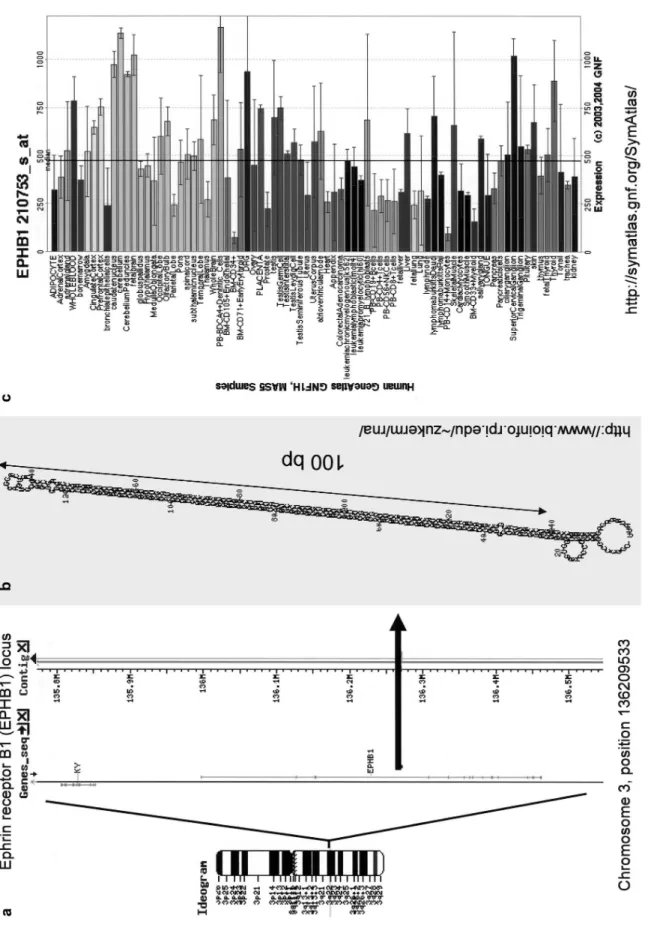
Figur
e 6.
Example of an L1 in
v
er
ted repeat lik
el y generating a long RN A hair pin. ( a ) Schematic o v er vie w of the locus. The in v er
ted repeat is localized in an intron of the Ephrin receptor B1
(EPHB1) gene on the human chromosome 3. (
b
) MFOLD-predicted str
ucture of the L1 hair
pin has an
~
100-bp, almost perfect stem (http://www
.bioinfo.r pi.edu/~zuk er m/r na/ [146]). ( c) GNF database entr y sho wing ubiquitous e
xpression of the host gene (http://symatlas.gnf.or
increases the degree of stringency of the search. However, we found about 50 unique human sequences capable of forming a stem-loop structure with a 50-bp minimal stem containing a maximum of three mismatches and a 5-nt loop. It would be interesting to test whether any of these sequences can be processed by miRNA/RNAi machinery. Apart from low complexity repeats (e.g. TAn), the
major-ity of long inverted repeats are derived from repetitive se-quences. In the human genome, the vast majority of long inverted repeats derive from SINE (Alu) sequences. The second most abundant source is L1 sequences. We have also found several long inverted repeats made of unique sequences (all sequences are available upon request). In the mouse, LINE/L1 inverted repeats are more abundant than SINEs presumably because mouse SINE sequences are not as uniform as human Alu sequences. A fraction of these inverted repeats is clearly transcribed as they side within introns; however, we did not find inverted re-peats among mammalian expressed sequence tags (ESTs) or mRNAs present in the GenBank database. Interest-ingly, the abundance of long inverted repeats in the hu-man genome correlates with the classification of huhu-man targets for adenosine deamination [132] and the observa-tion that RNA editing of Alu elements mostly targets in-tramolecular duplexes [133]. This likely reflects the fact that human Alu and L1 are so abundant that they have the highest probability of inverted repeat formation.
Our results also suggest that L1-derived inverted repeats could be a source of dsRNA, resulting in downregulation of L1 retrotransposition via RNAi. L1 retrotransposition occurs in germ cells or in early embryos [59, 134]. There is evidence for a role of RNAi during early mammalian development in silencing of the LTR retrotransposons IAP and MuERV-L, which potentially generate dsRNA [135]. At the same time, long hairpin expression from a transgene induces RNAi in mouse oocytes [120]. There are tens of L1 inverted repeats of various sizes and at least 10 of them reside within introns (Fig. 6). Since an inverted repeat ensures a high efficiency of dsRNA formation, it is tempting to speculate that this could be a source of dsRNA downregulating L1 retrotransposition in the ger-mline. However, small RNA cloning from mammalian species does not provide solid evidence that L1 sequences are processed by the RNAi machinery [65, 136]. There-fore, whether RNA from L1 inverted repeats is a substrate for ADARs or RNAi (or other dsRNA-responding path-ways) needs to be tested.
When we performed the same search on the
Caenorhab-ditis genomic DNA, the frequency of inverted repeats in
its genome surprisingly was about several hundred times higher than that in mammals (Table 1). More detailed analysis discovered that the difference is due to a high abundance of a special class of transposon-like DNA ele-ments, which are structured as long inverted repeats (~100–800 nt/arm) with short loops (~7–57 nt) [137].
This observation is somewhat counterintuitive, consider-ing that transposons are subjected to silencconsider-ing by RNAi in Caenorhabditis [138]. In any case, our search did not identify SINE, LINE or LTR inverted repeats in
Caeno-rhabditis; most of the inverted repeats originated from
DNA elements and unique sequences. Although these differences are remarkable, their biological significance is unclear.
Long RNA hairpins used for experimental gene silencing
Expression of a long RNA hairpin was the first approach to stable induction of RNA silencing by transgenes [118]. Today, long RNA hairpins are shadowed by short hairpin systems, and they are used only in special cases where a short hairpin RNA system cannot be used efficiently. Long hairpin RNA was successfully used to block gene function in several types of mammalian cells, but aside from mouse oocytes, it never acquired wider attention (reviewed in [139]). Long dsRNA expression from a large inverted repeat remains a common solution for transgenic RNAi approach in invertebrates and plants. The advan-tage of a long hairpin RNA is that it delivers a population of different siRNAs, ensuring a robust RNAi effect. It can also be combined with tissue-specific pol II promoters. Working with inverted repeats may be complicated (re-viewed in [139]), but despite all the possible pitfalls, transgenic RNAi in mouse oocytes has produced a func-tional knockdown in several instances [120, 140–142], and the list of successful knockdowns in Caenorhabditis and Drosophila is much longer.
Concluding remarks
In the present review we have attempted to provide a birds-eye view of the mechanisms that recognize and re-spond to RNA hairpin structures in metazoa. Naturally, our review cannot be comprehensive; however, some-times it is useful to zoom out and take a more panoramic view. RNA silencing is not completely isolated from many other pathways, which operate with RNA hairpins. Hair-pin structures and a combination of factors which interact with them will determine the final effect. It is difficult, if not impossible, to predict the effect of an RNA hairpin predicted from an RNA sequence. While we cannot pro-vide a key to this problem, at least we can remind you of the possible options that exist.
Acknowledgments. We thank Witold Filipowicz and Jody Filkowski for help in preparing of the manuscript. This work was supported by an EMBO Long Term Fellowship to P. S. (ALTF 2003-199). The Friedrich Miescher Institute is part of the Novartis Research Foun-dation.
1 Behrens S. E., Tomei L. and De Francesco R. (1996) Identifi-cation and properties of the RNA-dependent RNA polymerase of hepatitis C virus. EMBO J. 15: 12–22
2 Steitz T. A. (1999) RNA recognition by proteins. In: The RNA World, pp. 427–450, Gesteland R. and Atkins J. (eds.) Cold Spring Harbor Laboratory Press, Cold Spring Harbor, NY 3 Borovjagin A. V., Ezrokhi M. V., Rostapshov V. M., Ugarova
T., Bystrova T. F. and Shatsky I. N. (1991) RNA – protein in-teractions within the internal translation initiation region of encephalomyocarditis virus RNA. Nucleic Acids Res. 19: 4999–5005
4 Ye L., Timani K. A., Kong L., Yang X., Liao Q. and Wu J. (2005) Two cis-acting elements in negative RNA strand of He-patitis C virus involved in synthesis of positive RNA strand in vitro. Acta Virol. 49: 83–90
5 Krol A. (2002) Evolutionarily different RNA motifs and RNA-protein complexes to achieve selenoprotein synthesis. Biochimie 84: 765–774
6 Kato J. and Niitsu Y. (2002) Recent advance in molecular iron metabolism: translational disorders of ferritin. Int. J. Hematol.
76: 208–212
7 Marzluff W. F., Gongidi P., Woods K. R., Jin J. and Maltais L. J. (2002) The human and mouse replication-dependent his-tone genes. Genomics 80: 487–498
8 Marzluff W. F. (2005) Metazoan replication-dependent his-tone mRNAs: a distinct set of RNA polymerase II transcripts. Curr. Opin. Cell Biol. 17: 274–280
9 Ma T., Van Tine B. A., Wei Y., Garrett M. D., Nelson D., Adams P. D. et al. (2000) Cell cycle-regulated phosphoryla-tion of p220(NPAT) by cyclin E/Cdk2 in Cajal bodies pro-motes histone gene transcription. Genes Dev. 14: 2298–2313 10 Dominski Z., Zheng L. X., Sanchez R. and Marzluff W. F.
(1999) Stem-loop binding protein facilitates 3¢-end formation by stabilizing U7 snRNP binding to histone pre-mRNA. Mol. Cell Biol. 19: 3561–3570
11 Dominski Z. and Marzluff W. F. (1999) Formation of the 3¢ end of histone mRNA. Gene 239: 1–14
12 Battle D. J. and Doudna J. A. (2001) The stem-loop binding protein forms a highly stable and specific complex with the 3¢ stem-loop of histone mRNAs. RNA 7: 123–132
13 Shopland L. S., Byron M., Stein J. L., Lian J. B., Stein G. S. and Lawrence J. B. (2001) Replication-dependent histone gene expression is related to Cajal body (CB) association but does not require sustained CB contact. Mol. Biol. Cell 12: 565–576 14 Dominski Z., Yang X. C. and Marzluff W. F. (2005) The polyadenylation factor CPSF-73 is involved in histone-pre-mRNA processing. Cell 123: 37–48
15 Erkmann J. A., Sanchez R., Treichel N., Marzluff W. F. and Kutay U. (2005) Nuclear export of metazoan replication-de-pendent histone mRNAs is dereplication-de-pendent on RNA length and is mediated by TAP. RNA 11: 45–58
16 Kaygun H. and Marzluff W. F. (2005) Regulated degradation of replication-dependent histone mRNAs requires both ATR and Upf1. Nat. Struct. Mol. Biol. 12: 794–800
17 Dominski Z., Yang X. C., Kaygun H., Dadlez M. and Marzluff W. F. (2003) A 3¢ exonuclease that specifically interacts with the 3¢ end of histone mRNA. Mol. Cell 12: 295–305 18 Kennedy S., Wang D. and Ruvkun G. (2004) A conserved
siRNA-degrading RNase negatively regulates RNA interfer-ence in C. elegans. Nature 427: 645–649
19 Hong J., Qian Z., Shen S., Min T., Tan C., Xu J. et al. (2005) High doses of siRNAs induce eri-1 and adar-1 gene expres-sion and reduce the efficiency of RNA interference in the mouse. Biochem. J. 390: 675–679
20 Palacios I. M. and St Johnston D. (2001) Getting the message across: the intracellular localization of mRNAs in higher eu-karyotes. Annu. Rev. Cell Dev. Biol. 17: 569–614
21 Jansen R. P. (2001) mRNA localization: message on the move. Nat. Rev. Mol. Cell Biol. 2: 247–256
22 Driever W. and Nusslein-Volhard C. (1988) A gradient of bi-coid protein in Drosophila embryos. Cell 54: 83–93 23 Driever W. and Nusslein-Volhard C. (1988) The bicoid protein
determines position in the Drosophila embryo in a concentra-tion-dependent manner. Cell 54: 95–104
24 Johnstone O. and Lasko P. (2001) Translational regulation and RNA localization in Drosophila oocytes and embryos. Annu. Rev. Genet. 35: 365–406
25 Stauber M., Jackle H. and Schmidt-Ott U. (1999) The anterior determinant bicoid of Drosophila is a derived Hox class 3 gene. Proc. Natl. Acad. Sci. USA 96: 3786–3789
26 MacDonald P. M. (1990) Bicoid mRNA localization signal: phylogenetic conservation of function and RNA secondary structure. Development 110: 161–171
27 Schnorrer F., Bohmann K. and Nusslein-Volhard C. (2000) The molecular motor dynein is involved in targeting swallow and bicoid RNA to the anterior pole of Drosophila oocytes. Nat. Cell Biol. 2: 185–190
28 Arn E. A., Cha B. J., Theurkauf W. E. and Macdonald P. M. (2003) Recognition of a bicoid mRNA localization signal by a protein complex containing Swallow, Nod and RNA binding proteins. Dev. Cell 4: 41–51
29 St Johnston D., Beuchle D. and Nusslein-Volhard C. (1991) Staufen, a gene required to localize maternal RNAs in the Drosophila egg. Cell 66: 51–63
30 Roegiers F. and Jan Y. N. (2000) Staufen: a common compo-nent of mRNA transport in oocytes and neurons? Trends Cell Biol. 10: 220–224
31 Micklem D. R., Adams J., Grunert S. and St Johnston D. (2000) Distinct roles of two conserved Staufen domains in oskar mRNA localization and translation. EMBO J. 19: 1366–1377 32 Kim Y. K., Furic L., Desgroseillers L. and Maquat L. E.
(2005) Mammalian Staufen1 recruits Upf1 to specific mRNA 3¢UTRs so as to elicit mRNA decay. Cell 120: 195–208 33 MacDougall N., Clark A., MacDougall E. and Davis I. (2003)
Drosophila gurken (TGFalpha) mRNA localizes as particles that move within the oocyte in two dynein-dependent steps. Dev. Cell 4: 307–319
34 Neuman-Silberberg F. S. and Schupbach T. (1993) The Drosophila dorsoventral patterning gene gurken produces a dorsally localized RNA and encodes a TGF alpha-like protein. Cell 75: 165–174
35 Van De Bor V., Hartswood E., Jones C., Finnegan D. and Davis I. (2005) gurken and the I factor retrotransposon RNAs share common localization signals and machinery. Dev. Cell 9: 51–62
36 Zehner Z. E., Shepherd R. K., Gabryszuk J., Fu T. F., Al-Ali M. and Holmes W. M. (1997) RNA-protein interactions within the 3¢untranslated region of vimentin mRNA. Nucleic Acids Res. 25: 3362–3370
37 Nury D., Chabanon H., Levadoux-Martin M. and Hesketh J. (2005) An eleven nucleotide section of the 3¢-untranslated re-gion is required for perinuclear localization of rat metalloth-ionein-1 mRNA. Biochem. J. 387: 419–428
38 Chabanon H., Mickleburgh I., Burtle B., Pedder C. and Hes-keth J. (2005) An AU-rich stem-loop structure is a critical fea-ture of the perinuclear localisation signal of c-myc mRNA. Biochem. J. 392: 475–483
39 Reddy K. K., Oitomen F. M., Patel G. P. and Bag J. (2005) Perinuclear localization of slow troponin C m RNA in muscle cells is controlled by a cis-element located at its 3¢ untrans-lated region. RNA 11: 294–307
40 Mickleburgh I., Burtle B., Nury D., Chabanon H., Chrza-nowska-Lightowlers Z. and Hesketh J. E. (2004) Isolation and identification of a protein binding to the localization element of Metallothionein-1 mRNA. Biochem. Soc. Trans. 32: 705–706 41 Morris E. J., Evason K., Wiand C., L’Ecuyer T. J. and Fulton
A. B. (2000) Misdirected vimentin messenger RNA alters cell morphology and motility. J. Cell Sci. 113: 2433–2443
42 Al-Maghrebi M., Brule H., Padkina M., Allen C., Holmes W. M. and Zehner Z. E. (2002) The 3¢ untranslated region of human vimentin mRNA interacts with protein complexes con-taining eEF-1gamma and HAX-1. Nucleic Acids Res. 30: 5017–5028
43 John B., Enright A. J., Aravin A., Tuschl T., Sander C. and Marks D. S. (2004) Human MicroRNA targets. PLoS Biol. 2: e363
44 Fialcowitz E. J., Brewer B. Y., Keenan B. P. and Wilson G. M. (2005) A hairpin-like structure within an AU-rich mRNA-destabilizing element regulates trans-factor binding selectivity and mRNA decay kinetics. J. Biol. Chem. 280: 22406–22417 45 Mickleburgh I., Burtle B., Hollas H., Campbell G.,
Chrza-nowska-Lightowlers Z., Vedeler A. et al. (2005) Annexin A2 binds to the localization signal in the 3¢ untranslated region of c-myc mRNA. FEBS J. 272: 413–421
46 Bakheet T., Williams B. R. and Khabar K. S. (2006) ARED 3.0: the large and diverse AU-rich transcriptiome. Nucleic Acids Res. 34: D111–D114
47 Wilusz C. J., Wormington M. and Peltz S. W. (2001) The cap-to-tail guide to mRNA turnover. Nat Rev Mol. Cell Biol. 2: 237–246
48 Lal A., Mazan-Mamczarz K., Kawai T., Yang X., Martindale J. L. and Gorospe M. (2004) Concurrent versus individual binding of HuR and AUF1 to common labile target mRNAs. EMBO J. 23: 3092–3102
49 Espel E. (2005) The role of the AU-rich elements of mRNAs in controlling translation. Semin. Cell Dev. Biol. 16: 59–67 50 Donnini M., Lapucci A., Papucci L., Witort E., Jacquier A.,
Brewer G. et al. (2004) Identification of TINO: a new evolu-tionarily conserved BCL-2 AU-rich element RNA-binding protein. J. Biol. Chem. 279: 20154–20166
51 Jing Q., Huang S., Guth S., Zarubin T., Motoyama A., Chen J. et al. (2005) Involvement of microRNA in AU-rich element-mediated mRNA instability. Cell 120: 623–634
52 Selby M. J., Bain E. S., Luciw P. A. and Peterlin B. M. (1989) Structure, sequence and position of the stem-loop in tar deter-mine transcriptional elongation by tat through the HIV-1 long terminal repeat. Genes Dev. 3: 547–558
53 Feng S. and Holland E. C. (1988) HIV-1 tat trans-activation requires the loop sequence within tar. Nature 334: 165– 167
54 Gatignol A., Buckler-White A., Berkhout B. and Jeang K. T. (1991) Characterization of a human TAR RNA-binding pro-tein that activates the HIV-1 LTR. Science 251: 1597–1600 55 Cosentino G. P., Venkatesan S., Serluca F. C., Green S. R.,
Mathews M. B. and Sonenberg N. (1995) Double-stranded-RNA-dependent protein kinase and TAR RNA-binding pro-tein form homo- and heterodimers in vivo. Proc. Natl. Acad. Sci. USA 92: 9445–9449
56 Williams B. R. (1999) PKR: a sentinel kinase for cellular stress. Oncogene 18: 6112–6120
57 Eckmann C. R. and Jantsch M. F. (1997) Xlrbpa, a double-stranded RNA-binding protein associated with ribosomes and heterogeneous nuclear RNPs. J. Cell Biol. 138: 239–253 58 Dorin D., Bonnet M. C., Bannwarth S., Gatignol A., Meurs
E. F. and Vaquero C. (2003) The TAR RNA-binding protein, TRBP, stimulates the expression of TAR-containing RNAs in vitro and in vivo independently of its ability to inhibit the dsRNA-dependent kinase PKR. J. Biol. Chem. 278: 4440– 4448
59 Park H., Davies M. V., Langland J. O., Chang H. W., Nam Y. S., Tartaglia J. et al. (1994) TAR RNA-binding protein is an inhibitor of the interferon-induced protein kinase PKR. Proc. Natl. Acad. Sci. USA 91: 4713–4717
60 Maitra R. K., McMillan N. A., Desai S., McSwiggen J., Hov-anessian A. G., Sen G. et al. (1994) HIV-1 TAR RNA has an intrinsic ability to activate interferon-inducible enzymes. Vi-rology 204: 823–827
61 Zhong J., Peters A. H., Lee K. and Braun R. E. (1999) A dou-ble-stranded RNA binding protein required for activation of repressed messages in mammalian germ cells. Nat. Genet. 22: 171–174
62 Lee K., Fajardo M. A. and Braun R. E. (1996) A testis cyto-plasmic RNA-binding protein that has the properties of a translational repressor. Mol. Cell Biol. 16: 3023–3034 63 Erard M., Barker D. G., Amalric F., Jeang K. T. and Gatignol
A. (1998) An Arg/Lys-rich core peptide mimics TRBP bind-ing to the HIV-1 TAR RNA upper-stem/loop. J. Mol. Biol.
279: 1085–1099
64 Dostie J., Mourelatos Z., Yang M., Sharma A. and Dreyfuss G. (2003) Numerous microRNPs in neuronal cells containing novel microRNAs. RNA 9: 180–186
65 Houbaviy H. B., Murray M. F. and Sharp P. A. (2003) Embry-onic stem cell-specific MicroRNAs. Dev. Cell 5: 351–358 66 Kim J., Krichevsky A., Grad Y., Hayes G. D., Kosik K. S.,
Church G. M. et al. (2004) Identification of many microRNAs that copurify with polyribosomes in mammalian neurons. Proc. Natl. Acad. Sci. USA 101: 360–365
67 Haase A. D., Jaskiewicz L., Zhang H., Laine S., Sack R., Gatignol A. et al. (2005) TRBP, a regulator of cellular PKR and HIV-1 virus expression, interacts with Dicer and functions in RNA silencing. EMBO Rep. 6: 961–967
68 Chendrimada T. P., Gregory R. I., Kumaraswamy E., Norman J., Cooch N., Nishikura K. et al. (2005) TRBP recruits the Dicer complex to Ago2 for microRNA processing and gene si-lencing. Nature 436: 740–744
69 Wightman B., Ha I. and Ruvkun G. (1993) Posttranscriptional regulation of the heterochronic gene lin-14 by lin-4 mediates temporal pattern formation in C. elegans. Cell 75: 855–862 70 Lee R. C., Feinbaum R. L. and Ambros V. (1993) The C.
ele-gans heterochronic gene lin-4 encodes small RNAs with anti-sense complementarity to lin-14. Cell 75: 843–854
71 Fire A., Xu S., Montgomery M. K., Kostas S. A., Driver S. E. and Mello C. C. (1998) Potent and specific genetic interfer-ence by double-stranded RNA in Caenorhabditis elegans. Na-ture 391: 806–811
72 Zamore P. D., Tuschl T., Sharp P. A. and Bartel D. P. (2000) RNAi: double-stranded RNA directs the ATP-dependent cleavage of mRNA at 21 to 23 nucleotide intervals. Cell 101: 25–33
73 Ambros V., Bartel B., Bartel D. P., Burge C. B., Carrington J. C., Chen X. et al. (2003) A uniform system for microRNA annotation. RNA 9: 277–279
74 Doench J. G., Petersen C. P. and Sharp P. A. (2003) siRNAs can function as miRNAs. Genes Dev. 17: 438–442
75 Hutvagner G. and Zamore P. D. (2002) A microRNA in a mul-tiple-turnover RNAi enzyme complex. Science 297: 2056– 2060
76 Yekta S., Shih I. H. and Bartel D. P. (2004) MicroRNA-di-rected cleavage of HOXB8 mRNA. Science 304: 594–596 77 Meins F., Si-Ammour A. and Blevins T. (2005) RNA silencing
systems and their relevance to plant development. Annu. Rev. Cell Dev. Biol. 21: 297–318
78 Rehwinkel J., Behm-Ansmant I., Gatfield D. and Izaurralde E. (2005) A crucial role for GW182 and the DCP1:DCP2 decap-ping complex in miRNA-mediated gene silencing. RNA 11: 1640–1647
79 Lee Y., Kim M., Han J., Yeom K. H., Lee S., Baek S. H. et al. (2004) MicroRNA genes are transcribed by RNA polymerase II. EMBO J. 23: 4051–4060
80 Rodriguez A., Griffiths-Jones S., Ashurst J. L. and Bradley A. (2004) Identification of mammalian microRNA host genes and transcription units. Genome Res. 14: 1902–1910 81 Cai X., Hagedorn C. H. and Cullen B. R. (2004) Human
microRNAs are processed from capped, polyadenylated tran-scripts that can also function as mRNAs. RNA 10: 1957– 1966