FOCUS o n
Cellular Biochemistry
PROTOPLASMA 9 Springer-Verlag 2000 Prhlted in Austria
Immunity against extracellular pathogens
John Garfield, Giorgio Ferrari, and Jean Pieters*Basel Institute for Immunology, Basel Received July 29, 1999
Accepted August 31, 1999
Summary. Eukaryotic cells live in a relatively comfortable equilib- rium with a wide variety of microbes. However, while many of the cohabiting microorganisms are harmless or even beneficial to the eukaryotic host, a number of prokaryotes have evolved the capac- ity to invade and replicate within host cells, thereby becoming potentially pathogenic. To be able to cope with potential pathogens, most organisms have developed several host defense mechanisms. First, microbes can be internalized and destroyed by a number of cell types of an innate immune system in a rather aspecific manner. Second, more complex organisms possess additionally an adaptive immune system that is capable of eliminating hazardous microbes in a highly specific manner. This review describes recent progress in our understanding of how pathogens interact with cells of the immune system, resulting in activation of the immune system or, for certain microorganisms, in the evasion of host defense reactions. Keywords: Antigen processing; Antigen presentation; Phagocytosis; Host defense subversion.
Introduction
In the fight against microbes, various types of host responses occur, which fall into two classes. First, several nonspecific host defense mechanisms exist (innate immune response), and second, the host pos- sesses various mechanisms that are directed at a par- ticular invader (adaptive immunity). U n d e r normal circumstances, the first line of defense is provided by the cells of the innate immune system, the macro- phages, neutrophils, dendritic cells, and natural killer cells. These cells are attracted to the site of infection through multiple stimuli; macrophages and neu- trophils accumulate at these sites in response to bac- terial products, while cytokines secreted by these cells attract m o r e macrophages and neutrophils as well
* Correspondence and reprints: Basel Institute for Immunology, Grenzacherstrasse 487, CH-4005 Basel, Switzerland.
as dendritic cells and natural killer cells. The innate immune system ensures an effective response by efficiently degrading the microbes (antigens) either through intracellular proteolysis following uptake or via the release of degradative enzymes after release of their granule content (Brown et al. 1994, Janeway 1989).
A m o r e specialized response to invading pathogens is provided by cells of the adaptive immune system, comprising the B ceils (originating in the bone marrow, hence the name B lymphocytes) and the T cells (originating in the thymus, t h e r e f o r e t e r m e d T lymphocytes). Importantly, activation of T lympho- cytes depends on the recognition by the T cell recep- tors of peptides derived from pathogenic organisms. These peptides are not recognized alone, but in a complex with specialized molecules, the class I and class II molecules encoded by the major histocompat- ibility complex ( M H C ) (Zinkernagel 1997).
Presentation of microbial antigens to the immune system
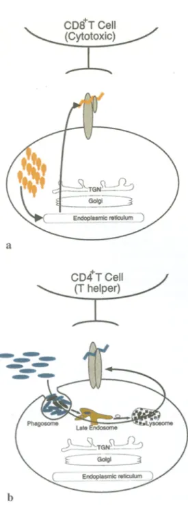
The two classes of peptide presenting molecules ( M H C class I and class II molecules) have probably evolved as a result of the existence of two types of pathogens that can invade the eukaryotic cell. One type, such as viruses, occupies the cytoplasmic com- partment, and viral peptides are presented by M H C class I molecules. The o t h e r type, most extracellularly residing bacteria, enters the endosomal-lysosomal organelles, and bacteria-derived antigens are being presented by M H C class II molecules. A n overview of the M H C class I and class II pathways is provided in Fig. 1.
Fig. l a , b. The MHC class I and class II pathways, a MHC class I molecules acquire antigenic peptides that are generated from infec- tious agents residing in the cytosol of infected cells (by, e.g., viruses). These peptides are formed by the proteasome and then translocated into the endoplasmic reticulum by the TAP complex. The MHC class I-peptide complex is transported through the biosynthetic pathway directly to the plasma membrane, for the activation of CD8 § T lymphocytes, b Peptides to be presented by MHC class II molecules are usually derived from antigens that are present extra- cellularly (such as most bacteria). This extracellularly residing mate- rial enters the cell through endocytosis or phagocytosis and is degraded within endosomal-lysosomal organelles. The resulting peptides are loaded onto MHC class II molecules in specialized organelles, the so-called MHC class II compartments, prior to trans- port of the class II-peptide complex to the cell surface for trigger- ing of CD4 § T helper lymphocytes (see also Janeway and Travers 1996)
Both MHC class I and class II molecules, being transmembrane molecules, are synthesized on endo- plasmic-reticulum-associated ribosomes and cotrans- lationally inserted in the endoplasmic-reticulum mem- brane (Dobberstein et al. 1979, Ploegh et al. 1979). The MHC-encoded molecules are generally highly polymorphic, ensuring that a wide variety of different peptides can be presented by these molecules (Be- nacerraf and McDevitt 1972, Kaufman et al. 1984, Mengle-Gaw and McDevitt 1985, Snell 1986, Trows- dale and Campbell 1992).
MHC class I pathway
MHC class I molecules are expressed by all cells and associate in the endoplasmic reticulum with another molecule, [3-2-microglobulin (Heemels and Ploegh 1995). Proteins derived from viruses and bacteria that reside in the cytosol are degraded by the cytosolic pro- teolytic machinery, the proteasome, and the resulting peptides are transported into the endoplasmic reticu- lure by transporter molecules, the TAP molecules (transporter associated with antigen processing) (Dev- erson et al. 1990, Spies et al. 1990). MHC class I mole- cules become loaded with antigenic peptides in the endoplasmic reticulum and are then transported via the secretory pathway through the Golgi complex to the cell surface where they can be recognized by a subset of T lymphocytes, the cytotoxic T cells (Pamer and Cresswell 1998, Townsend et al. 1989). Triggering of T cell receptors on cytotoxic T cells usually results in the death of the antigen-presenting cell, thus effec- tively eliminating the source of the infection (see Fig. 1).
MHC class H pathway
Class II molecules, in contrast to MHC class I mole- cules, are expressed by only a subset of cells special- ized in the presentation of extracellularly present antigens, including B cells, macrophages, and dendritic cells. Peptides destined to be presented by class II molecules are usually derived from exogenous anti- gens. Therefore, deposition of peptide-loaded MHC class II molecules on the cell surface of antigen- presenting cells involves not only organelles of the secretory pathway but also of the endocytic pathway (Pieters 1999a, Wolf and Ploegh 1995).
During the past decade, work from a variety of laboratories has delineated the pathways followed by MHC class II complexes. Directly after biosynthesis
and insertion into the membrane of endoplasmic re- ticulum, the class II molecules assemble noncovalently with a third molecule, the so-called invariant chain (Ii). In contrast to the class II molecules, the Ii is non- polymorphic and in fact encoded outside the M H C (Koch et al. 1989, Machamer and Cresswell 1982). The Ii performs three important functions during the biogenesis of surface-deposited class II molecules. First, the Ii acts as a chaperone in the endoplasmic reticulum, to aid in the folding and the egress of the class II/Ii complex from the endoplasmic reticulum (Anderson and Miller 1992). Second, the Ii contains a sequence (termed CLIR for class II-associated invariant-chain peptide) that binds directly to the class II peptide-binding groove, in a manner identical to antigenic peptides (Ghosh et al. 1995). In the endo- plasmic reticulum, the Ii therefore shields the class II peptide-binding groove from binding peptides that should, as mentioned above, bind to the M H C class I molecules (Eynon et al. 1999, Roche and Cresswell 1990, Teyton et al. 1990).
Targeting of M H C class H molecules to the endocytic pathway
The third important function of Ii is to provide a tar- geting signal for the class II molecules to be trans- ported to organelles of the endocytic pathway. Indeed, peptides destined to be presented by class II molecules are usually derived from exogenous antigens that have entered the endocytic pathway of the antigen- presenting cell (Cresswell 1994a, Pieters 1997b), and the molecule responsible for getting the class II com- plex into the endosomes is the associated invariant chain. The signals that are responsible for this target- ing event reside within the cytoplasmic tail and consist of two di-leucine-based motifs (Bakke and Dobber- stein 1990, Lotteau et al. 1990, Pieters et al. 1993). These signals mediate targeting of the class II com- plex from the trans-Golgi network to the endosomal pathway and, in addition, mediate the internalization of complexes that have arrived at the plasma mem- brane (Pieters et al. 1993).
The M H C class H compartments
Whereas initially the site where M H C class II mole- cules combine with antigenic peptides remained elu- sive, over the past few years the precise subcellular organelles to which class II/Ii complexes are targeted have been characterized relatively well. By immuno-
cytochemistry on cryosections, it was shown that class II molecules, Ii as well as internalized material could be found within the same vesicles (Pieters et al. 1991). These organelles had a typical multivesicular mor- phology and were therefore referred to as multivesic- ular bodies (Pieters et al. 1991), or, because of their high class lI content, M H C class II compartments (Peters et al. 1991, Pieters 1997a).
After the initial morphological identification of the MHC class II compartments, their subsequent bio- chemical isolation from various cell types allowed their in-depth characterization (Amigorena et al. 1994, Tulp et al. 1994, West et al. 1994). This work established the class II compartments as distinct organelles, sepa- rate from the endosomal-lysosomal pathway (Schmid and Jackson 1994).
More recently, it was established that the MHC class II compartments actually consisted of two phy- sically and functionally distinct vesicle populations (Ferrari et al. 1997). Newly synthesized class II/li com- plexes travel through these organelles in a sequential manner: due to the endosomal targeting signals within the Ii cytoplasmic tail, the complex is first targeted to an organelle where the Ii is degraded from its C- terminal, lumenal side, resulting in a class II complex still associated with the CLIP peptide. The class II- CLIP complex, most likely due to a sorting signal within the class II molecules themselves, are then tar- geted to a second organelle, where CLIP is released from class II molecules. Interestingly, this latter com- partment contains the H L A - D M molecules, which function as facilitators of peptide loading onto class II molecules (Cresswell 1994b; Mellins et al. 1990; Roche 1995, 1996). Indeed, peptides are readily loaded in this latter organelle, which therefore has been termed com- partment of peptide loading (CPL) (Ferrari et al. 1997, Pieters 1999a) (see also Fig. 1). From the compartment of peptide loading, peptide-loaded class II molecules travel to the cell surface by as yet undefined mecha- nisms, where they can trigger T cell receptors on T lym- phocytes leading to their activation. Activated T cells can then aid in the elimination of the invaded micro- organism, by either promoting production of specific antibodies by B cells or by activating phagocytic cells to more efficiently ingest and destroy microbes.
Entry of microorganisms into eukaryotic cells
Thus far we have discussed the mechanisms involved in the transfer of antigens to the M H C molecules that
present these to the T lymphocytes of the immune system. But an important question is how these anti- gens end up in the appropriate cells. Extracellular pathogens, whose peptides are being presented by MHC class II molecules, usually enter the antigen- processing and -presenting cell via endocytosis. Several forms of endocytosis exist. First, soluble mate- rial (e.g., that released by the microbes) can enter the cell through pinocytosis ("cell drinking"; more recently renamed endocytosis), which is the vesicular uptake of fluid and macromolecules. Pinocytosis can be receptor mediated or occur via fluid-phase uptake, either in large ("macropinocytosis", i.e., the uptake of fluid into large, 0.15-0.5 gm diameter vesicles) as well as in small (ca. 100 nm diameter) vesicles. In addition, professional antigen-presenting cells such as the den- dritic cells and the macrophages have the capacity to internalize entire microorganisms through the process of phagocytosis. Phagocytosis ("cell eating") refers to the internalization of large particles and microorgan- isms into large (_250 nm diameter) vesicles.
The endocytic pathway
Regardless of the mode of uptake, material that is internalized enters the endocytic pathway, is trans- ported from early to late endosomes, and may even- tually reach lysosomes (Kornfeld and Mellman 1989). The molecular mechanisms involved in transfer of material through the endosomal-lysosomal pathway is only beginning to be unraveled. Whereas the processes governing receptor-mediated uptake are now rela- tively well understood, those involved in phagocytosis and fluid-phase endocytosis remain less well charac- terized and will be subject to review elsewhere in this series. Here, we will focus on the processes involved in entry and degradation of internalized microorganisms as well as microbial products.
During receptor-mediated endocytosis, receptors that are engaged by ligands can become clustered at the plasma membrane due to receptor cross-linking. During this process a scaffold protein, clathrin, is recruited to the cytoplasmic domain of the receptor and binds via a series of adaptor proteins that are spe- cific for certain signals residing within receptor cyto- plasmic portions (Kirchhausen et al. 1997, Pearse and Robinson 1990, Robinson 1994, Schmid 1997). The current model proposes that clathrin-containing coats initiate the plasma membrane invaginations contain- ing reeeptor-ligand complexes at the cytosolic side of
the plasma membrane and aid in the formation of clathrin-coated vesicles from the plasma membrane (Kirchhausen 1999, Sever et al. 1999). These clathrin- coated vesicles are then uncoated, and their content is transferred to consecutive stages of the endocytic pathway (Schmid 1997). Recent work suggests that at several levels of the endocytic pathway, sorting events may occur that are dictated by the presence of specific sorting determinants, processes that may be regulated through various coat proteins (Stoorvogel et al. 1996, Whitney et al. 1995). Sorting of proteins within the endocytic pathway may lead to transport of proteins either back to the plasma membrane, to the trans- Golgi network, to late endosomes, or to lysosomes (Gu and Gruenberg 1999, Keller and Simons 1997, Kirchhausen et al. 1997).
Phagocytosis
Phagocytosis is defined as the internalization of entire microorganisms or relatively large particles rather than small molecular compounds. As a consequence, the mechanisms and machinery involved in these processes are likely to be distinct from those involved in endocytosis. During phagocytosis, relatively large pieces of the plasma membrane that surround the microorganism are internalized. Subsequently, this phagocytosed material has to be transferred to later stages of the endocytic pathway, followed by delivery to lysosomes (or formation of a so-called phagolyso- some through fusion of nascent phagosomes with lyso- somes) where this material can be degraded.
A number of receptor molecules have been impli- cated in the internalization of microbes by phagocytes and other cells. These include Fc receptors, mannose receptors, scavenging receptors, and various integrins (Brown 1991, Daeron 1997, Ezekowitz et al. 1991, Fanger et al. 1996). Furthermore, several microorgan- isms have developed sophisticated mechanisms to gain entry into eukaryotic cells (Falkow 1991, Finlay and Cossart 1997). Some of these make use of host cell components, for example in the case of lysosome recruitment by trypanosomes (Tardieux et al. 1992), or express proteins that function as a bona fide receptor for a eukaryotic-cell surface component. Examples of these are the invasins expressed by Escherichia coli or Yersinia pseudotuberculosis and binding to integrins (Isberg and Leong 1990, Leong et al. 1995); internalin expressed by Listeria spp. and binding to E-cadherin (Mengaud et al. 1996); Tir expressed by enteropatho-
genic E. coIi (EPEC), which is directly inserted into the host plasma membrane (Kenny et al. 1997, Rosen- shine et al. 1996), and the type III secretion mecha- nisms present in, e.g., Salmonella spp., Shigella spp., and Yersinia spp., that generate a translocation system that is inserted into the host plasma membrane and interacts with the host cytoskeleton (Bliska et al. 1993, Cornelis 1998, Galan and Bliska 1996). Thus, it seems that many different pathways exist to internalize a microorganism into phagocytes; the precise mode of uptake may have evolved as a result of the long-term coevolution of microbes with phagocytic cells (Falkow 1991).
Macrophage activation
One result of phagocytosis of particulate material as well as living microorganisms is activation of the macrophages. Whether or not this activation is similar to the above mentioned T-cell-induced macrophage activation is not clear. In either case, the overall effect of macrophage activation is an enhanced capacity of the cell to inactivate and degrade phagocytosed micro- organisms as well as cellular material including tumor cells (Adams and Hamilton 1992, Crawford et al. 1994). In addition, a plethora of cytokines that are released at the site of infection upregulate expression of M H C class II molecules and can also activate the T and B lymphocytes of the acquired immune system, thereby contributing to elimination of the infection.
The cell biological processes responsible for and accompanying macrophage activation are still poorly understood. Most likely, multiple signals are involved in generating an "activated macrophage", and which signals dominate may depend on the location of the macrophage within the organism, the type of infection, and the presence of accessory cells that can secrete activating cytokines. Whereas virtually every known cytokine has been implicated in macrophage activa- tion, the best studied is gamma interferon (IFN-7). INF-7, when added to macrophages, upregulates MHC molecules, enhances phagocytosis and for some microorganisms results in an increased killing (Fowles et al. 1973). IFN-ydoes not fully activate macrophages, but rather induces a "primed" state (Adams and Hamilton 1992). Primed macrophages can then be fully activated by other compounds such as low amounts of lipopolysaccharide (LPS, also called endo- toxin), a glycolipid that is a component of the gram- negative bacterial cell wall (Adams and Hamilton
1984). LPS binds to a macrophage cell surface recep- tor and thereby activates a signal transduction cas- cade resulting in the secretion of various cytokines that together contribute to the activation of the macrophage. Very recently, the receptor that is acti- vated upon LPS addition to macrophages has been determined to be a member of the Toll-like receptors (TLR) (Kirschning et al. 1998, Poltorak et al. 1998, Yang et al. 1998). These receptors bear homology to Toll, a molecule conserved from plants to mammals, and are thought to play a role in host defense (Fearon 1997, Lemaitre et al. 1996, Medzhitov et al. 1997).
LPS does not bind directly to Toll-like receptors but does so via the GPI(glycosyl phosphatidylinositol)- linked cell surface molecule CD14 (Gerard 1998). LPS that is shed from the bacteria within a host usually aggregates, and these aggregates bind to a serum protein called LPS-binding protein (LBP). LBP breaks down these aggregates allowing the binding of LPS to the cell surface molecule CD14. But because CD14 lacks a transmembrane and cytosolic domain (being a GPI-linked molecule), it cannot directly transmit activation signals to the cytosol. The molecules that seem to be responsible for transmitting signals are the aforementioned TLRs, which are activated upon engagement by LPS in the presence of LBP and CD14 (Gerard 1998; Medzhitov and Janeway 1997, 1998; Medzhitov et al. 1997; Yang et al. 1998).
Apart from LPS, virtually every known cytokine has been shown to act in macrophage activation, alone or in concert with other reagents, and to varying effi- ciency with regard to microbicidal activity against different microorganisms (Crawford etal. 1994). However, it is currently unknown which components induce activation in vivo. The availability of mice lacking one or several cytokines and/or cell surface receptor molecules may help to elucidate the precise molecules involved.
Macrophage activation leads to several cellular processes that aid in the elimination of the microor- ganisms. First, activated macrophages upregulate their phagocytic capacity. Second, the capacity of macro- phages to kill the phagocytosed microbes is drastically enhanced, through the upregulation of radical forma- tion in the phagosome on the one hand, and through the stimulation of phagosome-lysosome fusion on the other. Third, activated macrophages upregulate the expression of MHC class II molecules, allowing the adaptive immune system to contribute to the elim- ination of the invading microorganisms.
Evasion of host defense mechanism
The microbicidal activities of macrophages, together with the capacity of the adaptive immune system to generate a strong antibody-mediated response, is usually sufficient to eradicate any microbe that has invaded the host organism. However, many microor- ganisms have coevolved with their eukaryotic host for a considerable time, and it is therefore not surprising that various microbes have developed mechanisms allowing them to circumvent host defense responses. In fact, one reason why some microbes have become pathogenic is precisely because they have evolved ways to survive and replicate within their mammalian host. Many pathogens do this by finding a suitable site within the host cell where they can thrive and at the same time circumvent host defense mechanisms (Falkow et al. 1992, Galan and Bliska
1996,
Pieters 1999b, Small et al. 1994).Subversion of phagocytos& by mycobacteria
One particularly successful example of pathogen-host interaction is provided by the interaction of mycobac- teria with macrophages. Macrophages, as mentioned, are among the most professional phagocytes, which, in addition, can present antigenic peptides bound to MHC class II molecules to T lymphocytes (Silverstein 1995, Unanue 1984). Indeed, macrophages do ingest mycobacteria and are also able to stimulate T lym- phocytes to a certain degree (de Chastellier et al. 1995, Inaba et al. 1993, Kaufmann 1993). But at the same time, mycobacteria have the capacity to resist destruc-
tion by the macrophage, and as a result, these bacilli can survive and even replicate within the macrophage (Russell 1995). In doing so, mycobacteria can also become sequestered from the activity of T lympho- cytes, thus allowing these bacteria to escape immune defense mechanisms (Pancholi etal. 1993). These highly efficient escape mechanisms are believed to be involved in the pathogenicity of mycobacterial in- fections, which include tuberculosis, leprosy, as well as various opportunistic infections (Bloom 1992, Murray and Salomon 1998).
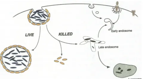
The mechanism by which mycobacteria resist destruction in the macrophage is linked to the capac- ity of these bacteria to inhibit phagosome-lysosome fusion (Armstrong and D'Arcy Hart 1971, Barker et al. 1997, Hasan et al. 1997). The molecular basis of this capacity has however remained unclear until recently, when it was demonstrated that within macro- phages, living mycobacteria recruit and retain a host protein, TACO (tryptophane aspartate-containing coat protein). As a result of this retention, the myco- bacterial phagosome is unable to fuse with or mature into lysosomes (Ferrari et al. 1999). In contrast, killed mycobacteria cannot retain TACO and are therefore rapidly delivered to lysosomes followed by their degradation (see Fig. 2). Although the normal cellular function of TACO is still unknown, it might be involved in generating the forces that are needed to internalize large particles, including (living) bacteria. Upon phagocytosis, TACO forms a coat around the phagosome that has to be released to allow the fusion machinery to be recruited at the phagosomal mem- brane. Apparently, during the long-standing coevolu-
Fig. 2. Evasion of macrophage host defense mechanisms by mycobacteria. Phagocytosis of mycobacteria (blue) into macrophages triggers the recruitment of TACO (yellow) around the nascent phagosome. In case of killed mycobacteria, the TACO coat has to be removed in order to allow delivery of the phagosomal content to lysosomes, where this material is degraded. In contrast, liv- ing mycobacteria that have entered the macrophage have somehow gained the ca- pacity to retain TACO at the phagosomal membrane, thereby preventing their deliv- ery to lysosomes and atlowing them to survive within the phagosome
t i o n of m y c o b a c t e r i a with t h e i r h o s t o r g a n i s m s , t h e y h a v e m a n a g e d to actively r e c r u i t a n d r e t a i n T A C O for t h e i r o w n b e n e f i t . B y h i d i n g i n s i d e m a c r o p h a g e p h a g o s o m e s , m y c o - b a c t e r i a n o t o n l y m a n a g e to c i r c u m v e n t t h e n o r m a l b a c t e r i c i d a l m e c h a n i s m s of t h e s e cells b u t , i n a d d i t i o n , b e c o m e i n v i s i b l e for o t h e r h o s t d e f e n s e m e c h a n i s m s , such as t h e g e n e r a t i o n of a n t i m y c o b a c t e r i a l a n t i b o d i e s t h a t of c o u r s e c a n o n l y d e a l w i t h e x t r a c e l l u l a r l y resid- i n g antigens. I n a d d i t i o n , b e c a u s e of t h e n o n d e g r a d a - tive e n v i r o n m e n t of t h e m y c o b a c t e r i a l p h a g o s o m e , a n t i g e n i c f r a g m e n t s c a n n o t b e g e n e r a t e d , p r e v e n t i n g t h e g e n e r a t i o n o f a p r o p e r M H C - d e p e n d e n t T cell r e s p o n s e ( P a n c h o l i et al. 1993). T h e s t r a t e g y e m p l o y e d b y m y c o b a c t e r i a to c i r c u m - v e n t d e s t r u c t i o n w i t h i n m a c r o p h a g e s is o n c e m o r e a n i n t r i g u i n g e x a m p l e of h o w m i c r o b e s h a v e e v o l v e d i n c o n c e r t with t h e i r host o r g a n i s m s . I n a d d i t i o n , k n o w l - e d g e of t h e s e strategies m i g h t a l l o w t h e d e s i g n of r a t i o n a l a g e n t s to c o m b a t p a t h o g e n s t h a t e s c a p e t h e n o r m a l i m m u n e d e f e n s e r e a c t i o n s . A c k n o w l e d g m e n t s
We thank Fraser McBlane and Christian Wimmer for critical reading of the manuscript. The Basel Institute for Immunology was founded and is supported by F. Hoffmann-La Roche Ltd., Basel, Switzerland.
R e f e r e n c e s
Adams DO, Hamilton TA (1984) The cell biology of macrophage activation. Annu Rev Immunol 2:283-318
- - (1992) Molecular basis of macrophage activation: diversity and origin. In: Lewis CE, McGee JOD (eds) The macrophage. Oxford University Press, Oxford, pp 75-114
Amigorena S, Drake JR, Webster R Mellman I (1994) Transient accumulation of new MHC molecules in a novel endocytic com- partment in B lymphocytes. Nature 369:113-120
Anderson MS, Miller J (1992) Invariant chain can function as a chaperone protein for class II major histocompatibility complex molecules. Proc Natl Acad Sci USA 89:2282-2286
Armstrong J, D'Arcy Hart P (1971) Response of cultured macrophages to Mycobacterium tuberculosis, with observations on fusion of lysosomes with phagosomes. J Exp Med 134:713-740 Bakke 0, Dobberstein B (1990) MHC class II-associated invariant chain contains a sorting signal for endosomal compartments. Cell 63:707-716
Barker LE George KM, Falkow S, Small PL (1997) Differential trafficking of live and dead Mycobacterium marinum organisms in macrophages. Infect Immun 65:1497-1504
Benacerraf B, McDevitt HO (1972) Histocompatibility-linked immune response genes. Science 175:273-279
Bliska JB, Galan JE, Falkow S (1993) Signal transduction in the mammalian cell during bacterial attachment and entry. Cell 73: 903-920
Bloom BR (1992) Tuberculosis: back to a frightening future. Nature 358:538-539
Brown E, Atkinson JP, Fearon DT (1994) Innate immunity: 50 ways to kilt a microbe. Curr Opin Immunol 6:73-74
Brown EJ (1991) Complement receptors and phagocytosis. Curr Opin Immunol 3:76-82
Cornelis GR (1998) The Yersinia deadly kiss. J Bacteriol 180: 5495-5504
Crawford RM, Leiby DA, Green S J, Nacy CA, Fortier AH, Meltzer MS (1994) Macrophage activation: a riddle of immunological resistance. In: Zwilling BS, Eisenstein TK (eds) Macrophage- pathogen interactions. Marcel Dekker, New York
Cresswell P (1994a) Antigen presentation: getting peptides into MHC class II molecules. Curr Biol 4:541-543
- (1994b) Assembly, transport, and function of MHC class II mole- cules. Annu Rev Immunol 12:259-293
Daeron M (1997) Fc receptor biology. Annu Rev Immunol 15: 203-234
de Chastellier C, Lang T, Thilo L (1995) Phagocytic processing of the macrophage endoparasite, Mycobacterium avium, in comparison to phagosomes which contain Bacillus subtilis or latex beads. Eur J Cell Biol 68:167-182
Deverson EV, Gow IR, Coadwell WJ, Monaco JJ, Butcher GW, Howard JC (1990) MHC class II region encoding proteins related to the multidrug resistance family of transmembrane transporters. Nature 348:738-741
Dobberstein B, Garoff H, Warren G, Robinson PJ (1979) Cell-free synthesis and membrane insertion of mouse H-2Dd histocompat- ibility antigen and beta2-microglobulin. Cell 17:759-769 Eynon EE, Schlax C, Pieters J (1999) A secreted form of the MHC
class II associated invariant chain inhibiting T cell activation. J Biol Chem 279:26266-26271
Ezeko,adtz RA, Williams DJ, Koziet H, Armstrong MY, Warner A, Richards FF, Rose RM (1991) Uptake of Pneumocystis carinii
mediated by the macrophage mannose receptor. Nature 351: 155-158
Falkow S (1991) Bacterial entry into eukaryotic cells. Cell 65: 1099-1102
- Isberg RR, Portnoy DA (1992) The interaction of bacteria with mammalian cells. Annu Rev Cell Biol 8:333-363
Fanger NA, WardwelI K, Shen L, Tedder TF, Guyre PM (1996) Type I (CD64) and type II (CD32) Fc gamma receptor-mediated phagocytosis by human blood dendritic cells. J Immunol 157: 541-548
Fearon DT (1997) Seeking wisdom in innate immunity Nature 388: 323-324
Ferrari G, Knight AM, Watts C, Pieters J (1997) Distinct intracellu- lar compartments involved in invariant chain degradation and antigenic peptide loading of major histocompatibility complex (MHC) class II molecules. J Cell Biol 139:1433-1446
- Naito M, Langen H, Pieters J (1999) A coat protein on phago-
somes involved in the intracellular survival of mycobacteria. Cell 97:435-447
Finlay BB, Cossart P (1997) Exploitation of mammalian host cell functions by bacterial pathogens. Science 276:718-725
Fowles RE, Fajardo IM, Leibowitch JL, David JR (1973) The enhancement of macrophage baeteriostasis by products of acti- vated lymphocytes. J Exp Med 138:952-964
Galan JE, Bliska JB (1996) Cross-talk between bacterial pathogens and their host cells. Annu Rev Cell Dev Biol 12:221-255 Gerard C (1998) Bacterial infection: for whom the bell tolls. Nature
395:217 and 219
Ghosh P, Amaya M, Mellins E, Wiley DC (1995) The structure of an intermediate in class II MHC maturation: CLIP bound to HLA- DR3. Nature 378:457-462
Gu F, Gruenberg J (1999) Biogenesis of transport intermediates in the endocytic pathway. FEBS Lett 452:61-66
Hasan Z, Schlax C, Kuhn L, Lefkovits I, Young D, Thole J, Pieters J (1997) Isolation and characterization of the mycobacterial phago- some: segregation from the endosomal/lysosomal pathway. Mol Microbiol 24:545-553
Heemels MT, Ploegh H (1995) Generation, translocation, and pre- sentation of MHC class I-restricted peptides. Annu Rcv Biochem 64:463-491
Inaba K, Inaba M, Naito M, Steinman R (1993) Dendritic cell pro- genitors phagocytose particulates, including Bacillus Calmette- Guerin organisms, and sensitize mice to mycobacterial antigens in vivo. J Exp Med 178:479-488
Isberg RR, Leong JM (1990) Multiple beta 1 chain integrins are receptors for invasin, a protein that promotes bacterial penetra- tion into mammalian cells. Cell 60:861-871
Janeway CA Jr (1989) Approaching the asymptote? Evolution and revolution in immunology. Cold Spring Harb Syrup Quant Biol 54:1-13
- T r a v e r s P (1996) Immunobiology. Current Biology Ltd and Garland Publishing, London
Kaufman JF, Auffray C, Korman A J, Shackelford DA, Strominger J (1984) The class II molecules of the human and murine major histocompatibility complex. Cell 36:1-13
Kaufmann SH (1993) Immunity to intracellular bacteria. Annu Rcv Immunol 11:129-163
Keller R Simons K (1997) Post-Golgi biosynthetic trafficking. J Cell Sci 110:3001-3009
Kenny B, DeVinney R, Stein M, Reinscheid D J, Frey EA, Finlay BB (1997) Enteropathogenic E. coIi (EPEC) transfers its receptor for intimate adherence into mammalian cells. Cell 91:511-520 Kirchhausen T (1999) Cell biology: Boa constrictor or rattlesnake?
Nature 398:470-471
- Bonifacino JS, Riezman H (1997) Linking cargo to vesicle for- mation: receptor tail interactions with coat proteins. Curr Opin Cell Biol 9:488-495
Kirschning C J, Wesche H, Merrill Ayres T, Rothe M (1998) Human toll-like receptor 2 confers responsiveness to bacterial lipopo- lysaccharide. J Exp Med 188:2091-2097
Koch N, Lipp J, Pessara U, Schenck K, Wraight C, Dobberstein B (1989) MHC class II invariant chains in antigen processing and presentation. Trends Biochem Sci 14:383-386
Kornfeld S, Mellman I (1989) The biogenesis of lysosomes. Annu Rev Cell Biol 5:483-525
Lemaitre B, Nicolas E, Michaut L, Reichhart JM, Hoffmann JA (1996) The dorsoventral regulatory gene cassette spatzle/Toll/ cactus controls the potent antifungal response in Drosophila adults. Cell 86:973-983
Leong JM, Morrissey PE, Marra A, Isberg RR (1995) An aspartate residue of the Yersinia pseudotuberculosis invasin protein that is critical for integrin binding. EMBO J 14:422-431
Lotteau V, Teyton L, Peleraux A, Nilsson T, Karlsson L, Schmid SL, Quaranta V, Peterson PA (1990) Intracellular transport of class II MHC molecules directed by invariant chain. Nature 348:600-605 Machamer CE, Cresswell P (1982) Biosynthesis and glycosylation of
the invariant chain associated with H L A - D R antigens. J Immunol 129:2564-2569
Medzhitov R, Janeway CA Jr (1997) Innate immunity: the virtues of a nonclonal system of recognition. Cell 91:295-298
- - (1998) An ancient system of host defense. Curr Opin Immunol 10:12-15
- Preston-Hurlburt R Janeway CA Jr (1997) A human homologue
of the Drosophila Toll protein signals activation of adaptive immunity. Nature 388:394-397
Mellins E, Smith L, Arp B, Cotner T, Celis E, Pious D (1990) De-
fective processing and presentation of exogenous antigens in mutants with normal HLA class II genes. Nature 343:71-74 Mengaud J, Ohayon H, Gounon E Mege RM, Cossart P (1996)
E-cadherin is the receptor for internalin, a surface protein required for entry of L. monocytogenes into epithelial cells. Cell 84:923-932
Mengle-Gaw L, McDevitt HO (1985) Genetics and expression of mouse Ia antigens. Annu Rev Immunol 3:367-396
Murray C J, Salomon JA (1998) Modeling the impact of global tuber- culosis control strategies. Proc Natl Acad Sci USA 95: 13881- 13886
Pamer E, Cresswell P (1998) Mechanisms of MHC class I-restricted antigen processing. Annu Rev Immunol 16:323-358
Pancholi R Mirza A, Bhardwaj N, Steinman RM (1993) Seques- tration from immune CD4 + T cells of mycobacteria growing in human macrophages. Science 260:984-986
Pearse BM, Robinson MS (1990) Clathrin, adaptors, and sorting. Annu Rev Cell Biol 6:151-171
Peters P J, Neefjes J J, Oorschot V, Ploegh HL, Geuze HJ (1991) Seg- regation of MHC class II molecules from MHC class I molecules in the Golgi complex for transport to lysosomal compartments. Nature 349:669-676
Pieters J (1997a) MHC class II compartments: specialized organelles of the endocytic pathway in antigen presenting ceils. Biol Chem 378:751-758
- (1997b) MHC class II restricted antigen presentation. Curr Opin Immunol 9:89-96
- (1999a) MHC class II restricted antigen processing and presenta- tion. Adv Immunol (in press)
- (1999b) Processing and presentation of phagocytosed antigens to the immune system. In: Gordon S (ed) Phagoeytosis and pathogens: the host. JAI Press, Stamford, Conn, pp 379-406 - Horstmann H, Bakke O, Griffiths G, Lipp J (1991) Intracellular
transport and localization of major histocompatibility complex class II molecules and associated invariant chain. J Cell Biol 115: 1213-1223
- Bakke O, Dobberstein B (1993) The MHC class II-associated invariant chain contains two endosomal sorting signals within its cytoplasmic tail. J Cell Sci 106:831-846
Ploegh HL, Cannon LE, Strominger JL (1979) Cell-free trans- lation of the mRNAs for the heavy and light chains of HLA-A and HLA-B antigens. Proc Natl Acad Sci USA 76: 2273- 2277
Poltorak A, He X, Smirnova I, Liu MY, Huffel CV, Du X, Birdwell D, Alejos E, Silva M, Galanos C, Freudenberg M, Ricciardi- Castagnoli P, Layton B, Beutler B (1998) Defective LPS signaling in C3H/HeJ and C57BL/10ScCr mice: mutations in Tlr4 gene. Science 282:2085-2088
Robinson MS (1994) The role of clathrin, adaptors and dynamin in endocytosis. Curr Opin Cell Biol 6:538-544
Roche PA (1995) HLA-DM: an in vivo facilitator of MHC class II peptide loading. Immunity 3:259-262
- (1996) Out, damned CLIP! Out, I say! Science 274:526-527 - Cresswell P (1990) Invariant chain association with H L A - D R
molecules inhibits immunogenic peptide binding. Nature 345: 615-618
Rosenshine I, Ruschkowski S, Stein M, Reinscheid D J, Mills SD, Finlay BB (1996) A pathogenic bacterium triggers epithelial signals to form a functional bacterial receptor that mediates actin pseudopod formation. EMBO J 15:2613-2624
Russell D G (1995) Mycobactcrium and Leishmania: stowaways in the endosomal network. Trends Cell Biol 5:125-128
Schmid SL (1997) Clathrin-coated vesicle formation and protein sorting: an integrated process. Annu Rev Biochem 66: 511- 548
- Jackson MR (1994) Making class II presentable. Nature 369: 103-104
Sever S, Muhlberg AB, Schmid SL (1999) Impairment of dynamin's GAP domain stimulates receptor-mediated endocytosis. Nature 398:481-486
Silverstein SC (1995) Phagocytosis of microbes: insights and prospects. Trends Cell Biol 5:141-142
Small PLC, Ramakrishnan L, Falkow S (1994) Remodeling schemes of intracellular pathogens. Science 263:637-639
Snell GD (1986) Some recollections of Peter Gorer and his work on this fiftieth anniversary of his discovery of H-2. Immunogenetics 24:33%340
Spies T, Bresnahan M, Bahram S, Arnold D, Blanck G, Mellins E, Pious D, DeMurs R (1990) A gene in the human major histo- compatibility complex class II region controlling the class I antigen presentation pathway. Nature 348:744-747
Stoorvogel W, Oorschot V, Geuze HJ (1996) A novel class of clathrin-coated vesicles budding from endosomes. J Cell Biol 132: 21-33
Tardieux I, Webster R Ravesloot J, Boron W, Lunn JA, Heuser JE, Andrews NW (1992) Lysosome recruitment and fusion are early events required for trypanosome invasion of mammalian cells. Cell 71:1117-1130
Teyton L, O'Sullivan D, Dickson PW, Lotteau V, Sette A, Fink R Peterson PA (1990) Invariant chain distinguishes between the exogenous and endogenous antigen presentation pathways. Nature 348:3%44
Townsend A, Ohlen C, Bastin J, Ljunggren H-G, Foster L, Karre K (1989) Association of class I major histocompatibility heavy and light chains induced by viral peptides. Nature 340:443-448 Trowsdale J, Campbell RD (1992) Complexity in the major histo-
compatibility complex. Eur J Immunogenet 19:45-55
Tulp A, Verwoerd D, Dobberstein B, Ploegh HL, Pieters J (1994) Isolation and characterization of the intracellular MHC class II compartment. Nature 369:120-126
Unanue ER (1984) Antigen-presenting function of the macrophage. Annu Rev Immunol 2:395-428
West MA, Lucocq JM, Watts C (1994) Antigen processing and class II MHC peptide-loading compartments in human B-lymphoblas- toid cells. Nature 369:147-151
Whitney JA, Gomez M, Shelf D, Kreis TE, MelIman I (1995) Cyto- plasmic coat proteins involved in endosome function. Cell 83: 703--713
Wolf PR, Ploegh HL (1995) How MHC class II molecules acquire peptide cargo: biosynthesis and trafficking through the endocytic pathway. Annu Rev Cell Dev Biol 11:267-306
Yang RB, Mark MR, Gray A, Huang A, Xie MH, Zhang M, Goddard A, Wood WI, Gurney AL, Godowski PJ (1998) Toll-like receptor- 2 mediates Iipopolysaccharide-induced cellular signalling. Nature 395:284-288
Zinkernagel RM (1997) The Nobel Lectures in Immunology The Nobel Prize for Physiology or Medicine, 1996. Cellular immune recognition and the biological role of major transplantation anti- gens. Scand J Immunol 46:42t-436