HAL Id: hal-02556739
https://hal.inrae.fr/hal-02556739
Submitted on 28 Apr 2020HAL is a multi-disciplinary open access archive for the deposit and dissemination of sci-entific research documents, whether they are pub-lished or not. The documents may come from teaching and research institutions in France or abroad, or from public or private research centers.
L’archive ouverte pluridisciplinaire HAL, est destinée au dépôt et à la diffusion de documents scientifiques de niveau recherche, publiés ou non, émanant des établissements d’enseignement et de recherche français ou étrangers, des laboratoires publics ou privés.
Distributed under a Creative Commons Attribution - NonCommercial - NoDerivatives| 4.0 International License
Alfalfa leaf curl virus is transmitted by Aphis craccivora
in a highly specific circulative manner
Faustine Ryckebusch, Nicolas Sauvion, Martine Granier, Philippe Roumagnac,
Michel Peterschmitt
To cite this version:
Faustine Ryckebusch, Nicolas Sauvion, Martine Granier, Philippe Roumagnac, Michel Peterschmitt. Alfalfa leaf curl virus is transmitted by Aphis craccivora in a highly specific circulative manner. Virology, Elsevier, 2020, 546, pp.98-108. �10.1016/j.virol.2020.04.004�. �hal-02556739�
Alfalfa leaf curl virus is transmitted by Aphis craccivora in a highly
specific circulative manner
Ryckebusch F., Sauvion N., Granier M., Roumagnac, P., Peterschmitt M.*
CIRAD, UMR BGPI, Montpellier, France
BGPI, INRA, CIRAD, Montpellier SupAgro, Univ Montpellier, Montpellier, France
*Corresponding author
Abstract
Two members of the genus Capulavirus (Geminiviridae) are transmitted by aphids including Alfalfa leaf curl virus (ALCV) transmitted by Aphis craccivora. The capulavirus Euphorbia caput-medusae latent virus was shown here to be transmitted also by A. craccivora, using the population EuphorbiaSA. ALCV was transmissible by several A. craccivora populations including Robinia, but not EuphorbiaSA population, reflecting a high transmission specificity. Typical of the circulative-persistent mode of transmission, ALCV persists through insect molts. ALCV accumulation and localization were analyzed in whole insects, midguts, hemolymphs, and heads of aphids from vector and non-vector populations of A. craccivora and from the non-vector species Acyrthosiphon pisum. Vector and non-vector populations could be distinguished by contrasted virus accumulations and midgut intracellular localization
consistent with a gut barrier to the transmission of ALCV in A. pisum and a primary salivary gland barrier in A. craccivora.
Keywords
geminivirus, taxonomy, South Africa, luteovirus, Luteoviridae, Hemiptera, Aphididae, species complex
1. Introduction
Nearly 55% of reported plant pathogenic viruses are transmitted by piercing-sucking insects of the Hemiptera order (Hogenhout et al., 2008). In the viral family Geminiviridae, a large range of hemipterans were identified as vectors. Geminiviruses of the genus Begomovirus are transmitted by whiteflies (i.e., Aleyrodidae), viruses of the genera Becurtovirus, Curtovirus, Mastrevirus, Turncurtovirus are transmitted by leafhoppers (i.e, Cicadellidae) (Heydarnejad et al., 2013; Razavinejad et al., 2013) and those belonging to the genera Grablovirus and Topocuvirus are transmitted by treehoppers (i.e., Membracidae) (Bahder et al., 2016). Interestingly, although Aphididae is the insect group with the highest number of species reported as vector of plant viruses, aphid transmission was discovered only in 2015 when aphids of the species Aphis craccivora Koch, 1854 (Aphididae) were shown to transmit Alfalfa leaf curl virus (ALCV), a geminivirus belonging to the newly defined genus Capulavirus (Roumagnac et al., 2015; Varsani et al., 2017). Since then, Plantago lanceolata latent virus (PlLV) another capulavirus was shown to be transmitted by the aphid Dysaphis plantaginea (Passerini, 1860) (Susi et al., 2019) suggesting that aphid transmission is a
taxonomic criterion of the new genus which comprises two additional members, Euphorbia caput-medusae latent virus (EcmLV) and French bean severe leaf curl virus (FbSLCV).
Contrasted transmission specificities were observed between genera within the family Geminiviridae. While begomoviruses, irrespective of their geographic origin, are transmitted by whiteflies of the species complex Bemisia tabaci Gennadius, 1889 (Bedford et al., 1994; De Barro et al., 2011), mastreviruses are transmitted by leafhoppers of various genera depending on their geographic origin. Therefore, considering the scarcity of transmission data about capulaviruses, their transmission specificity is quite unpredictable. While it is clear that unlike the Begomovirus genus, there are more than one vector species associated with the Capulavirus genus, species and population-level specificities of capulavirus vectors have not been adequately studied.
The first objective of this study was to further support that aphids are vectors of capulaviruses and thus confirm that it may be a taxonomic criterion of the new geminivirus genus. The second objective was to assess the specificity of the aphid transmission. The identification of an aphid vector of a third capulavirus (EcmLV) confirmed that aphid transmission can be considered as a taxonomic criterion of this new genus. Moreover, the transmission tests of ALCV performed with various aphid species and populations showed that only A. craccivora transmitted this virus, but not all tested A. craccivora subpopulations. According to viral DNA persistence and distribution monitored with qPCR and FISH in vector and non-vector aphids, we detected two barriers to transmission, salivary glands and gut.
2. Material and Methods
2.1.1. Preparation of agroinfectious clones and agroinoculation
The agroinfectious clones of EcmLV and ALCV were reported previously (Bernardo et al., 2013, Roumagnac et al., 2015). The ALCV clone belongs to genotype A, which is the most widespread genotype and whose capsid protein is the most common among ALCV populations (Davoodi et al. 2018). An agroinfectious clone of the reported PILV clone (Genbank accession number KT214390) (Susi et al., 2017) was prepared as follows. Its genome was released from its Pjet1.2 vector by PstI restriction and ligated as a tandem repeat into the corresponding restriction site of the binary vector pCambia 2300. Agrobacterium tumefaciens strain C58-MP90 was transformed with the recombinant plasmid by electroporation. Agrobacteria were grown overnight at 28°C in LB medium containing gentamicin and kanamycin until an optical density (OD600) of 2-3. Bacterial suspensions were then centrifuged at 1000 g for 25 min. The pellets were resuspended in ultrapure water (Milli-Q) containing MgCl2 (10mM) and acetosyringone (150mM). Agroinoculations were performed with a syringe by repeated needle injections at the base of the stem on ten-day-old broad bean plants (Vicia faba, cv. ‘Sevilla’), 14-day old tomato plants (Solanum lycopersicum L.), one-month-old buckhorn plantain (Plantago lanceolata L.) or 6-year old euphorbia Medusa’s head plants (Euphorbia caput-medusae L.).
2.1.2. DNA extraction
The infection of ALCV, EcmLV, and PlLV inoculated plants was monitored 4 to 6 weeks after inoculation by symptom observation (ALCV) and/or by PCR-mediated detection of viral DNA (ALCV, EcmLV, and PlLV) in total plant DNA extracts. From each plant, four leaf disks of 4 mm diameter were collected from young leaves, one disc per leaf, and stored at – 20°C; due to the small size of the leaves of E. caput medusa plants, 4 whole leaves were collected per plant. Total DNA was extracted by grinding the leaf material in 400 µ L of
modified Edwards buffer containing 200 mM Tris-HCl (pH 7.5), 25 mM EDTA, 250 mM NaCl, 0.5% SDS, 1% PVP40 and 0.2% ascorbic acid. The extract was incubated at 65°C for 10 min and centrifuged at 15700 g for 10 min. One volume of isopropanol was added to the supernatant before a 20 min centrifugation at 15700 g. After resuspension of the pellet in 500 µ L of 70% ethanol, nucleic acids were recovered by centrifugation (15700 g, 15 min) and resuspended in 50 µ L sterile distilled water. The DNA extracts were stored at −20°C before use.
2.1.3. PCR detection of viral DNA
PCR primers designed to detect ALCV and EcmLV prime the amplification of a 174 bp fragment from the Rep gene: ALCV2cEcmLVF, 5' GAG GAA TTC GGA CTT GGA TG -3' and ALCV2cEcmLV-R, 5'- TTC TTC GAC ATC AAG GAC CC --3'. PCR primers designed to detect PlLV prime the amplification of a 293 bp fragment from the CP gene: PlLV_729-F, 5’- AAG GGA AAG GCT GGT TAT GG -3’ and PlLV_1013-R, 5’- GAA TCT CTT CTC TGA ATC GTG GTC -3’. The cycling protocol for all primers was as follow: 2 min denaturation at 95°C, 1 min primer annealing at 60°C and 50 sec DNA extension at 72°C followed by 30 cycles each consisting of 1 min at 94°C, 1 min at 60°C, and 50 sec at 72°C. The PCR program was terminated by a 5 min incubation at 72°C. The PCR products were resolved by electrophoresis in a 1% agarose gel and ethidium bromide staining.
2.2. Species and populations of aphids
2.2.1. Origin of aphids
The aphids used for transmission tests were from rearings initiated with aphids collected in France, South Africa, or Switzerland. Aphids of the following species were from France: A. craccivora; D. plantaginea; Acyrthosiphon pisum (Harris, 1776); Aphis fabae Scopoli,
1763; Aphis gossypii Glover, 1877; Myzus persicae (Sulzer, 1776); Therioaphis trifolii (Monell, 1882). Full description of aphids is provided in Supplemental Table S1. A. craccivora aphids were from three populations collected on Fabaceae species near Montpellier (France), namely Robinia pseudoacacia L. (false acacia), Vicia sativa (L.) Bernh. (common vetch), and Medicago sativa L. (alfalfa). These populations were respectively named population Robinia, Vicia, and Medicago. The vector candidate for a potential aphid transmission of EcmLV was a black-backed aphid observed on euphorbia Medusa’s head plants, the natural host of EcmLV. A rearing of this aphid was established in a P3 containment chamber with individuals collected in 2015 in the Buffelsfontein Game and Nature Reserve (Darling region of the Western Cape, South Africa) where EcmLV was detected for the first time (Bernardo et al., 2013). It was identified here with molecular and morphological criteria. Taxonomy and nomenclature were as described by Remaudière and Remaudière, 1997, Blackman and Eastop, 2000, and Favret, 2014.
2.2.2. Molecular identification of aphids
The total DNA was purified from individual aphids using the CTAB method as described in Peccoud et al., 2013. DNA was recovered in 50 µl of ultra-pure H2O. A 658-bp fragment of the mitochondrial cytochrome c oxidase subunit I gene (COI) was amplified with a mix of degenerate primers adapted from the LCO1490/HCO2198 universal primers (Folmer et al., 1994) to Sternorrhyncha (Isabelle Meusnier, com. pers.), the suborder of the Hemiptera that include Aphididae. They consist of two forward primers namely LCO1490stern1_t1 (5’-TGTAAAACGACGGCCAGTTTASAACTAACCACAAARMTATTGG-3’),
LCO1490stern2_t1
(5’-TGTAAAACGACGGCCAGTTTTCAACTAATCATAARGATATTGG-3’), and two reverse
primers namely HCO2198Stern1_t1
HCO2198Stern2_t1 (5’-CAGGAAACAGCTATGACTAMACCTCAGGATGHCCAAAAAATCA-3’). The PCR was performed in a final volume of 30 μL containing: 3 μL of QIAGEN Coraload buffer (containing 45 pmol MgCl2), 0.1 mM of each dNTP, 0.5 mM of MgCl2 (Qiagen), 0.2 μM of each primer, 0.625 U of Taq DNA Polymerase (Qiagen) and 2 μL of DNA extract. The PCR cycles were as follows: initial denaturation at 94°C for 2 min; followed by 5 cycles at 94°C for 30 s, 45°C for 40 s and 72°C for 1 min, and then 35 cycles at 94°C for 30 s, 51°C for 40 s and 72°C for 1 min, with a final 10-min extension period at 72°C. PCR products were purified and Sanger-sequenced in both directions by Beckman Coulter Genomics (Takeley, UK) with M13 primers complementary to the 5’ ends of the PCR primers. Chromatograms were aligned by the Muscle algorithm, cleaned and visually checked under Geneious Pro 10.2.6 (http://www.geneious.com). All sequences were deposited in GenBank. Sequences were aligned with Genbank or BOLD (http://www.barcodinglife.org) COI sequences from aphids of the A. craccivora group from different geographical origins, and of other species, in particular species previously described as closely related to the A. craccivora group (e.g. Aphis coronillae Ferrari, 1872; Aphis intybi Koch, 1855; Aphis lhasaensis Zhang, 1981). Aphids from which COI sequences were downloaded from Genbank or BOLD are described in Supplemental Table S2. A neighbor joining tree (Saitou and Nei, 1987) was constructed from Jukes and Cantor’s distance matrix using the PAUP* 4.0b10 (Swofford, 2001) plugin in Geneious. Node support was calculated from 10,000 bootstrap replicates.
2.3. Transmission tests
Broad bean, buckhorn plantain, and alfalfa plants were kept in P2 containment chambers under 16 h light at 26±2 °C, and 8 h dark at 24±2 °C. Tomato and euphorbia Medusa’s head plants were maintained in a P3 containment chamber with the same temperatures but with 14 h light. The duration of the acquisition access period (AAP) was adapted to each test (Table 1)
and carried out with 50 aphids per source plant. The duration of the inoculation access period (IAP) and the number of insects transferred to each test plant for the IAP were also adapted to each test. The IAP was stopped by spraying the test plants with the insecticide Pirimor G (1g/L in water).
2.3.1. ALCV transmission
Aphids of A. fabae, A. gossypii, M. persicae, A. pisum, T. trifolii and four populations of A. craccivora (Robinia, Medicago, Vicia, and EuphorbiaSA), were tested for their ability to transmit ALCV. The virus acquisition feeding was performed on broad bean plants 4 to 6 weeks after their agroinfection with ALCV. Virus inoculation was performed on 8-day-old broad bean or one-month-old alfalfa plants. The transmission success was assessed by symptom observation and detection of ALCV DNA by PCR as described above. In all the transmission tests, A. craccivora Robinia was used as a positive control of ALCV transmission.
2.3.2. EcmLV transmission
Aphid transmission of EcmLV was tested with A. gossypii, M. persicae, D. plantaginea and A. craccivora EuphorbiaSA (Table 1). The virus acquisition feeding was performed on tomato, buckhorn plantains, and euphorbia Medusa’s head plants agroinfected with EcmLV. Plants used for virus inoculation by aphids were 2-week old tomato plants, one-month-old buckhorn plantain plants, or 6-year old euphorbia Medusa’s head plants. The transmission success was assessed by PCR-mediated detection of EcmLV DNA. All the transmission tests of EcmLV were carried out in a P3 containment chamber.
Aphid transmission of PlLV was tested with D. plantaginea and A. gossypii. Aphids were given access to PlLV by rearing them on buckhorn plantain plants one month after their agroinoculation with PlLV. Aphids thus exposed to PlLV, were shifted onto 1-month-old buckhorn plantain plants. The transmission success was assessed by PCR-mediated detection of PlLV DNA as described above.
2.4. Virus persistence and localization in aphids
2.4.1. Testing virus persistence
Adults of A. pisum, A. craccivora Robinia and A. craccivora EuphorbiaSA were given a 3-day period on healthy broad bean plants for larvae delivery. Groups of 50 L1-L2 larvae were given a 3-day AAP on broad bean plants one month after their agroinoculation with ALCV. L3-L4 larvae were then moved on about 15 eight-day-old healthy plants - 10 individuals per plant- , and allowed for an IAP period of 2 days. The same individuals were shifted two more times to healthy plants for two additional 2-day IAPs. During the first and possibly second IAP the L3-L4 larvae from the AAP became adults by molting. Individuals were sampled before AAP, after AAP and at the end of the third IAP, i.e. 6 days after the end of the AAP. It is noteworthy that each individual underwent at least one molting between the end of the AAP and before the third IAP. The growth conditions were 14h light at 26±2°C, and 10h dark at 24±2 °C.
2.4.2. Sampling of insect material
After collection, aphids were stored at -20°C until use. Some aphids were dissected by pulling the insect’s head with forceps under a binocular microscope. Gut and heads were separated in a water bath to prevent contaminations and subsequently grouped by 10 in 100 µL Edwards buffer. One drop of hemolymph was collected from each aphid with a glass capillary after
pulling a leg. The DNA extraction was performed by grinding 10 individuals or organs in 1.5 mL microtubes with 30 rotations of small pestles. The crude extracts were centrifuged at 4000 g for 5 min. Supernatants were transferred on the filter of 200 µ L filtered-tips with a PCR plate below and then centrifuged 10 min at 1500 g. One volume of isopropanol was added to the filtered extract. After several tubes inversions, the mix was centrifuged 25 min at 5000 g. Pellets were resuspended with 70% EtOH, and after centrifugation at 15700 g for 10 min, pellets were dried at 60°C and resuspended in 50µ L H2O. DNA extracts were stored at -20°C before use.
2.4.3. qPCR conditions
Amplification was performed with the LightCycler FastStart DNA Master Plus SYBR Green I kit (Roche) and the LightCycler 480 thermocycler (Roche). Primers were those described above for ALCV detection by PCR (ALCV2cEcmLV-F & ALCV2cEcmLV-R). They were used at a final concentration of 0.6 μM. Viral DNA detection was done by the addition of a 2
μL volume of extracted DNA to each well containing the Master-mix. The cycling protocol was as follows: an initial cycle consisting of 10 min at 95°C, 30 sec at 60°C and 20 sec at 72°C; 40 cycles consisting of 15 sec at 95°C, 30 sec at 60°C and 20 sec at 72°C; and finally a melting curve. DNA accumulations were reported with fluorescence values adjusted for amplification efficiencies with the LinRegPCR program (Ruijter et al., 2013). They were also presented as copy numbers of viral DNA with standard curves derived from 10 fold serial dilutions of recombinant plasmids containing the viral genome.
2.4.4. Fluorescent in situ hybridization (FISH)
A fluorescent probe complementary to the CP gene of ALCV was prepared by random priming with the BioPrime DNA labeling system (Invitrogen) and Alexa Fluor 488-labeled dUTP. The template DNA was PCR amplified from the recombinant plasmid containing the
ALCV genome, with the following primer pair: ALCV_FISH_620-F, 5'- GAA GAG GGC GAG AAC GAC AG-3' and ALCV_FISH_1025-R, 5'- GTG GTC TAT TTC AGC AGT TGC C -3'. Just before use, 10 μL probe was diluted with 290 μL hybridization buffer (see below), denatured 10 min at 100°C and rapidly cooled on ice for 15 min. Individuals of A. pisum, A. craccivora Robinia and A. craccivora EuphorbiaSA were each divided into two groups raised for 2 weeks either on broad bean plants agroinfected with ALCV or on non-infected plants. Three days before use, aphids were shifted to healthy plants. Individuals were dissected in 1x phosphate-buffered saline (PBS) under a stereomicroscope. Pairs of salivary glands and digestive tracts were detached from the whole body by pulling the head with forceps. The dissected organs were fixed for 20 min at room temperature (RT) in embryo dishes containing 4% paraformaldehyde (PFA) diluted in PBS. Fixation was stopped by a 15-minute incubation in PBS containing 0,1M glycine. To improve the permeability of the tissues, they were incubated in H2O2 for 15 min. The dissected organs were then soaked 3 times 5 min in 20 mM Tris-HCl hybridization buffer (pH8) containing 0.9M NaCl, 0.01% SDS and 30% formamide. Organs were then incubated overnight at 37°C in the diluted and heat-denatured probe solutions (see above) in embryo dishes sealed with parafilm membranes. After three washing steps of 5 min with hybridization buffer and two with PBS, organs were mounted on microscope slides in Vectashield antifade mounting medium containing 1.5μg/mL DAPI for staining nuclei. Observations were performed using a Zeiss Confocal microscope and acquired in a stack mode. A minimum of thirty midguts and pairs of primary salivary glands were observed for ALCV exposed aphids of the three populations. Non-exposed aphids were observed as negative controls. The size of ALCV aggregates observed in midguts and salivary glands was estimated with the ImageJ software. To do this, the areas of 250 virtual confocal sections of aggregates were measured in midguts and salivary glands of 3 viruliferous
individuals of A. craccivora Robinia and in the midgut of 3 viruliferous individuals of A. craccivora EuphorbiaSA.
2.5. Statistical analysis
All statistical analyses were conducted with the R software v3.6.1 (R Core Team, 2017). Data were compared using the Kruskal–Wallis rank sum test (function krustal.test of the package stats). When the null hypothesis of mean equality was rejected, the means of each pair of modalities were compared using the multiple comparison method based on the Benjamini and Yekutieli, 2001 procedure (function pairwise.t.test() with p-value adjustment method: BY).
3. Results
3.1. Aphid transmission is a common feature of capulaviruses
Aphid transmission of PlLV, previously shown with a non-cloned virus (Susi et al., 2017) and a Finnish D. plantaginea population (Susi et al., 2019), was tested here with an agroinfectious clone and a French population of D. plantaginea. Fifty percent of the Buckhorn plantain plants agroinoculated with PlLV were PCR positive, and as expected from the latent status of this virus (Susi 2017, 2019), none of them exhibited any particular symptom. Some of the agroinfected plants were used as source plants for a transmission test (Suppl. Table S1). The transmission was successful for nine of the ten test plants (Table 1) confirming that D. plantaginea is a vector of PlLV, and together with Susi et al. results, showed that transmission was possible irrespective of the geographic origin of the aphids, Finland or France.
The aphid candidate for the potential transmission of EcmLV was a black-backed aphid from South Africa collected on plants of the species E. caput-medusae, the natural host of EcmLV.
These aphids were able to transmit EcmLV from an agroinoculated source plant to two E. caput-medusae plants obtained from seeds in controlled conditions (Table 1).
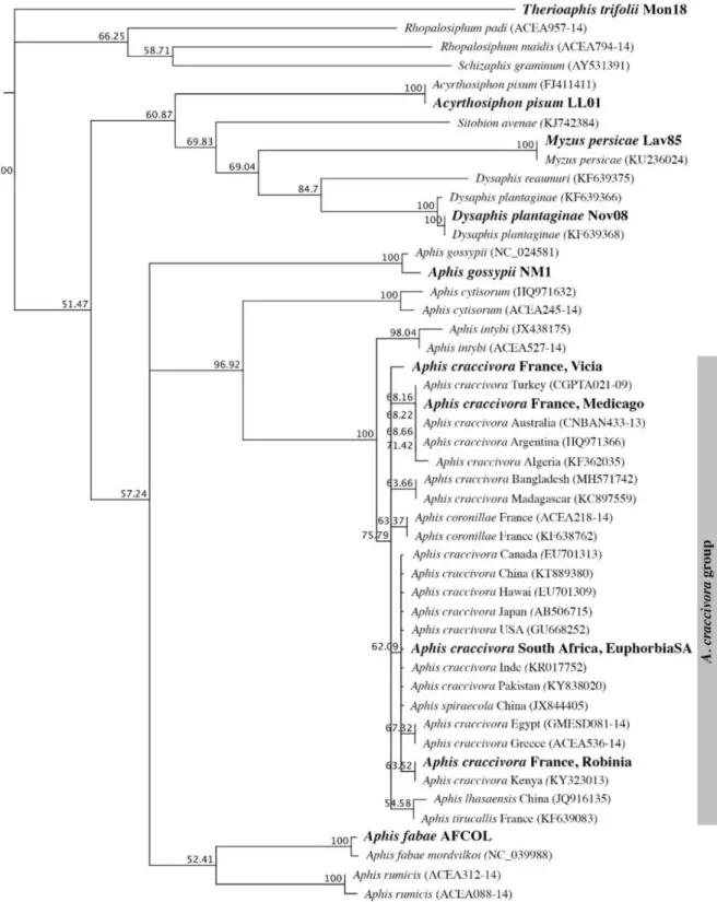
3.2. The aphid vectors of EcmLV belongs to the A. craccivora group
According to COI gene sequences, individuals of the rearing population derived from black-backed aphids collected on E. caput-medusae in South-Africa clustered with representatives of the A. craccivora group, including Aphis tirucallis Hille Ris Lambers, 1954, a species known in Africa on Euphorbia spp. plants (Fig. 1). There are numerous morphologically similar Aphis spp. on Euphorbia that look like A. craccivora and which are recognized to be very difficult to distinguish from each other (Blackman and Eastop, 2000). As shown by morphological measures of specimens mounted on slides (Supplemental Fig. S1), the black-backed aphid specimens collected on Euphorbia Medusa’s head plants are distinguishable from individuals of the A. craccivora group, sensu stricto, particularly by siphunculi length and a contrasting length ratio between the processus terminalis and the base of the last antennal segment (Supplemental Fig.S1), two previously reported discriminating features between specimens of the A. craccivora group, sensu stricto, and specimens of other Aphis living on Euphorbia, in particular, Aphis euphorbiae Kaltenbach, 1843 (Blackman and Eastop, 2000). Morphometric data suggest that our specimens probably belong to A. tirucallis or A. euphorbiae species but with low certainty because it cannot be excluded that the morphological differences are adaptive. Thus, as suggested by Coeur d’Acier et al. (2014) in this case, we adopted a pragmatic and conservative point of view and considered that it is safe to identify the black-backed aphids from South Africa as members of the A. craccivora group according to CO1 sequences. Hence, the name EuphorbiaSA was given to this South African population that refers both to its host plant and its geographical origin. Its adaptation to spurges was confirmed with branches cut from Euphorbia nicaeensis All. and Euphorbia
serrata L. plants of the area of Montpellier, on which EuphorbiaSA individuals developed readily.
3.3. Transmission of capulaviruses by aphids is highly specific
Aphids of five non- A. craccivora species were tested for their ability to transmit ALCV (Table 1). They were selected according to at least one of the following criteria: regularly found on Fabaceae, phylogenetically close to the known vectors of capulaviruses, and reported to transmit other viruses. No transmission was detected with any of these non- A. craccivora species.
The transmission tests for EcmLV and PlLV were conducted with A. gossypii and M. persicae, the only aphid species available in the laboratory that develop on the few reported hosts of these viruses. EcmLV was not transmissible by A. gossypii and M. persicae and PlLV was not transmissible by A. gossypii (Table 1). These failures support the concept of a high transmission specificity, with only one vector species per capulavirus species.
Transmission specificity was explored further by testing if an aphid vector of one capulavirus can transmit another one. Due to a limited number of compatible host combinations of virus and aphid species (Table 2A), only one combination could be tested, i.e A. craccivora EuphorbiaSA and ALCV which are both hosted by broad bean. According to symptom observations and PCR test, all the broad bean plants exposed to EuphorbiaSA aphids that were given access to ALCV infected plants, were negative for the presence of ALCV (Table 1: 0/12, 0/45, 0/15). The transmission was also unsuccessful in test plants exposed to 30-150 viruliferous aphids (0/6). Aphids of the A. craccivora Robinia population tested in parallel as positive controls did successfully transmit ALCV (Table 1: 8/15, 9/30).
To explore other combinations of capulaviruses and known capulavirus vectors suitable for transmission tests, we tested if capulaviruses available in the laboratory have common host plants (Table 2B). The only virus/vector combination was EcmLV with D. plantaginea but no transmission occurred (Table 1), suggesting that EcmLV is not transmitted by D. plantaginea; nonetheless, we cannot exclude that the failure results from a poor adaptation of EcmLV on this host.
3.4. High transmission specificity of ALCV within the A. craccivora group
The results presented above show that while aphids of the Robinia population of A. craccivora were able to transmit ALCV, those of the EuphorbiaSA population, were not (Table 1). This indicates that the transmission of ALCV by aphids is highly specific. Hence, the specificity within the A. craccivora group was investigated further by testing two more aphid populations, Medicago and Vicia (Supplemental Table S1). Aphids of Robinia, Vicia and Medicago populations transmitted ALCV from broad bean agroinfected plants to 7 of 13 (53.8%), 7 of 20 (35%) and 1 of 20 (5%) test plants, respectively (Table 3A). The relatively low transmission success with the Medicago population may be explained by its low affinity for broad bean plants because some individuals ran away during IAP. This hypothesis was confirmed with a distinct test in which individuals of the Medicago population exposed to ALCV infected broad bean plants were shifted to alfalfa plants; in this case, the transmission rate was 62%, similar to that obtained with the Robinia population on broad bean plants (Table 3A).
3.5. ALCV persist in vector and non-vector populations of the A. craccivora group
Since geminiviruses are transmitted in a circulative persistent manner (Nault, 1997, Whitfield et al., 2015), we wanted to verify if this is also the case for capulaviruses. To do this, we determined ALCV persistence in the vector population A. craccivora Robinia and compared it
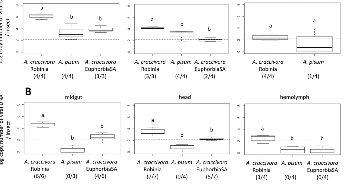
to that in non-vector populations of A. craccivora (EuphorbiaSA) and A. pisum. The transmission procedure consisted of a 3-day AAP on broad bean plants agroinfected with ALCV, followed by three sequential 2-day IAPs on three batches of broad bean plants. While 70% of the plants exposed to individuals of the Robinia population were symptomatic in the three batches, none of the plants exposed to A. pisum and EuphorbiaSA populations exhibited symptoms, confirming their non-vector status (Table 3B). The transmission of ALCV to the three sequential batches by Robinia aphids indicates the persistence of transmissibility for at least 4 days after the end of AAP. To determine to what extent ALCV persists in the non-vector A. pisum and A. craccivora EuphorbiaSA, viral DNA content was assessed by qPCR. At the end of the 3-day AAP, ALCV DNA was detected in vector and non-vector aphids indicating that all had access to the virus (Fig. 2A). The number of viral DNA copies per insect in A. craccivora Robinia (1.5x10⁶) was significantly higher than that of A. pisum (1.6x10⁵; pairwise.t.test() p-value = 6.9 x 10⁻⁴) and A. craccivora EuphorbiaSA (5.6x10⁴; p-value = 4.6 x 10⁻⁵) (Fig. 2A). Six days after the end of the AAP, the viral amount only slightly decreased in A. craccivora Robinia (7.5x10⁵ viral DNA copies per insect), remained constant in A. craccivora EuphorbiaSA (6.4x10⁴), and was undetectable in A. pisum (Fig. 2B)
The persistence of ALCV in the non-vector A. craccivora EuphorbiaSA was further investigated, analyzing the localization of the viral DNA in the insect body. To do this, a second transmission test was carried out, this time quantifying ALCV on dissected insect organs, i.e. the digestive tract, the head with the salivary glands, and hemolymph. Like in the first test, symptoms occurred only after exposure to viruliferous Robinia aphids (Table 3B). At the end of the AAP, viral DNA was detected in the digestive tract in all the treatments (Fig. 3A) which shows that aphids of the three populations had access to the virus. Although the body of A. pisum aphids is much larger than that of EuphorbiaSA aphids (Supplemental Fig. S1), the average ALCV DNA content was similar (1.75x10⁴ and 1.4x10⁴, respectively).
The guts of Robinia aphids contained 2.0x10⁶ viral DNA copies per insect on average, which is 145 times higher than the content estimated for A. pisum and EuphorbiaSA. At the end of the three sequential 2-day IAPs, the digestive tract of A. pisum aphids was qPCR negative for ALCV (Fig. 3B). On the contrary, the samples of Robinia aphids were qPCR positive (6.7x10⁴ viral DNA copies per individual) as well as 4 of the 6 samples of EuphorbiaSA aphids (5.7x10² viral DNA copies per individual). The detection pattern of ALCV in head samples was similar to that observed in gut samples. Indeed, while the head samples of A. pisum were all qPCR negative, those of Robinia aphids were positive (5.8x10³ viral DNA copies per individual) as well as 5 of 7 samples of EuphorbiaSA aphids (1.9x10²). Hemolymph samples were qPCR-positive only for Robinia aphids, with 5.1x10² viral DNA copies per individual in average.
3.6. Intracellular ALCV aggregates were localized in vector and non-vector A. craccivora
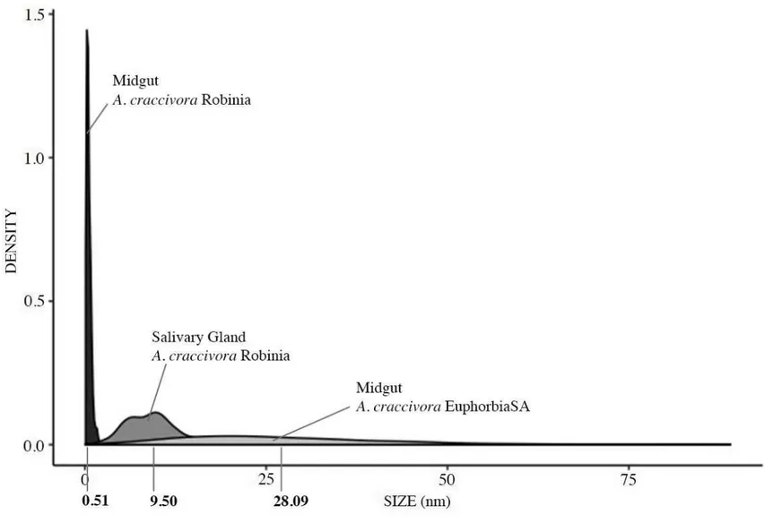
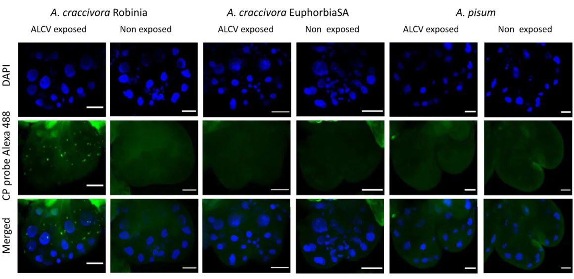
To evaluate if the persistence of ALCV detected with the non-vector population of A. craccivora (EuphorbiaSA) might be associated with internalization and persistence in midgut cells, we used fluorescent in-situ hybridization (FISH) using A. craccivora Robinia and A. pisum as positive and negative controls, respectively. Aphids of the three populations were allowed 15-day AAPs followed by 48-hour IAPs before dissection and FISH analysis. In Robinia aphids, fluorescent aggregates were observed all around the nuclei of epithelial cells of the anterior midgut (Fig. 4) and of the beginning of the posterior midgut (data not shown). Their area estimated from confocal virtual sections was 0.52 ± 0.02 μm2 on average (Fig. 5). These aggregates were detected neither in aphids of the A. pisum population nor in aphids that fed on healthy plants only. In EuphorbiaSA aphids, virus-specific aggregates were also detected but were distinguishable from those observed with Robinia aphids based on their distribution and size. Indeed, unlike those of Robinia aphids, they did not circle the nuclei but
seemed to be concentrated towards the apical and basal side of epithelial cells. Their area was 28.10 ± 1.01 μm2 on average, significantly larger than the Robinia aggregates (Fig. 5, Benjamini and Yekutieli’s method: p-value < 0.01). Noteworthy, the size range of the aggregates is larger for EuphorbiaSA aphids than for Robinia aphids. Additionally, while virus aggregates were observed in a majority of Robinia individuals (68%), they were observed in only 33% of EuphorbiaSA individuals (Table 4).
Virus-specific aggregates were detected in primary salivary glands of A. craccivora Robinia aphids (Fig. 6) and only in 13% of individuals (Table 4). Interestingly, their size was larger than the Robinia midgut aggregates, with a mean area of 9.50 ± 0.62 μm2 (Fig. 5). No virus-specific aggregates were detected in salivary glands of A. craccivora EuphorbiaSA and A. pisum aphids.
4. Discussion
4.1. Aphid transmission is a taxonomic criterion of the genus Capulavirus
ALCV and PlLV were previously reported to be transmitted by aphids. By demonstrating that a third capulavirus (EcmLV) is transmitted by aphids, aphid transmission is validated as a taxonomic criterion of the new genus. Additionally, the successful transmission of a cloned PlLV by the aphid D. plantaginea not only confirmed previous transmission results with a non-cloned PlLV isolate (Susi et al., 2019), but showed, together with the transmission of ALCV (Roumagnac et al., 2015) and EcmLV (this study) clones, that their monopartite genome determines by itself aphid transmissibility without any helper DNA or virus. It is assumed that CP is a major player in aphid transmission of capulaviruses as demonstrated previously for the CP of non-aphid transmitted geminviruses (Azzam et al., 1994, Briddon et al., 1990).
Consistent with these reports, CPs of ALCV and EcmLV, the two capulaviruses transmitted by aphids of the A. craccivora group, are more similar to each other (75% amino acid identity) than to the CP of PlLV (47% and 53% aa identity respectively) which is transmitted with a non-craccivora aphid, i.e. D. plantaginea. Hence, as the CP of FbSLCV is highly similar to ALCV and EcmLV CPs (78% and 72%, respectively) (Varsani et al., 2017), it may be predicted that it will be also transmitted by A. craccivora aphids.
4.2. Transmission results and taxonomy of A. craccivora populations
The transmission tests showed that vectors of EcmLV and ALCV are all belonging to the A. craccivora group. While the EuphorbiaSA population was distinguished from other A. craccivora populations by its adaptation to spurges and by morphological differences, it could not be distinguished according to COI gene comparisons (Fig. 1); this later result is consistent with previous results showing that sub-species or populations are not easily distinguished within the very cosmopolitan A. craccivora group (Blackman and Eastop, 2000, Wang et al., 2011; Song et al., 2016; Coeur d’Acier et al., 2014). According to aphid literature, morphological variations are not necessarily heritable (Mehrparvar, 2012). Therefore, as recommended by these authors, the taxonomy has to be confirmed with genetic and biological data. Unexpectedly, the transmission results may further support the distinction between the EuphorbiaSA population and the other three A. craccivora populations used in this study. Indeed, the intra-species transmission specificity previously detected with various populations of Schizaphis graminum (Rondani, 1852), an aphid vector of luteoviruses (Gray et al., 2002), was shown to be a heritable trait regulated by multiple genes acting in an additive fashion (Burrows et al., 2007).
EcmLV was identified in South Africa and ALCV in Europe, Argentina, the Middle East and China (Davoodi et al., 2018, Guo et al. 2019) which suggests that A. craccivora is a global vector of capulaviruses. This prediction is consistent with the global distribution of A. craccivora and with the fact that FbSLCV was isolated from French bean in India, a highly conducive host of A. craccivora. Thus, with the exception of PlLV, the transmission of capulaviruses may be comparable to the transmission of begomoviruses, both having a unique vector species. .
While A. craccivora is polyphagous, D. plantaginea is specialized on buckhorn plantain and apple tree. This difference is consistent with the large range of viruses transmitted by A. craccivora, including representatives of families Tombusviridae, Potyviridae, Bromoviridae, and Nanoviridae, whereas PlLV is the first virus reported to be transmitted by D. plantaginea.
4.4. ALCV is transmitted by A. craccivora in a highly specific and persistent circulative manner
The persistence of ALCV in A. craccivora individuals was not interrupted by molts, which is typical of the persistent circulative mode of transmission. Consistently with this result, ALCV DNA was detected in hemolymph and in epithelial cells of the midgut and the primary salivary glands.
ALCV was not transmissible by the EuphorbiaSA population of A. craccivora, even with a high number of individuals per test plants (30-150). Thus, the transmission failure of ALCV by EuphorbiaSA individuals is expected to be due to incompatible virus-aphid interactions rather than to a potentially lower amount of inoculated virus due to their smaller size compared to Robinia individuals (Supplemental Fig. S1). The extremely high transmission specificity of ALCV by certain populations of A. craccivora is similar to that reported with
barley yellow dwarf virus - MAV (BYDV-MAV) and BYDV-PAV differentially transmitted by S. graminum aphid populations (Gray et al., 2002). However, it was not detected in other geminivirus genera, not even within B. tabaci populations that are thought to be vector of all begomoviruses. The molecular interactions that drive ALCV recognition for transmission are expected to be highly specific. Hence, it seems highly unlikely that non-aphid vectors would transmit capulaviruses, which further supports that aphid transmission is a taxonomic criterion of the genus Capulavirus.
The inability of A. craccivora EuphorbiaSA to transmit ALCV may be related to its specialization in the transmission of EcmLV. It would be interesting to validate this hypothesis with a symmetric transmission of EcmLV by A. craccivora populations that are vectors of ALCV. Unfortunately, besides EuphorbiaSA, A. craccivora populations available in our laboratory did not develop on reported hosts of EcmLV (Bernardo et al., 2013). However, such test may be carried out in the future with African A. craccivora populations reported from spurges (Remaudière, 1985).
4.5. Transmission barriers to ALCV in aphids
ALCV DNA was detected in some hemolymph samples of the non-vector A. pisum at the end of the AAP. However, their low percentage combined with the lack of ALCV signals by FISH in the midgut are consistent with a hemolymph contamination. These results indicate a transmission barrier located at the gut level similarly to the barriers detected with non-vector leafhoppers of mastreviruses (Lett et al., 2002) and non-vector whiteflies of begomoviruses (Rosell et al., 1999; Czosnek et al., 2001; Ohnishi et al., 2009). The viral persistence in A. craccivora EuphorbiaSA but not in A. pisum may be explained by a selective accessibility to gut cells.
The results on ALCV persistence and localization in A. craccivora EuphorbiaSA individuals suggest that the transmission barrier is in the salivary glands. Indeed, viral DNA was detected by FISH in midgut epithelial cells, and at 6 days post-AAP, some head samples were detected virus-positive. Interestingly, salivary glands were previously reported to be the major barrier for the aphid-transmitted barley yellow dwarf disease associated viruses (family Luteoviridae) (Gildow and Gray, 1993). Indeed, these authors showed that the basal lamina (BL) surrounding the accessory salivary glands acts as a viral barrier. While BL acts as an absolute barrier to the transmission of BYDV-MAV in the non-vector aphid Rhopalosiphum maidis (Fitch, 1856), it is slightly permeable in the non-efficient vector Rhopalosiphum padi Linnaeus, 1758 and attractive as well as permeable in the efficient vector Sitobion avenae (Fabricius, 1775).
ALCV DNA was readily localized by FISH in the midgut of vector aphids of A. craccivora. Its rare observation in the salivary glands may be explained by difficult access to salivary glands due to the BL barrier as reported for BYDV-MAV in R. maidis (Gildow and Gray, 1993). Indeed, as these authors estimated the size exclusion of BL in cereal aphids to 20 to 30 nm (Peiffer et al., 1997), a potentially similar size exclusion in A. craccivora may reduce the capulavirus transit through the BL due to their size, 20 x 36 nm in the case of EcmLV (Roumagnac et al., 2015).
The standard size of the virus aggregates detected by FISH in midgut cells of A. craccivora Robinia and their defined perinuclear distribution reflect a well-established mechanism associated with virus transit. On the contrary, the large range of sizes of the aggregates observed in midgut cells of A. craccivora EuphorbiaSA and their apparently undefined distribution seems to reflect non-established virus-aphid interactions that nevertheless allow some virus persistence. Further tests will be necessary to characterize the specific and non-specific interactions and confirm the transmission barriers of capulaviruses.
Acknowledgements
We are very thankful to Armelle Coeur d’Acier for her valuable expertise in aphid identification. We thank Myriam Siegwaert (Inra-Avignon) for providing D. plantaginea aphids, Sylvaine Boissinot (Inra-Colmar) and Christoph Vorburger (ETHzürich) for providing A. fabae aphids and Josep Vicens Fandos (Lab. de Botánica, Fac. de Farmacia, Univ. de Barcelona) for confirming the species identification of the spurges collected near Montpellier. The design of the CO1 primers was from Isabelle Meunier (Inra-Montpellier). The study was carried out during the Ph.D. project of Faustine Ryckebusch that was funded by the Agropolis Fondation (E-Space flagship program) grant number 1504-004.
References
Azzam, O., Frazer, J., de la Rosa, D., Beaver, J.S., Ahlquist, P., Maxwell, D.P., 1994. Whitefly transmission and efficient ssDNA accumulation of bean golden mosaic geminivirus require functional coat protein. Virology 204, 289–296. https://doi.org/10.1006/viro.1994.1533
Bahder, B.W., Zalom, F.G., Jayanth, M., Sudarshana, M.R., 2016. Phylogeny of geminivirus coat protein sequences and digital PCR aid in identifying Spissistilus festinus as a vector of Grapevine red blotch-associated virus. Phytopathology 106, 1223–1230. https://doi.org/10.1094/PHYTO-03-16-0125-FI
Bedford, I.D., Briddon, R.W., Brown, J.K., Rosell, R.C., Markham, P.G., 1994. Geminivirus transmission and biological characterization of Bemisia tabaci (Gennadius) biotypes from
different geographic regions. Annals of Applied Biology 125, 311–325. https://doi.org/10.1111/j.1744-7348.1994.tb04972.x
Benjamini, Y., Yekutieli, D., 2001. The control of the false discovery rate in multiple testing under dependency. Annals of Statistics. 29, 1165–1188. https://doi.org/10.1214/aos/1013699998
Bernardo, P., Golden, M., Akram, M., Naimuddin, Nadarajan, N., Fernandez, E., Granier, M., Rebelo, A.G., Peterschmitt, M., Martin, D.P., Roumagnac, P., 2013. Identification and characterisation of a highly divergent geminivirus: Evolutionary and taxonomic implications. Virus Research 177, 35–45. https://doi.org/10.1016/j.virusres.2013.07.006
Blackman, R. L., & Eastop, V. F., 2000. Aphids on the world's crops: an identification and information guide (No. Ed. 2). John Wiley & Sons Ltd.
Briddon, R.W., Pinner, M.S., Stanley, J., Markham, P.G., 1990. Geminivirus coat protein gene replacement alters insect specificity. Virology 177, 85–94. https://doi.org/10.1016/0042-6822(90)90462-z
Burrows, M.E., Caillaud, M.C., Smith, D.M., Gray, S.M., 2007. Biometrical genetic analysis of luteovirus transmission in the aphid Schizaphis graminum. Heredity 98, 106–113. https://doi.org/10.1038/sj.hdy.6800909
Coeur d’acier, A., Cruaud, A., Artige, E., Genson, G., Clamens, A.-L., Pierre, E., Hudaverdian, S., Simon, J.-C., Jousselin, E., Rasplus, J.-Y., 2014. DNA barcoding and the associated PhylAphidB@se website for the identification of european aphids (Insecta: Hemiptera: Aphididae). PLoS ONE 9, e97620. https://doi.org/10.1371/journal.pone.0097620
Czosnek, H., Ghanim, Miriam, Ghanim, Murad, 2001. The circulative pathway of begomoviruses in the whitefly vector Bemisia tabaci — insights from studies with Tomato yellow leaf curl virus. Annals of Applied Biology. 2002, 140:215-231
Davoodi, Z., Bejerman, N., Richet, C., Filloux, D., Kumari, S., Chatzivassiliou, E., Galzi, S., Julian, C., Samarfard, S., Trucco, V., Giolitti, F., Fiallo-Olivé, E., Navas-Castillo, J., Asaad, N., Moukahel, A., Hijazi, J., Mghandef, S., Heydarnejad, J., Massumi, H., Varsani, A., Dietzgen, R., Harkins, G., Martin, D., Roumagnac, P. 2018. The westward journey of Alfalfa Leaf Curl Virus. Viruses 10, 542. https://doi.org/10.3390/v10100542
De Barro, P.J., Liu, S.-S., Boykin, L.M., Dinsdale, A.B., 2011. Bemisia tabaci: a statement of species status. Annual Review of Entomology 56, 1–19. https://doi.org/10.1146/annurev-ento-112408-085504
Favret, C., 2014. Cybertaxonomy to accomplish big things in aphid systematics: Cybertaxonomy in aphid systematics. Insect Science 21, 392–399. https://doi.org/10.1111/1744-7917.12088
Folmer, O., Black, M., Hoeh, W., Lutz, R., Vrijenhoek, R., 1994. DNA primers for amplification of mitochondrial cytochrome c oxidase subunit I from diverse metazoan invertebrates. Molecular marine biology and biotechnology. 3, 294–299.
Gildow, F.E., Gray, S.M., 1993. The aphid salivary gland basal lamina as a selective barrier associated with vector-specific transmission of barley yellow dwarf luteovirus. Phytopathology 83, 1293-1302.
Gray, S.M., Smith, D.M., Barbierri, L., Burd, J., 2002. Virus transmission phenotype is correlated with host adaptation among genetically diverse populations of the aphid Schizaphis graminum. Phytopathology 92, 970–975. https://doi.org/10.1094/PHYTO.2002.92.9.970
Guo, Z.P., Zhang, J.X., Wang, M.L., Guan, Y.Z., Qu, G., Liu, J.Y., Guo, Y.X., Yan, X.B., First Report of alfalfa leaf curl virus infecting alfalfa (Medicago sativa) in China. Plant Disease https://doi.org/10.1094/PDIS-02-19-0318-PDN
Heydarnejad, J., Keyvani, N., Razavinejad, S., Massumi, H., Varsani, A., 2013. Fulfilling Koch’s postulates for beet curly top Iran virus and proposal for consideration of new genus in the family Geminiviridae. Archives of Virology 158, 435–443. https://doi.org/10.1007/s00705-012-1485-6
Hogenhout, S.A., Ammar, E.-D., Whitfield, A.E., Redinbaugh, M.G., 2008. Insect vector interactions with persistently transmitted viruses. Annual Review of Phytopathology 46, 327– 359. https://doi.org/10.1146/annurev.phyto.022508.092135
Kvarnheden, A., Lett, J.M., Peterschmitt, M., 2016. Mastreviruses: Tropical and temperate leafhopper-borne geminiviruses, in: Brown, J.K. (Ed.), Vector-mediated transmission of plant pathogens. The American Phytopathological Society, St. Paul, Minnesota, USA, pp. 231-241.
Lett, J.-M., Granier, M., Hippolyte, I., Grondin, M., Royer, M., Blanc, S., Reynaud, B., Peterschmitt, M., 2002. Spatial and temporal distribution of geminiviruses in leafhoppers of the genus Cicadulina monitored by conventional and quantitative Polymerase Chain Reaction. Phytopathology 92, 65–74. https://doi.org/10.1094/PHYTO.2002.92.1.65
Mehrparvar, M., 2012. Morphometric discrimination of Black Legume Aphid, Aphis craccivora Koch (Hemiptera: Aphididae) populations associated with different host plants. North-Western Journal of Zoology 8 (1): 172-180
Nault, L.R., 1997. Arthropod Transmission of Plant Viruses: a New Synthesis. Annals of the Entomological Society of America 90, 521–541. https://doi.org/10.1093/aesa/90.5.521
Ohnishi, J., Kitamura, T., Terami, F., Honda, K., 2009. A selective barrier in the midgut epithelial cell membrane of the nonvector whitefly Trialeurodes vaporariorum to Tomato yellow leaf curl virus uptake. Journal of General Plant Pathology 75, 131–139. https://doi.org/10.1007/s10327-009-0147-3
Peccoud, J., Labonne, G., Sauvion, N., 2013. Molecular test to assign individuals within the
Cacopsylla pruni complex. PLOS ONE 8, e72454.
https://doi.org/10.1371/journal.pone.0072454
Peiffer, M.L., Gildow, F.E., Gray, S.M., 1997. Two distinct mechanisms regulate luteovirus transmission efficiency and specificity at the aphid salivary gland. Journal of General Virology 78 ( Pt 3), 495–503. https://doi.org/10.1099/0022-1317-78-3-495
R Core Team, 2017. R: A language and environment for statistical computing. R Foundation for Statistical Computing, Vienna, https://www.r-project.org
Razavinejad, S., Heydarnejad, J., Kamali, M., Massumi, H., Kraberger, S., Varsani, A., 2013. Genetic diversity and host range studies of turnip curly top virus. Virus Genes 46, 345–353. https://doi.org/10.1007/s11262-012-0858-y
Remaudière, G., & Remaudière, M., 1997. Catalogue of the world's Aphididae: Homoptera Aphidoidea. Institut National de la Recherche Agronomique (INRA).
Remaudière, G., 1985. Contribution à l'écologie des aphides africains (Vol. 64). Food & Agriculture Organisation, Rome.
Rosell, R.C., Torres-Jerez, I., Brown, J.K., 1999. Tracing the geminivirus-whitefly transmission pathway by Polymerase Chain Reaction in whitefly extracts, saliva, hemolymph, and honeydew. Phytopathology 89, 239–246. https://doi.org/10.1094/PHYTO.1999.89.3.239
Roumagnac, P., Granier, M., Bernardo, P., Deshoux, M., Ferdinand, R., Galzi, S., Fernandez, E., Julian, C., Abt, I., Filloux, D., Mesléard, F., Varsani, A., Blanc, S., Martin, D.P., Peterschmitt, M., 2015. Alfalfa Leaf Curl Virus: an aphid-transmitted geminivirus. Journal of Virology 89, 9683–9688. https://doi.org/10.1128/JVI.00453-15
Ruijter, J.M., Pfaffl, M.W., Zhao, S., Spiess, A.N., Boggy, G., Blom, J., Rutledge, R.G., Sisti, D., Lievens, A., De Preter, K., Derveaux, S., Hellemans, J., Vandesompele, J., 2013. Evaluation of qPCR curve analysis methods for reliable biomarker discovery: Bias, resolution, precision, and implications. Methods 59, 32–46. https://doi.org/10.1016/j.ymeth.2012.08.011
Saitou, N., Nei, M., 1987. The neighbor-joining method: a new method for reconstructing phylogenetic trees. Molecular Biology and Evolution 4, 406–425. https://doi.org/10.1093/oxfordjournals.molbev.a040454
Song, N., Zhang, H., Li, H., Cai, W., 2016. All 37 mitochondrial genes of aphid Aphis craccivora obtained from transcriptome sequencing: implications for the evolution of aphids. PLOS ONE 11, e0157857. https://doi.org/10.1371/journal.pone.0157857
Susi, H., Filloux, D., Frilander, M.J., Roumagnac, P., Laine, A.-L., 2019. Diverse and variable virus communities in wild plant populations revealed by metagenomic tools. PeerJ 7, e6140. https://doi.org/10.7717/peerj.6140
Susi, H., Laine, A.-L., Filloux, D., Kraberger, S., Farkas, K., Bernardo, P., Frilander, M.J., Martin, D.P., Varsani, A., Roumagnac, P., 2017. Genome sequences of a capulavirus infecting Plantago lanceolata in the Åland archipelago of Finland. Archives of Virology 162, 2041– 2045. https://doi.org/10.1007/s00705-017-3298-0
Swofford, D.L., 2001. PAUP*: Phylogenetic Analysis Using Parsimony (and other methods) 4.0.b5.
Varsani, A., Roumagnac, P., Fuchs, M., Navas-Castillo, J., Moriones, E., Idris, A., Briddon, R.W., Rivera-Bustamante, R., Murilo Zerbini, F., Martin, D.P., 2017. Capulavirus and Grablovirus: two new genera in the family Geminiviridae. Archives of Virology 162, 1819– 1831. https://doi.org/10.1007/s00705-017-3268-6
Wang, J., Jiang, L.-Y., Qiao, G.-X., 2011. Use of a mitochondrial COI sequence to identify species of the subtribe Aphidina (Hemiptera, Aphididae). ZooKeys 122, 1–17. https://doi.org/10.3897/zookeys.122.1256
Whitfield, A.E., Falk, B.W., Rotenberg, D., 2015. Insect vector-mediated transmission of plant viruses. Virology, 60th Anniversary Issue 479–480, 278–289. https://doi.org/10.1016/j.virol.2015.03.026
Zerbini, F.M., Briddon, R.W., Idris, A., Martin, D.P., Moriones, E., Navas-Castillo, J., Rivera-Bustamante, R., Roumagnac, P., Varsani, A., 2017. ICTV Virus Taxonomy Profile:
Geminiviridae. Journal of General Virology 98, 131–133. https://doi.org/10.1099/jgv.0.000738
Legends
Figure 1. Neighbor joining tree showing the genetic distances among members of the Aphis craccivora group and members of related species, based on the cytochrome oxidase 1 gene. Sequences are identified by a species name and an accession number for those uploaded from Genbank or BOLD. Members of the A. craccivora group are identified further with the country of origin. The description of sequences generated in this study is in bold. A detailed description of the sequenced specimens is provided in Supplemental Tables S1 and S2. Numbers associated with nodes represent the percentage of 10,000 bootstrap iterations supporting the nodes; only percentages > 50 % are indicated. Therioaphis trifolii was used as an outgroup.
Figure 2. Box-plots showing the amount of ALCV DNA in vector aphids (A. craccivora Robinia) and non-vector aphids (A. craccivora EuphorbiaSA and A. pisum) following a 3-day AAP on broad bean plants agroinfected with ALCV (A), and after three sequential 2-day IAPs on non-infected plants (B). In each treatment, the content of ALCV DNA was determined by qPCR in 4 pools of 10 individuals. Accumulations are reported as logarithm 10 of the number of viral DNA copies. The dotted lines represent the highest values obtained with non-viruliferous pools of 10 aphids sampled before the 3-day AAP; the post-AAP and IAPs pools for which the number of estimated DNA copies is above this threshold are considered positive for the presence of ALCV DNA. The ratio of positive pools is indicated below aphid names. Letters
above box-plots indicate significant differences (p-value < 0.01) between the modalities according to the multiple comparison method based on Benjamini and Yekutieli’s procedure.
Figure 3. Box-plots showing amount of ALCV DNA in the midgut, head, and hemolymph in vector aphids (A. craccivora Robinia) and non-vector aphids (A. craccivora EuphorbiaSA and A. pisum), following 3-day AAP on broad bean plants agroinfected with ALCV (A) and after three sequential 2-day passages on non-infected plants (B). The content of ALCV DNA was determined by qPCR in 3-7 pools of 10 dissected fractions. Accumulations are reported as logarithm 10 of the number of viral DNA copies. The dotted lines represent the highest value obtained with non-viruliferous pools of 10 aphids sampled before the 3-day AAP; the post-AAP and IAPs pools for which the number of estimated DNA copies is above this threshold are considered positive for the presence of ALCV DNA. The ratio of positive pools is indicated below the aphid names. Presentation of accumulations and statistical analysis as in Fig. 2.
Figure 4. Localization of ALCV DNA by FISH in dissected anterior midguts of vector and non-vector aphids of ALCV. Aphids were exposed during 15 days to broad bean plants agroinfected with ALCV and then shifted to non-infected plants for three days before FISH analysis. Non exposed aphids underwent the same procedure except that broad bean plants of the 15 day period were non-infected. A. craccivora Robinia is a vector aphid of ALCV whereas A. craccivora EuphorbiaSA and A. pisum are non-vector aphids. The ALCV specific DNA probe is labeled with a green Alexa 488 fluorochrome. Nuclei are DAPI-blue stained. Preparations were examined with confocal microscopy. Horizontal bars = 30 µm.
Figure 5. Density plots showing the frequency distribution of section areas of fluorescent ALCV-specific aggregates observed in dissected midguts and salivary glands of vector and non-vector A. craccivora aphids. FISH was performed on aphids that were given a 15-day AAP on broad bean plants agroinfected with ALCV, followed by a 3-day period on healthy plants. Areas of aggregate sections were measured in midguts and primary salivary glands of three vector aphids (A. craccivora Robinia) and in midguts of three non-vector aphids (A. craccivora Euphorbia SA). Areas of 250 sections of aggregates were measured per modality. A graph was obtained with the libraries ggplot2 and function ggplot in R.
Figure 6. Localization of ALCV DNA by FISH in dissected primary salivary glands of vector and non-vector aphids of ALCV. Aphid species, figure layout, probe and DAPI staining as in Fig. 4. Horizontal bars = 30 µm.
Table 1. Transmission tests of three capulaviruses with aphids of various species. The rate of infected plants is in bold for successful transmissions. PlLV, Plantago lanceolata latent virus; EcmLV, Euphorbia caput medusae latent virus; ALCV: alfalfa leaf curl virus; AAP, acquisition access period; IAP, inoculation access period.
Table 2. Detection of compatible plant/aphid/capulavirus trios to assess the transmission specificity of capulaviruses by aphids. (A) reported host plants of capulaviruses that may be hosts of capulavirus vectors. + aphids were able to stay for at least several days on the plant;
- aphids were not able to survive on the plant. (B) Detection of reported host plants of capulaviruses that may host other capulaviruses. Capulaviruses were agroinoculated to host plants. nd = not done : * = transitory virus infection.
Table 3. Transmission tests of ALCV with A. craccivora populations. (A) Comparison of populations from Robinia pseudoacacia L. (Robinia), Vicia sativa (L.) Bernh. (Vicia), and Medicago sativa L. (Medicago) (B) Test of the persistency of the infectivity in A. craccivora Robinia. The persistency was assessed with individuals that were given a 3-day AAP on broad bean plants agroinfected with ALCV and subsequently shifted sequentially to three batches of plants each exposed to 2-day IAPs. The rate of transmission successes is mentioned in the chronological order of the three passages. Aphids of the EuphorbiaSA population of A. craccivora and of A. pisum were used as negative controls particularly for the persistence of ALCV DNA determined in the whole body (Experiment 1, Fig. 2) and in dissected fractions (Experiment 2, Fig. 3).
Table 4. Frequency of midgut and salivary glands in which ALCV-specific aggregates were detected by FISH in vector and non-vector aphids (Figs 2 and 3). Vector aphids were from the Robinia population of A. craccivora and the non-vector aphids were from the EuphorbiaSA population of A. craccivora and from A. pisum. Aphids were analyzed after a 15-day AAP on broad bean plants agroinfected with ALCV followed by a 3-day IAP on non-infected plants.
Supplemental Figure 1. Morphological comparisons between specimens of a population of A. craccivora sensu stricto (a, a’) and specimens of a euphorbia population of black-backed South African aphids (EuphorbiaSA) (d, d’) collected on Euphorbia caput-medusae, the natural host species of EcmLV. The lengths of the processus terminalis, the base of the last antennal segment, the siphunculi and the cauda were measured in A. craccivora specimens (b, c) and in EuphorbiaSA specimens (e, f). (g) shows the absence of rhinaria on the IVth antennal segment of an alate EuphorbiaSA aphid. Based on these discriminating morphological features, EuphorbiaSA was identified as a member of the formerly described Aphis tirucallis (Blackman and Eastop, 2000).
Supplemental Table 1. Description of aphid species and populations used in this study and associated references.
Supplemental Table 2. Description of aphid species and populations mentioned in this study and associated references.
Virus species Aphid species Rate of infected plants Host plant AAP (days) IAP (days) Number aphids / IAP plant PlLV Dysaphis plantaginea Nov08 9/10 Plantago lanceolata 27 5 10
Aphis gossypii NM1 0/20 Plantago lanceolata 13 5 10
0/20 21 5 10
EcmLV Aphis craccivora EuphorbiaSA 2/2 Euphorbia caput-medusae weeks 35 50
Aphis gossypii NM1 0/9 Solanum lycopersicum 2 5 10
Myzus persicae Lav85 0/24 Solanum lycopersicum 2 5 10
Dysaphis plantaginea Nov08 0/12 Plantago lanceolata 3 3 10
ALCV Aphis craccivora Robinia 8/15 Vicia faba 2 5 10
9/30 2 5 10
Aphis fabae A06-405 0/19 Vicia faba 2 5 5
Aphis fabae AFCOL 0/3 Vicia faba 2 5 5
0/5 2 5 5
0/10 2 5 10
Aphis gossypii NM1 0/20 Vicia faba 2 5 5
Myzus persicae Lav85 0/6 Medicago sativa 2 5 20
Acyrthosiphon pisum LL01 0/20 Vicia faba 2 5 10
Therioaphis trifolii Mon18 0/1 Medicago sativa 15 5 10
Aphis craccivora EuphorbiaSA 0/45 Vicia faba 2 5 10
0/15 2 5 10
0/6 3 5 30-150
0/12 7 weeks 10
Table 1. Transmission tests of three capulaviruses with aphids of various species. The rate of infected plants is in bold for successful
A
B
Host plant Virus species
ALCV EcmLV PlLV
Vicia faba 10/10 0/22 0/26
Euphorbia caput-medusae nd 3/10 nd
Plantago lanceolata 1/20* 1/19 5/6
Host plant Aphid species
Aphis craccivora Robinia Aphis craccivora EuphorbiaSA Dysaphis plantaginea
Vicia faba + +
-Euphorbia caput-medusae - +
-Plantago lanceolata - - +
Table 2. Detection of compatible plant/aphid/capulavirus trios to assess the transmission specificity of capulaviruses by aphids. (A)
reported host plants of capulaviruses that may be hosts of capulavirus vectors. + aphids were able to stay for at least several days on
the plant; - aphids were not able to survive on the plant. (B) Detection of reported host plants of capulaviruses that may host other
capulaviruses. Capulaviruses were agroinoculated to host plants; the rate of infected plants is in bold for successful transmissions. nd
= not done : * = transitory virus infection.
Table 3. Transmission tests of three capulaviruses by aphids belonging to various species. (A) Confirmation of the transmission of PlLV
by D. plantaginae and discovery of the aphid transmission of EcmLV. (B) Test of a range of aphid species for their ability to transmit
capulaviruses. (C) Test of a range of A craccivora population for their ability to be vector of ALCV. (D) Test of the persistency of the
infectivity of a vector population of A. craccivora (Robinia) following a 3-day AAP on broad bean plants agroinfected with ALCV. The
EuphorbiaSA population of A. craccivora and a population of A. pisum were used as negative controls for the persistence of ALCV DNA.
A
B
Virus species Experiment Aphid species Rate of infected plants Host plant AAP (days) IAP (days) Number of aphids / plant ALCV
1.
Aphis craccivora Robinia 10/14 ; 9/13 ; 9/12 Vicia faba 3 2 10
Aphis craccivora EuphorbiaSA 0/15 ; 0/15 ; 0/15 Vicia faba 3 2 10
Acyrthosiphon pisum 0/15 ; 0/15 ; 0/15 Vicia faba 3 2 10 ALCV
2.
Aphis craccivora Robinia 3/15 ; 4/19 ; 1/15 Vicia faba 3 2 10
Aphis craccivora EuphorbiaSA 0/15 ; 0/18 ; 0/20 Vicia faba 3 2 10
Acyrthosiphon pisum 0 /15 ; 0/15 ; 0/15 Vicia faba 3 2 10 Virus
species
Aphid species Rate of infected plants
Source plant Test plant AAP (days) IAP (days) Number of aphids / plant
ALCV Aphis craccivora Robinia 7/13 Vicia faba Vicia faba 3 5 10
Aphis craccivora Vicia 7/20 Vicia faba Vicia faba 3 5 10
Aphis craccivora Medicago 1/20 Vicia faba Vicia faba 3 5 10
Aphid species Aphid organs
Anterior midgut Primary salivary gland
Aphis craccivora Robinia 62/91 (68,13%) 12/97 (12,37%)
Aphis craccivoraEuphorbiaSA 13/39 (33,33%) 0/32
Acyrthosiphon pisum 0/30 0/34