HAL Id: tel-01690745
https://tel.archives-ouvertes.fr/tel-01690745
Submitted on 23 Jan 2018HAL is a multi-disciplinary open access archive for the deposit and dissemination of sci-entific research documents, whether they are pub-lished or not. The documents may come from teaching and research institutions in France or abroad, or from public or private research centers.
L’archive ouverte pluridisciplinaire HAL, est destinée au dépôt et à la diffusion de documents scientifiques de niveau recherche, publiés ou non, émanant des établissements d’enseignement et de recherche français ou étrangers, des laboratoires publics ou privés.
innovative shorter anti-tuberculosis regimens
Daniel Atwine
To cite this version:
Daniel Atwine. Improving TB management and control through innovative shorter anti-tuberculosis regimens. Human health and pathology. Université Montpellier, 2017. English. �NNT : 2017MONTT041�. �tel-01690745�
Devant le jury composé de
Cécile GOUJARD, PU-PH, Hôpitaux Universitaires Paris-Sud, Bicêtre, AP-HP Président
Nicolas VEZIRIS, PU-PH, Hôpitaux Universitaires de l’Est Parisien, Centre National de Référence
des Mycobactéries Rapporteur
Rodolphe GARRAFFO, MCU-PH, Hôpitaux Universitaires de Nice Rapporteur
Sylvain GODREUIL, PU-PH, Hôpitaux Universitaires Arnaud de Villeneuve/Université de Montpellier
Examinateur
DE L’UNIVERSITÉ DE MONTPELLIER
En Biologie de la Santé
École doctorale CBS2
Unité de recherche UMI233 TRANSVIHMI
IMPROVING TB MANAGEMENT AND
CONTROL THROUGH INNO VATIVE SHORTER
ANTI-TUBERCULOSIS REGIMENS
Présentée par Daniel ATWINE
Le 24 novembre 2017
Sous la direction de Maryline BONNET Directeur de thèse
et Anne-Marie TABURET co-Directeur de thèse
3
DEDICATION
TO ALL PATIENTS BATTLING TUBERCULOSIS
,
WHOF
OR THE HOPE OF A MUCH SHORTER AND SAFER TREATMENT,
DESPITE BEING TOTHE BENEFIT OF FUTURE PATIENTS
,
SELFLESSLY OFFERED TO PARTICIPATE IN5
ACRONYMS
3TC Lamivudine
ABC Abacavir
ADH Antidiuretic Hormone
AE Adverse Event
AFB Acid-Fast Bacilli
AIDS Acquired Immuno-Deficiency Syndrome ALT Alanine Aminotransferase
ANRS French National Agency For Research On AIDS And Viral Hepatitis
ART Antiretroviral Therapy ARV Antiretroviral
AST Aspartate Aminotransferase ATT Anti-Tuberculosis Treatment
AUC Area Under Concentration Versus Time Curve
c Cobicistat
C12 Mid-Dose Concentration
CI Confidence Interval Cmax Maximum Concentration
Cmin/C24 Minimum Concentration/24-Hour Concentration
CNS Central Nervous System
CR Control Regimen
CXR Chest X-Ray
CYP Cytochrome P450
DAIDS Division Of AIDS
DILI Drug Induced Liver Injury DNA Deoxyribonucleic Acid DOT Directly Observed Treatment
DOTS Directly Observed Treatment Short Course Strategy DR-TB Drug Resistant Tuberculosis
DST Drug Susceptibility Test DS-TB Drug Susceptible Tuberculosis
DTG Dolutegravir
DTH Delayed-Type Hypersensitivity DTM Domiciary Treatment Monitor
E Ethambutol
EBA Early Bactericidal Activity
EFV Efavirenz
EHRZ Ethambutol, Isoniazid, Rifampicin And Pyrazinamide EMA European Medicines Agency
EMRC Epicentre Mbarara Research Centre FDA Food And Drug Administration FDC Fixed Dose Combination FI Fusion Inhibitor
FTC Emtricitabine
GCP Good Clinical Practice
6 GMR Geometric Mean Ratio
H Isoniazid
HBC High-Burden Country HBV Hepatitis B Virus HCV Hepatitis C Virus
HIV Human Immunodeficiency Virus IC Infection Control
ICF Intensified Case Finding
INSTI Integrase Strand Transfer Inhibitors IPT Isoniazid Preventive Treatment IQR Interquartile Range
IRIS Immune Reconstitution Inflammatory Syndrome IU/L International Units Per Litre
LAM Lipoarabinomannan LJ Löwenstein Jensen
LMICs Low And Middle Income Countries LPV/r Ritonavir Boosted Lopinavir LTBI Latent Tuberculosis Infection MAH Mono-Acetyl Hydrazine
MDR-TB Multi-Drug Resistant Tuberculosis MGIT Mycobacteria Growth Indicator Tube mITT Modified Intention To Treat Population
mm Millimeter
MRC Medical Research Council MSF Médecins sans frontières
MTB Mycobacterium Tuberculosis Complex
NA Not Applicable
NALC-NaOH N-Acetyl-L-Cysteine-Sodium Hydroxide NAT2 N-Acetyltransferase Type 2
NDA National Drug Authority ng/ml Nanogram Per Milliliter
NNRTI Non-Nucleoside Reverse Transcriptase Inhibitor NRTI Nucleoside Reverse Transcriptase Inhibitor
NVP Nevirapine
OR Odds Ratio
PCR Polymerase Chain Reaction PI Protease Inhibitor
PK Pharmacokinetics
POC Point-Of-Care
PPD Purified Protein Derivative PTB Pulmonary Tuberculosis
R Rifampicin
r Ritonavir
RCT Randomized Clinical Trials RCT Randomized Clinical Trial RH Rifampicin And Isoniazid RNA Ribonucleic Acid
RR Risk Ratio
7 SAE Serious Adverse Event
SGOT Serum Glutamic Oxaloacetic Transaminase
SR Study Regimen
TB Tuberculosis
TDF Tenofovir Disoproxil Fumarate
Tmax Time To Reach Maximum Concentration
TTD Time-To-Detection ULN Upper Limit of Normal
UNAIDS United Nations Programme On HIV/AIDS
UNCST Uganda National Council of Science and Technology USPHS United States Public Health Service
VL Viral Load
VOT Video Observed Treatment WHO World Health Organization
XDR-TB Extensively Drug-Resistant Tuberculosis
Z Pyrazinamide
9
Table of Contents
ACRONYMS ... 5 LIST OF FIGURES ... 11 LIST OF TABLES... 11 CHAPTER 1 INTRODUCTION ... 131.1 MICROBIOLOGY, PHYSIOPATHOLOGY, IMMUNOLOGY, RISK FACTORS AND CLINICAL PRESENTATION OF TUBERCULOSIS ... 15
1.1.1 Microbiology ... 15
1.1.2 Physiopathology and immunology ... 16
1.1.3 Risk-factors of tuberculosis ... 17
1.1.4 Clinical features of tuberculosis ... 18
1.2 EPIDEMIOLOGY OF TUBERCULOSIS ... 19
1.2.1 Global TB burden ... 19
1.2.2 Burden of HIV-TB co-infection ... 21
1.3 TUBERCULOSIS DIAGNOSIS ... 22
1.4 TREATMENT OF TUBERCULOSIS ... 24
1.4.1 History of tuberculosis drug development ... 24
1.4.2 Treatment of drug-susceptible TB... 25
1.4.2.2 Anti-tuberculosis drug induced liver injury ... 27
1.4.2.3 Treatment outcomes of drug susceptible tuberculosis ... 31
1.4.2.4 Adherence to TB treatment ... 31
1.4.3 Drug-resistant tuberculosis ... 32
1.4.3.1 Burden of drug-resistant tuberculosis ... 32
1.4.3.2 Multi and extensive-drug resistant tuberculosis ... 32
1.4.3.3 Treatment of drug-resistant tuberculosis ... 33
1.5 TREATMENT OF TB-HIV CO-INFECTION ... 34
1.5.1 Anti-retroviral treatment ... 34
1.5.2 Drug interaction between anti-tuberculosis and antiretroviral therapy. ... 36
1.5.2.1 Mechanism of interaction between anti-tuberculosis s and antiretroviral drugs ... 36
1.5.3 TB-HIV coinfection treatment recommendation ... 37
1.5.4 Key Safety considerations for ART and ATT co-administration ... 38
1.5.4.1 CNS toxicity ... 38
1.5.4.2 Drug-induced liver injury during ART and ATT co-administration... 39
1.5.4.3 TB associated Immune reconstitution inflammatory syndrome (TB-IRIS) ... 39
1.6 STRATEGIES FOR TB CONTROL ... 41
CHAPTER 2 JUSTIFICATION, OBJECTIVES AND METHODS ... 45
2.1 JUSTIFICATION ... 47
2.1.1 Justification for a shorter treatment regimen ... 47
2.1.2 Justification for high-dose rifampicin ... 48
2.2 OBJECTIVES ... 50
2.2.1 General objective ... 50
2.2.2 Specific objectives ... 50
2.3 MATERIALS AND METHODS ... 51
2.3.1.1 Mbarara... 51
2.3.1.2 Epicentre ... 52
CHAPTER 3 STUDIES ... 55
CHAPTER 3.1 SAFETY OF HIGH-DOSE RIFAMPICIN AMONG HIV-NEGATIVE TB PATIENTS ... 57
3.1.1 Justification and objectives ... 59
3.1.2 Methods, Results and Conclusion ... 59
3.1.3 Involvement in this work ... 60
CHAPTER 3.2 PHARMACOKINETICS AND SAFETY OF EFAVIRENZ DURING CO-ADMINISTRATION WITH ANTI-TUBERCULOSIS TREATMENT IN HIGH HIV AND TUBERCULOSIS BURDEN COUNTRIES: A SYSTEMATIC REVIEW ... 71
3.2.1 Justification and objectives ... 73
3.2.2 Methods, results and conclusion ... 73
3.2.3 Involvement in this work ... 73
CHAPTER 3.3 ... 101
SAFETY AND PHARMACOKINETICS OF HIGH-DOSE RIFAMPICIN AND EFAVIRENZ DURING CO-ADMINISTRATION AMONG HIV-POSITIVE TB PATIENTS ... 101
10
3.3.1 Justification and objectives ... 103
3.3.2 Methods ... 103 3.3.2.1 Study population ... 103 3.3.2.2 Randomization ... 104 3.3.2.3 Treatment allocation ... 104 3.3.2.4 Follow-up ... 105 3.3.2.6 Trial Endpoints ... 107 3.3.2.7 Statistical analysis ... 107 3.3.3 Results ... 108
3.3.3.1 Characteristics of the patients ... 108
3.3.3.2 Pharmacokinetics of efavirenz ... 110
3.3.3.3 Safety ... 114
3.3.3.4 Efficacy of antiretroviral treatment ... 116
3.3.3.5 Efficacy of tuberculosis treatment ... 117
3.3.4 Discussion and Conclusion ... 117
3.3.4.1 Pharmacokinetics ... 117
3.3.4.2 Safety of high-dose R in HIV-TB co-infected patients ... 118
3.3.4.3 Efficacy of antiretroviral therapy ... 119
3.3.4.4 Efficacy of anti-tuberculosis therapy ... 119
3.3.4.5 Limitations ... 119
3.3.4.6 Conclusion ... 119
3.3.5 Involvement in this work ... 120
CHAPTER 3.4 CHALLENGES RELATED TO EARLY SURROGATE MARKERS OF TB CHEMOTHERAPY EFFICACY WITHIN PHASE 2 TRIALS. ... 125
3.4.1 Justification and objectives ... 127
3.4.2 Methods, results and conclusion ... 127
3.4.3 Involvement in this work ... 128
CHAPTER 4 DISCUSSION ... 137
4.1 SAFETY OF HIGH-DOSE RIFAMPICIN ... 139
4.2 DRUG-DRUG INTERACTION BETWEEN RIFAMPICIN AND EFAVIRENZ AND EFFECT OF DOUBLING THE DOSE OF RIFAMPICIN ... 140
4.3 EFFICACY OF HIGH DOSE RIFAMPICIN ... 141
4.4 EARLY EFFICACY MARKERS FOR TUBERCULOSIS CHEMOTHERAPEUTIC TRIALS ... 141
4.4.1 Does the performance of month-2 culture conversion differ across HIV status? ... 142
4.4.2 What influences month-2 culture conversion among both TB and HIV-TB co-infected populations? ... 143
4.4.3 What is the impact of the culture media type used on early treatment response indicators, and in context of shorter treatment regimens? ... 143
4.5 PERSPECTIVE OF TB TREATMENT SHORTENING ... 144
4.6 CONCLUSION ... 145 SUMMARY... 147 SHORT SUMMARY ... 149 RESUME ... 151 RESUME COURT ... 152 REFERENCES ... 153 ACKNOWLEDGEMENTS ... 171 PHD PORTFOLIO ... 173
11 List of Figures
FIGURE 1.TB SPECTRUM FROM MYCOBACTERIUM TUBERCULOSIS INFECTION TO ACTIVE (PULMONARY)TB DISEASE, FROM:PAI M, ET
AL.2016[13] ... 17
FIGURE 2.RISK-FACTORS OF TB INFECTION AND DISEASE, FROM NARASIMHAN P, ET AL.2013[14] ... 18
FIGURE 3.ESTIMATED INCIDENCE RATES,2015, FROM WHOTB REPORT 2016. HTTP://WWW.WHO.INT/TB/PUBLICATIONS/GLOBAL_REPORT/EN/[16] ... 20
FIGURE 4.GLOBAL TRENDS IN THE ESTIMATED NUMBER OF DEATHS CAUSED BY TB AND HIV(IN MILLIONS),2000-2015, FROM WHO TB REPORT,2016. HTTP://WWW.WHO.INT/TB/PUBLICATIONS/GLOBAL_REPORT/EN/[16] ... 20
FIGURE 5.THE THREE HIGH BURDEN COUNTRIES’(HBC) LISTS (TB,MDR-TB AND TB/HIV) OF 30 COUNTRIES EACH THAT WILL BE USED BY WHO2016–2020, FROM WHO,2015. HTTP://WWW.WHO.INT/TB/PUBLICATIONS/GLOBAL_REPORT/HIGH_TB_BURDENCOUNTRYLISTS2016-2020.PDF?UA=1[3] ... 21
FIGURE 6.ESTIMATED HIV PREVALENCE IN NEW AND RELAPSE TB CASES,2015, FROM WHOTB REPORT 2016. HTTP://WWW.WHO.INT/TB/PUBLICATIONS/GLOBAL_REPORT/EN/[16] ... 22
FIGURE 7.SCHEMATIC REPRESENTATION SHOWING THE DIFFERENT FORMS OF TUBERCULOSIS-ASSOCIATED IRIS AND ART-ASSOCIATED TUBERCULOSIS, FROM MEINTJES G., ET AL.2008[200]... 40
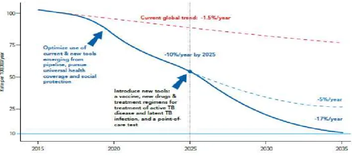
FIGURE 8.DESIRED DECLINE IN GLOBAL TB INCIDENCE RATES TO REACH THE 2035 TARGETS, FROM WHO:THE END TB STRATEGY. URL: HTTP://WWW.WHO.INT/TB/END_TB_BROCHURE.PDF [207] ... 42
FIGURE 9.LIST OF ON-GOING CLINICAL TRIALS, FROM WORKING GROUP ON NEW TB DRUGS [224] ... 47
FIGURE 10.EBA OVER 2 DAYS RELATED TO RIFAMPICIN DOSE SIZE, FROM DIACON A.H, ET AL.2007[241] ... 49
FIGURE 11.STUDY SCHEME ... 105
FIGURE 12.STUDY PROFILE ... 109
FIGURE 13.EFAVIRENZ AUC PER TREATMENT ARM ... 112
FIGURE 14.SHOWS INDIVIDUAL CMIN ON AND OFF R IN THE THREE TREATMENT ARMS. ... 113
FIGURE 15.ONSET OF GRADE 3 OR 4 ELEVATED TRANSAMINASES DURING FIRST 8 WEEKS BEFORE AND AFTER ART INITIATION PER TREATMENT ARM. ... 115
FIGURE 16.EVOLUTION IN MEDIAN ALT PER TREATMENT REGIMEN DURING STUDY INTERVENTION (FIRST 8 WEEKS) ... 115
List of Tables TABLE 1.MAIN TUBERCULOSIS DRUGS IN CLINICAL USE, THEIR YEAR OF DISCOVERY AND TARGETS, FROM ZUMLA A., ET AL.2013 [43]. ... 25
TABLE 2.RECOMMENDED DOSES OF FIRST-LINE ANTI-TUBERCULOSIS DRUGS FOR ADULTS, FROM WHO REPORT,2017[37] ... 26
TABLE 3.ADVERSE EFFECTS OF FIRST LINE ANTI-TUBERCULOSIS DRUGS, FROM ARBEX MA, ET AL.2010[52]. ... 27
TABLE 4.WHO RECOMMENDED FIRST-LINE ART REGIMENS FOR ADULTS, PREGNANT OR BREASTFEEDING WOMEN, ADOLESCENTS AND CHILDREN, FROM WHO REPORT,2016[34] ... 35
TABLE 5.ADVERSE EVENTS ATTRIBUTED TO ANTI-TUBERCULOSIS AND ANTIRETROVIRAL DRUGS, FROM ZUMLA A. ET AL.2015[119] 38 TABLE 6.SUMMARY OF THE DIFFERENT STUDIES CONDUCTED PER STUDY OBJECTIVE ... 54
TABLE 7.BASELINE PARTICIPANT’S CHARACTERISTICS (MITT POPULATION)... 110
TABLE 8.PHARMACOKINETIC PARAMETERS OF EFV ... 111
TABLE 9.GEOMETRIC MEAN RATIO (GMR) WEEK8/WEEK28 OF THE CMIN AND AUC. ... 111
TABLE 10.PATIENTS WITH AT LEAST ONE SERIOUS ADVERSE EVENT ... 114
TABLE 11.NON-HEPATIC GRADE 3 AND 4 ADVERSE EVENTS ... 116
TABLE 12.LEVEL OF REDUCTION IN HIV1RNA AT 4,12 AND 24 WEEKS AFTER ART INITIATION, ACROSS TREATMENT ARMS– MITT POPULATION AFTER EXCLUSION OF 3 PATIENTS WITH BASELINE RESISTANCE TO NNRTI ... 116
TABLE 13.NUMBER OF PATIENTS WITH POSITIVE MTB CULTURE AT WEEK 8 AND END OF TB TREATMENT OUTCOMES – MITT POPULATION ... 117
TABLE 14.MONTH-2 CULTURE CONVERSION RATES REPORTED WITHIN 2 HIGH R DOSE TRIALS, THAT IS, AMONG HIV NEGATIVE TB PATIENTS (RIFATOX TRIAL) AND HIV-TB CO-INFECTED PATIENTS (RIFAVIRENZ TRIAL) IN UGANDA (EXTRACTED FROM CHAPTERS 3.1 AND 3.3). ... 142
13
C
HAPTER
1
15
Tuberculosis (TB) is the most important infectious disease of all time, a global plague, with far reaching devastating effects to its victims, affecting the marginalized persons of lower socioeconomic status and with roots in both the history and present of humanity [1, 2]. The disease is not selective across age groups. In 1993, TB was declared a health emergency by the World Health Organization (WHO). It is one of the world's major infectious diseases with 2-3 billion people infected, 10.4 million new (incident) TB cases worldwide (56% and 34% in males and females respectively). It is most prevalent in poor countries and countries affected by the human immunodeficiency virus (HIV) pandemic, particularly in sub-Saharan Africa and Asia. In about 80% of cases, it manifests itself in a pulmonary form at the origin of its transmission. The disease is curable if treated with appropriate treatment and with optimal adherence. Unfortunately, TB management has two important obstacles: the lack of simple and effective point-of-care diagnostic tests and the length of TB treatment. A long treatment creates problems of adherence and promotes the emergence of the resistant forms of the disease. It is unlikely that new drugs will be available in the near future for shortening TB treatment for drug-susceptible TB so as to avert the poor treatment outcomes which include treatment failure, drug-resistance, drug toxicity, TB recurrence, mortality due to TB, and increases in TB-related costs at household and health-facility level among low and middle income countries with dual epidemic of TB and HIV.
Our work was conducted in Uganda, one of the TB-HIV high-burden countries [3], so as to evaluate the potential of using high rifampicin doses as a strategy towards shortening of drug-susceptible pulmonary TB treatment duration to lower than the current 6 months among adult TB and TB-HIV co-infected patients .
1.1 Microbiology, physiopathology, immunology, risk factors and clinical presentation of tuberculosis
1.1.1 Microbiology
Tuberculosis is caused by a mycobacterium belonging to the tuberculosis complex consisting of Mycobacterium (M) tuberculosis, M. bovis, M. africanum, M. canettii and M. pinipedii. M. tuberculosis or bacillus of Koch, named after the scientist who discovered it in 1882, is the principal agent responsible for human tuberculosis. M. bovis is responsible for about 1% of infections. M. africanum infection occurs mainly in West and Central Africa where it accounts for between 20% and 50% of cases of tuberculosis. Mycobacteria are aerobic bacilli, microaerophilic, acid-fast bacilli (AFB), having the general structure of gram positive bacilli and characterized by a thick outer parietal layer rich in mycolic acids. They are characterized by their culture requirement and the slow growth with an average split time of 20 hours, the cultures being positive only after one to several weeks of incubation at 37°C [4]. Secondary identification makes it possible to differentiate mycobacteria from the tuberculosis complex from other non-tuberculous mycobacteria. These atypical mycobacteria from the environment are usually non-pathogenic but can sometimes give clinical manifestations simulating those of TB in immunosuppressed patients or in patients with chronic bronchial conditions [5]. The sequence of the genome of M. tuberculosis has recently been identified. It is composed of approximately 4 000 genes and is characterized by the importance of the coding sequences dedicated
16
to the production of enzymes involved in the synthesis and degradation of lipids [6]. The deciphering of the genome has allowed the development of molecular diagnostic tests for TB and tests for the identification of mutations associated with anti-tuberculosis drug resistance.
The tuberculosis complex has as its essential reservoir the patients with TB, besides M. bovis whose reservoir is animal (domestic or wild bovids). The transmission is human-airborne and all the more important it is related to the density of the bacilli in the air breathed. Patients with "pulmonary caverns" that are very rich in bacilli (100 million bacilli for a cavern about 2 cm in diameter) are the most contagious.
1.1.2 Physiopathology and immunology
After inhalation, the bacilli enter the pulmonary alveolus and are phagocytosed by the alveolar macrophages within which they multiply. Other macrophages and monocytes are attracted, and thus participate in the process of defense against infection. The infectious focus thus constituted is the initial focus. The bacilli and the antigens they release are drained by the macrophages to the lymph node satellite. Within the ganglion, T lymphocytes identify M. tuberculosis (MTB) antigens and transform into specific T lymphocytes, resulting in the release of lymphokines and activation of macrophages that inhibit the growth of phagocytic bacilli. At the level of the initial focus an inflammatory tissue is formed, then fibrous scar tissue, in which the macrophages containing bacilli are isolated and die. This initial focus or "chancre inoculation" is then the site of a case-specific necrosis specific to TB. There are then between 1 000 and 10 000 bacilli which gradually lose their viability and have a very slow multiplication. Bacilli called "quiescent" can persist for several years [7]. The same evolution occurs in the lymph node, resulting in the formation of the caseous ganglion which evolves spontaneously, in the majority of cases,
towards the fibrous healing then the calcification.
It is on average 2 to 3 weeks after infection that the delayed hypersensitivity reaction to humoral mediation and cell-mediated immunity occur. Delayed cell-mediated hypersensitivity is conventionally evidenced by the intradermal injection of tuberculin (inactivated MTB). All these clinical and immunological phenomena observed after the contamination of a healthy subject constitute the primary TB infection. This phase is usually asymptomatic but may, in rare cases, be accompanied by clinical manifestations of hypersensitivity. This patient with non-symptomatic TB infection is considered to have Latent TB infection (LTBI) In 90% of cases (in HIV negative patients), there is no further development with the disappearance of lung lesions, except for the persistence of calcifications [8]. It confers on the infected subject a certain degree of immunity. It translates into tuberculin conversion. The intradermal reaction (IDR) to tuberculin becomes positive 6 to 12 weeks after the infecting contact. This tuberculin conversion is evidence of recent infection and reflects the resulting immunity [7]. The recent identification of genome regions encoding specific proteins of MTB (CFP-10 and EAST-6) has led to the development of novel diagnostic tests for TB infection. These tests are based on the measurement of the release of interferon gamma in the blood of patients after sensitization by these specific antigens [9, 10].
17
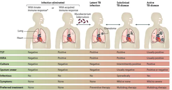
In 10% of the patients, the infection will evolve towards active TB disease, pulmonary or extra-pulmonary, or even disseminated after passage of the bacilli in the blood. Half of the active forms occur in the first year after primary infection. The other half may occur years after the primary infection following reactivation of the dormant or quiescent bacilli in the wake of a decline in immunity e.g following HIV infection, immunosuppressive treatments, malnutrition, elderly, etc) [8]. HIV infection is a major risk factor for progression to TB disease. The HIV-positive patient after primary infection has a 5-10% risk of developing active TB per year, compared with 0.2% in an HIV-negative patient [11]. After 5 years, untreated, pulmonary TB (PTB) spontaneously evolves towards healing in 20-25% of patients; It is fatal in 50-60% of cases and it progresses to a chronic active form in the remaining 20-25% [12]. Below is the figurative summary described by Pai M, et al, to demonstrate the TB spectrum from infection to disease [13] (Figure 1).
Figure 1. TB spectrum from Mycobacterium tuberculosis infection to active (pulmonary) TB disease, from: Pai M, et al. 2016 [13]
1.1.3 Risk-factors of tuberculosis
A review involving the 22 TB high-burden countries summarizes the common risk-factors of TB infection and disease[14]. This review notes that the progression from exposure to the TB bacilli to the development of active disease is a two-stage process governed by both exogenous and endogenous risk factors (see Figure 2). Exogenous factors play a key role in accentuating the progression from exposure to infection among which the bacillary load in the sputum and the proximity of an individual to an infectious TB case are key factors. Similarly endogenous factors lead in progression from infection to active TB disease. Along with well-established risk-factors (such as HIV, malnutrition, and young age), emerging variables such as diabetes, indoor air pollution, alcohol, use of immunosuppressive drugs, and tobacco smoke, play a significant role at both the individual and population level. Socioeconomic and behavioral factors are also shown to increase the susceptibility to infection. Specific groups
18
such as health care workers and indigenous population are also at an increased risk of TB infection and disease [14].
Figure 2. Risk-factors of TB infection and disease, from Narasimhan P, et al. 2013 [14]
1.1.4 Clinical features of tuberculosis
The clinical features of TB vary from classical to specific based on the part of the body affected. The disease can also be asymptomatic [15]. The classical clinical features associated with active pulmonary TB (commonest type of TB in 2/3 of patients) are: cough, hemoptysis, chest pain, and constitutional signs (weight loss/anorexia, fever, night sweats, and fatigue). Chest pain in patients with TB can also result from acute TB pericarditis. Pericardial TB can lead to cardiac tamponade or constriction. Elderly individuals with TB may not display typical signs and symptoms of active TB disease, because they may not mount a good immune response. Active TB disease in this age group may manifest as non-resolving pneumonitis. Extra-pulmonary TB symptoms and biological signs may be nonspecific or they can include leukocytosis, anemia, and hyponatremia due to the release of ADH (antidiuretic hormone)-like hormone from affected lung tissue. Patients with TB meningitis may present with a headache that has been either intermittent or persistent for 2-3 weeks. Subtle mental status changes may progress to coma over a period of days to weeks. Fever may be low grade or absent. In patients with skeletal TB, the most commonly affected skeletal site is the spine (Pott disease); symptoms include back-pain or stiffness. Lower-extremity paralysis occurs in up to half of patients with undiagnosed Pott disease. TB arthritis usually involve only one joint. Although any joint may be involved, the hips and knees are affected most commonly, followed by the ankle, elbow, wrist, and shoulder. Pain may precede radiographic changes by weeks to months. For genitourinary TB, symptoms may include flank pain, dysuria, and frequent urination. In men, genital TB may manifest as a painful scrotal mass, prostatitis, orchitis, or epididymitis. In women, genital TB may mimic pelvic inflammatory disease. TB is the cause of approximately 10% of sterility cases in women worldwide and of approximately 1% in industrialized countries [15].
19
Physical examination findings associated with TB also depend on the organs involved. Patients with pulmonary TB have abnormal breath sounds, especially over the upper lobes or involved areas. Rales or bronchial breath signs may be noted, indicating lung consolidation. Signs of extra-pulmonary TB differ according to the tissues involved. They may include the following: confusion, coma, neurologic deficit, chorioretinitis, lymphadenopathy, and cutaneous lesions. Lymphadenopathy in TB occurs as painless swelling of 1 or more lymph nodes. Lymphadenopathy is usually bilateral and typically involves the anterior and posterior cervical chain or supraclavicular nodes. The absence of any significant physical findings does not exclude active TB. Classical symptoms are often absent in high-risk patients, particularly those who are immunocompromised or elderly. Up to 20% of patients with active TB may be asymptomatic. Therefore, further investigations are essential when chest radiographic findings are suggestive of TB [15]. It is important to note that atypical presentation of TB occurs in HIV infected people.
1.2
Epidemiology of tuberculosis
1.2.1 Global TB burden
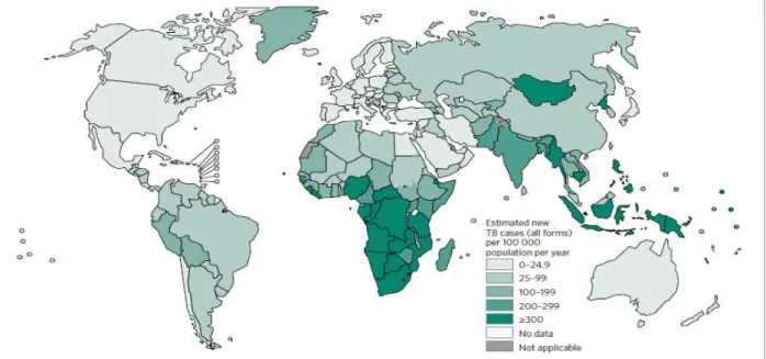
TB is one of the world's major infectious diseases with 2-3 billion infected people, 10.4 million new (incident) TB cases worldwide (56% and 34% in males and females respectively). Sub-Saharan Africa and Asia bear the largest incidence rates (See Figure 3). Six countries accounting for 60% of the new cases are: India, Indonesia, China, Nigeria, Pakistan and South Africa. Global progress depends on major advances in TB prevention and care in these countries[16]. In 2005, the World Health Organization (WHO) Regional Committee for Africa comprising health ministers from 46 Member States declared tuberculosis an emergency in the African region [17]. About 1.4 million TB deaths, and an additional 0.4 million deaths resulting from TB disease among people living with HIV have been reported (See Figure 4). Despite a fall in TB deaths by 22% between 2000 and 2015, TB has remained one of the top 10 causes of death worldwide. With the global rate of decline in TB incidence remaining at only 1.5% from 2014 to 2015, a rate far below the needed 4–5% annual decline by 2020 set to ensure attainment of the first milestones of the End TB Strategy [16], there is need for new advances in TB prevention and management [16].
20
Figure 3. Estimated incidence rates, 2015, from WHO TB report 2016.
http://www.who.int/tb/publications/global_report/en/ [16]
Figure 4. Global trends in the estimated number of deaths caused by TB and HIV (in millions), 2000-2015, from WHO TB report, 2016. http://www.who.int/tb/publications/global_report/en/ [16]
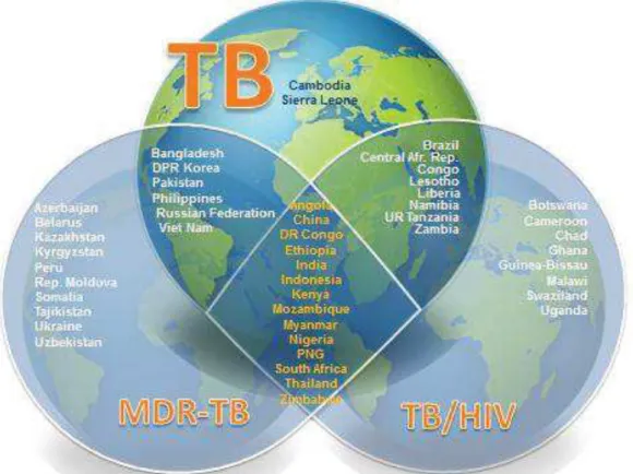
Since 2015, WHO has classified countries into 3 groups: 1) high-burden TB countries; 2) high-burden TB/HIV countries; 3) high-burden multi-drug resistant TB (MDR-TB) countries (See Figure 5).
21
Figure 5. The three high burden countries’ (HBC) lists (TB, MDR-TB and TB/HIV) of 30 countries each that will be used by WHO 2016–2020, from WHO, 2015.
http://www.who.int/tb/publications/global_report/high_tb_burdencountrylists2016-2020.pdf?ua=1 [3]
1.2.2 Burden of HIV-TB co-infection
HIV infection is still a global public health concern, especially in Africa and Asia. Since the beginning of the epidemic, more than 70 million people have been infected with the HIV virus and about 35 million people have
died [18]. Globally, 36.7 million [34.0–39.8 million] people were living with HIV at the end of 2015. The burden
of the epidemic continues to vary considerably between countries and regions with the Sub-Saharan Africa being
the most severely affected, with nearly 1 in every 25 adults (4.4%) living with HIV and accounting for nearly 70% of the people living with HIV worldwide [18].
The estimated risk of developing active TB in people living with HIV ranges between 26 and 31 times greater than in those without HIV infection [19]. Overall, people living with HIV account for 1.2 million (11%) of all new TB cases. The proportion of TB cases living with HIV is highest in the WHO African Region (31%), and exceeds 50% in parts of southern Africa [16] (See Figure 6).
22
Figure 6. Estimated HIV prevalence in new and relapse TB cases, 2015, from WHO TB report 2016.
http://www.who.int/tb/publications/global_report/en/ [16]
The overall TB mortality rates among HIV patients is still highest in the same regions with African region experiencing about 6 times higher mortality (30 per 100,000 population) as compared to the global average (5.3 per 100,000 population), with approximately 75% of all deaths occurring in Sub-Saharan Africa. In countries with a high prevalence of HIV infection, TB is a major cause of mortality and morbidity [20]. In addition to diagnosis and treatment of TB, early antiretroviral treatment (ART) of HIV infection has long been noted as the best protection against the risk of death from TB [21, 22]. Co-infection with HIV greatly increases mortality and risk of treatment failure [23]. These important co-morbidities clearly demonstrate that TB management needs an integrated approach and that every TB patient should be investigated for HIV, and every patient with HIV should be screened for active TB disease [24].
1.3
Tuberculosis diagnosis
Diagnosing active TB is the first step to being able to treat it and prevent transmission. In as much as diagnosis of TB should be “available free of charge to all persons with TB and populations at risk” [25], yet an estimated over four million people with TB went undiagnosed or unreported to national treatment programs in 2015, with India, Indonesia, and Nigeria accounting for almost half of this gap [16]. Even among those who do receive a TB diagnosis, often do so only after multiple health care visits and lengthy delays. Average delays of 28 to 30 days from when patients first contact a health care provider to diagnosis, even when patients present with overt TB symptoms have been reported [26]. Drug-susceptibility testing (DST) is not widely available and is used far too infrequently, even among those diagnosed with TB and living in countries with a high burden of drug-resistant TB (DR-TB). The end result is that 40% of people with TB do not receive a diagnosis or are not reported to national program, and DR-TB is detected in only 23% of people thought to have it [26].
23
Microscopy has been the commonly used method that detects the AFB although not specific as in to distinguish between live and dead bacilli, but also atypical mycobacteria from MTB. It is also simple, fast, and inexpensive. However, microscopy has low and variable sensitivity (20 to 60%) [27], especially among patients co-infected with HIV [28]. This low sensitivity is due to low level of detection. Nevertheless, it is widely implemented in limited resource countries.
On the other-hand, culture methods are the definitive diagnostic tests of TB, given their ability to distinguish between alive from dead bacilli. Its use is hindered by the long turn-around time. On average 3 weeks may be needed for mycobacterium growth to occur. This hinders its use as a point-of-care test. More so, the lack of appropriate laboratory infrastructure with good infection control mechanisms in many resource limited settings limit its use and left only in reference laboratories. Two types of culture media exist: 1) solid media e.g. Lowenstein-Jensen (LJ) and mycobacteria growth indicator tube (MGIT). Specifically, LJ tend to exhibit a slower growth and lower sensitivity as compared to MGIT [29, 30].
Molecular diagnostic methods can provide the data needed more rapidly, and in many cases it is more cost-effective than traditional culture methods [31, 32]. Nucleic acid amplification assays including polymerase chain reaction (PCR) have revolutionized the detection of MTB. It is a useful and sensitive tool for the early diagnosis of MTB in variety of clinical samples[33]. Xpert MTB/RIF is an automated PCR test (that is, a molecular test) utilizing the GeneXpert platform (Cepheid, Sunnyvale, CA, United States). Xpert MTB/RIF is a single test that can detect both MTB complex and rifampicin resistance within 2 hours after starting the assay, with minimal hands-on technical time. Given that all steps in the assay are automated and contained within its cartridge, that fact that it requires limited infrastructure and expertise to do, increases its access even in low and middle-income countries (LMICs). The fact that the assay’s sample reagent, used to liquefy sputum, is tuberculocidal, it largely eliminates concerns about biosafety during the test procedure and allow the technology to be decentralized for use nearer to patients[34].
In an effort to enhance early detection of TB and prompt initiation of appropriate treatment, there has been a switch in the recommendations to Xpert MTB/RIF use rather than conventional microscopy, culture and DST as the initial diagnostic test in adults and children suspected of having TB or MDR-TB [35]. This also applies in case of non-respiratory specimens like cerebrospinal fluid, lymph nodes and other tissues from patients suspected of having TB. However, microscopy still remain key for treatment monitoring given that it is readily available in remote settings and despite not being able to differentiate alive from dead bacilli, it can show reduction in bacillary load with treatment [36].
Chest X-ray (CXR) is used to diagnose intra-thoracic lesions due to TB. CXR has high sensitivity for PTB and thus is a valuable tool to diagnose PTB and allow also to identify differential diagnosis of TB. However, CXR has poor specificity; although some CXR abnormalities are rather specific for PTB (for example, cavities), many CXR abnormalities that are consistent with pulmonary TB are also common in several other lung pathologies. Moreover, there is significant intra- and inter-observer variation in the reading of CXRs. Relying only on CXR for
24
TB diagnosis leads to over-diagnosis, as well as under-diagnosis [37, 38]. Rigorous efforts should always be made to base a TB diagnosis on bacteriological confirmation. WHO classifies TB diagnosis into bacteriologically confirmed TB, if it is based on bacteriological confirmation, or clinically diagnosed TB, if it is based on clinical assessment including CXR, only , the latter receiving an empirical TB treatment [37]. However, it should be noted that the low access to X-ray in limited resource countries, issues of poor quality and the fact that its costs are often not covered by National TB programs (NTP) limits its use among patients that would need it. In 2016, the WHO issued a summary of its existing recommendations on CXR as a screening tool for TB disease, indicating its sensitivity, its importance for diagnosing childhood TB, its additive value with Xpert MTB/RIF, its use in diagnosing TB in people with HIV, and its role in ruling out active TB before treating LTBI [37].
It is noteworthy that the lack of antibody or antigen sputum-based rapid test or non-sputum based point of care test is a limitation in the effort to diagnose TB, especially in patients with advanced HIV infection and children. This leaves many patients being started on empirical TB treatment.
However, recently, the WHO endorsed use of urine lipoarabinomannan (LAM) test for active TB screening and diagnosis among HIV/AIDS patients with advance disease or with severe immune-suppression. LAM antigen is a lipopolysaccharide present in mycobacterial cell walls, which is released from metabolically active or degenerating bacterial cells and appears to be present only in people with active TB disease. Urine-based testing would have advantages over sputum-based testing because urine is easy to collect and store, and lacks the infection control risks associated with sputum collection. However, it suffers poor sensitivity[39].
1.4
Treatment of Tuberculosis
The aims of TB treatment are: to cure the patient and restore quality of life and productivity; to prevent death from active TB or its late effects; to prevent relapse of TB; to reduce transmission of TB to others; and to prevent the development and transmission of drug resistance [40]. Anti-TB treatment (ATT) is a combination of antibiotics, to avoid the risk of selection of resistant strains of MTB. It is of long duration so as to increase the chances of sterilization of quiescent bacilli and reduce the risk of TB recurrence. Given the long waiting times for the results of DST and their low access in countries with a high TB prevalence, TB treatment has for long been an empirical standardized treatment [41].
1.4.1 History of tuberculosis drug development
The first antibiotic to be discovered with proven activity against MTB was streptomycin. It is an antibiotic purified from Streptomyces griseus, discovered almost 70 years ago, thus providing the first hope of a TB-specific therapy [42, 43]. Nevertheless, uncertainties remained with regard to its ability to consistently cure patients, and this was closely followed by the realization that drug resistance develops rapidly when a single agent is used for the treatment of
25
TB. Following the launch of the Medical Research Council (MRC) TB unit in the United Kingdom in 1946, the unit conducted the first recorded randomized, controlled clinical trial designed to compare streptomycin plus bed rest versus bed rest alone [44]. The study showed that streptomycin plus bed rest achieved greater clinical improvement but only modest pathological improvement (as assessed by CXR) in comparison to bed rest alone. Importantly, improvement in TB was greatest in the first 3 months of therapy, after which many patients began to deteriorate due in part to the emergence of streptomycin resistance. In the 1950s, several other TB drugs with different mechanisms of action were discovered and developed (see Table 1), including para-amino salicylic acid, isoniazid (H), pyrazinamide (Z), cycloserine and kanamycin. This paved the way for combination therapy that, at that time, had a treatment duration of 18 months or more. In collaboration with the United States Public Health Service (USPHS) the MRC TB unit spent the next four decades developing the current short-course therapeutic regimen comprising H, rifampicin (R), Z and ethambutol (E). The introduction of R into clinical practice in the 1960s was a major breakthrough that allowed treatment duration to be shortened to 9 months, and then 6 months especially when used in a regimen that also contained Z. Through these developments, the USPHS (now known as the Center for Disease Control TB Trials Consortium) and the MRC TB unit firmly established the concept of the randomized, controlled clinical trial as the gold-standard methodology for the assessment of new drugs [45].
Table 1. Main tuberculosis drugs in Clinical use, their year of discovery and targets, from Zumla A., et al. 2013 [46].
1.4.2 Treatment of drug-susceptible TB
Management of a patient diagnosed with active TB depends on whether or not the patient has already received anti-TB drugs in the past. Thus, the newly diagnosed drug-susceptible TB patient receives a six-month regimen that breaks down into an intensive two-month phase involving H, R, Z and E followed by four months of continuation phase with R and H with doses based on body weight [40]. (See Table2).
26
Table 2. Recommended doses of first-line anti-tuberculosis drugs for adults, from WHO report, 2017 [40] Drug Daily recommended dose
Dose and range (mg/kg body weight) Maximum (mg)
H 5 (4-6) 300
R 10 (8-12) 600
Z 25 (20-30) --
E 15 (15-20) --
In patients who require retreatment due to treatment interruption or recurrence of disease, the current recommendation is that DST should be conducted first so as to inform the choice of treatment regimen [47]. Both R and H are the back-bone drugs within the standard 6-month WHO recommended first-line TB treatment regimen [47, 48]. It is important to note that the potency of this regimen is attributed mainly to rifampicin’s strong bactericidal activity [49, 50] that is also based on its ability to inhibit transcription by binding with high affinity to bacterial DNA-dependent RNA polymerase [51-53]. The 6-month regimen (2HRZE/4RH) has a good efficacy with a TB recurrence rate of 5% [50, 54]. This regimen is recommended for use as a fixed dose combination (FDC), and administered once daily for both TB only and TB-HIV co-infected patients [47].
1.4.2.1 General Safety description of first line anti-tuberculosis drugs
A number of adverse effects have been reported in respect to each of the first line anti-TB drugs. These are summarized in Table 3. Pyridoxine is administered throughout the duration of TB treatment to prevent peripheral neuropathy due to H. In programmatic conditions, it is not necessary to systematically monitor liver function test or other laboratory tests for safety purposes.
27
Table 3. Adverse effects of first line anti-tuberculosis drugs, from Arbex MA, et al. 2010 [55].
Drug Adverse effects
Isoniazid Minor: Nausea, vomiting, and epigastric pain. Transitory and asymptomatic increase in
hepatic enzyme levels, Arthralgia, Headache, insomnia, euphoria, agitation, anxiety, Somnolence, Acne, Cutaneous pruritus or fever
Major: Psychosis, convulsive seizures, mental, confusion, coma, attempted suicides, hematological alterations or vasculitis, peripheral neuropathy, Clinical hepatitis and Lupus-like syndrome
Rifampicin Minor: Gastrointestinal reactions: Nausea, anorexia, and abdominal pain. Orange-colored tears, sweat, and urine. Skin reaction: Pruritus, with or without Erythema. Flu-like syndrome, fatigue, dizziness, headache, dyspnea, and ataxia
Major: Exanthema, hepatotoxicity, immunological reactions like Thrombocytopenia, leukopenia, eosinophilia, hemolytic anemia, agranulocytosis, vasculitis, acute interstitial nephritis, and septic shock. Pyrazinamide Minor: Gastrointestinal symptoms: Nausea, Vomiting, and anorexia. Hyperuricemia and arthralgia in
non-gouty individuals. Exanthema, pruritus and dermatitis.
Major: Severe exanthema, pruritus, Rhabdomyolysis with myoglobinuria and kidney failure. Acute arthritis in gouty individuals and hepatotoxicity
Ethambutol Retro-bulbar neuritis with symptoms of blurred vision, decrease in visual acuity, presence of scotomas, and loss of the ability to discern the color green and, in some cases, the color red. Peripheral fiber impairment which manifests as a reduction in the visual field. Peripheral neuritis.
Gastrointestinal symptoms (nausea, vomiting, abdominal pain, and hepatotoxicity), hematological symptoms (eosinophilia, neutropenia, and thrombocytopenia), cardiovascular symptoms (myocarditis and pericarditis),
neurological symptoms (headache, dizziness, and mental confusion),
hyperuricemia/gout (due to a reduction in the excretion of uric acid by the kidney), hypersensitivity (skin rash, arthralgia, and fever), and (occasionally) pulmonary Infiltrates.
1.4.2.2 Anti-tuberculosis drug induced liver injury
Drug-induced liver injury (DILI) is an important consideration during administration of ATT. DILI is ultimately a clinical diagnosis of exclusion given that histologic specimens of the liver are often not obtained. Its diagnosis is made plausible after the exclusion of other causes of liver injury, such as acute viral hepatitis[56]. Detection of DILI is based on measurement of hepatic enzymes [56]. More specifically, an increase in ALT, is more specific for hepatocellular injury than an increase in aspartate aminotransferase (AST) or serum glutamic oxaloacetic transaminase [SGOT]), which can also signify abnormalities in muscle, heart, or kidney [57, 58]. Increases in alkaline phosphatase and/or bilirubin with little or no increase in ALT indicate cholestasis. Jaundice is usually detectable on the physical examination when serum bilirubin exceeds 3.0 mg/dl [56]. Overall, the strongest confirmation of the DILI is based on: a re-challenge with the suspected offending agent in cases with more than two-fold serum alanine aminotransferase (ALT) elevation, and discontinuation leading to a fall in ALT [59]. DILI may result from direct toxicity of the primary compound, a metabolite, or from an immunologically mediated response, affecting hepatocytes, biliary epithelial cells, and/or liver vasculature[56]. There are two types of DILI that have been reported, that is: 1) Predictable DILI: characterized by certain dose-related injury in experimental animal models, has a higher attack rate, and tends to occur rapidly. Injurious free radicals cause hepatocyte necrosis in zones farthest from the hepatic arterioles, where metabolism is greatest and antioxidant detoxifying capacity is
28
the least [60, 61]. 2) Unpredictable or idiosyncratic reactions: comprise most types of DILI and these hypersensitivity or metabolic reactions occur largely independent of dose and relatively rarely for each drug, and may result in hepatocellular injury and/or cholestasis. Hepatocyte necrosis is often distributed throughout hepatic lobules rather than being zonal, as is often seen with predictable DILI [62]. Metabolic idiosyncratic reactions may result from genetic or acquired variations in drug biotransformation pathways, with synthesis or abnormally slow detoxification of a hepatotoxic metabolite. Metabolic idiosyncratic reactions may have a widely variable latent period, but recur within days to weeks after re-exposure [63].
DILI may occur with all currently recommended regimens for the treatment of TB infection or disease [64]. This can occur with R, Z and H [65-67]. Metabolic idiosyncratic reactions appear to be responsible for most DILI from the ATT [56]. The onset of DILI bears some differences depending on the type of TB drug responsible.
Rifampicin-related DILI
R has been reported to occasionally cause dose-dependent interference with bilirubin uptake, resulting in subclinical, unconjugated hyperbilirubinemia or jaundice without hepatocellular damage. This may be transient and occur early in treatment or in some individuals with preexisting liver disease [68-71]. R occasionally can cause hepatocellular injury and potentiate hepatotoxicities of other anti-TB medications [72, 73]. The conjugated hyperbilirubinemia occasionally reported during treatment of TB using R-based combinations, is probably caused by R inhibiting the major bile salt exporter pump [74]. Asymptomatic elevated bilirubin may also result from dose-dependent competition with bilirubin for clearance at the sinusoidal membrane or from impeded secretion at the canalicular level [63, 68, 69]. Rare hepatocellular injury appears to be a hypersensitivity reaction, and it may be more common with large, intermittent doses [68]. Hypersensitivity reactions have been reported in combination with renal dysfunction, hemolytic anemia, or “flulike syndrome” [75, 76]. R-induced hepatotoxicity is characterized by cholestasis which may be insidious. Idiosyncratic hypersensitivity reaction to R, manifest as anorexia, nausea, vomiting, malaise, fever, mildly elevated ALT, and elevated bilirubin, usually occurs in the first month of treatment initiation [68, 76-78].
Isoniazid-related DILI
Two potential explanations for onset of isoniazid DILI have been raised. The first one is based on acetylation rate, a claim that is still controversial. . In contrast to early studies [79-81], slow acetylators may actually have greater cumulative mono-acetyl hydrazine (MAH) exposure, which could be further metabolized to other toxic intermediaries especially reactive metabolites[45]. Slow acetylators also had higher peak ALT than did fast acetylators and, when rechallenged with H, more frequently developed transaminase elevation of at least three times the upper limit of normal (ULN) [79]. Secondly, metabolic idiosyncratic mechanisms have been implicated in H-induced hepatotoxicity. The H metabolite acetyl-hydrazine covalently binds to liver macromolecules, a process mediated by microsomal enzymes [80]. Patients with homozygous cytochrome P450 2E1 c1/c1 host gene polymorphism, who have enhanced cytochrome P450 2E1 activity, in one study had a higher risk of
29
hepatotoxicity, particularly in slow acetylators [82]. R appears to enhance a metabolic hepatocellular idiosyncratic reaction in patients receiving H, perhaps by promoting the formation of toxic H metabolites [83, 84].
At presentation, some individuals may be asymptomatic, whereas others may experience symptomatic hepatotoxicity at varying serum transaminase concentrations. Constitutional symptoms may be seen early in severe hepatotoxicity, and may last from days to weeks. Nausea, vomiting, and abdominal pain are seen in 50 to 75% of patients with severe illness, whereas fever is noted in 10% and rash in 5% of patients. Overt jaundice, dark urine, and clay-colored stools are late signs of clinical worsening. Coagulopathy, hypoalbuminemia, and hypoglycemia signify life-threatening hepatic dysfunction. The regression of H hepatotoxicity usually takes weeks. Recovery is complete in most after discontinuation of H [85].
H induced hepatotoxicity occurs generally within weeks to months rather than the days to weeks of onset seen with hypersensitivity reactions [85, 86]. Unlike a classical hypersensitivity reaction, H re-challenge does not always elicit rapid recurrence of hepatotoxicity [85]. Approximately 60% of the hepatotoxicity incidence is reported to occur in the first 3 months of treatment, and 80% of the incidence in the first 6 months [87-89]. A retrospective case fatality review found that the median interval from treatment initiation to symptom onset was 16 weeks [90]. Most H-associated hepatotoxicity is age associated, with symptomatic transaminase elevation: ranging from 0% in <14 years to 0.28% in those >65 years in the Seattle study [91]; 0.44% in <35years to 2.08% in >49 years having AST elevation more than five times the ULN in the Tennessee study, a difference that was statistically significant [89]. However, the differences in the findings among these studies have been attributed to differing definitions of hepatotoxicity, patient selection, and inability to exclude confounding causes of hepatotoxicity [56]. The severity of H-related hepatitis has been reported to also increase with age, with higher mortality in those older than 50 years [85, 90, 92].
Combined Rifampicin and Isoniazid related DILI
The rate of symptomatic hepatitis with the combination of R and H has been estimated at 2.55% in a meta-analysis that included patients with TB disease, a higher incidence than in regimens containing one or the other drug [93]. It is thought that the observed elevation in transaminases may partly or fully be due to H, a phenomenon that is supported by studies reporting transaminase elevations above the upper-limit of normal ranges being seen more commonly in people with LTBI on H alone as compared to those on R alone [73], or even the absence of significant transaminase elevations even with intake of R alone for 4-months [72]. Secondly, that perhaps the observed ALT elevations may actually be equivalent to what we would normally expect even without any treatment administration. This is supported by results of a study that showed a non-significant difference in geometric mean of serum ALT between the placebo and R groups [94].
Pyrazinamide-related DILI
Transaminase elevation more than four times the ULN were reported with administration of Z and either E (58%) or levofloxacin (18%) or ofloxacin (41%) for treatment of LTBI after exposure to MDR TB [65, 66]. The
half-30
life (t1/2) of Z is notably longer than that of either H or R, approximately 10 hours [82]. In patients with preexisting hepatic disease, t1/2 is increased to 15 hours (101). Z, a nicotinic acid derivative, is de-amidated to pyrazinoic acid in the liver and subsequently metabolized to 5-hydroxy-pyrazinoic acid by xanthine oxidase [95], aldehyde oxidase [96], and xanthine dehydrogenase [97, 98]. In addition, 5-hydroxy-pyrazinamide may be generated during metabolism [99]. The kidneys clear metabolites of Z, hence requiring intermittent dosing in patients with renal insufficiency [100]. Z may exhibit both dose-dependent and idiosyncratic hepatotoxicity. Historically, daily use of Z at 40 to 50 mg/kg commonly resulted in hepatotoxicity, and a relationship to dose was noted [101]. It is thought that there might be shared mechanisms of injury for H and Z, because there is some similarity in molecular structure. This is implied in the evidence that patients who previously had hepatotoxic reactions with H have had more severe reactions with R and Z given for LTBI [102]. Z may induce hypersensitivity reactions with eosinophilia and liver injury [102] or granulomatous hepatitis [103].
There has been few reports of ethambutol-related liver cholestatic jaundice, with unclear circumstances [104]. Otherwise, E has not been linked to liver injury.
Factors associated with onset of DILI during TB treatment
An increased risk of hepatotoxicity has been associated with: being woman [105-108] though contradicted in other studies [87, 109]; alcohol use [87, 110, 111], contrary to other two studies [106, 109]; having abnormal baseline transaminases [107]; NAT2 slow acetylators as determined by phenotypic assays [83, 110, 112] or by genotypic assays [79, 113]; malnutrition or hypoalbuminemia [110, 114-116] ; presence of HLA-DQB1*0201 [116]; gene polymorphisms at loci of genes coding for cytochrome P450 2E1 and for glutathione S-transferase [79, 117]; higher EFV concentrations [113, 118]; CYP2B6*6 genotype [113, 118]; extensive TB disease [105, 110]; and patients under-going liver transplant [119]. In a meta-analysis, the presence of R in a multidrug treatment regimen increased the incidence of significant hepatotoxicity for adults from 1.6 to 2.55% and in children from 1.0 to 6.9% [93]. Z is considered as a contributor to increased incidence or severity of hepatotoxicity [107, 109, 120, 121], although some studies could not demonstrate this link [122-125]. The role of hepatitis B infection on incidence of TB DILI has been demonstrated in some studies [126, 127] but not on others [110]. Hepatitis B virus carriage has been linked to incidence of more severe hepatic disease from treatment-associated liver injury [126, 128]. Additional studies are needed, but the limited data leave sufficient concern that hepatitis B may be a risk factor for more frequent or severe hepatotoxicity during treatment of TB disease[45]. The role of Hepatitis C Virus infection on DILI during treatment for TB disease was shown in a study conducted in Florida, in which patients received at least 5 days of H, R or rifabutin, or Z, and had not received alcohol or drugs of abuse for at least 10 days before starting anti-TB therapy. Hepatotoxicity was reported in 30% versus 11% of patients with and without hepatitis C–infection respectively. Hepatitis C was independently associated with fivefold risk of transaminase elevation of at least 120 U/L, or of serum bilirubin of at least 1.5 mg/dl. Co-infection with both hepatitis C and HIV elevated the risk of hepatotoxicity more than 14-fold [129].
31
1.4.2.3 Treatment outcomes of drug susceptible tuberculosis
Assessment of therapeutic response is based on systematic microscopic examination of sputum at the end of the intensive phase (two months), at five months and at the end of treatment (6 months)[130]. A patient is considered cured if he has received full treatment and is smear-negative twice between the end of the intensive phase and the end of treatment. One is considered to have treatment failure if smear-positive at five months or later [41]. Needless to say, microscopy is poorly adapted to this evaluation of therapeutic response given its inability to differentiate live from a dead bacillus. Indeed, smear-positive persistence is not uncommon after five months of treatment in patients with large pulmonary cavities [131]. Although accessibility to Xpert MTB/RIF is increasing, it is also not adequate in differentiating dead from live bacilli and therefore not recommended for treatment monitoring. The only test to assess response is culture but it is poorly available and with long delays that make difficult treatment decision.
There is no effective early surrogate markers of TB treatment efficacy yet. Culture conversion at week 8 though used commonly in TB chemotherapeutic trials, has been reported to be a poor surrogate marker for treatment efficacy [132-134] and therefore create a challenge in precise estimation of new anti-tuberculosis drug or regimen efficacy. The delayed culture conversion beyond week 8 in East African populations [135, 136], has challenged the use of week-8 culture conversion rate, with researchers suggesting the use of months-3 instead of month-2 culture conversion endpoint as a surrogate of treatment efficacy within the phase II East African trials [133]. Because of the weakness of month-2 culture conversion, phase 3 confirmatory trials still rely on relapse free cure as the primary endpoint for efficacy which implies very long post treatment follow-up for a minimum of 12 months, which is time consuming and costly, something that could explain why there are so few randomized clinical trials (RCT) in TB.
1.4.2.4 Adherence to TB treatment
The effectiveness of the 6-month TB treatment regimen is based on attaining good patient adherence. This is also key in prevention of onset of rifampicin resistance [137, 138]. Directly Observed Treatment (DOT) and the use of FDC, is strongly promoted by WHO to enhance adherence [41]. Treatment administration approaches currently being recommended by the WHO are: a) Community- or home-based DOT is recommended over health facility-based DOT or unsupervised treatment; b) DOT administered by trained lay providers or health-care workers is recommended over DOT administered by family members or unsupervised treatment; c) Video observed treatment (VOT) can replace DOT when the video communication technology is available and can be appropriately organized and operated by health-care providers and patients [47].
In addition to DOT, patients are entitled to effective care and support interventions, which are: a) Health education and counselling on the disease and treatment adherence; b) A package of treatment adherence intervention such as social support (e.g. food, financial incentive, and transport fee); psychological support; home
32
visit or digital health communication (e.g. SMS, telephone call); medication monitor. The interventions should be selected on the basis of the assessment of individual patient's needs, provider's resources and conditions for implementation; c) Support in selection of a suitable treatment administration option, which include DOT, VOT, non-daily DOT (e.g. not every dose supervised treatment, weekly or a few times per week supervision), or unsupervised treatment. All of this is good in theory but represents a significant cost for the TB programs, which explains why it is often poorly implemented [47].
1.4.3 Drug-resistant tuberculosis
1.4.3.1 Burden of drug-resistant tuberculosis
Development of resistance against any of the anti-tuberculosis drugs arises by bacterial chromosomal mutations[29]. While these mutations are rare events, a mutation early in multiplication produces a clone of resistant bacilli that are found more frequently. The mutation rates were thus found to be about 2.6 × 10⁻⁸ for H
and 2.2 × 10⁻¹⁰ for R, while the more useful estimates of the highest proportion of mutants that can be expected
in an unselected bacterial population were found to be 3.5 × 10⁻⁶ for H and 3.3 × 10⁻⁸ for R[139]. A recent
calculation has made these proportions somewhat greater [140]. During the initial phase of treatment, when bacterial populations are large, treatment is almost always with Z, known to prevent resistance emerging [141], and sometimes E as well as R and H. The origins of drug resistance appear to be due to 1) irregularity in drug taking by mechanisms described elsewhere [142]; while attempts to simulate the emergence of resistance by irregular drug taking have failed [143], resistance does emerge, although rarely, in relapse cultures [144, 145]; 2) inadequate dosage, particularly of R [146], leading to a slow response and eventual resistance; and 3) the prescription of single-drug treatment for financial reasons by private practitioners, a common but regrettable practice in some countries[147]. Resistant strains therefore have their own epidemiology, and are capable of creating disastrous epidemics, with treatment that is expensive and of low efficacy.
1.4.3.2 Multi and extensive-drug resistant tuberculosis
Multi-drug resistant TB (MDR-TB) is defined as resistance to rifampicin and isoniazid, the two most effective anti-TB drugs. In 2015, WHO estimated 480 000 new cases of MDR-TB and an additional 100 000 people with R-resistant TB (RR-TB) who were also newly eligible for MDR-TB treatment. India, China and the Russian Federation accounted for 45% of the combined total of 580 000 cases. There were about 250 000 (range, 160 000–340 000) deaths from TB in 2015[16]. In May 2016, WHO issued guidance [148] that people with RR-TB should be treated with second line drugs.
There is evidence on the growing resistance against second line anti-TB drugs and Z. By the end of 2015, extensively drug-resistant TB (XDR-TB) had been reported by 117 WHO Member States. XDR-TB is defined as
33
MDR-TB plus resistance to at least one fluoroquinolone and a second-line injectable agent. The average proportion of MDR-TB cases with XDR-TB was 9.5% (95% CI: 7.0–12.1%), similar to estimates for previous years (9.7% in 2014 and 9.0% in 2013) [16]. The proportion of MDR-TB cases with resistance to any fluoroquinolone for which testing was done – including ofloxacin, levofloxacin and moxifloxacin – was 21.0% (95% CI: 8.8–33.3%). A total of 51% (30–70%) of patients with MDR-TB have resistance to a fluoroquinolone or a second-line injectable agent, or both [16]. In a recent multi-country project assessing the level of resistance to both Z and fluoroquinolone in five countries – Azerbaijan, Bangladesh, Belarus, Pakistan and South Africa and coordinated by WHO, substantial variations in levels of resistance were noted among the different settings (3.1–42.1%) [149]. Resistance to Z was reported to be significantly associated with R resistance (0.5–4.2% among R-susceptible cases and 36.7–81.3% among R-resistant cases). Resistance ranged from 1.0% to 16.6% for ofloxacin, from 0.5% to 12.4% for levofloxacin and from 0.9% to 14.6% for moxifloxacin when tested at 0.5 μg/ml. The study recommended that the presence of R resistance, which currently is easily identified because of the wide availability of new rapid molecular technology, should prompt attention to the possibility of the simultaneous presence of resistance to Z and, in some settings, the earlier generation fluoroquinolones. Resistance to the latest generation fluoroquinolones at the clinical breakpoint is still uncommon, a finding that supports current WHO recommendations to use moxifloxacin or gatifloxacin in the treatment of MDR-TB [149].
1.4.3.3 Treatment of drug-resistant tuberculosis
The currently recommended medicines for RR-TB and MDR-TB treatment are categorized into groups A-D, as summarized below. Group A. Fluoroquinolones (Levofloxacin, Moxifloxacin, Gatifloxacin); Group B. Second-line injectable agents (Amikacin, Capreomycin, Kanamycin, Streptomycin); Group C. Other core second-Second-line agents (Ethionamide / prothionamide, Cycloserine / terizidone, Linezolid, Clofazimine); Group D. Add-on agents and not part of the core MDR-TB regimen (D1:Z, E, high-dose H; D2:Bedaquiline, Delamanid; D3: p-aminosalicylic acid, Imipenem–cilastatin, Meropenem, Amoxicillin-clavulanated, (Thioacetazone))[150]. WHO recommends both shorter and longer MDR-TB regimens based on different circumstances[150]. Most of these drugs are repurposed or old TB drugs abandoned before due to toxicity..
The longer MDR-TB regimens of 18-24 months apply to both adults and children and covers all forms of RR-TB, that is those who are H susceptible, MDR-RR-TB, resistant to other medicines from firstline group (poly-resistant). In patients with RR-TB or MDR-TB, a regimen with at least five effective TB medicines during the intensive phase is recommended, including Z and four core secondline TB medicines – one chosen from Group A, one from Group B, and at least two from Group C6 (conditional recommendation, very low certainty in the evidence). If the minimum number of effective TB medicines cannot be composed as given above, an agent from Group D2 and other agents from Group D3 may be added to bring the total to five. In patients with RR-TB or MDR-TB, it is recommended that the regimen be further strengthened with high-dose H and/or E (conditional recommendation, very low certainty in the evidence). It is recommended that any patient – child or adult – with
34
RR-TB in whom H resistance is absent or unknown be treated with a recommended MDR-TB regimen. It could either be a shorter MDR-TB regimen, or a longer MDR-TB regimen to which isoniazid is added [150].
A shorter MDR-TB regimen of 9–12 months may be used instead of the longer regimens in adults and children with RR-TB or MDR-TB who were not previously treated with second-line drugs and in whom resistance to fluoroquinolones and second-line injectable agents was excluded or is considered highly unlikely. The shorter MDR-TB treatment regimens are standardized in content and duration and split into two distinct parts. The first is an intensive phase of four months (extended up to a maximum of six months in case of lack of sputum smear conversion) and included the following drugs: gatifloxacin (or moxifloxacin), kanamycin, prothionamide, clofazimine, high-dose H, Z, and E. This is followed by a continuation phase of five months with the following medicines: gatifloxacin (or moxifloxacin), clofazimine, pyrazinamide and ethambutol (with or without prothionamide kept in the continuation phase in earlier studies).
Bedaquilline and delamanid were approved by FDA after phase 2 trials, and have received a conditional recommendation by WHO for use in the treatment of adults with drug-resistant TB in 2013 and 2014 respectively [151, 152].
Among patients with H resistance or populations with known or suspected high levels of H mono-resistance, administration of 7RZE or 9RE as therapy in the continuation phase is an alternative to HR to prevent relapses [47].
Surgery has also been reported to have a role especially among patients with lung cavities and tuberculomas. Its need is demonstrated in the study from Tbilisi Georgia, that described pulmonary surgery in 137 patients with half having MDR/XDR TB. The study recommended the revisiting of the need for surgery especially in patients with extensive pulmonary lesions even those with bacterial cure or patients where bacterial cure is not possible [153].
1.5
Treatment of TB-HIV co-infection
1.5.1 Anti-retroviral treatment
Since 1987, more than 25 antiretroviral (ARV) drugs in 6 mechanistic classes for treatment of HIV infection have been approved by Food and Drug Administration (FDA) [154]. These 6 classes include the nucleoside/nucleotide reverse transcriptase inhibitors (NRTIs), non-nucleoside reverse transcriptase inhibitors (NNRTIs), protease inhibitors (PIs), a fusion inhibitor (FI), a CCR5 antagonist, and integrase strand transfer inhibitors (INSTIs). In addition, two drugs, ritonavir (r) and cobicistat (COBI or c), potent CYP3A inhibitors, are used solely as pharmacokinetic (PK) enhancers (i.e. boosters) to improve the PK profiles of some ARV drugs highly metabolized by CYP3A (e.g. PIs and the INSTI elvitegravir [EVG])[155]. The two commonly used NNRTIs as part of the treatment of HIV patients in low-resource settings, that is efavirenz and nevirapine, received FDA approval in
![Figure 2. Risk-factors of TB infection and disease, from Narasimhan P, et al. 2013 [14]](https://thumb-eu.123doks.com/thumbv2/123doknet/13606991.424476/19.892.55.664.132.471/figure-risk-factors-tb-infection-disease-narasimhan-p.webp)


![Figure 7. Schematic representation showing the different forms of tuberculosis-associated IRIS and ART-associated tuberculosis, from Meintjes G., et al.2008 [205]](https://thumb-eu.123doks.com/thumbv2/123doknet/13606991.424476/41.892.61.770.67.277/schematic-representation-different-tuberculosis-associated-associated-tuberculosis-meintjes.webp)
![Figure 10. EBA over 2 days related to rifampicin dose size, from Diacon A. H, et al.2007 [246]](https://thumb-eu.123doks.com/thumbv2/123doknet/13606991.424476/50.892.55.698.375.671/figure-eba-days-related-rifampicin-dose-size-diacon.webp)
