HAL Id: hal-02931857
https://hal.archives-ouvertes.fr/hal-02931857
Submitted on 17 Dec 2020HAL is a multi-disciplinary open access archive for the deposit and dissemination of sci-entific research documents, whether they are pub-lished or not. The documents may come from teaching and research institutions in France or abroad, or from public or private research centers.
L’archive ouverte pluridisciplinaire HAL, est destinée au dépôt et à la diffusion de documents scientifiques de niveau recherche, publiés ou non, émanant des établissements d’enseignement et de recherche français ou étrangers, des laboratoires publics ou privés.
Dynamic control of the high-affinity iron uptake
complex in root epidermal cells
Amanda Martín Barranco, Julien Spielmann, Guillaume Dubeaux, Grégory
Vert, Enric Zelazny
To cite this version:
Amanda Martín Barranco, Julien Spielmann, Guillaume Dubeaux, Grégory Vert, Enric Zelazny. Dy-namic control of the high-affinity iron uptake complex in root epidermal cells. Plant Physiology, American Society of Plant Biologists, 2020, 184, pp.1236 - 1250. �10.1104/pp.20.00234�. �hal-02931857�
Short title: Arabidopsis iron uptake complex 1
2
Dynamic control of the high-affinity iron uptake complex in root epidermal
3cells
45
Amanda Martín-Barranco1, Julien Spielmann2, Guillaume Dubeaux3, Grégory Vert2, and 6
Enric Zelazny1,4 7
8 1
Institute for Integrative Biology of the Cell (I2BC), UMR9198 CNRS/CEA/Univ. Paris Sud, 9
Université Paris-Saclay, 91198 Gif-sur-Yvette, France. 10
2
Plant Science Research Laboratory (LRSV), UMR5546 CNRS/University of Toulouse 3, 11
31320 Auzeville Tolosane, France. 12
3
Division of Biological Sciences, Cell and Developmental Biology Section, University of 13
California San Diego, La Jolla, CA 92093, USA. 14
4
Current address: BPMP, CNRS, INRAE, Montpellier SupAgro, Univ Montpellier, 2 Place 15
Viala, 34060 Montpellier Cedex 2, France. 16
17
Corresponding authors: [email protected] ; [email protected] 18
19 20
The authors responsible for distribution of materials integral to the findings presented in this 21
article in accordance with the policy described in the Instructions for Authors 22
(www.plantphysiol.org) are: Enric Zelazny ([email protected]) and Grégory Vert 23
([email protected]). 24
25
Author contributions: A.M.B., G.V. and E.Z. designed the research; A.M.B. J.S. and E.Z. 26
performed the experiments; G.D. performed the quantification of co-localization and 27
statistical analyses; A.M.B., J.S., G.D., G.V. and E.Z. analyzed data; A.M.B., G.V. and E.Z. 28
wrote the manuscript. 29
30
Funding: This work has benefited from a French State grant (reference ANR-10-LABX-31
0040-SPS, to E.Z.) managed by the French National Research Agency under an Investments 32
for the Future program (reference n°ANR-11-IDEX-0003-02). This work was also funded by 33
Marie Curie Actions (PCIG-GA-2012-334021, to G.V.), by the French National Research 34
Agency (ANR-13-JSV2-0004-01, to G.V.) and the French Laboratory of Excellence project 35
“TULIP” (ANR-10-LABX-41 and ANR-11-IDEX-0002-02 to G.V.). Guillaume Dubeaux is 36
supported by an EMBO long-term postdoctoral fellowship (ALTF334-2018). 37
38 39
One-sentence summary: The three main actors of iron acquisition in Arabidopsis root, 40
namely IRT1, FRO2, and AHA2, assemble into a dedicated protein complex. 41 42 43 44 45 46 47 48 49 50 51 52 53 54 55
ABSTRACT 56
In plants, iron uptake from the soil is tightly regulated to ensure optimal growth and 57
development. Iron absorption in Arabidopsis root epidermal cells requires the IRT1 58
transporter that also allows the entry of certain non-iron metals, such as Zn, Mn, and Co. 59
Recent work demonstrated that IRT1 endocytosis and degradation are controlled by IRT1 60
non-iron metal substrates in an ubiquitin-dependent manner. To better understand how metal 61
uptake is regulated, we identified IRT1-interacting proteins in Arabidopsis roots by mass 62
spectrometry and established an interactome of IRT1. Interestingly, the AHA2 proton pump 63
and the FRO2 reductase, both of which work in concert with IRT1 in the acidification-64
reduction-transport strategy of iron uptake, were part of this interactome. We confirmed that 65
IRT1, FRO2, and AHA2 associate through co-immunopurification and split-ubiquitin 66
analyses, and uncovered that they form tripartite direct interactions. We characterized the 67
dynamics of the iron uptake complex and showed that FRO2 and AHA2 ubiquitination is 68
independent of the non-iron metal substrates transported by IRT1. In addition, FRO2 and 69
AHA2 are not largely endocytosed in response to non-iron metal excess, unlike IRT1. Indeed, 70
we provide evidence that the phosphorylation of IRT1 in response to high levels of non-iron 71
metals likely triggers dissociation of the complex. Overall, we propose that a dedicated iron-72
acquisition protein complex exists at the cell surface of Arabidopsis root epidermal cells to 73
optimize iron uptake. 74
75
Keywords: IRT1, iron, Arabidopsis, transport, endocytosis, phosphorylation 76 77 78 79 80 81 82 83 84
85
INTRODUCTION 86
Iron is essential for plant growth and development as it plays fundamental roles in 87
many cellular processes, including photosynthetic and respiratory electron transfer reactions. 88
However, iron is also toxic when present in excess because it induces oxidative stress. Iron 89
bioavailability to plants is often limited, such as in calcareous soils in which iron is present in 90
the form of insoluble complexes (Briat et al., 2015).Iron is a limiting factor for plant biomass 91
production and hence is required as an important component of agriculture productivity. To 92
maintain iron homeostasis, plants must tightly regulate iron absorption from the soil. In non-93
graminaceous plants, including the model plant Arabidopsis thaliana, iron absorption by root 94
epidermal cells is achieved through the so-called strategy I, which requires three successive 95
steps. Firstly, soil ferric chelates are solubilized by local rhizosphere acidification via the 96
release of protons by the proton pump H+-ATPase2 (AHA2). Solubilized Fe3+ ions are then 97
reduced to Fe2+ by the Ferric Reduction Oxydase2 (FRO2) reductase and finally transported 98
into the cell by the iron transporter Iron Regulated Transporter1 (IRT1) (Palmer and Guerinot, 99
2009; Thomine and Vert, 2013; Jeong et al., 2017). During this acidification-reduction-100
transport mechanism, reduction of Fe3+ ions by FRO2 has been proposed to be the rate-101
limiting step in iron acquisition (Robinson et al., 1999; Connolly et al., 2003). In Arabidopsis, 102
the expression of IRT1, FRO2, and AHA2 genes is activated under iron-limited conditions 103
through the direct action of the basic Helix-Loop-Helix (bHLH) transcription factor FER-like 104
Iron Deficiency-Induced Transcription Factor (FIT) that can form heterodimers with other 105
bHLH proteins (Colangelo and Guerinot, 2004; Jakoby et al., 2004; Yuan et al., 2008). Apart 106
from the predominant role of IRT1, FRO2, and AHA2, another membrane protein called 107
ATP-Binding Cassette G37 (ABCG37/PDR9) was demonstrated to be involved in 108
Arabidopsis iron acquisition by exporting coumarins in the rhizosphere under iron deficiency 109
(Fourcroy et al., 2014). These excreted phenolic compounds chelate Fe3+ and facilitate iron 110
availability for reduction by FRO2 (Fourcroy et al., 2016). Similar to the other components of 111
the Arabidopsis iron-acquisition machinery, the ABCG37/PDR9 gene is transcriptionally 112
induced in response to iron deficiency in a FIT-dependent manner (Rodriguez-Celma et al., 113
2013). 114
IRT1 is a major player in the regulation of plant iron homeostasis, as attested by the 115
severe chlorosis and the lethality of the irt1-1 knock-out mutant (Vert et al., 2002). 116
Interestingly IRT1 is a broad-spectrum transporter, which also allows the absorption of metals 117
such as zinc, manganese, cobalt, and cadmium, in addition to iron (Rogers et al., 2000; Vert et 118
al., 2001; Vert et al., 2002). The dynamics of IRT1 protein and its role in the maintenance of 119
metal homeostasis in Arabidopsis have been widely investigated. IRT1 localizes to early 120
endosomes in root epidermal cells and rapidly cycles between this compartment and the cell 121
surface to perform iron absorption (Barberon et al., 2011). Importantly, IRT1 dynamics 122
requires clathrin-mediated endocytosis and is controlled by ubiquitination on two cytosol-123
exposed lysine residues (Barberon et al., 2011), a process mediated by the E3 ubiquitin ligase 124
IRT1 DEGRADATION FACTOR1 (IDF1) (Shin et al., 2013; Dubeaux et al., 2018). 125
Surprisingly, IRT1 ubiquitination and endocytosis are not regulated by iron, the primary 126
substrate of the IRT1 transporter, but rather by its secondary metal substrates (Zn, Mn, and 127
Co, herein called non-iron metal substrates). These non-iron metal substrates, which are 128
highly reactive and toxic when present in excess in plant cells, were recently demonstrated to 129
regulate IRT1 endocytosis (Barberon et al., 2014). In the presence of physiologically relevant 130
concentrations of non-iron metal substrates, a functional IRT1-mCitrine fusion protein is 131
localized in early endosomes and to some extent at the plasma membrane of root epidermal 132
cells. Interestingly, in the presence of an excess of non-iron metal substrates, IRT1-mCitrine 133
is targeted to late endosomes and then reaches the vacuole for degradation, whereas it is 134
exclusively localized to the plasma membrane in the absence of such metals. The sensing of 135
the excess of non-iron metal substrates is directly mediated by IRT1 through the binding of 136
metals on a histidine-rich stretch located in IRT1 large cytosolic loop (Dubeaux et al., 2018). 137
Hence, IRT1 was proposed to act as a transceptor, combining transporter and receptor 138
properties (Cointry and Vert, 2019). Non-iron metal binding to IRT1 allows the recruitment 139
of Calcineurin B-like (CBL)-interacting serine/threonine-protein kinase 23 (CIPK23) and 140
subsequent phosphorylation of IRT1. This in turn allows the lysine-63 polyubiquitination of 141
IRT1 by IDF1, triggering IRT1 endocytosis and targeting to the vacuole (Dubeaux et al., 142
2018). The control of IRT1 degradation by its secondary substrates certainly constitutes a 143
protective mechanism allowing limitation of the absorption of readily available Zn2+, Mn2+, 144
and Co2+ ions that, contrary to Fe3+, do not need prior reduction by FRO2 to be transported 145
(Zelazny et al., 2011). Interestingly, the plasma membrane pool of IRT1 is present at the outer 146
polar domain of root epidermal cells, which is necessary for proper radial transport of metals 147
in the root (Barberon et al., 2014). IRT1 physically interacts with FYVE1, a protein recruited 148
to the Endosomal Sorting Complexes Required for Transport (ESCRT) in late endosomes 149
(Barberon et al., 2014; Gao et al., 2014). FYVE1 drives the maintenance of IRT1 lateral 150
polarity, through a mechanism that is not fully understood. Interference with FYVE1 151
expression induces apolar localization of IRT1 at the plasma membrane and disturbs metal 152
uptake in Arabidopsis (Barberon et al., 2014). IRT1 recycling from endosomes to the plasma 153
membrane was shown to require Sorting Nexin 1 (SNX1) (Ivanov et al., 2014). Recently, the 154
peripheral membrane protein ENHANCED BENDING1 (EHB1) was demonstrated to interact 155
with IRT1 in a calcium-dependent manner and was proposed to act as an inhibitor of IRT1-156
mediated iron transport (Khan et al., 2019). 157
Since IRT1 is an essential determinant of root metal uptake in low-iron conditions, we 158
sought to identify additional proteins involved in IRT1 dynamics/activity or working in 159
concert with IRT1 to better characterize how plant metal homeostasis is controlled. Using co-160
immunopurification (co-IP) of IRT1 and identification of IRT1-interacting proteins by mass 161
spectrometry, we shed light on the existence of a dedicated protein complex composed of 162
IRT1, FRO2, and AHA2 that likely optimizes iron uptake in root epidermal cells. We also 163
uncovered that IRT1 is selectively removed from the complex in response to non-iron metal 164
excess, in a process involving its phosphorylation. 165
166
RESULTS 167
IRT1 interactome in Arabidopsis root epidermal cells 168
To better understand how the non-iron metal-mediated internalization of IRT1 is 169
controlled in root epidermal cells, we searched for proteins interacting with IRT1 upon non-170
iron metal excess. To this purpose, a functional IRT1-mCitrine fusion protein expressed under 171
the control of the IRT1 promoter in Arabidopsis irt1-1 null mutant (Dubeaux et al., 2018) was 172
immunopurified, and co-purified proteins were identified by mass spectrometry. irt1-173
1/IRT1::IRT1-mCitrine transgenic line and Ws wild-type (WT) plants, used as a negative 174
control, were initially grown for nine days on MS/2 medium containing iron, transferred for 175
five days onto a medium lacking iron and containing physiological concentrations of non-iron 176
metals to induce IRT1-mCitrine expression, and finally transferred to a medium without iron 177
and containing an excess of non-iron metal substrate for two days. To purify the 178
transmembrane IRT1-mCitrine protein, root protein extracts from IRT1-mCitrine-expressing 179
plants were solubilized with a soft non-ionic detergent, n-Dodecyl -D-maltoside (DDM), to 180
preserve the interactions between IRT1 and its partners. Then, IRT1-mCitrine and the 181
associated proteins were immunopurified with anti-GFP antibodies coupled to magnetic 182
microbeads, the same procedure was performed on DDM-solubilized proteins from WT plant 183
roots. Immunopurified proteins from IRT1-mCitrine and WT plants were separated by SDS-184
PAGE (Supplemental Fig. 1) and analyzed by mass spectrometry. Proteins were considered as 185
interacting with IRT1 when specifically identified in IRT1-mCitrine immunopurified fraction 186
with at least two different peptides. This approach allowed the identification of 142 putative 187
IRT1 interactants (Supplemental Dataset 1). Among these, 31 were found in the two 188
independent experiments whereas 111 were detected in only one of the two replicates. Since 189
the control of IRT1 trafficking is of the utmost importance for the regulation of this 190
transporter, we specifically looked for IRT1-interacting proteins that are linked to the 191
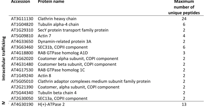
secretory or endocytic pathways (Table1). Clathrin was found as putatively interacting with 192
IRT1, which is in accordance with our previous results showing that IRT1 undergoes clathrin-193
mediated endocytosis (Barberon et al., 2011; Barberon et al., 2014). Interestingly SEC13a and 194
SEC31b proteins were identified as IRT1 putative interactants, suggesting a role of coat 195
protein complex II (COPII) machinery in the export of IRT1 from the endoplasmic reticulum 196
(ER) to the Golgi apparatus (Chung et al., 2016). Conversely, IRT1 probably also undergoes 197
retrograde transport from the Golgi to the ER since it interacts with components of COPI 198
vesicles (Table 1) (Yorimitsu et al., 2014). Another interesting group of IRT1 interactants are 199
proteins linked to Arabidopsis metal homeostasis such as the Pleiotropic drug resistance 8 / 200
Penetration 3 protein that was proposed to act as a cadmium extrusion pump (Kim et al., 201
2007) (Table 1). IRT1 also associates with two iron transporters from the VIT family; 202
however, the relevance of this interaction remains unclear since these proteins were described 203
as localizing to the ER body membrane (Yamada et al., 2013). Recently, rhizosphere-excreted 204
coumarins were shown to be important for iron acquisition in an IRT1-dependent manner 205
(Fourcroy et al., 2016). Interestingly, the Feruloyl-Coenzyme A 6’-Hydroxylase 1 (F6'H1) 206
(Schmid et al., 2014) and the Cytochrome P450/CYP82C4 (Rajniak et al., 2018), which are 207
both involved in coumarin biosynthesis, were identified as putative IRT1-interacting proteins 208
(Table 1). In addition, FRO2 and AHA2, which are known to both act in concert with IRT1 in 209
the acidification-reduction-transport strategy for iron uptake in Arabidopsis thaliana, were 210
also identified (Table 1). Mass spectrometry analyses performed on IRT1 co-immunopurified 211
fractions indeed identified peptides specific to the AHA2 isoform, but also identified peptides 212
common to AHA2 and other AHA proteins, mostly AHA1. However, since no peptide 213
specific to other AHA proteins than AHA2 were found, and because rhizosphere acidification 214
is chiefly mediated by AHA2 in lack of iron (Santi and Schmidt, 2009), we decided to focus 215
our attention on AHA2. These observations indicate that IRT1, FRO2, and AHA2 likely 216
associate to drive iron uptake. To gain further insight into the regulation of the iron uptake 217
machinery, we characterized in more detail the interaction and the spatial organization of the 218
three major actors of iron acquisition, namely FRO2, AHA2, and IRT1. 219
220
IRT1 directly interacts with FRO2 and AHA2 221
To validate the observations obtained from mass spectrometry analyses, the 222
interactions between IRT1 and AHA2/FRO2 were first investigated in Arabidopsis roots by 223
performing co-IP combined with immunodetections. We analyzed the interaction between 224
IRT1-mCitrine, expressed under the control of IRT1 promoter, and endogenous AHA2 protein 225
by using a previously described antibody raised against Plasma Membrane H+-ATPase 2 226
(PMA2) from Nicotiana plumbaginifolia that recognizes AHA2 and also other Arabidopsis 227
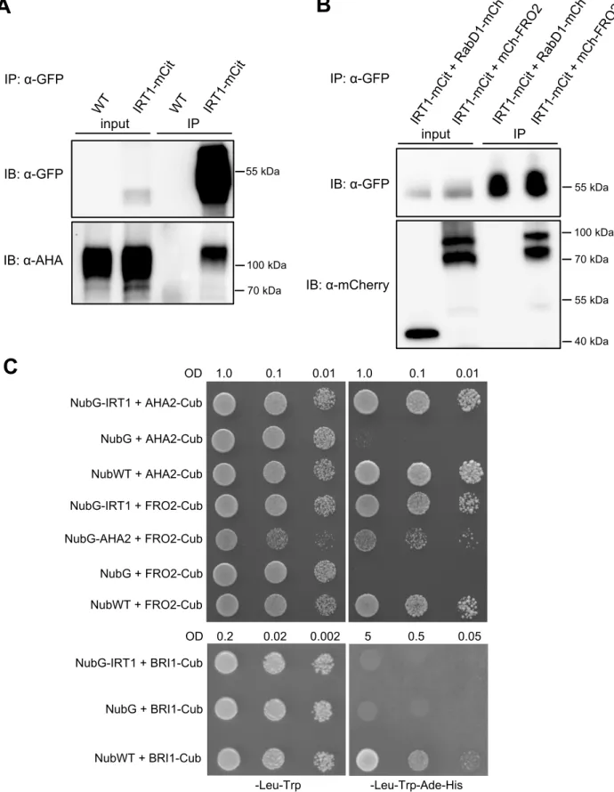
AHA proteins. When IRT1-mCitrine IP fraction was probed with anti-PMA2 antibodies, a 228
strong signal corresponding to the expected size of AHA2 (104 kDa) was detected, whereas 229
no signal was observed in the IP fraction from WT plant roots used as a negative control (Fig. 230
1A). Although this signal may correspond in part to other AHA isoforms than AHA2 due to a 231
lack of specificity of the anti-PMA2 antibodies, this result suggests that endogenous AHA2 232
likely associates with IRT1 in root epidermal cells. The existence of an IRT1-AHA2 complex 233
is substantiated by other protein-protein interaction assays described hereafter. Since no 234
antibody raised against FRO2 was available, we generated a translational fusion of FRO2 235
expressed under control of the FRO2 promoter in the previously described fro2 mutant named 236
frd1-1 (Robinson et al., 1999). Expression of FRO2 fused at its N-terminal end to the 237
mCherry fluorescent protein (mCherry-FRO2) complemented the hypersensitivity of frd1-1 to 238
low iron, even for transgenic lines expressing low levels of mCherry-FRO2 (Supplemental 239
Fig. 2A and B). This clearly shows that mCherry-FRO2 fusion protein is fully functional. 240
Consistently, mCherry-FRO2 protein was only detected in root epidermal cells (Supplemental 241
Fig. 2C), highlighting the specificity of FRO2 promoter, as previously reported (Connolly et 242
al., 2003). To analyze the interaction between FRO2 and IRT1 in Arabidopsis roots by co-IP, 243
we generated transgenic lines co-expressing mCherry-FRO2 and IRT1-mCitrine under control 244
of the FRO2 and IRT1 promoters, respectively. Transgenic lines co-expressing IRT1-mCitrine 245
and RabD1-mCherry, the latter co-localizing with IRT1 in endosomes (Supplemental Fig. 3), 246
were used as a negative control. mCherry-FRO2 was co-immunopurified with IRT1-mCitrine 247
whereas RAbD1-mCherry was not, proving that FRO2 and IRT1 form a protein complex in 248
root epidermal cells (Fig. 1B and Supplemental Fig. 4). 249
The interaction between IRT1 and AHA2/FRO2 was also confirmed by a split-250
ubiquitin assay, which allows the detection of direct interactions between membrane proteins 251
in yeast. Such an approach has been successfully implemented with IRT1 and IDF1 (Shin et 252
al., 2013; Dubeaux et al., 2018). IRT1 fused to the mutated N-terminal half of ubiquitin 253
(NubG), which is unable to spontaneously reassemble with the C-terminal part of ubiquitin 254
(Cub), was co-expressed in yeast with AHA2 or FRO2 fused to Cub linked to the chimeric 255
transcription factor ProteinA-LexA-VP16 (PLV) (Fig. 1C). Physical interactions between 256
IRT1 and AHA2/FRO2 were tested through the ability to rescue yeast growth on a selective 257
medium. Yeast co-expressing NubG-IRT1 with AHA2-Cub or FRO2-Cub grew on a selective 258
medium, similarly to the respective positive controls expressing NubWT with AHA2-259
Cub/FRO2-Cub. However, no growth was observed for the respective negative controls 260
expressing NubG with AHA2-Cub/FRO2-Cub. As an additional negative control for split-261
ubiquitin, NubG-IRT1 was co-expressed with an unrelated transmembrane protein, 262
specifically the brassinosteroid receptor BRI1, fused to Cub. Whereas yeast co-expressing 263
NubWT with BRI1-Cub (positive control of interaction) grew on a selective medium, no 264
growth was observed when BRI1 and IRT1 were co-expressed, indicating that these two 265
proteins do not interact. Interestingly, our split-ubiquitin assay also revealed that AHA2 and 266
FRO2 could physically associate with each other (Fig. 1C). This result was confirmed in 267
plants by co-IP approaches showing that endogenous AHA2 is co-immunopurified with 268
mCherry-FRO2 from Arabidopsis root protein extracts (Supplemental Fig. 5). Altogether, 269
these observations validate the existence of a protein complex in root epidermal cells that 270
gathers the different actors of the high-affinity iron uptake machinery in close proximity. 271
272
Differential regulation of the iron uptake system by ubiquitination 273
We recently demonstrated that IRT1 endocytosis is controlled by the non-iron metal 274
substrates of IRT1 (Dubeaux et al., 2018). Upon an excess of these metals, IRT1 275
ubiquitination strongly increases leading to the endocytosis and the degradation of IRT1 in 276
the vacuole. Interestingly, proteomic analyses allowed the identification of AHA2 and FRO2 277
as part of the Arabidopsis ubiquitinome (Kim et al., 2013; Johnson and Vert, 2016; Walton et 278
al., 2016). Since AHA2 and FRO2 belong to an IRT1-containing protein complex, we 279
wondered whether ubiquitination of these proteins could be co-regulated by non-iron metal 280
availability. First, we analyzed the ubiquitination profile of AHA2 and FRO2 by performing 281
IP of AHA2-GFP and mCherry-FRO2 expressed in Arabidopsis roots, followed by the 282
immunodetection of ubiquitination with the P4D1 general anti-ubiquitin antibodies. In the 283
presence of physiological concentrations of non-iron metal substrates, AHA2-GFP and 284
mCherry-FRO2 IP fractions probed with P4D1 antibodies exhibited high-molecular-weight 285
smears that are typical of ubiquitinated proteins as observed for IRT1-mCitrine used as a 286
control (Fig. 2). To quantify the effect of non-iron metal regime on the ubiquitination of the 287
investigated proteins, the signal intensity observed for the anti-ubiquitin immunoblots 288
performed on IRT1-mCitrine, AHA2-GFP, or mCherry-FRO2 immunopurified proteins was 289
measured and normalized to the corresponding immunopurified proteins (Fig. 2C). As 290
previously observed (Dubeaux et al., 2018), a short treatment with an excess of non-iron 291
metals led to a strong increase in IRT1-mCitrine ubiquitination (Fig. 2A and C). By contrast, 292
the pool of ubiquitinated AHA2-GFP and mCherry-FRO2 remained unchanged in both metal 293
regimes (Fig. 2A, B and C, and Supplemental Fig. 6). Hence, although AHA2, FRO2, and 294
IRT1 belong to the same complex involved in a common mechanism i.e. iron acquisition, 295
ubiquitination of these proteins is differentially regulated by metal availability. 296
297
Selective endocytosis of IRT1 in response to non-iron metal excess 298
Although the intracellular dynamics of IRT1 and AHA2 were previously 299
independently investigated (Barberon et al., 2011; Dubeaux et al., 2018; Haruta et al., 2018), 300
the subcellular distribution of FRO2 has not previously been determined. Thus, the respective 301
localization of IRT1-mCitrine and FRO2/AHA2 expressed as mCherry fusion proteins was 302
investigated. We used root-tip epidermal cells as i) these cells are well suited to analyze the 303
precise localization of endocytosed plasma membrane proteins and ii) the metal-triggered 304
endocytosis of IRT1 was already characterized in such cells (Dubeaux et al., 2018). 305
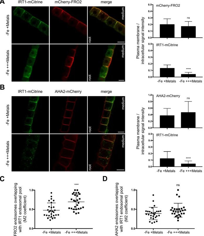
Interestingly, in the absence of iron and in the presence of physiologically relevant amounts 306
of non-iron metal substrates, mCherry-FRO2 was present at the plasma membrane in the outer 307
polar domain facing the rhizosphere, as observed for IRT1-mCitrine (Fig. 3A, left panel) 308
(Barberon et al., 2014; Dubeaux et al., 2018). FRO2 also co-localized with IRT1 in 309
intracellular vesicles (Mander’s coefficient, M2 = 0.48) (Fig. 3A, left panel and 3C), which 310
correspond to early endosomes since IRT1 constitutes a marker of these compartments in 311
such metal conditions (Barberon et al., 2011; Dubeaux et al., 2018). In contrast to IRT1 and 312
FRO2, AHA2-mCherry displayed an apolar plasma membrane localization in epidermal cells 313
in the same metal conditions and although AHA2-mCherry was observed in a limited number 314
of endosomes, these were mostly co-labeled with IRT1-mCitrine (M2 = 0.39) (Fig. 3B, left 315
panel and 3D). 316
The intracellular dynamics of FRO2 and AHA2 were then investigated after a short-317
term treatment with an excess of non-iron metal substrates that triggers IRT1 endocytosis and 318
its subsequent degradation (Dubeaux et al., 2018). Upon non-iron metal excess, IRT1-319
mCitrine was depleted from the cell surface and accumulated in late endosomes (Fig. 3A and 320
3B, left panels). mCherry-FRO2 and AHA2-mCherry were, however, mostly detected at the 321
plasma membrane, even though they were also found to co-localize with IRT1-mCitrine in 322
late endosomes (Fig. 3A and 3B, left panels). To quantify the response to metals, the ratio of 323
plasma membrane to intracellular fluorescence signal intensities was measured for the three 324
fusion proteins under physiological non-iron metal provision or in the presence of an excess 325
of these metals (Fig. 3A and 3B, right panels). As previously reported, the plasma 326
membrane/intracellular ratio highly decreased for IRT1-mCitrine in response to non-iron 327
metal excess (Dubeaux et al., 2018). For mCherry-FRO2 and AHA2-mCherry, however, no 328
significant difference was observed, indicating that FRO2 and AHA2 are not largely 329
endocytosed in response to non-iron metal excess. However, the level of co-localization of 330
FRO2 and IRT1 in endosomes significantly increased with non-iron metal excess (M2 = 0.69) 331
compared to control conditions (M2 = 0.48) (Fig. 3C), suggesting a minor effect of non-iron 332
metal status on FRO2 endocytosis. On the other hand, the co-localization level between 333
AHA2 and IRT1 in endosomes was not significantly modified by non-iron metal substrates 334
(Fig. 3D). 335
The absence of a massive internalization of FRO2 and AHA2 in response to non-iron 336
metal excess suggests that the IRT1/FRO2/AHA2 complex dissociates prior to IRT1 337
endocytosis. Since the phosphorylation of residues in the large cytosolic loop of IRT1 was 338
shown to be the trigger of IRT1 ubiquitin-dependent endocytosis upon non-iron metal 339
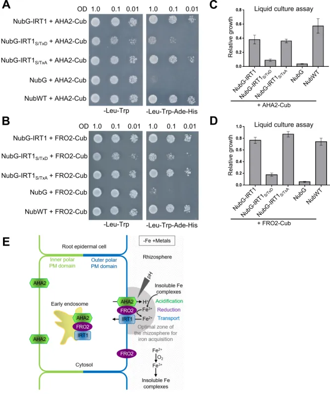
substrate excess (Dubeaux et al., 2018), we investigated whether phosphomimic (IRT1S/TxD) 340
or non-phosphorylatable (IRT1S/TxA) variants of IRT1 would display modified association 341
with AHA2 and FRO2. Using split-ubiquitin approaches, we observed that yeast co-342
expressing NubG-IRT1S/TxD and AHA2- or FRO2-Cub grew slower on the selective medium 343
than yeast co-expressing NubG-IRT1 or NubG-IRT1S/TxA together with AHA2- or FRO2-Cub 344
(Fig. 4A, B). To better quantify the effect of the respective IRT1 variants, we measured yeast 345
growth in liquid cultures. We consistently observed reduced growth for yeast co-expressing 346
FRO2 or AHA2 with IRT1S/TxD compared to their wild-type or IRT1S/TxA counterparts (Fig. 347
4C, D), although NubG-IRT1S/TxD protein accumulated to the same extent as wild-type or 348
IRT1S/TxA when expressed in yeast cells (Supplemental Fig. 7). These results show that the 349
phosphomimic variant of IRT1 displays a reduced interaction with AHA2 and FRO2. 350
Altogether, these observations clearly indicate a role for IRT1 phosphorylation in dissociation 351
of the high-affinity iron uptake complex upon non-iron metal excess. 352
DISCUSSION 353
IRT1 interactome: new insight into IRT1 dynamics and metal uptake 354
IRT1 mediates the absorption of iron but also non-iron metal substrates such as Zn, 355
Mn, and Co in root epidermal cells and is a major actor of metal nutrition in Arabidopsis. 356
Previous studies revealed that the tight control of IRT1 intracellular dynamics by IDF1, 357
CIPK23, FYVE1, or SNX1 is essential to maintain metal homeostasis and to ensure optimal 358
plant growth and development (Shin et al., 2013; Barberon et al., 2014; Ivanov et al., 2014; 359
Dubeaux et al., 2018). To better understand how IRT1 is controlled in the cell, we searched 360
for further IRT1-interacting proteins by performing IP of the functional IRT1-mCitrine fusion 361
protein expressed under the control of IRT1 promoter coupled to mass spectrometry analyses. 362
Co-IP has the great advantage to reveal physiologically relevant protein-protein interactions 363
in plant cells and regarding false positives, co-IP does not generate more than the yeast two-364
hybrid or the split-ubiquitin techniques and results in less than using bimolecular fluorescence 365
complementation (BiFC) (Xing et al., 2016). Besides, co-IP was successfully used in the past 366
to identify interactants of channels and receptors (Karlova et al., 2006; Bellati et al., 2016). 367
One must consider that an interactome provides an overview of proteins putatively interacting 368
with a bait, but that each candidate protein has to be validated by an independent protein-369
protein interaction technic before further investigation to avoid putative artifacts. Here, we 370
decided to (i) use the IRT1 endogenous promoter to maximize the detection of interactions 371
between IRT1 and its interactants in relevant cell types, i.e. root epidermal cells, and (ii) 372
validate each candidate of interest using the split-ubiquitin technic. Two independent co-373
IP/mass spectrometry experiments were performed, which provides qualitative data on 374
proteins interacting with IRT1. Using another replicate would have certainly allowed us to 375
obtain more quantitative data. Besides, since several very interesting candidates such as FRO2 376
were identified in one of the two experiments, we decided to include the proteins that were 377
identified in only one replicate in the IRT1 interactome. Among 142 proteins putatively 378
interacting with IRT1, a group of proteins related to intracellular trafficking emerged, which 379
included clathrin, tubulin, and actin (Table 1). An association between clathrin and IRT1 is in 380
agreement with the previously reported clathrin-mediated internalization of IRT1 from the 381
plasma membrane (Barberon et al., 2014). The co-IP approach does not allow the 382
determination of whether two proteins directly interact or not; however, according to what is 383
known for other cargo proteins, it is likely that the interaction between IRT1 and clathrin is 384
mediated by an unknown adaptor protein. So far the role of actin and tubulin in IRT1 385
dynamics is poorly understood; however, since these proteins are known to be involved in 386
plant endocytosis (Fan et al., 2015), their presence in the IRT1-interactome also opens 387
interesting perspectives. Two small G proteins from the Rab class, namely RAB GTPase 388
homolog A1D and RAB GTPase homolog 1C, were also identified as putative interactants of 389
IRT1 (Table 1). Although the role of these two proteins in intracellular trafficking is still 390
elusive, similar Rab proteins were described to act in plant endocytic pathways (Qi and 391
Zheng, 2013). Intriguingly, the IDF1 E3 ubiquitin ligase and the CIPK23 kinase that are 392
important for IRT1 endocytosis and were demonstrated to interact with IRT1 in split-ubiquitin 393
and yeast-two hybrid candidate approaches, respectively (Shin et al., 2013; Dubeaux et al., 394
2018), were not co-purified with IRT1 in the present study. Given the functions of IDF1 and 395
CIPK23, their interactions with IRT1 are probably very transient and may be lost during the 396
co-IP procedure contrary to other systems such as split-ubiquitin and yeast-two hybrid where 397
these associations would be stabilized (Xing et al., 2016). Until now, studies of IRT1 398
trafficking in plant cells mainly described endocytic mechanisms including IRT1 399
internalization from the plasma membrane in an ubiquitin-dependent process or IRT1 400
recycling from endosomes to the plasma membrane. How IRT1 traffics along the secretory 401
pathway to reach the plasma membrane remains largely unknown, even though Malus 402
xiaojinesis IRT1 was proposed to exit the ER in COPII vesicles (Tan et al., 2018). The 403
Arabidopsis IRT1 interactome provides interesting clues on these aspects since IRT1 was 404
found to putatively interact with several components of the COPII machinery that is 405
sequentially recruited to the surface of the ER membrane to induce the formation of transport 406
vesicles and ensure the delivery of cargo proteins to the Golgi apparatus (Chung et al., 2016) 407
(Table 1). Membrane proteins exit the ER via the recognition of specific cytoplasmic export 408
motifs by the COPII machinery; these signals include diacidic motifs corresponding to 409
(D/E)x(D/E), with x representing any amino acid residue (Zelazny et al., 2009). A diacidic 410
motif (EDD) located in the large cytosolic loop of IRT1 at the position 180 may be involved 411
in the packaging of IRT1 in COPII vesicles and its export from the ER, although this remains 412
to be experimentally determined. 413
Besides the IRT1 interactants linked to intracellular trafficking, proteins involved in 414
metal homeostasis represent a very interesting group of candidates (Table 1). Recently, 415
coumarins, which are excreted in the rhizosphere by PDR9, were demonstrated to be 416
important for Arabidopsis iron acquisition by chelating Fe3+ and as a result facilitating iron 417
availability for FRO2 (Fourcroy et al., 2014; Fourcroy et al., 2016). Intriguingly, IRT1 418
potentially interacts with F6'H1 and Cytochrome P450/CYP82C4 that are both involved in 419
coumarin biosynthesis (Schmid et al., 2014; Rajniak et al., 2018), but the meaning of these 420
interactions remains unclear, notably because PDR9 was not identified as interacting with 421
IRT1. However, solubilization of this large transmembrane protein may not be optimal in the 422
conditions we used for the co-IP and other approaches will be necessary to determine whether 423
PDR9 and IRT1 could work in concert among a common protein complex. Interestingly, 424
FRO2 and AHA2, which act with IRT1 in the acidification-reduction-transport strategy for 425
iron acquisition, were found to interact with IRT1. 426
IRT1, FRO2, and AHA2 form an iron-acquisition complex to optimize iron uptake in 427
Arabidopsis roots
428
By combining co-IP analyses and split-ubiquitin assays, we showed that IRT1 429
associates with FRO2 and AHA2 proteins, but also that FRO2 and AHA2 interact together 430
(Fig. 1). Importantly, the tripartite physical interactions between IRT1, FRO2, and AHA2 are 431
direct, suggesting the existence of a dedicated protein complex at the cell surface for iron 432
uptake. Interaction between IRT1 and FRO2/AHA2 is probably not required for the intrinsic 433
activity of IRT1, since heterologous expression of IRT1 alone allows phenotypic 434
complementation of the fet3 fet4 yeast mutant defective in iron uptake (Eide et al., 1996). 435
Similar to IRT1, FRO2 was also observed at the outer polar domain of the plasma membrane 436
in epidermal cells from the root tip (Fig. 3) and from the differentiated zone of the root 437
(Supplemental Fig. 8A). The co-polarity between FRO2 and IRT1 proteins in this domain of 438
the plasma membrane highlights the specificity of their functions achieved at the interface 439
between the root surface and the rhizosphere. On the other hand, AHA2 distribution in the 440
plasma membrane of root epidermal cells was homogenous (Fig. 3 and Supplemental Fig. 441
8B), suggesting that AHA2 does not obligatory associate with FRO2 and IRT1. This result is 442
in accordance with AHA2 function not being restricted to iron acquisition (Yuan et al., 2017; 443
Hoffmann et al., 2018; Pacifici et al., 2018). Even though the performed interaction tests do 444
not provide any information on the localization of the IRT1/FRO2/AHA2 complex, co-445
localization analyses suggest that IRT1 may interact with FRO2 and AHA2 in the plasma 446
membrane and in endosomes (Fig. 3 and Supplemental Fig. 8). In the future, Förster 447
resonance energy transfer-fluorescence lifetime imaging microscopy (FRET-FLIM), which 448
allows the detection of protein-protein interactions in living plant cells with a high spatial and 449
temporal resolution (Zelazny et al., 2007), may reveal where IRT1 interact with FRO2 and 450
AHA2 in the cell. 451
IRT1 intracellular dynamics is regulated by ubiquitination notably in response to an 452
excess of non-iron metal substrates, following which IRT1 ubiquitination is enhanced, thus 453
triggering its endocytosis and degradation in the vacuole (Dubeaux et al., 2018). Our 454
biochemical analyses revealed that FRO2 and AHA2 are also ubiquitinated; however, their 455
ubiquitination is not modulated by the non-iron metal provision (Fig. 2 and Supplemental 456
Fig.6). This is consistent with the endocytosis of both proteins being rather constitutive and 457
not regulated by non-iron metals (Fig. 3). In this context, the ubiquitination of FRO2 and 458
AHA2 appears to be involved in a non-degradative process. First, FRO2 and AHA2 may 459
undergo mono-ubiquitination that is known to promote internalization from the cell surface 460
but to be insufficient for vacuolar targeting (Lauwers et al., 2010). Second, the internalized 461
pool of AHA2 and FRO2 may be rapidly de-ubiquitinated in endosomes allowing their 462
recycling to the plasma membrane. Third, since ubiquitination is not only involved in 463
endocytosis and degradation but is also implicated in other processes such as the allosteric 464
regulation of proteins (Komander, 2009), we may speculate that FRO2 and AHA2 465
ubiquitination may convey this type of regulation. Although the identity of the E3 ubiquitin 466
ligase at stake is unknown, IDF1 represents a possible candidate for constitutive 467
ubiquitination of AHA2 and FRO2 that will have to be tested in the future. The fact that 468
AHA2 and FRO2 likely carry out other functions independent of iron nutrition may provide 469
an explanation for their not being degraded upon non-iron metal excess. Indeed, AHA2 was 470
previously reported to contribute to acidic growth or phosphorus uptake (Yuan et al., 2017; 471
Hoffmann et al., 2018; Pacifici et al., 2018). FRO2 may play a role in copper reduction since 472
frd1-1 mutants lack low iron-inducible copper chelate reductase activity and 35S::FRO2 473
plants display elevated copper reduction (Yi and Guerinot, 1996; Robinson et al., 1999; 474
Connolly et al., 2003). The absence of internalization of FRO2 and AHA2 from the plasma 475
membrane in root epidermal cells in response to non-iron metal excess indicates that the 476
IRT1/FRO2/AHA2 complex must disassemble prior to IRT1 endocytosis to release AHA2 477
and FRO2 pools that may engage in other processes. In response to non-iron metal excess, 478
IRT1 is phosphorylated by CIPK23 in its large cytosolic loop, boosting the interaction with 479
the E3 ubiquitin ligase IDF1 and yielding polyubiquitinated IRT1 (Dubeaux et al., 2018). We 480
demonstrated in the present study that phosphorylation of IRT1 at the same residues also 481
controls the disassembly of the root high-affinity iron uptake complex (Fig. 4). 482
Phosphorylation of IRT1 therefore has two opposite effects: dissociation of the 483
IRT1/AHA2/FRO2 complex and recruitment of IDF1. Although phosphorylation is often 484
considered as a post-translational modification allowing the recruitment of downstream 485
factors, there is mounting evidence that it also controls the disassembly of protein complexes 486
(Zhang et al., 2010; Couto et al., 2016). The fact that AHA2 and FRO2 were identified in our 487
co-IP/mass spectrometry analysis as interacting with IRT1 in non-iron metal excess is rather 488
surprising, since IRT1 phosphorylation in response to non-iron metal excess induces the 489
dissociation of the IRT1/FRO2/AHA2 complex. However, this may not induce total 490
dissociation of the complex, as evidenced by the residual interaction observed between the 491
phosphomimic IRT1 and AHA2/FRO2. This is also supported by the partial co-localization of 492
FRO2 and AHA2 with IRT1 in late endosomes under non-iron metal excess. In addition, we 493
showed that the level of co-localization between FRO2 and IRT1 in late endosomes slightly 494
increased with non-iron metal excess, suggesting that a small proportion of FRO2 is still able 495
to associate with IRT1 in these conditions. Therefore, our co-IP/mass spectrometry analysis 496
carried out when plants experience non-iron metal excess likely allowed us to identify the 497
small pool of FRO2 and AHA2 still interacting with IRT1 in endosomes. This may explain 498
why a limited number of FRO2 peptides were identified by proteomics. 499
The biological significance of iron uptake using a specific platform at the cell surface 500
is unclear but likely relies in the chemistry of iron. Although iron is abundant in most soils, its 501
bioavailability to plants is often limited. This is especially true for calcareous soils, which 502
represent one third of cultivated lands, where iron is present under the form of insoluble 503
complexes (Briat et al., 2015). During the iron-acquisition process, rhizosphere acidification 504
by the root is essential to increase iron availability, indeed the solubility of iron increases 505
1000-fold for every one unit drop in pH (Olsen et al., 1981). However, this acidification 506
process, mainly mediated by AHA2 under iron deficiency (Santi and Schmidt, 2009), is very 507
local, which likely impacts on the efficiency of iron uptake. Moreover, the presence of 508
oxygen in most soils likely provokes the rapid reoxidation of Fe2+ produced by FRO2 into 509
Fe3+ that is not transported by IRT1. Thus, we propose that the tripartite protein complex 510
gathering IRT1, FRO2, and AHA2 together creates a local environment of pH and Fe2+ 511
concentration in the rhizosphere that favors an optimal acquisition of iron (Fig. 4E). To 512
experimentally validate this model, mutated versions of these proteins that do not interact 513
with each other but that conserve their activities need to be generated to evaluate the 514
functional outcome on iron uptake. However, this requires deep knowledge about the 515
structure or interaction domains between these highly hydrophobic membrane proteins, which 516
is currently missing. Alternatively to the local environment theory, we can also speculate that 517
reduced iron is directly transferred from FRO2 to IRT1 by a channeling mechanism, similar 518
to what has been described for metabolic pathways. Channeling consists in the transfer of the 519
product of a proximal activity as substrate to a distal activity without equilibration with bulk 520
solvent, which increases the efficiency of the kinetic process (Kwok et al., 2006). Such 521
mechanism requires the close proximity of the donor and acceptor sites. Interestingly, 522
channeling of iron was described in yeast between the multicopper oxidase Fet3p, which 523
oxidizes Fe2+ to Fe3+, and the iron permease Ftr1p that transports Fe3+ into the cells, with both 524
proteins forming a hetero-oligomeric complex (Kwok et al., 2006; Singh et al., 2006). Further 525
work will elucidate whether FRO2 and IRT1 use a similar mechanism.Whether the formation 526
of an iron-acquisition complex comprising IRT1, FRO2, and AHA2 is conserved in plants 527
other than Arabidopsis remains to be determined. This complex is probably not present in rice 528
(Oryza sativa) that combines two strategies to take up iron from the soil: a phytosiderophore-529
based system allowing the acquisition of Fe3+ (Inoue et al., 2009; Lee et al., 2009) and the use 530
of Fe2+ transporters such as OsIRT1 (Ishimaru et al., 2006). Indeed, Fe3+ chelate reductase 531
activity has been shown to not be required for Fe2+ uptake under iron deficiency in rice, 532
suggesting that OsIRT1 works independently of OsFRO2-like proteins (Ishimaru et al., 2006). 533
In paddy fields, where rice plants are grown, Fe2+ is abundant due to the low redox potential 534
and therefore rice plants do not need to reduce Fe3+ to Fe2+ (Ishimaru et al., 2007).So far, the 535
description of protein complexes aimed at optimizing nutrient uptake in plant remains scarce. 536
To our knowledge, only the interaction between glutamine synthase, the principal ammonia 537
assimilatory enzyme, and the aquaglyceroporin Nodulin 26, a transporter of NH3, was 538
proposed to promote efficient assimilation of nitrogen in soybean (Masalkar et al., 2010). 539
Although experimental evidence is still needed, the co-localization between FRO2/AHA2 and 540
IRT1 in early endosomes in the presence of physiologically relevant levels of non-iron metals 541
suggests that the iron-acquisition complex may exist in this compartment in addition to the 542
plasma membrane (Fig.4E). This complex may help plant metal uptake by translocating iron 543
from endocytic vesicles to the cytosol. Alternatively, the AHA2/FRO2/IRT1 complex may 544
simply cycle between early endosomes and the plasma membrane in a constitutive manner or 545
in response to some undetermined stimulus. Finally, since early endosomes/trans-Golgi 546
network constitute a crossroad between endocytic and secretory pathways in plants (Dettmer 547
et al., 2006), the presence of the IRT1/FRO2/AHA2 complex in early endosomes may also 548
reflect a step in the delivery of a pre-formed complex to the plasma membrane. Future work 549
will be needed to discriminate between these different scenarios. 550
551
MATERIAL AND METHODS 552
Plant material and growth conditions 553
Arabidopsis thaliana wild-type (WT) plants (Col-0, Col-gl1, and Ws), the fro2 loss-of-554
function mutant named frd1-1 (Robinson et al., 1999), the previously described irt1-555
1/IRT1::IRT1-mCitrine line (Dubeaux et al., 2018), and the various transgenic plants 556
generated in this study were vertically grown in sterile conditions at 21°C with 16 h light/8 h 557
dark cycleswith a light intensity of 90 µmol m-2 s-1 using Philips 17W F17T8/TL741 bulbs. 558
The plant growth medium used was half-strength Murashige and Skoog (MS/2) medium 559
containing 1% sucrose (w/v), 1% (w/v) agar, and various concentrations of metals. Hence, 560
depending on the experiment (see below), plants were grown in the absence of iron and in the 561
presence of physiological concentrations of IRT1 secondary substrates Zn (15 µM), Mn (50 562
µM), and Co (0.05 µM) (-Fe +Metals) or in the presence of 10-fold more Zn, Mn, and Co (-Fe 563
+++Metals) corresponding to an excess of non-iron metal substrates, as previously described 564
(Dubeaux et al., 2018). Plants have also been grown in iron-replete conditions using MS/2 565
medium containing 50 μM or 100 μM Fe-EDTA (+Fe). 566
For immunopurifications followed by mass spectrometry analyses, irt1-1/IRT1::IRT1-567
mCitrine transgenic lines and Ws WT plants were initially grown for 9 days on MS/2 medium 568
containing 50 μM Fe-EDTA, transferred for 5 days onto a -Fe +Metals medium to induce 569
IRT1-mCitrine expression, and finally subjected to a -Fe +++Metals treatment for 48 h. To 570
confirm the interactions between IRT1, FRO2, and AHA2 by co-immunopurifications, the 571
various genotypes were grown for 11 days on MS/2 medium containing 50 μM Fe-EDTA, 572
and then transferred for 4 days on a -Fe +Metals medium supplemented with 300 μM of the 573
iron chelator Ferrozine [3-(2-pyridyl)-5,6-diphenyl-1,2,4-triazine sulfonate] to ensure a rapid 574
and strong expression of genes under the control of IRT1 and FRO2 promoters. 575
To analyze mCherry-FRO2, AHA2-GFP, and IRT1-mCitrine ubiquitination profiles, the 576
appropriate transgenic lines as well as WT plants used as negative controls were grown for 11 577
days on -Fe +Metals MS/2 solid medium. Then, plants were transferred for 2 hin -Fe +Metals 578
(control) or –Fe +++Metals MS/2 liquid medium as previously described (Dubeaux et al., 579
2018). 580
For microscopy analyses, transgenic lines expressing IRT1/AHA2/FRO2 fusion proteins 581
under the control of IRT1 promoter, were first grown for 11 days on a -Fe +Metals MS/2 582
medium to ensure protein expression. Then, before observation, plants were transferred for 2 583
h in -Fe +Metals (control) or -Fe +++Metals MS/2 liquid medium. The localization of 584
mCherry-FRO2 protein in frd1-1/FRO2::mCherry-FRO2 transgenic lines was performed on 585
plants grown for 11 days in -Fe +Metals condition. 586
For mCherry-FRO2 functionality test, frd1-1/FRO2::mCherry-FRO2 transgenic lines, frd1-1 587
mutant, and Col-gl1 WT plants were grown for 11 days on MS/2 lacking iron (-Fe +Metals) 588
or on MS/2 supplemented with 100 μM Fe-EDTA (control conditions). Roots from iron-589
starved transgenic lines and frd1-1 (negative control) were collected to analyze mCherry-590
FRO2 protein accumulation by Western blot analysis as detailed below. 591
592
Constructions and generation of Arabidopsis transgenic lines 593
All the constructions described in this section were obtained using the MultiSite Gateway® 594
Three-Fragment Vector Construction system. The FRO2 promoter corresponding to a 595
sequence of 1,845 bp upstream of the FRO2 start codon was amplified from Arabidopsis 596
thaliana genomic DNA using the attB4.promoFRO2 forward and attB1r.promoFRO2 reverse 597
primers (supplemental Table 1) and subsequently cloned into the pDONR.P4P1R entry 598
vector. The FRO2 open reading frame (ORF) was amplified from Arabidopsis cDNAs with 599
the attB2r.FRO2 forward and attB3.FRO2 reverse primers (supplemental Table 1) and cloned 600
into the pDONR.P2RP3 entry vector. The AHA2 ORF without the stop codon was amplified 601
from Arabidopsis cDNA with the AHA2.F forward and AHA2.R reverse primers and was 602
cloned into the pDONR.221 entry vector. The mCherry sequence without stop codon was 603
amplified with attB1.mCherry forward and attB2.mCherry reverse primers and also cloned 604
into the pDONR.221 entry vector (supplemental Table 1). Entry vectors carrying the IRT1 605
and 35S promoters (pDONR.P4P1R-IRT1 and pDONR.P4P1R-35S) or the GFP and the 606
mCherry coding sequence allowing C-terminal fusions (pDONR.P2RP3-GFP and 607
pDONR.P2RP3-mCherry) were previously described (Marques-Bueno et al., 2016; Dubeaux 608
et al., 2018). Final destination vectors for expression in plants were obtained by multisite 609
Gateway® recombination using the entry vectors described above and the pH7m34GW and 610
pK7 m34GW destinations vectors used for mCherry and GFP fusions, respectively. The 611
following constructs were generated: FRO2::mCherry-FRO2, IRT1::mCherry-FRO2, 612
IRT1::AHA2-mCherry, and 35S::AHA2-GFP. 613
The previously described irt1-1/IRT1::IRT1-mCitrine line (Dubeaux et al., 2018) was 614
transformed with FRO2::mCherry-FRO2, IRT1::mCherry-FRO2, and IRT1::AHA2-mCherry 615
constructions by the floral-dipping technique using Agrobacterium tumefaciens. The frd1-1 616
mutant and Col-0 plants were transformed with the FRO2::mCherry-FRO2 and 35S::AHA2-617
GFP constructs, respectively. The irt1-1/IRT1::IRT1-mCitrine transgenic line was crossed 618
with the Wave marker line number 25 expressing RabD1-mCherry fusion protein under the 619
control of UBQ10 promoter (Geldner et al., 2009). 620
Immunopurifications 621
Immunopurifications (IP) were performed on approximatively 500 mg of Arabidopsis roots, 622
mostly as previously described (Dubeaux et al., 2018). Briefly, for IRT1-mCitrine IP followed 623
by mass spectrometry, for co-IP analyses between IRT1-mCitrine and mCherry-FRO2, as 624
well as for co-IP analyses between mCherry-FRO2 and endogenous AHA2, roots were 625
ground in liquid nitrogen and resuspended in IRT1 solubilization buffer (50 mM Tris-HCl 626
(pH 7.4), 150 mM NaCl, 5 mM EDTA, 1% (w/v) n-Dodecyl -D-maltoside (DDM), and 627
plant-specific protease inhibitors (Sigma-Aldrich)). For co-IP analyses between IRT1-628
mCitrine and endogenous AHA2, roots were ground and resuspended in RIPA buffer (50 mM 629
Tris-HCl (pH 7.5), 150 mM NaCl, 0.5% (w/v) sodium deoxycholate, 1% (v/v) IGEPAL®CA-630
630, 0.1% (w/v) SDS, and plant-specific protease inhibitors (Sigma-Aldrich)). After two 631
successive centrifugations at 3,800 × g for 10 min at 4°C, the resultant supernatants were 632
collected and solubilization of membrane proteins was continued for 1 h 30 min at 4°C on a 633
rotating wheel. Samples were then centrifuged at 100,000 × g for 1 h at 4°C to remove 634
unsolubilized material and supernatants containing solubilized proteins were recovered for 635
IPs. This ultracentrifugation step avoids the immuno-capture of proteins present in patches of 636
residual non-solubilized membranes, allowing the IP to be carried out on solubilized protein 637
complexes only. IPs of GFP and mCitrine fusion proteins were performed using µMACS GFP 638
isolation kit (Miltenyi Biotec) whereas IP of mCherry fusion proteins was performed using 639
RFP-Trap®_MA magnetic beads (Chromotek), following the instructions of the 640
manufacturers. Before elution, extensive washes were performed with IRT1 solubilization 641
buffer or RIPA buffer depending on the IP type. Co-IP analyses followed by mass 642
spectrometry were performed twice. Co-IP combined with immunodetections were performed 643
thrice with similar results. 644
To analyze the ubiquitination profile of AHA2-GFP and IRT1-mCitrine, the solubilization of 645
fusion proteins as well as the IP procedure were performed exactly as previously described 646
(Dubeaux et al., 2018). The same protocol was used for mCherry-FRO2 except that the 647
protein was immunopurified with RFP-Trap®_MA magnetic beads (Chromotek). Three 648
independent analyses of ubiquitination profiles were performed. 649
Mass spectrometry analysis 650
For sample preparation, proteins from each eluate were separated by SDS-PAGE in order to 651
fractionate the protein samples into two fractions, including proteins 10–63 kDa and above 63 652
kDa, respectively, to exclude abundant contaminating IRT1-mCitrine protein at 63 kDa. After 653
Coomassie-Blue staining, each gel fraction was cut into bands and subjected to in-gel trypsin 654
digestion with the Progest robot (Genomic Solutions) using standard conditions including 655
reduction and alkylation as described previously (Blanchet et al., 2014). Tryptic peptides 656
extracted from the different bands of each gel fraction were pooled, vacuum dried, and 657
resuspended in 0.1% (v/v) formic acid prior to nanoLC-MS/MS mass spectrometry analyzes. 658
The same cutting pattern of the SDS-PAGE lane was performed for each eluate. 659
Tryptic peptides from the two or three SDS-PAGE fractions from each eluate were analyzed 660
separately by nanoLC-MS/MS with the Triple-TOF 4600 mass spectrometer (ABSciex) 661
coupled to the nanoRSLC ultra performance liquid chromatography (UPLC) system (Thermo 662
Scientific) equipped with a trap column (Acclaim PepMap100C18, 75 μmi.d.× 2 cm, 3 μm) 663
and an analytical column (Acclaim PepMapRSLCC18, 75 μmi.d.× 50 cm, 2 μm, 100 Å). 664
Peptides were loaded at 5 µl/min with 0.05% (v/v) TFA in 5% (v/v) acetonitrile and peptide 665
separation was performed at a flow rate of 300 nl/min with a 5–35% (v/v) solvent B gradient 666
in 40 min. Solvent A was 0.1% (v/v) formic acid in water, and solvent B was 0.1% (v/v) 667
formic acid in 100% acetonitrile. NanoLC-MS/MS experiments were conducted in a Data 668
Dependent acquisition method by selecting the 20 most intense precursors for CID 669
fragmentation with Q1 quadrupole set at low resolution for better sensitivity. 670
Protein identification was performed by processing raw data with MS Data Converter 671
software (AB Sciex) for generating .mgf data files and protein identification were performed 672
using the MASCOT search engine (Matrix science, London, UK) against the Swissprot and 673
TAIR10 databases with carbamidomethylation of cysteines set as fixed modification and 674
oxidation of methionines as variable modifications. Peptide and fragment tolerance were set 675
at 20 ppm and 0.05 Da, respectively. Results were analyzed with Scaffold 3.6.5 software 676
(Proteome Software). Proteins were validated when identified with at least two unique 677
peptides and 95% probability levels for both peptides and proteins. 678
679
Extraction of total proteins and immunoblots 680
Total proteins were extracted from around 100 mg of Arabidopsis roots ground in liquid 681
nitrogen and directly resuspended in 2X SDS sample buffer. Samples were heated at 65°C for 682
10 min, centrifuged 10 min at 20,000 × g and finally supernatants were collected and directly 683
used for SDS-PAGE. Protein extraction from yeast was carried out as previously described 684
(von der Haar, 2007). 685
Immunoblot analyses were performed as previously described (Barberon et al., 2011). 686
Immunodetection of GFP and mCitrine fusion proteins was performed using an anti-GFP 687
antibody conjugated to horseradish peroxidase (HRP) (Miltenyi Biotec 130-091-833, 688
1/5,000). mCherry fusion proteins were monitored with a rabbit anti-DsRed antibody 689
(Clontech 632496, 1/5,000). Endogenous AHA2 protein was immunodetected using a rabbit 690
antibody initially raised against the Plasma Membrane H+-ATPase 2 (PMA2) from Nicotiana 691
plumbaginifolia (W1C) diluted 1/15,000 (Morsomme et al., 1998). Ubiquitin modifications 692
were detected with the P4D1 mouse anti-ubiquitin antibody (Millipore 05-944, 1/4,000). The 693
detection of NubG-IRT1 protein from yeast used anti-HA antibodies (Miltenyi Biotec 130-694
091-972, 1/7,000). Anti-tubulin antibodies were used as loading control (Agrisera AS10 681, 695
1/5,000). The anti-rabbit IgG or anti-mouse IgG secondary antibodies coupled to HRP were 696
both diluted 1/20,000. Detection of HRP chemiluminescence was performed using 697
SuperSignal West Dura Extended Duration Substrate (Thermo Scientific) in a Chemidoc 698
Touch Imaging system (Bio-Rad). Stain-Free protein staining (Bio-Rad) was used as a 699
loading control as previously described (Dubeaux et al., 2018). To quantify IRT1-mCitrine, 700
AHA2-GFP, and mCherry-FRO2 ubiquitination levels under different metal regimes, signal 701
intensity observed with anti-ubiquitin immunoblots performed on IRT1-mCitrine, AHA2-702
GFP, or mCherry-FRO2 immunopurified proteins was measured using Image Lab 6.0.1 and 703
normalized to the quantity of immunopurified proteins detected in IP with GFP or anti-704
DsRed antibodies. To facilitate comparisons, the ubiquitination level measured in the 705
presence of physiological concentrations of non-iron metal substrates was arbitrary fixed to 1. 706
707
Constructions and split-ubiquitin assay 708
Split-ubiquitin vectors were generated using the Gateway® technology. First, the FRO2 ORF 709
without the stop codon and the AHA2 ORF with the stop codon were amplified with 710
FRO2.F/FRO2.R and AHA2.F/AHA2stop.R primers, respectively (supplemental Table 1), 711
and were both cloned into the pDONR.221 entry vector. pDONR.221-BRI1 without stop was 712
previously generated (Martins et al., 2015) and pDONR.221-AHA2 without stop was created 713
in this study as mentioned above. Then, FRO2, AHA2, and BRI1 ORFs without the stop codon 714
were inserted into pMetYC-DEST destination vector (Hachez et al., 2014) to produce 715
methionine-repressible constructs FRO2-Cub-PLV, AHA2-Cub-PLV, and BRI1-Cub-PLV, 716
respectively, where Cub corresponds to the C-terminal part of ubiquitin and PLV to a 717
chimeric transcription factor. AHA2 ORF with the stop codon was cloned into the pNX35-718
DEST destination vector to generate the NubG-AHA2 fusion wherein NubG corresponds to 719
the mutated N-terminal part of ubiquitin. The NubG-IRT1, NubG-IRT1S/TxD, and NubG-720
IRT1S/TxA constructs were previously described (Dubeaux et al., 2018). It is important to note 721
that NubG and Cub were fused to a cytosolic part of IRT1, FRO2, AHA2, and BRI1, 722
according to the known or predicted topology of these proteins. The wild-type ubiquitin N-723
terminal fragment (NubWT) expressed by the pNubWT-Xgate vector and the NubG fragment 724
expressed by the non-recombined pNX35-DEST vector were used as a positive and negative 725
control, respectively (Hachez et al., 2014). 726
Split-ubiquitin assay was performed as previously described (Dubeaux et al., 2018). Briefly, 727
THY.AP4 yeast strain was co-transformed with the Nub and Cub constructs of interest and 728
transformed cells were selected on SD medium lacking Leu and Trp. Then, yeast co-729
expressing Cub-PLV fusion proteins with NubG fusion proteins or NubG (negative control of 730
interaction) or NubWT (positive control of interaction) were dropped in serial dilutions (O.D. 731
1, 0.1, 0.01) onto SD medium without Leu and Trp (control medium) or onto SD medium 732
lacking Leu, Trp, His, Ade (selective medium) supplemented with 500 µM methionine (250 733
µM methionine for IRT1/BRI1 interaction test) to limit the expression of the Cub-PLV fusion 734