HAL Id: hal-03047226
https://hal.archives-ouvertes.fr/hal-03047226
Submitted on 8 Dec 2020
HAL is a multi-disciplinary open access
archive for the deposit and dissemination of
sci-entific research documents, whether they are
pub-lished or not. The documents may come from
teaching and research institutions in France or
abroad, or from public or private research centers.
L’archive ouverte pluridisciplinaire HAL, est
destinée au dépôt et à la diffusion de documents
scientifiques de niveau recherche, publiés ou non,
émanant des établissements d’enseignement et de
recherche français ou étrangers, des laboratoires
publics ou privés.
Eliza Martin, Célia Vicari, Louis Tsakou-Ngouafo, Pierre Pontarotti, Andrei
Petrescu, David Schatz
To cite this version:
Eliza Martin, Célia Vicari, Louis Tsakou-Ngouafo, Pierre Pontarotti, Andrei Petrescu, et al..
Identifi-cation of RAG-like transposons in protostomes suggests their ancient bilaterian origin. Mobile DNA,
BioMed Central, 2020, 11 (1), �10.1186/s13100-020-00214-y�. �hal-03047226�
R E S E A R C H
Open Access
Identification of RAG-like transposons in
protostomes suggests their ancient
bilaterian origin
Eliza C. Martin
1†, Célia Vicari
2†, Louis Tsakou-Ngouafo
2, Pierre Pontarotti
2,3*, Andrei J. Petrescu
1*and
David G. Schatz
4*Abstract
Background: V(D) J recombination is essential for adaptive immunity in jawed vertebrates and is initiated by the RAG1-RAG2 endonuclease. The RAG1 and RAG2 genes are thought to have evolved from a RAGL (RAG-like) transposon containing convergently-oriented RAG1-like (RAG1L) and RAG2-like (RAG2L) genes. Elements resembling this presumptive evolutionary precursor have thus far only been detected convincingly in deuterostomes, leading to the model that the RAGL transposon first appeared in an early deuterostome.
Results: We have identified numerous RAGL transposons in the genomes of protostomes, including oysters and mussels (phylum Mollusca) and a ribbon worm (phylum Nemertea), and in the genomes of several cnidarians. Phylogenetic analyses are consistent with vertical evolution of RAGL transposons within the Bilateria clade and with its presence in the bilaterian ancestor. Many of the RAGL transposons identified in protostomes are intact elements containing convergently oriented RAG1L and RAG2L genes flanked by terminal inverted repeats (TIRs) and target site duplications with striking similarities with the corresponding elements in deuterostomes. In addition, protostome genomes contain numerous intact RAG1L-RAG2L adjacent gene pairs that lack detectable flanking TIRs. Domains and critical active site and structural amino acids needed for endonuclease and transposase activity are present and conserved in many of the predicted RAG1L and RAG2L proteins encoded in protostome genomes.
(Continued on next page)
© The Author(s). 2020 Open Access This article is licensed under a Creative Commons Attribution 4.0 International License, which permits use, sharing, adaptation, distribution and reproduction in any medium or format, as long as you give appropriate credit to the original author(s) and the source, provide a link to the Creative Commons licence, and indicate if changes were made. The images or other third party material in this article are included in the article's Creative Commons licence, unless indicated otherwise in a credit line to the material. If material is not included in the article's Creative Commons licence and your intended use is not permitted by statutory regulation or exceeds the permitted use, you will need to obtain permission directly from the copyright holder. To view a copy of this licence, visithttp://creativecommons.org/licenses/by/4.0/. The Creative Commons Public Domain Dedication waiver (http://creativecommons.org/publicdomain/zero/1.0/) applies to the data made available in this article, unless otherwise stated in a credit line to the data.
* Correspondence:pierre.pontarotti@univ-amu.fr;andrei.petrescu@biochim.ro;
david.schatz@yale.edu
†Eliza C. Martin and Célia Vicari contributed equally to this work. 2Evolutionary biology team, Aix Marseille Université IRD, APHM, MEPHI, IHU
Méditerranée Infection, Marseille, France
1Department of Bioinformatics and Structural Biochemistry, Institute of
Biochemistry of the Romanian Academy, Splaiul Independentei 296, 060031 Bucharest, Romania
4Department of Immunobiology, Yale School of Medicine, 300 Cedar Street,
Box 208011, New Haven, CT 06520-8011, USA
(Continued from previous page)
Conclusions: Active RAGL transposons were present in multiple protostome lineages and many were likely transmitted vertically during protostome evolution. It appears that RAGL transposons were broadly active during bilaterian evolution, undergoing multiple duplication and loss/fossilization events, with the RAGL genes that persist in present day protostomes perhaps constituting both active RAGL transposons and domesticated RAGL genes. Our findings raise the possibility that the RAGL transposon arose earlier in evolution than previously thought, either in an early bilaterian or prior to the divergence of bilaterians and non-bilaterians, and alter our understanding of the evolutionary history of this important group of transposons.
Keywords: Recombination activating genes, RAG, Evolution, Transposon, Adaptive immunity, Transposon molecular domestication
Background
The powerful adaptive immune systems found in verte-brates rely on highly diverse antigen receptors encoded by genes that are non-functional in the germline and as-sembled by recombination during lymphocyte develop-ment [1, 2]. In jawed vertebrates, this assembly reaction is known as V(D) J recombination and operates on ar-rays of V, D, and J gene segments of immunoglobulin and T-cell receptor loci [3]. V(D) J recombination is ini-tiated by the RAG1/RAG2 endonuclease (RAG), which cleaves DNA at a conserved recombination signal se-quence (RSS) that flanks each gene segment and consists of heptamer and nonamer elements separated by a 12 or 23 bp spacer [4]. RAG1 is a multi-domain protein that makes extensive DNA contacts and cleaves DNA using an RNaseH-domain DDE active site similar to that found in many DNA transposases and retroviral integrases [5,
6]. RAG2 assists RAG1 in DNA binding and cleavage and is composed of a kelch-type 6-bladed beta propeller connected to a plant homeodomain (PHD) finger by an acidic“hinge” region [4–6]. High resolution structures of the RAG heterotetramer from mouse and zebrafish, ei-ther alone or in complex with the RSS, have provided extensive mechanistic insights into the DNA binding and cleavage steps of the recombination reaction [5–8].
The RAG recombination machinery is present only in jawed vertebrates. For decades, this raised debate regard-ing its evolutionary origins. Early observations linked RAG to cut-and-paste DNA transposases, revealing that RAG1and RAG2 invariably exist as a closely-juxtaposed, convergently transcribed gene pair (Fig. 1a), that RAG performs DNA cleavage by a nick-hairpin mechanism similar to that used by transposases, and that RAG pos-sesses transposase activity in cell-free reactions [11]. Subsequently, sequence similarity was noted between RAG1’s essential core region and Transib transposases, and between RSSs and Transib terminal inverted repeats (TIRs) [12]. Functional [13,14] and structural [15] stud-ies have provided evidence that RAG1 and Transib transposase share a common ancestor [12, 16]. Transib elements, however, only contain a single gene encoding
a protein similar to RAG1 and hence did not fully ex-plain the origin of RAG1-RAG2 gene pairs.
Since the discovery of Transib, multiple pairs of conver-gently oriented RAG1-like (RAG1L) and RAG2-like (RAG2L) genes have been discovered in invertebrate deuterostomes including echinoderms (purple sea urchin, bat star, and green sea urchin) [10,17], cephalochordates (amphioxus) [18, 19], and hemichordates (acorn worm) [9]. TIRs flanking these gene pairs, when present, have sequence similarity to the RSS heptamer and the Transib TIR, and target site duplica-tions (TSDs) are almost always 5 bp in length [9, 19], as is the case for transposition mediated by RAG and Transib [12,
13, 20, 21]. We refer to invertebrate RAG-like proteins as RAG1L and RAG2L, and transposons containing RAG1L-RAG2Lgene pairs as RAGL transposons. Notably, the BbeR-AG1L and BbeRAG2L proteins, encoded by the amphioxus ProtoRAG transposon of Branchiostoma belcheri (Fig. 1a), possess TIR-dependent endonuclease and transposase activ-ities similar to those of RAG [19]. Furthermore, despite lim-ited amino acid sequence identity, the BbeRAG1L/2 L and RAG1/2 complexes exhibit striking structural similarities [22]. Together, these findings provide compelling evidence for the hypothesis that the RAG1-RAG2 gene pairs and RSSs of jawed vertebrates evolved from the transposase genes and TIRs, respectively, of a RAGL transposon [23].
Outside the Deuterostomia superphylum, there has been virtually no evidence for RAG1L-RAG2L gene pairs or for any gene encoding the combination of a kelch do-main and PHD finger that uniquely identifies RAG2/ RAG2L proteins [17]. RAG1L genes, in the context of Transibtransposons, are widespread, having been found in protostomes, cnidarians, fungi, and echinoderms [12]. Nevertheless, there were hints that RAG1-RAG2 gene pairs might not be limited to deuterostomes. Solitary RAG1L loci that resembled deuterostome RAG1L more than arthropod Transib transposase were described in cnidarians [12], and intriguingly, the deteriorated rem-nants of a RAG1L-RAG2L gene pair were reported in the protostome Crassostrea gigas, the pacific oyster [10].
Here, we report the identification of numerous RAG1L-RAG2L gene pairs in the protostome clade, both
in mollusks and in phylum Nemertea. Many of these ele-ments exhibit all of the expected components of a RAGL transposon: convergent RAG1L-RAG2L genes flanked by TIRs that resemble the RSS heptamer, which in turn are flanked by 5 bp TSDs. Many of the identified RAGL
coding sequences can be translated into full length RAG1L and RAG2L proteins in which key functional do-mains and amino acids are present, and sequence analysis reveals a new RAG-like transposon family in the nemertean Notospermus geniculatus. Phylogenetic
Fig. 1 Genomic organization and phylogenetic tree of RAG and RAGL transposons. a Genomic organization of the mouse RAG locus and the amphioxus RAGL (ProtoRAG) transposon. The legend for panels (a), (b), and (c) is provided at the bottom of panel (c). b Tree depicting phyla within which RAG loci or RAGL or Transib transposons have been identified. Blue, orange and green shading indicate Deuterostomia, Protostomia and Cnidaria, respectively. Branches lacking evidence for RAG/RAGL sequences were omitted. With the exception of Cnidaria, all phyla with identified RAGL sequences contain at least one complete RAGL transposon with the configuration TSD-TIR5′-RAG1L-RAG2L-TIR3′-TSD. Prior to this study, potentially active RAG1-RAG2 gene pairs and RAGL transposons had been reported only in deuterostomes [9], while outside this phylum, only as a single deteriorated RAG1L-RAG2L locus (in C. gigas) was previously reported [10]. c Genomic organization of the most complete RAGL copies detected in Mollusca, Nemertea, and Cnidaria. Predicted RAG1L/RAG2L coding regions, TIRs, and TSDs are depicted, using symbols explained in the legend at bottom. Supporting transcriptomic data are indicated along with corresponding TSA entry (Additional file8: File S1 and Additional file9: S2). Green and gray arrows indicate transcripts corresponding to coding and untranslated regions, respectively. Unmapped regions of transcripts are shown unfilled while the black star in GFRY01002319.1 indicates a frameshift caused by an 8 bp deletion
analyses suggest that in protostomes, as in deutero-stomes [9], the RAGL transposon evolved primarily in a vertical manner. We propose that the RAGL transposon arose very early in animal evolution, in a bilaterian if not earlier, and that subsequently, through duplication and vertical transmission, this transposon gave rise to several transposon families in protostomes and deuterostomes, with some extant elements retaining the potential to be active transposons.
Results
Distribution ofRAG1L-RAG2L gene pairs across phyla
To search for additional RAGL transposons in disparate phyla, RAG1L and RAG2L amino acid sequences from in-vertebrate deuterostomes [9, 17, 19] were used to scan re-cently updated databases including Whole-Genome Shotgun Contigs (WGS), High Throughput Genomic Sequences (HTGS) and Transcriptomic Shotgun Assembly (TSA). Scans were performed on all metazoan invertebrate and jaw-less vertebrate projects available before February 2019. RAG1L proteins, possibly because of their direct role in ca-talysis, exhibit greater evolutionary conservation than RAG2L proteins, and detecting evolutionarily-distant RAG2L homo-logues using existing genome-wide blast techniques is chal-lenging. For example, using tblastn [24, 25] and mouse or human RAG2 as query does not detect RAG2L from amphi-oxus WGS data (data not shown), and standard blast searches failed to identify RAG2L sequences in the purple sea urchin genome [17]. We overcame these difficulties using an iterative blast search approach (see Methods), which allowed the identification of new RAG1L-RAG2L gene pairs in protostomes, in both the Mollusca and Nemertea phyla, and in cnidarians (Fig. 1b, c). Notably, most RAG2L se-quences identified were found to reside adjacent to RAG1L sequences, and in such gene pairs, the two genes invariably resided in transcriptionally convergent (tail-to-tail) configur-ation, an organization characteristic of RAGL transposons and jawed-vertebrate RAG loci (Fig.1a).
In Mollusca, RAG1L-RAG2L gene pairs were found in class Bivalvia, subclass Pteriomorphia, in oysters (eastern oyster (Crassostrea virginica), pacific oyster (Crassostrea gigas - as previously reported by Kapitonov and Koonin [10]) and sydney rock oyster (Saccostrea glomerata)), mussels (Philippine horse mussel (Modiolus philippi-narum) and the deep-sea mussel (Bathymodiolus plati-frons)) and in Pterioida (akoya pearl oyster (Pinctada imbricata)) (Fig. 1c and Additional file 1: Figure S1). In Nemertea, numerous RAG1L-RAG2L gene pairs were detected in the ribbon worm Notospermus geniculatus, the only species with DNA or mRNA sequence data re-ported from this phyla. In N. geniculatus, several of the RAGL genes identified were supported by mRNA tran-scriptomic data (Fig.1c and Additional file1: Figure S1).
We also identified new RAG1L-RAG2L gene pairs in recently-sequenced invertebrate deuterostome genomes from the amphioxus Branchiostoma lanceolatum and the sea urchin Hemicentrotus pulcherrimus; these new elements are quite similar in sequence (> 60% protein se-quence identity within the RAG1L core region) to those previously identified in amphioxus [19] and purple sea urchin [17], respectively (Additional file 1: Figure S1, and see below). Application of our search strategy to available sequence data from agnathans (jawless verte-brates) failed to identify RAG1L or RAG2L sequences.
Finally, WGS scans detected RAG1L-RAG2L gene pairs outside the Bilaterian clade in three species in the phylum Cnidaria: anthozoan stony coral Porites rus, mountainous star coral Orbicella faveolata, and moon jellyfish Aurelia aurita from the jellyfish Scyphozoan group (Fig. 1c and Additional file1: Figure S1). Only a single RAG1L-RAG2L gene pair was detected in each species of coral, while in the moon jellyfish, one intact and two degenerate pairs were detected. Given the current low quality of the WGS data from these species, we did not attempt further ana-lyses and interpret these findings cautiously. The intact RAG1L-RAG2L element from A. aurita is predicted to en-code RAG1L and RAG2L proteins with striking conserva-tion of funcconserva-tionally-important domains and sequence features (see below).
These findings indicate that RAG1L-RAG2L gene pairs are present not only in deuterostomes, but in proto-stomes and potentially non-bilaterians as well (Fig.1and Additional file1: Figure S1).
Identification of TIRs and TSDs flanking protostomeRAGL transposons
TIRs define the boundaries of a transposable element and serve as sites that direct the binding and cleavage of transposase during transposition [26]. We searched for TIRs flanking RAG1L-RAG2L gene pairs using a hom-ology variation-based approach that was based on the expectation that copies of a transposable element inserted in different sites in the genome should share a higher degree of homology within the elements than in their flanking regions (see Methods). We also required the presence of TSDs, short direct repeats flanking the 5′-TIR and 3′-TIR that arise as a consequence of stag-gered attack of the transposon ends on target DNA dur-ing transposition [26]. While many of the new RAG1L-RAG2L gene pairs identified failed to satisfy our strin-gent TIR criteria, multiple elements with TIRs and TSDs were found in three protostome species: eastern oyster (C. virginica), akoya pearl oyster (P. imbricata), and rib-bon worm (N. geniculatus) (Fig.1 and Additional file1: Figure S1).
While within a species TIRs contain extended regions of sequence similarity, between species the sequence
similarity is largely confined to the outside termini of the TIRs, in a region of about 13 to 15 bp (Fig.2a, Add-itional file 2: Figure S2a). The first 3 bp of protostome TIRs are 5′-CAC, matching the invariant and function-ally critical first 3 bp of the RSS and the TIRs of Transib and deuterostome RAG transposons including Proto-RAG. The protostome TIR consensus, CACTWM-CAAACKTYKBB, also includes a highly conserved AAA sequence at positions 8–10 that aligns with an A-rich re-gion in deuterostome RAGL transposon TIRs [9, 19] (Fig. 2a). The TSDs found flanking protostome RAGL transposons are invariably 5 bp in length (Fig.2a), simi-lar to the predominant length of TSDs of ProtoRAG, Transib, and RAG [9,10,12,19–21].
In addition to complete RAGL transposons with the structure TSD-5’TIR-RAG1L-RAG2L-3’TIR-TSD, nu-merous incomplete RAGL transposons were identified which lack one or both TIRs (and hence TSDs) and/or contain a solitary RAG1L or RAG2L gene (Fig. 1c and Additional file 1: Figure S1). We also detected a small number of 5′-3′ TIR pairs in which the intervening DNA lacked an intact RAG1L or RAG2L gene, structures which are known as non-autonomous transposable elements [29]. Such non-autonomous RAGL elements, often flanked by TSDs, were found in the oyster P. imbricata and in several deuterostomes (Additional file
2: Figure S2b). The occurrence of complete and incom-plete RAGL transposon configurations indicates the ex-istence of potentially active, fossilized, and possibly domesticated RAGL transposon copies.
Curiously, for the mussel M. philippinarum, WGS data contain a single RAG1L-RAG2L gene pair in which the genes are incomplete and contain stop codons, as well as unpaired RAG1L and RAG2L loci that have the potential to encode full length, intact protein products (Fig. 1c and Additional file1: Figure S1).
Phylogenetic analysis of protostome RAGL sequences suggests vertical transmission
Phylogenetic analysis of RAG1L sequences is more in-formative than that of RAG2L sequences because of RAG1L’s greater sequence conservation between line-ages [11]. We constructed phylogenetic trees of RAG1L protein sequences using several different algorithms (Fig.
2b and Additional file3: Figure S3, Additional file4: Fig-ure S4), which consistently yielded a tree structFig-ure simi-lar to that of species phylogeny (Fig. 3). This finding suggests that the RAGL transposon evolved primarily by vertical transmission in deuterostomes and protostomes, with many duplication and loss events within clades, consistent with our previous analysis of deuterostome RAG1L sequences [9]. While our results are fully con-sistent with the hypothesis that the RAGL transposon was present in the bilaterian ancestor, we cannot rule
out alternative scenarios that include horizontal gene transfer events. As expected, the phylogenetic analysis of RAG2L protein sequences was largely uninformative, with the only branch with > 50% bootstrap support being consistent with the observations deduced from the RAG1L proteins tree (Additional file3: Figure S3e).
Our prior phylogenetic analyses indicate the existence of several families of RAGL proteins in deuterostomes [9]. The RAGL_A family, in the hemichordate P. flava, is the closest relative of vertebrate RAG. RAGL_C family members have also only been identified in P. flava. The RAGL_B family appears to be more widespread, with members identified in cephalochordates (including Pro-toRAG), in several echinoderm lineages, and in P. flava [9]. Inclusion of protostome RAGL sequences in our analysis revealed four families: the RAG_A family com-posed of RAG in jawed vertebrates (Gnathostomata) and the RAGL_A family in P. flava; the RAGL_B family in all clades except jawed vertebrates; the RAGL_C family in P. flava; and the RAGL_D family in the nemertean N. geniculatus(Fig. 2b). Because of the relative lack of data, little can be concluded about families A, C, and D except that the RAG_A family was probably present in the deuterostomian ancestor. The most widespread and con-served family is RAGL_B, which is present in numerous copies in every species examined thus far except for jawed vertebrates. This distribution is consistent with a RAG1L_B-RAG2L_Bgene pair being present in the bila-terian ancestor and its subsequent loss in jawed vertebrates.
RAG1L sequences from the class Pteriomorphia of bi-valves, which includes P. imbricata, form a monophy-letic group belonging to the RAGL_B family (Fig.2b and Additional file3: Figure S3c). Overall, the RAG1L phyl-ogeny corresponds to the consensus species phylphyl-ogeny (Fig. 3) except for some RAG1L sequences from the pearl oyster P. imbricata. The findings suggest that a du-plication occurred in the Bivalvia clade, which led to the RAGL_B_Bivalvia1 and RAGL_B_Bivalvia2 subfamilies (Additional file3: Figure S3c). Based on the RAG1L fam-ily tree, two distinct RAGL families were found in N. geniculatus, RAGL_B and RAGL_D (Figs. 1c,2b). Inter-pretation of our findings (see Discussion) is limited by the availability of data in the databases (Fig.3 and Add-itional file 6: Table S1). For example, we cannot be confident about the absence of RAGL sequences in some groups, as the number of sequenced species is not homogenous among clades.
Protostomes encode intact and potentially functional RAG1L and RAG2L proteins
The ProtoRAG-encoded BbeRAG1L-BbeRAG2L proteins from the cephalochordate amphioxus together constitute an active endonuclease/transposase in vitro and in vivo
Fig. 2 TIRs, TSDs, and phylogenetic tree (a) TIRs and TSDs of protostome and deuterostome RAGL transposons. Sequences of 5’-TIR and 3’-TIR (reverse complement) pairs are aligned with Transib TIRs and the consensus RSS heptamer. Protostome consensus TIR, shown at top, was generated using only nonredundant TIR sequences (asterisk). Most intact TIR sequence pairs detected in protostomes are flanked by 5 bp TSDs, displayed at right, with TIRs indicated black triangles and identities indicated with dark gray shading. b Phylogenetic trees of RAG1 and RAG1L. Phylogenetic trees were built using the Maximum Likelihood as described in Methods using MEGA X [27] and WAG with Freqs. (+) correction model [28]. The bootstrap numbers next to branches are the percentage of branches in which the associated proteins clustered together. Branches with a bootstrap value below 50% were collapsed together. Branch lengths represent the number of substitutions per site, with positions with gaps or missing data being ignored. The protein sequences used for these analyses were chosen as representative of the monophyletic group to which they belong. Bold indicates proteins from protostomes
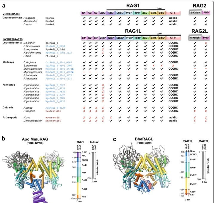
and contain domains corresponding to all of the func-tionally essential “core” subdomains of jawed vertebrate RAG1 and RAG2 (Fig. 4) [19, 22]. These core subdo-mains are: the nonamer binding domain (NBD), the dimerization and DNA binding domain (DDBD), the
Pre-RNase H domain (PreR), the catalytic RNase H do-main (RNH), two zinc binding dodo-mains (ZnC2 and ZnH2) that together coordinate a zinc ion, and the C-terminal Domain (CTD) (Fig. 4). The RAG1 C-terminal tail (CTT) is not required for catalytic activity [4], while
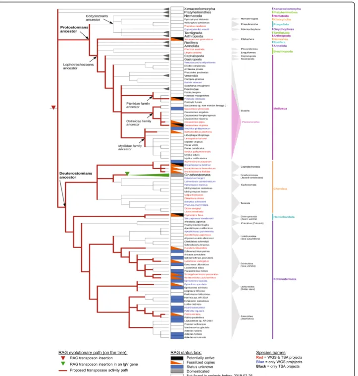
Fig. 3 Bilaterian evolutionary tree and RAG evolutionary history. Tree was built with species for which there are at least one WGS and/or TSA project in the NCBI database. The species in which there are no RAG-like sequences found were regrouped into larger groups (Xenacoelomorpha, Platyhelminthes, Nematoda, Tardigrada, Arthropoda phyla), and the Gnathostome species were grouped as well, as RAG is domesticated in all of these species. Several species contain RAG1L-RAG2L copies with different statuses (e.g., one copy is potentially active and another is fossilized), and in such cases, the species are annotated as having more than one status within the status box. Red lines indicate branches in which RAGL transposon activity might have been present
the corresponding domain from BbeRAG1L (CTT*; an asterisk specifies domains derived from RAG1L proteins) is critical for full activity and is part of the BbeRAG1L core region [19,22]. Both RAG1 and BbeRAG1L contain zinc finger motifs C1/C1*, C2/C2*, and C3/C3*, as well
as the ring zinc finger dimerization domain (ZDD/ ZDD*) in their non-essential N-terminal regions (Fig.
4a). The RAG2 core domain is a 6-bladed β-propeller composed of 6 kelch repeats [5]; this region, but not the RAG2 C-terminal domain with its acidic hinge and plant
Fig. 4 Protein domains found in protostome RAGL proteins. a Predicted domains present in the best preserved RAG1L and RAG2L proteins identified in this study (blue font) compared with RAG and RAGL protein sequences from deuterostomes (black font) and representative Transib transposase proteins (red font). All RAG1L and RAG2L proteins shown exist in tandem pairs with the exception of M. philippinarum RAG1L and RAG2L. Black lines, RAG1(L) and RAG2(L) core regions. Domains are not depicted to scale. RAG1L from S. glomerata is intact except for a premature stop codon in the ZnH2 domain. b, c Cartoon representations of the apo mouse RAG (i.e. in the absence of DNA) and B. belcheri RAGL (with DNA removed) tetramer structures, with domains colored as indicated and darker and lighter tones used to discriminate between subunits. Boundaries between domains are indicated with residue numbers. RAG1(L) domain abbreviations used: N-terminal zinc finger motifs, C1(*), C2(*), C3(*); ring zinc finger dimerization domain, ZDD(*); nonamer binding domain, NBD(*); dimerization and DNA binding domain, DDBD(*); pre-RNase H domain, PreR (*); catalytic RNase H domain, RNH(*); zinc finger ZnC2(*), zinc finger ZnH2(*),C-terminal domain, CTD(*); and C-terminal tail, CTT(*), that contains either the CCGHC motif of invertebrate RAG1L or acidic amino acids of vertebrate RAG1. RAG2(L) domain abbreviations used: 6-bladed kelch-type beta propeller domain, 6-Kelch(*); and plant homeodomain, PHD(*)
homeodomain (PHD) finger, is found in BbeRAG2L (Fig.4) [19].
We analyzed the sequences of the predicted RAG1L and RAG2L proteins from protostomes to determine if these species, like amphioxus, had the potential to encode active RAGL complexes. Indeed, the phyla Mollusca and Nemer-tea each harbor multiple pairs of intact RAG1L-RAG2L open reading frames able to encode the kelch repeat do-main of RAG2/BbeRAG2L and all of the essential core subdomains found in RAG1 and BbeRAG1L, including CTT* of BbeRAG1L (Fig.4a; Figs.5and6show sequence alignments of selected RAG1L and RAG2L proteins, re-spectively, while alignments of all of the RAG1L and RAG2L protein sequences identified are shown in Add-itional file7: Alignment S1a,b). In Mollusca, such pairs are observed in two species (C. virginica and P. imbricata), while the nemertean N. geniculatus harbors at least six dif-ferent intact RAG1L-RAG2L protein pairs. Conservation of the core domains is also observed in the RAG1L-RAG2L pair identified in the cnidarian A. aurita (Figs.4,7
and Additional file5: S5a,b).
Many protostome RAG1L proteins also exhibit sub-stantial conservation with the RAG1 N-terminal non-core region. The three N-terminal zinc finger motifs are well conserved among most of the newly detected RAG1L homologs while ZDD* is readily identified in RAG1L sequences from Mollusca (Fig.4a, Additional file
7: Alignment S1a). However, as in RAG1L proteins from echinoderms [9,10,17], ZDD* is absent from all RAG1L proteins from N. geniculatus and from RAG1L of A. aur-ita(Fig. 4a, Additional file 7: Alignment S1a). This sug-gests that this domain, which in RAG1 forms a tight dimer with E3 ubiquitin ligase activity [30], has under-gone at least two independent loss events during RAG1L evolution.
With the exception of RAG2L from amphioxus [19], all invertebrate RAG2L proteins contain a C-terminal PHD finger (Figs.4a,6and Additional file5: S5b).
Patterns of sequence conservation in RAG1L and RAG2L domains
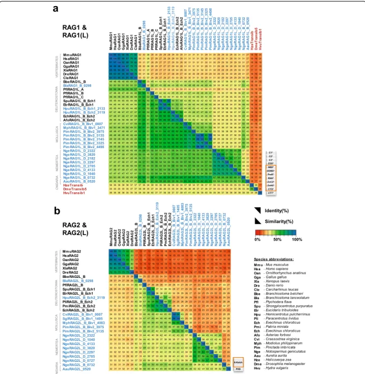
We analyzed the patterns of conservation of key amino acid residues and domains of protostome RAG1L and RAG2L proteins to provide further insight into their potential functional properties and evolutionary relationships. Analysis of levels of se-quence identity within the core region of RAG1L proteins reveals a broad correspondence with species phylogeny, with levels of identity highest between the RAG1L_B family sequences from protostomes and deuterostome invertebrates (Fig. 7a). And in general, core RAG1L sequences from invertebrates exhibit greater identity to one another than to core RAG1 sequences from jawed vertebrates, with the
exception of the RAG1L_A family member from P. flava. Transib sequences diverge most strongly, exhi-biting less than 22% sequence identity with the RAG1 and RAG1L proteins analyzed. Transib’s low sequence identity with RAG1/RAG1L and absence of elements corresponding to the RAG1 N-terminal non-core region allow one to distinguish between Transib and RAG1/RAG1L proteins. As was the case for RAG1/RAG1L, RAG2 and RAG2L core region se-quences exhibit higher levels of identity within inver-tebrates than between jawed vertebrates and invertebrates (Fig. 7b). As expected, overall levels of sequence identity are lower for RAG2/RAG2L than for RAG1/RAG1L.
Numerous stretches of conservation are observed be-tween protostome and deuterostome core RAG1L/RAG1 sequences beginning with the preR domain and extend-ing to the CTD (Fig. 5 and Additional file7: Alignment S1a). In addition, numerous functionally/structurally important residues are highly conserved in RAG1L se-quences from protostomes. These include critical cata-lytic residues [4] and four zinc-coordinating residues from ZnC2 and ZnH2 that stabilize domain folding (Fig.
5, Additional file 5: Figure S5a and Additional file 7: Alignment S1a). The Cx2Cx3GHx4C motif that defines
CTT* of BbeRAG1L is found in essentially all RAG1L sequences from protostomes and deuterostome inverte-brates (Fig.5 and Additional file7: Alignment S1a). Nu-merous other potential zinc coordinating residues are also conserved in protostome RAG1L sequences includ-ing many in ZDD*, C1*, C2*, and C3* (Fig.4a, and Add-itional file 7: Alignment S1a). Most protostome RAG1L proteins contain valine at the position equivalent to mouse RAG1 E649, and mutation of E649 to V or A in-creases the propensity of RAG to perform asynchronous, or “uncoupled” cleavage in vitro and in cells [22, 31] (Additional file 5: Figure S5a). Virtually all protostome RAG1L proteins contain a hydrophobic amino acid at the position equivalent to mouse RAG1 R848, a change that strongly activates the transposition activity of RAG in vitro and in cells [22] (Additional file 5: Figure S5a). Furthermore, invertebrate RAG2L proteins reported here and previously lack the acidic linker that exists be-tween the RAG2 core and the PHD in jawed vertebrate RAG2 proteins (Fig. 6 and Additional file7: Alignment S1b), and this acidic region has been shown to inhibit RAG-mediated transposition in cells [22]. These latter observations are consistent with the idea that proto-stome RAGL enzymes are, or evolved from, active transposases.
The core region of protostome RAG2L proteins preserves the structural features of a kelch-type do-main including the GG motif that typifies the second β-strand of each kelch repeat (Fig. 6 and Additional
file 7: Alignment S1b). The core region of protostome RAG2L is invariably followed by a cysteine-rich PHD, but the pattern of C and H zinc-coordinating residues found in protostome and deuterostome invertebrate PHDs (Cx4-7Cx14-16Cx2-4Cx4Hx2Cx11-18Cx2C) differs
considerably from that seen in vertebrate RAG2 PHDs (CCx2Cx22Cx5-6Hx2Hx2Cx19Cx2H) (Additional file 5:
Figure S5b and Additional file 7: Alignment S1b). The remarkable conservation of the C/H pattern in inver-tebrate RAG2L PHDs and its divergence from the pattern observed in its vertebrate counterparts suggest structural and functional differences that are as yet largely unexplored. The RAG2L PHD from the purple
sea urchin S. purpuratus is capable of binding the tail of histone H3 when lysine 4 is methylated, although its pref-erence for dimethylated lysine differs from the trimethyla-tion preference of the mouse RAG2 PHD [32].
Together, these sequence analyses argue that many RAG1L-RAG2L protein pairs from protostomes have the potential to be active endonucleases with transposase activity.
Analysis of protein-DNA and protein-protein interaction surfaces
The availability of RAG1L-RAG2L sequences from pro-tostomes provided an opportunity for a broad
Fig. 5 Alignment of RAG1L sequences from protostomes and deuterostomes. RAG1L sequences from 3 deuterostomes (the cephalochordate amphioxus (Bbe), echinoderm purple sea urchin (Spu), and hemichordate P. flava (Pfl)), 2 mollusk RAGL_B subfamilies (eastern oyster (Cvi) and pearly oyster (Pim)), and a nemertean N. geniculatus RAGL_D family representative (Nge) were aligned to mouse (Mmu) RAG1. Domains, sequence motifs, secondary structure assignment (helices - wavy lines; beta sheet - arrows, other - straight line), protein-protein and protein-DNA contact interactions (within 5 Å) displayed above the alignment derive from the BbeRAG1L cryo-EM structure (PDB: 6B40). Acidic catalytic residues, red; active site residue mouse H795, purple; zinc coordinating residues within ZDD (*) and ZnC2 and ZnH2 (#) are indicated above the sequences. Locations at which coding sequences span exon boundaries are underlined. Amino acid color code: hydrophobic aliphatic, yellow; hydrophobic aromatic, orange; positively charged, blue; negatively charged, red; neutral polar, light blue; glycine and prolines, grey; cysteine, purple; histidine, dark purple.Sequences displayed are BbeRAG1L_B (GenBank: KJ748699.1), PflRAG1L_B (TSA:GDGM01438088.1), SpuRAG1L_B_Ech1 (Uniprot: Q45ZT6), and CviRAG1L_B_Biv1_0007, PimRAG1L_B_Biv2_3145, and NgeRAG1L_D_2322 from this study (Additional file7: Alignment S1a).
evolutionary examination of the conservation of inter-action surfaces in the complexes formed by these pro-teins with each other and with DNA. This in silico analysis involved mapping representative RAG1L and RAG2L sequences from protostomes and deuterostomes (the six species whose sequences are shown in Figs. 5
and 6) onto the recently reported BbeRAG1L-BbeRAG2L three-dimensional structure, which closely resembles that of vertebrate RAG1-RAG2 [22]. This re-vealed strong conservation of the RAG1L DNA binding groove in regions that interact with both the TIR
heptamer and the TIR heptamer-flanking region (Fig.8a, b). This binding region contains numerous basic amino acid residues, creating a positively charged surface for DNA interaction [22]. This observation, combined with the high sequence conservation that surrounds the active site residues D701, E764, D811, H894 and E1063 in BbeRAG1L (Fig. 5, Additional file 5: Figure S5a and Additional file 7: Alignment S1a), suggests that proto-stome RAGL proteins have the potential to interact with and cleave DNA in a manner similar to that of RAG and BbeRAGL.
Fig. 6 Alignment of RAG2L sequences from protostomes and deuterostomes. RAG2L sequences are aligned and displayed as in Fig.5. Domains, beta sheet regions of each kelch-type blade, the GG motif, secondary structure (helixes - wavy lines; beta sheet - arrows, other - straight line), protein-protein interactions (5 Å threshold) displayed above the alignment derive from the BbeRAG2L cryo-EM structure (PDB: 6B40). Sequences displayed are: BbeRAG2L_B (GenBank: KJ748699.1), PflRAG2L_B (TSA:GDGM01438088.1), SpuRAG2L_B_Ech1 (Uniprot: Q45ZT5) and CviRAG2L_B_Biv1_0007, PimRAG2L_B_Biv2_5135, and NgeRAG1L_D_2322 from this study (Additional file7: Alignment S1b). These RAG2L proteins are the transposon pairs of the RAG1L sequences displayed in Fig.5except that PimRAG2L_B_Biv2_5135 was used instead of PimRAG2L_B_Biv2_3145 due to merge uncertainties in the 3145 sequence; these two sequences are 98% identical on their counterpart RAG1L core. Species abbreviations as in Fig.5
In contrast, the portions of RAG1L predicted to inter-act with RAG2L are less well conserved (Fig.8a, b), and reciprocally, the portions of RAG2L predicted to interact with RAG1L also show high variability (Fig. 8c, d). Hence, the predicted RAG1L-RAG2L protein-protein interaction surfaces appear to have evolved more rapidly than the central DNA binding groove.
Discussion
Over the last 15 years, multiple RAG1L-RAG2L gene pairs, some flanked by TIRs and TSDs, have been dis-covered in the genomes of invertebrate deuterostomes [9, 10, 17, 19], leading to the hypothesis that the RAGL transposon first arose in an early deuterostome [9, 16,
22]. Our finding of RAG1L-RAG2L gene pairs and
Fig. 7 Identity and similarity matrices of the a RAG1L and b RAG2L core regions. Identity (upper right region) and similarity (lower left region) percentages were computed using the protein multiple sequence alignment shown in Additional file7: Alignment S1a, b starting from the beginning of RAG1L NBD(*) until the end of CTD(*) and RAG2L kelch-type domain respectively, as described in Methods. Two sequences from Additional file7: Alignment S1a (SglRAG1L_B_Biv1_1405 and NgeRAG1L_D_0727) were not included because they are incomplete in the core region interval
potential RAGL transposons in protostomes calls this hy-pothesis into question. Phylogenetic analyses of RAG1L sequences suggest that RAGL transposon evolution has proceeded primarily through vertical transmission, sup-porting the possibility that the RAGL transposon arose in a bilaterian ancestor, if not earlier. Sequence and struc-tural analyses argue that at least some of the RAG1L-RAG2L protein pairs from protostomes, and even one from a non-bilaterian, have the potential to be active
endonucleases and transposases. Our findings have impli-cations for our understanding of the evolutionary history of the RAGL transposon and for the process of transposon molecular domestication, an important contributor to genome and species evolution [33–35].
Potentially activeRAGL transposons in protostomes
Numerous findings described here support the con-clusion that the RAGL transposon is present in the
Fig. 8 Sequence variability mapped onto BbeRAG1L/2 L cryo-EM structure (PDB: 6B40). a, b, c, d Surface representation of sequence variability of the protein-DNA and protein-protein contact interfaces of RAG1L (a, b) or RAG2L (c, d) in a lateral view of a RAG1L-RAG2L heterodimer (a, c) or a top view of the RAG1L-RAG2L tetramer (b, d). Jensen-Shannon divergence (JSD) conservation score is displayed using a rainbow color code as indicated with the scale bar, with blue and red indicating highly conserved and highly variable positions, respectively. RAG2L and RAG1L are shown in gray in (a, b) and (c, d), respectively, while TIR DNA and TIR flanking DNA are shown in black and white, respectively. e, f Alternative models for the evolutionary relationship between Transib and the RAGL transposon. In the current model [16] (e), Transib was the ancestral element and the RAGL transposon was derived from Transib through acquisition of a RAG2L gene. In the alternative model (f), the RAGL transposon was ancestral and the first Transib transposon arose from a RAGL transposon by loss of RAG2L
genomes of protostomes and has been active during protostome evolution: 1) Multiple elements with se-quence similarity to known RAG1/RAG1L and RAG2/ RAG2L genes of deuterostomes are present in proto-stome sequence databases; 2) protoproto-stome RAG1L and RAG2L genes often lie in close proximity in conver-gent transcriptional orientation; 3) many of these RAG1L-RAG2L gene pairs are flanked by TIRs and TSDs; 4) the TIRs resemble the sequence of the RSS heptamer and TIRs of deuterostome RAGL transpo-sons; 5) protostome RAGL transposon TSDs are five bp in length; 6) predicted protostome RAG1L and RAG2L proteins often contain critical active site and structurally important amino acid residues and all of the domains required for activity by RAG or BbeR-AGL; 7) conservation in protostome RAGL proteins extends to include non-essential but important regula-tory domains at the N-terminus of RAG1/RAG1L and the C-terminus of RAG2/RAG2L of deuterostomes; 8) two different families (RAGL_B and RAGL_D), several RAGL_B subfamilies, and multiple degenerate copies of RAG1L and RAG2L sequences are detected in the genomes of protostomes, arguing for instances of transposon movement followed by inactivation.
The TIRs associated with protostome RAGL transpo-sons exhibit two blocks of strong sequence conservation (Fig. 2a). The first is the perfectly conserved 5′-CAC
se-quence at the beginning of the heptamer, a region that is also rigidly conserved in jawed vertebrate RSSs. These res-idues are vital for cleavage [36], contributing to both a structural propensity for unwinding and sequence-specific protein-DNA contacts [7, 8]. The second conserved por-tion of the protostome TIR is an A-rich region from resi-dues 8–10. Conservation of this sequence and the 13–15 bp length of the protostome TIR consensus are notable in light of the findings that only the first 16–17 bp of the Pro-toRAG TIR are essential for cleavage and that CTT* of BbeRAG1L constitutes a novel DNA binding domain that interacts with TIR sequences that span this conserved AAA sequence [22]. CTT* from deuterostome RAG1L proteins contains a highly conserved Cx2Cx3GHx4C motif,
and the same motif is strongly conserved in CTT* in RAG1L proteins from protostomes.
We therefore propose that protostome RAG1L-RAG2L protein complexes recognize a core TIR se-quence of about 15 bp, with recognition of sese-quences flanking the heptamer mediated in part by CTT* [22]. If this is the case, then DNA recognition by inverte-brate RAGL complexes is likely to follow distinct rules from those of their vertebrate RAG relatives: in-vertebrate RAGL will rely predominantly on the ter-minal ~ 15–17 bp of the TIR and CTT* and will be relatively insensitive to TIR asymmetry, whereas ver-tebrate RAG relies on a bipartite and asymmetric
RSS, and in particular on a nonamer sequence sepa-rated from the heptamer by a spacer of 12 or 23 bp, with the nonamer recognized by a NBD on a flexible hinge [5–7]. Both protostome and deuterostome RAG1L proteins contain an NBD* domain with the potential to bind DNA—and in the case of BbeR-AG1L, there is evidence that this region does indeed interact with more distal TIR sequences—but because of the presence of CTT*, the BbeRAG1L NBD* do-main appears to serve a non-essential, auxiliary DNA binding function [22].
Evolutionary history of theRAGL transposon in bilaterians
We have assembled existing information concerning the presence or absence of TIRs, TSDs, and open reading frames capable of encoding intact and potentially func-tional RAG1L and RAG2L proteins to predict the status of RAGL across protostome and deuterostome species, with status characterized as potentially active, fossilized, domesticated, not found, or unknown (Fig. 3). In some species, including several protostomes, both potentially active and fossilized RAGL elements are present. We emphasize that attribution of status is highly influenced by the availability (Additional file6: Table S1) and qual-ity of existing sequence data.
From this information and the assumption of vertical transmission, a working model for the evolutionary his-tory of the RAGL transposon can be derived (red lines in Fig.3). On this model, the RAGL transposon was present and active in the common bilaterian ancestor, remained active in both deuterostomes and protostomes, and might still be active in nemerteans, oysters and pearl oysters as well as some deuterostome invertebrate spe-cies [9]. We emphasize that while there is as yet no dir-ect evidence for horizontal gene transfer of the RAGL transposon, such events cannot be ruled out, particularly between clades where the supporting sequence data re-main sparse and the corresponding phylogenetic ana-lyses provide lower levels of certainty. Our finding of several RAG1L and RAG2L sequences in cnidarians, in-cluding genes encoding a potentially active RAG1L-RAG2L protein pair, raises the possibility that the RAGL transposon arose prior to the origin of bilaterians, though again, horizontal gene transfer cannot be ruled out. Additional sequence data should allow testing of this idea and other predictions of the working model, providing a better understanding of the evolutionary events that led to the RAG recombinase.
Our findings suggest that four distinct RAGL protein families emerged during bilaterian evolution, two of which (RAGL_B and RAGL_D) are found in proto-stomes. The most widespread family, RAGL_B, is found in mollusks, nemerteans and many invertebrate deutero-stomes, and is further divided into subfamilies,
suggesting frequent duplication of the RAGL transposon in multiple clades. Many copies were subsequently lost while others were retained, some in fossilized form. This supports the idea that the RAGL transposon was broadly active during bilaterian evolution, giving rise to multiple families and subfamilies in some taxa.
The current model for RAGL transposon evolution posits that the first RAGL transposon was generated when a Transib transposon acquired a RAG2-like gene [16, 22]. This sequence of events, which places Transib prior to the RAGLtransposon, is based on the widespread distribution of Transib, which is found even in fungi [12]. However, many uncertainties remain regarding early events in RAGL/Transib evolution, including uncertainties regard-ing the extent to which Transib was spread by horizontal transmission. These considerations, together with our data consistent with the hypothesis that the RAGL transposon arose earlier than previously thought, suggest that we con-sider the alternative possibility that the RAGL transposon arose prior to Transib. Hence, in addition to the current model that the RAGL transposon arose from Transib by gain of RAG2L (Fig. 8e) [16], we suggest that a different scenario also be considered in which Transib arose from a RAGL transposon by loss of RAG2L (Fig. 8f). Sequence data from additional eukaryotes will help test the plausibil-ity of this second scenario.
Transposon molecular domestication and theRAGL transposon
Transposon molecular domestication refers to a process in which transposon-derived sequences are co-opted by the host to perform new functions [37]. The repurposing of the components of a RAGL transposon for jawed ver-tebrate V(D) J recombination illustrates the large evolu-tionary impact this process can have. Our finding that several protostomes harbor potentially active RAGL transposons expands the range of species within which domestication of RAGL transposons could have oc-curred. A switch in biological function from transposase to sequence-specific endonuclease appears to be a com-mon evolutionary event for multiple transposon families. In addition to the conversion of the RAGL transposase into the RAG recombinase, two such domestication events have been documented in the yeast Kluyvero-myces lactis, where the Kat1 andα3 endonucleases, de-rived from hAT family and MULE family transposases, respectively, trigger mating type switching [38–40]. In ciliates, multiple endonucleases derived from PiggyBac family transposases mediate the programmed DNA rear-rangements that remodel the somatic genome [41–43]. And PGBD5 and THAP9, factors derived from PiggyBac and Drosophila P-element family transposases, respect-ively, are active endonucleases expressed in humans whose domesticated function remains to be determined
[44, 45]. Our findings identify multiple examples of in-tact RAG1L and RAG2L genes, either in pairs or in isola-tion, that appear to lack one or both flanking TIRs (Fig.
1c and Additional file 1: Figure S1a) and hence are un-likely to retain the ability to transpose. These RAGL genes, which are found in protostomes and in the moon jellyfish A. aurita, join the previously identified RAG1L-RAG2L gene pair from the purple sea urchin [17] as potentially domesticated derivatives of the RAGL trans-poson. Biochemical and structural analyses of the RAGL proteins encoded by these loci might shed light on their putative novel biological functions.
Conclusion
The pivotal role played by a RAGL transposon in the evolution of the jawed vertebrate adaptive immune sys-tem represents a paradigmatic example of transposon molecular domestication. The findings reported here are consistent with a revised model for the evolutionary his-tory of the RAGL transposon in which this transposon was present and active in the bilaterian ancestor. Our findings strongly suggest that RAGL transposons were transmitted vertically and in active form in multiple protostome lineages, as is also thought to be the case in deuterostomes. Our findings also argue that intact and potentially active RAGL transposons exist in the ge-nomes of protostomes today, and similarly, that proto-stome genomes contain an assortment of intact RAG1L-RAG2L adjacent gene pairs that appear to lack flanking TIRs and are candidates for molecular domestication. Hence, the potential for RAGL transposons to have con-tributed novel gene functions during eukaryotic evolu-tion is substantially broader than previously anticipated. Methods
Genomic and Transcriptomic database screening
Detection of new RAG-like sequences was performed starting from a collection of previously reported RAG1-like and RAG2-RAG1-like sequences from deuterostomian or-ganisms B. belcheri [19], S. purpuratus [17], P. flava and P. miniata [9]. Queries were searched against all meta-zoan invertebrate and jawless vertebrate projects avail-able before February 2019 from multiple databases such as Whole-Genome Shotgun Contigs (WGS), High Throughput Genomic Sequences (HTGS) and Tran-scriptomic Shotgun Assembly (TSA), using TBLASTN [24,25], with a Blosum62, Blosum45 and PAM250 simi-larity matrices and a e-value threshold of 1e-08.
Regions containing RAG1L sequence signatures were further assessed for their potential to encode complete RAG1L proteins in the intron/exon context by analyzing the level and distribution of sequence similarity and the secondary structure profile match to RAG1L. The most complete newly detected RAG1L homologues were
further used iteratively as queries in order to detect more divergent RAG1L sequences that were initially below the detection threshold or to allow for detection of RAG1L N-terminal non-core domain regions (which are more variable and hence harder to detect) in already detected RAG1L sequences.
In a second step, the WGS/TSA regions bordering RAG1L (within ~ 10 kb) were further explored to find potential RAG2L sequences. Here, special attention had to be drawn to sequence analysis. Because of the low levels of similarity among RAG2L proteins, retrieved hits often exhibited only partial coverage or were detected by only one or two RAG2L homologs. In such cases, a pool of translation product predictions was extracted and trimmed based on conservation of Kelch domain struc-tural properties and its motif conservation.
The newly identified RAG1L and RAG2L sequences were further used independently as queries in an itera-tive manner to expand the detection threshold and find other more degraded copies within the same WGS pro-jects or new hits in new WGS / TSA propro-jects. In this step, the searches were performed in an unbiased fash-ion to identify not only RAG1L-RAG2L pairs, but also solitary loci. This resulted in only several solitary RAG1L or RAG2L that had the potential to be intact genes, sharing over 50% identity with their paralogues from a RAG1L-RAG2L tandem pair. These were also consid-ered for further analysis as some of them might be trans-latable to protein even if they were isolated from the counterpart RAGL locus. Detailed information on the detected loci is presented in Additional file 8: File S1 and Additional file9: File S2.
Prediction of protein translation products was per-formed starting from a FGENESH and FGENESH+ [46] and Augustus [47] pool of predicted products. The pre-dicted protein sequences that have different exon compos-ition were further trimmed based on mRNA sequence compatibility (when TSA entries were available) and based on the presence of highly conserved sequence motifs and subdomains that are found in full sized known deuterosto-mian homologs. Exon merging areas that are not covered in mRNA data are subjected to a higher degree of uncer-tainty and therefore are underlined where present.
Phylogenetic analysis
Alignments were created with MEGA X [27] using Clus-talW [48]. RAG trees (Fig.2, Additional file3: Figure S3) were built using MEGA X (Maximum Likelihood method, complete deletion, WAG with Freqs. (+) correc-tion model [28], Gamma distribution with 5 categories, 1000 bootstrap replicates) and were confirmed with PhyML [49, 50] (Maximum Likelihood method) using the WAG substitution model, or AIC-based and BIC-based model selection (Additional File4: Figure S4). The analyses
were done first on RAG1L sequences because it is more conserved than RAG2L, and then the RAG2L sequences were analyzed to complement the results. To identify new RAGL sequence families, all RAG1L fragments longer than approx. 100 bp were aligned. Thereafter, we selected the areas of the alignment that were sufficiently conserved to identify the most significant positions (substitutions) with which to build the tree. This revealed the significant mono-phyletic groups with bootstrap values greater than 50, allowing us to define the representative sequences in each monophyletic group. The previously identified RAG/RAGL A, B and C families [9] represent different duplications of the RAGL transposon. Whenever a sequence did not sig-nificantly form a monophyletic group with a known family, we defined it as a new family, together with other quences that form a monophyletic group with this se-quence, as for example the N. geniculatus RAGL_D sequences. In contrast, if a sequence formed a monophy-letic group significantly with an existing RAGL transposon family, we defined it as part of that family.
Data availability & Bilateria tree
We established an overview of the species for whom se-quence data was present in the WGS and TSA databases of NCBI on February 26, 2019 (Additional file 6: Table S1). While TSA projects are typically indicated as “TSA master”, some additional sequences marked as “Tran-scripts” are detected on a BLAST search against the TSA database (e.g., Branchiostoma lanceolatum), and some species that lack identifiable RAGL sequences might have been omitted inadvertently from Additional file 6: Table S1. From the available species in the NCBI database, a summary species tree was built using NCBI Taxonomy Common Tree [51, 52] and was edited with iTOL 4.3.2 [53]. The evolutionary tree timeline shown in Fig. 1b was obtained from TimeTree [54] and the tree editing was performed in online iTOL v4.3 [53].
Detection of TIR and TSD sequences
The detection of TIRs is challenging due to their small size, the high incidence of short inverted repeats in DNA sequences, and the sequence drift expected to occur between the two TIRs after elements become do-mesticated. Moreover, previously reported TIR pairs in Deuterostomia [9] exhibit substantial variation between the 5′-TIR and 3′-TIR, with strong similarity only present in the vicinity of the terminal heptamer-like re-gion. However, a significant drop in sequence identity is expected to occur at the tip of the TIR because the transposon cassette is expected to be similar in sequence to other transposon copies, while the flanking regions are ex-pected to be divergent. We therefore designated sequences as TIRs only when they satisfied the following three strin-gent conditions: 1) a significant homology drop was
detected on both sides of the RAG1L-RAG2L gene pair, 2) an inverted repeat, with greater than 50% identity between the two sequences, was present at the sites of the observed homology drops, and 3) a TSD was present flanking the terminal inverted repeats. The presence of TIR and TSD se-quences was investigated in DNA regions where a signifi-cant drop in homology was detected using in a similar approach to that described previously [9]. Margins of 2–3
kb adjacent to RAG1L and RAG2L loci were compared be-tween different cassette copies from the same or closely re-lated organisms using the Needleman–Wunsch [55, 56] and Lalign [56,57] pairwise alignment algorithms. In cases where a homology drop was detected at both ends flanking RAG1L and RAG2L loci, the homology boundaries were searched for inverted repeats. Furthermore, the presence of a 3–8 bp TSD adjacent to the identified inverted repeat was required and allowed us to discriminate between TIRs and a premature end of the transposon cassettes. The TIR pairs flanked by TSDs were then used to identify transposon margins containing solitary, unpaired TIRs using blastn [58,
59] and 150 bp TIR containing margins as queries of each WGS project data. Detailed information about the detected TIRs and TSDs are provided in Additional file 8: File S1 and Additional file9: File S2.
Sequence analysis and variability
Domains within each of the identified RAG1L and RAG2L pair were delineated using InterproScan [60], while RaptorX-property [61] was used to predict the local sec-ondary structure. Multiple sequence alignments were per-formed using T-coffee in psicoffee mode using a Uniref50 database for homology searching [62,63]. Due to the low homology between RAG2L sequences, the Kelch-type do-mains and the PHD dodo-mains were first aligned separately and subsequently merged into a single alignment.
Identity percentage matrices were computed excluding gaps, using Unipro Ugene v1.22.0 [64], as the ratio of identical amino acid pair counts over the length of the smallest sequences from the compared pair. Similarity percentages presented below the diagonal in the same figures were computed using an in-house implementa-tion of the Ugene algorithm, but using for counts the matrix of all BLOSUM62 positive substitutions, as used in the blast-like methods. Ugene was also used to gener-ate the graphics included in figures containing protein and nucleotide alignments, while AnnotationSketch [65] was used to generate genomic organization figures.
Given redundancies and the unbalanced distribution of RAGLs among the evolutionary branches, variability was computed only over a nonredundant set of 6 RAGL pairs sharing less than 50% protein sequence identity within the core RAG1L region. This set proves also to be represen-tative for the evolutionary clades and consists of three deu-terostome sequences: cephalochordata group (amphioxus),
echinodermata group (sea urchin), hemichordata clade (P. flava) and three protostome sequences: one from each mol-lusk RAGL_B subfamily, and one nemertean N. geniculatus RAGL_D family representative. P. flava RAGL_A and RAGL_C were discarded given the low confidence protein prediction for their RAG2L counterparts.
Conservation Jensen-Shannon divergence (JSD) was used to compute similarity scores for each position in the align-ment of the above six sequences and used to map the se-quence variability of RAGL. JSD was calculated using the implementation of [66] based on Blosum62 background probabilities with a gap penalty of 1 and window = 0.
Relative entropy logo was generated using WebLogo [67] and PyMOL Molecular Graphics System, Version 2.2.3 Schrödinger, LLC was used to represent all protein structures.
Supplementary information
Supplementary information accompanies this paper athttps://doi.org/10. 1186/s13100-020-00214-y.
Additional file 1: Figure S1. Genomic organization of RAGL and RAGL transposons identified in this study. (a) Genomic organization of RAGL copies identified in deuterostomes, mollusks, nemerteans and cnidarians. Only the most relevant RAG1L/RAG2L pairs are shown. The legend for panels (a) and (b) is provided at the bottom of panel (b). Loci that are likely to be pseudogenized are indicated with a white box. Supporting transcriptomic data are indicated along with corresponding TSA entry. Green and gray arrows indicate transcripts corresponding to coding and untranslated regions, respectively. Unmapped regions of transcripts are shown as unfilled rectangles outside of the gene track. (b) Genomic organization of incomplete and potentially pseudogenized RAG1L/RAG2L loci in cnidarians. Most of these regions either have stop codons or low sequence coverage and are therefore shown with vertical stripes. The P. damisconis locus is incomplete as it is located at the margin of the scaffold and might encode a complete protein. Assembly gaps near the detected loci are shown as black boxes.
Additional file 2: Figure S2. (a) Alignments of four groups of protostome RAGL transposon TIRs. Sequences of 5′-TIR and 3′-TIR (reverse complement) pairs are aligned from four groups: C. virginica (I), P. imbricata (II and III) and nemertean N. geniculatus (IV). Despite both being mollusks, the C. virginica and P. imbricata TIR sequences are very dissimilar both in sequence and length. None of the 4 groups contain an RSS nonamer-like region, however, the C. virginica 5′ and 3′ TIRs exhibit a length difference of 11 bp, reminiscent of the 11 bp difference in lengths of the spacers in the 12RSS and 23RSS that underlies the 12/23 rule of V(D) J recombination [3,6]. Similarity color code: dark grey, fully con-served; light grey, partially concon-served; white, no similarity. (b) Potential RAG-derived non-autonomous transposable elements in protostomes and deuterostomes. Summary of the potential RAG-derived non-autonomous elements identified in WGS database in organisms where the RAGL transposon was detected. The configuration of these elements, as well as TSD sequences (if present) are indicated. The e-values shown derive from blastn searches of the WGS project of each organism using previously detected RAGL TIR-containing margins (200 bp) as queries. Additional file 3: Figure S3. Additional phylogenetic analyses. (a-d) Detailed phylogenetic trees of RAG1 and RAG1L protein sequences including several from: (a) Cephalochordata - indicated with blue shading., (b) Echinodermata - blue shading, (c) Mollusca - orange shading and (d) including one from cnidaria - green shading (e) RAG2/ RAG2L phylogenetic trees. Trees were built using Maximum Likelihood and WAG substitution model as implemented in MEGA X [27] and are displayed as in Fig.2b except that branches with bootstrap numbers below 50% were not collapsed together. Trees were built from a variable
number of significant positions of their alignment: (a) 289, (b) 398, (c) 354, and (d) 469 respectively .
Additional file 4: Figure S4. Complementary RAG1L phylogenetic analyses using PhyML implementation [49,50]. Trees are displayed as in Fig.2b except that branches with bootstrap numbers below 50% were not collapsed together. Different substitution models were used as follows: (a) LG + G + I + F model selected via AIC minimization (b) LG + G + I model selected via BIC minimization (c) WAG substitution model. Additional file 5: Figure S5. (a) Conservation of functional relevant amino acids in RAG1 and RAG1L. The alignments depict the extensive conservation of some of the most important and well characterized amino acids in RAG1/RAG1L proteins (numbers given for BbeRAG1L): from left to right, catalytic carboxylates (D701, E764, D818, and E1063), residues implicated in controlling coupled versus uncoupled cleavage (A1064 and V751), a residue that facilitates transposition (M949), a histidine component of the active site (H894), and zinc-coordinating resi-dues (C830, C833, H1035 and H1040). Residue E649 in mouse RAG1 con-tributes to synchronous, or“coupled”, cleavage by RAG at two RSSs, in part through its ability to form a hydrogen bond with S963 [22,31]. The BbeRAG1L/2 L complex (BbeRAGL) exhibits less propensity for coupled cleavage in part because E649 has been replaced with V751 [22]. Valine is highly conserved at this position in RAG1L proteins from protostomes, suggesting that DNA cleavage by these proteins, if it occurs, would more likely resemble the uncoupled cleavage activity of BbeRAGL. Mutation of the charged residue R848 in mouse RAG1 to the hydrophobic residue methionine, as is found in BbeRAG1L, strongly activates the transposition activity of RAG [22]. Virtually all invertebrate RAG1L proteins, including those from protostomes, have a hydrophobic amino acid, most often me-thionine, at this position. (b) RAG2/RAG2L PHD domain alignment. The pattern of conserved cysteine and histidine residues (marked with X) are different between invertebrate RAG2L (top) and jawed vertebrate RAG2 (bottom). Variability logo (top) shows relative entropy (bits) calculated on the invertebrate alignment group. Amino acid color code as in Fig.5. Additional file 6: Table S1. Genomic and transcriptomic data availability for bilaterian invertebrates. List of the bilaterian species for which there are Transcriptome Shotgun Assembly (TSA) and/or Whole Genome Shotgun (WGS) projects in the NCBI database as of February 26, 2019. Gnathostomata species are not listed. The taxonomic identifier is given in the NCBI taxid column and corresponds to that used in NCBI databases. The number of projects available is indicated. In the Number of TSA projects column, transcriptomic projects that were not marked as “TSA project” are indicated in parentheses.
Additional file 7. Alignment S1 Multiple sequence alignment of (a) RAG1/RAG1L and (b) RAG2/RAG2L predicted proteins. Domains, sequence motifs, secondary structure assignment, protein-protein and protein-DNA contact interactions (within 5 Å) displayed above the align-ment derive from the BbeRAGL cryo-EM structure (PDB: 6B40). Addition-ally, for RAG1/RAG1L (a), acidic catalytic residues, red; active site residue mouse H795, purple; zinc coordinating residues within ZDD (*) and ZnC2 and ZnH2 (#) are indicated above the sequences, while for RAG2/RAG2L (b) the beta sheet regions of each kelch-type blade and the GG motifs are shown above the alignment. Locations at which coding sequences span exon boundaries are underlined. Sequence descriptions including references to genomic, transcriptomic or protein databases are shown at the end of the alignment, along with a legend of the symbols used. Additional file 8: File S1. Detailed presentation of the sequence information about the new RAGL loci identified in this study: detection relevant data (e-values and query-target alignments from TBLASTN), TIR and TSD detection information, predicted protein sequences, and add-itional relevant observations regarding some of the sequences. Additional file 9: File S2. Identified RAGL loci mapped onto nucleotide sequence.
Abbreviations
RAG:Recombination Activating Gene; RAGL: RAG-like; TIR: Terminal inverted repeat; TSD: Target site duplication; RSS: recombination signal sequence; ZDD: ring zinc finger dimerization domain; NBD: nonamer binding domain; DDBD: dimerization and DNA binding domain; PreR: pre-RNase H domain;
RNH: catalytic RNase H domain; ZnC2: zinc finger domain that contributes two cysteine residues; ZnH2: zinc finger domain that contributes two histidine residues; CTD: C-terminal domain; CTT: C-terminal tail; PHD: plant homeodomain
Acknowledgments
The authors thank Y. Zhang for input on RAG and BbeRAGL structures. Authors’ contributions
D.G.S and A.J.P. provided overall direction for the analyses. E.C.M. and C.V. performed the WGS searches. E.C.M. preformed the alignments, structural predictions and variability analysis. C.V., L.T.N. and P.P. performed
phylogenetic analyses. All authors contributed to the data interpretation and analysis and to the writing of the paper. The author(s) read and approved the final manuscript.
Funding
This work was supported by a public grant overseen by the French National Research Agency (ANR) as part of the second“Investissements d’Avenir” program (reference: ANR-17-RHUS-000X) (P.P.), UEFISCDI grant PN-III-ID-PCE-2016-0650 and Romanian Academy programs 1 & 2 of IBAR (E.C.M. and A.J.P), and NIH grant R01 AI137079 (D.G.S.).
Availability of data and materials
The datasets analyzed during the current study are available in the GenBank repository, specifically in Whole-Genome Shotgun Contigs (WGS) and Tran-scriptomic Shotgun Assembly (TSA). Additional file8: File S1 contains a de-tailed presentation of the sequence information about the new RAGL loci identified in this study: detection relevant data (e-values and the exact iden-tified segments alignments from TBLASTN), TIR/TSD detection information, predicted protein sequences and additional relevant observations regarding some of the sequences, while Additional file9: File S2 contains nucleotide sequence data.
Ethics approval and consent to participate Not applicable.
Consent for publication Not applicable. Competing interests
The authors declare that they have no competing interest. Author details
1Department of Bioinformatics and Structural Biochemistry, Institute of
Biochemistry of the Romanian Academy, Splaiul Independentei 296, 060031 Bucharest, Romania.2Evolutionary biology team, Aix Marseille Université IRD,
APHM, MEPHI, IHU Méditerranée Infection, Marseille, France.3SNC5039 CNRS,
19-21 Boulevard Jean Moulin, 13005 Marseille, France.4Department of
Immunobiology, Yale School of Medicine, 300 Cedar Street, Box 208011, New Haven, CT 06520-8011, USA.
Received: 18 December 2019 Accepted: 14 April 2020
References
1. Flajnik MF. Re-evaluation of the immunological big bang. Curr Biol. 2014; 24(21):R1060–5.
2. Litman GW, Rast JP, Fugmann SD. The origins of vertebrate adaptive immunity. Nat Rev Immunol. 2010;10(8):543–53.
3. Gellert M. V(D) J recombination: RAG proteins, repair factors, and regulation. Annu Rev Biochem. 2002;71:101–32.
4. Schatz DG, Swanson PC. V(D) J recombination: mechanisms of initiation. Annu Rev Genet. 2011;45:167–202.
5. Kim MS, Lapkouski M, Yang W, Gellert M. Crystal structure of the V(D) J recombinase RAG1-RAG2. Nature. 2015;518(7540):507–11.
6. Ru H, Chambers MG, Fu TM, Tong AB, Liao M, Wu H. Molecular mechanism of V(D) J recombination from synaptic RAG1-RAG2 complex structures. Cell. 2015;163(5):1138–52.
7. Kim MS, Chuenchor W, Chen X, Cui Y, Zhang X, Zhou ZH, et al. Cracking the DNA code for V(D) J recombination. Mol Cell. 2018;70(2):358–70 e4.