HAL Id: hal-00261171
https://hal.archives-ouvertes.fr/hal-00261171
Submitted on 6 Mar 2008HAL is a multi-disciplinary open access archive for the deposit and dissemination of sci-entific research documents, whether they are pub-lished or not. The documents may come from teaching and research institutions in France or abroad, or from public or private research centers.
L’archive ouverte pluridisciplinaire HAL, est destinée au dépôt et à la diffusion de documents scientifiques de niveau recherche, publiés ou non, émanant des établissements d’enseignement et de recherche français ou étrangers, des laboratoires publics ou privés.
Zinc adaptation and resistance to cadmium toxicity in
mammalian cells: molecular insight by proteomic
analysis.
Estelle Rousselet, Alain Martelli, Mireille Chevallet, Hélène Diemer, Alain van
Dorssealer, Thierry Rabilloud, Jean-Marc Moulis
To cite this version:
Estelle Rousselet, Alain Martelli, Mireille Chevallet, Hélène Diemer, Alain van Dorssealer, et al.. Zinc adaptation and resistance to cadmium toxicity in mammalian cells: molecular insight by proteomic analysis.. Proteomics, Wiley-VCH Verlag, 2008, 8 (11), pp.2244-55. �10.1002/pmic.200701067�. �hal-00261171�
Zinc adaptation and resistance to cadmium toxicity in mammalian
cells. Molecular insight by proteomic analysis.
Estelle Rousselet1,2,3, Alain Martelli1,2,3,4, Mireille Chevallet3,5,6, Hélène Diemer7, Alain
Van Dorsselaer7, Thierry Rabilloud3,5,6 and Jean-Marc Moulis1,2,3
1
CEA, DSV, IRTSV, Laboratoire de Chimie et Biologie des Métaux, Grenoble,
France.
2
LCBM, CNRS, Grenoble, France.
3
Université Joseph Fourier, Grenoble, France
4
present address: IGBMC, 1 rue Laurent Fries, BP 10142, 67404 Illkirch, France
5
CEA, DSV, IRTSV, Laboratoire de Biochimie et Biophysique des Systèmes
Intégrés; Grenoble, France
6
BBSI, CNRS, Grenoble, France
7
Laboratoire de Spectrométrie de Masse, CNRS UMR7178, Strasbourg, France Correspondence: Jean-Marc Moulis, CEA-Grenoble, IRSTV/LCBM, 17 rue des Martyrs,
F-38054 Grenoble Cedex 9, France. Tel: (33) 4 38 78 56 23, e-mail: jean-marc.moulis@cea.fr
Abbreviations: HZR: High Zinc Resistant HeLa cells; SERCA: sarco-endoplasmic reticulum
Ca2+-ATPase; ICP-OES: Inductively coupled plasma optical emission spectroscopy; LC50:
lethal concentration for 50% of the cells; HSP: Heat Shock Proteins; UPR: Unfolded Protein
Response; Hop: Hsp70/Hsp90 organizing protein; HPPD : 4-hydroxyphenylpyruvate
dioxygenase; NTBC: 2-(2-Nitro-4-Trifluoromethyl-Benzoyl)-1,3-Cyclohexanedione; HGO :
homogentisate 1,2-dioxygenase (EC E.C.1.13.11.5) ; ERAD: ER-associated protein
degradation; MIF: macrophage migration inhibitory factor.
ABSTRACT
To identify proteins involved in cellular adaptive responses to zinc, a comparative proteome
analysis between a previously developed high zinc- and cadmium- resistant human epithelial
cell line (HZR) and the parental HeLa cells has been carried out. Differentially produced
proteins included co-chaperones, proteins associated with oxido-reductase activities, and
ubiquitin. Biochemical pathways to which these proteins belong were probed for their
involvement in the resistance of both cell lines against cadmium toxicity. Among endoplasmic
reticulum stressors, thapsigargin sensitized HZR cells, but not HeLa cells, to cadmium
toxicity more acutely than tunicamycin, implying that these cells heavily relied on proper
intracellular calcium distribution. The similar sensitivity of both HeLa and HZR cells to
inhibitors of the proteasome, such as MG-132 or lactacystin, excluded improved proteasome
activity as a mechanism associated with zinc adaptation of HZR cells. The enzyme
4-hydroxyphenylpyruvate dioxygenase was overproduced in HZR cells as compared to HeLa
cells. It transforms 4-hydroxyphenylpyruvate to homogentisate in the second step of tyrosine
catabolism. Inhibition of 4-hydroxyphenylpyruvate dioxygenase decreased the resistance of
HZR cells against cadmium, but not that of HeLa cells, suggesting that adaptation to zinc
1 Introduction
Zinc is an essential metal as a component of a wide variety of proteins, including transcription
factors, metalloenzymes, and other proteins with various functions [1]. However, large or
improperly handled zinc concentrations in cells are toxic [2]. Reciprocally, too little zinc
induces apoptosis [3] since zinc is an inhibitor of caspase 3 [4]. Consequently zinc
homeostasis is precisely maintained [5].
Zinc and cadmium share a range of physico-chemical properties, but it is not known
whether their toxicity mechanisms are related or similar [6]. Cadmium is well recognized as
an industrial pollutant and a food contaminant which accumulates with a half-life of more
than 10 years in the body due to inefficient excretion mechanisms. Mercury is also a chemical
sharing the same electronic configuration with filled d orbitals. However some properties of
mercury, including very strong formation constants with various ligands, common two
ligand-coordination, large volatility of the element, or ease of alkylation, trigger different toxicity
and detoxification mechanisms in living cells and target different organs in mammals, such as
the central nervous system, as compared to zinc and cadmium [7]. Cadmium is deleterious to
HeLa cells, but adaptation to the toxic metal has been demonstrated [8]. A human epithelial
cell line derived from HeLa cells by increased resistance to zinc, called HZR, has been
previously developed [9] and it provides a convenient model to study mechanisms of
resistance to heavy metals such as zinc and cadmium. This adapted cell line is maintained in
200 µM of zinc in the culture medium, a concentration that is more than 10 times the average
zinc concentration (10-15 µM) found in human plasma [10].
Once inside cells, cadmium leads to a diverse and complex series of events that may
culminate with cell death. To attenuate cadmium toxicity, cells may respond with various
strategies. These include proteins and activities that: (i) sequester cadmium, such as
glutathione synthesis and detoxify reactive oxygen species that are generated by cadmium;
(iii) repair damage to cellular components such as DNA; and (iv) help folding or degrade
unfolded proteins [11]. In response to the endoplasmic reticulum (ER) stress that is induced
by impairment of protein folding, cells activate a pathway known as the unfolded protein
response (UPR) [12]. The UPR is a complex mechanism that includes increased synthesis of
heat shock proteins (HSP) acting as mediators of protein folding to maintain or recover their
activity. A mechanism that contributes to maintain cellular homeostasis is the elimination of
unfolded proteins through the energy-dependent, ubiquitin-proteasome degradation pathway
[13]. This pathway involves two steps. First, the target protein is conjugated with ubiquitin
molecules at lysine residues, then the ubiquitin-tagged substrate is transferred to the 26S
proteasome, a multisubunit complex consisting of a 20S barrel-shaped proteolytic core and a
19S cap-like regulatory complex.
A candid approach to gain insight into the differences between zinc-resistant HZR and
reference HeLa cells has been implemented by obtaining high resolution 2-DE of total cellular
extracts. Comparison of the two proteomes revealed a short list of proteins that differed
between the two cell lines: they pointed to some of the above mentioned biological functions
previously shown to be perturbed by cadmium. These functions have been probed by use of
specific inhibitors and no unique activity can be singled out to explain the resistant phenotype
of HZR cells. Rather, detailed analysis of the suggested pathways indicates a likely important
role for signaling pathways, as shown by the sensitivity of resistant cells to perturbed calcium
homeostasis. In addition, these studies also highlight the importance of
4-hydroxyphenylpyruvate dioxygenase, an enzyme of the tyrosine catabolic pathway, in the
adaptation to zinc and the resistance against cadmium toxicity in HZR cells.
2.1 Cell Culture and Preparation of cell lysates
Epithelial human cervix carcinoma HeLa cells were grown in DMEM (Sigma-Aldrich, St
Louis, MO, USA) with 2 mM L-glutamine and 5% heat-inactivated FBS at 37°C with a gas
mixture of 5% CO2 and 95% air. HZR cells [9] were routinely grown as HeLa cells in the
same medium supplemented with 200 µM zinc sulfate. Exchange of zinc sulfate for zinc
acetate did not change the behavior of HZR cells.
HeLa and HZR cells (40 x 106) were harvested by centrifugation, rinsed three times in
1 mL phosphate-bufferedsaline and pellets were suspended in homogenization buffer (0.25 M
sucrose,10 mM Tris-HCl, pH 7.5, 1 mM EDTA). A buffer volume approximatelyequal to the
packed cell volume was used. The suspension wastransferred to a polyallomer ultracentrifuge
tubes, and the cellswere lysed by the addition of 4 volumes (respective to the suspension
volume) of 8.75 M urea, 2.5 M thiourea, 25 mM spermine,and 50 mM dithiothreitol. After
1 hour at room temperature, theextracts were centrifuged (30 min at 200,000 x g). The
supernatant was collected and the protein content was determined bythe Bio-Rad protein
assay using bovine serum albumin as a standard. A total of 500 µg of proteins were diluted in
1 mL rehydratation buffer (7 M urea, 2 M thiourea, 4% CHAPS, 0.4% ampholytes, 20 mM
DTT). The proteinextracts were stored at –20 °C.
2.2 2-DE
The data reported herein were obtained with samples prepared from at least three independent
cultures of each cell line. For a given lysate, at least two gels were prepared, one to localize
differentially produced spots between the two cell lines by silver staining and a second to
confirm the first by visualizing with a mass spectrometry-compatible stain, and to extract
The first dimension of electrophoresiswas performed with immobilized pH gradients for
isoelectricfocusing. Non linear 4–8 and zoom 5.5-7.5 pH gradients were used.Homemade pH
gradient plates were cast and cut into 4-mm-widestrips [14, 15]. The samples were applied
onto the strips by in-gel rehydration overnight using a thiourea-urea mixture as denaturing
agent [16]. IEF was carried out for 60,000 Vh at a maximum of 3000 V using the Multiphor II
system (Amersham-Pharmacia, Sweden). Strips were then equilibrated for 20 min first in
0.15 M BisTris/0.1 M HCl, 6 M urea, 2.5% SDS, 30% glycerol, 0.5 M DTT and then in
0.15 M BisTris/0.1 M HCl, 6 M urea, 2.5% SDS, 30% glycerol, 0.3 M iodoacetamide. Strips
were placed on top of a SDS-polyacrylamide gel. After migration, the gels were stained either
with silver [17], orwith colloidal Coomassie Blue when protein identification was sought
[18]. Expression ratios were estimated by image analysis of pairs of gels with the Delta2D
software (DECODON GmbH, Greifswald, Germany). At least three pairs (HeLa vs HZR
cells) of gels run in parallel were analyzed and statistics reported in Table 1 are for between 3
and 6 measurements.
2.3 In Gel Digestion and MALDI-TOF-MS Analysis
Excising gel slices, rinsing, and reduction/alkylationsteps were performed on a MassPREP
station (Micromass, Manchester,UK) as described previously [19]. Gel pieces were
completelyvacuum-dried before digestion. Three volumes of freshly diluted 12.5 ng/µl
trypsin (Promega,Madison, WI) in 25 mM NH4HCO3 were added to the volume of the dried
gel.Digestion was performed at 35°C overnight. Then, thesamples were again vacuum-dried
for 5 min and 5 µlof 35% H2O:60% acetonitrile:5% HCOOH were added to extractpeptides.
The mixture was sonicated for 5 min and centrifugedfor 5 min. The supernatant was
Mass measurements were carried outon an UltraflexTM MALDI-TOF/TOF spectrometer
(Bruker-DaltonikGmbH, Bremen, Germany). The instrument was used at a maximum
accelerating potential of 20 kV and was operated in reflector-positivemode. Sample
preparation was performed with the dried droplet method using a mixture of 0.5 µl of sample
with 0.5 µl of matrix solution. The matrix solution was prepared from a saturated solution of
α-cyano-4-hydroxycinnamicacid in H2O:50% acetonitrile diluted 3 times in water. Internal
calibration was performed withtryptic peptides resulting from autodigestion of trypsin
(monoisotopic masses at m/z = 842.5; m/z = 1045.6; and m/z = 2211.1).
Monoisotopic peptide masses were assignedand used for databases searches using the
search engine MASCOT(Matrix Science, London, UK) [20]. All human proteins present in
Swiss-Prot were used without any pI and Mr restrictions. Thepeptide mass error was limited
to 70 ppm, one possible missedcleavage was accepted.
2.4 MTT cell viability and other assays
The cytotoxicity of cadmium acetate, in addition to different drugs, on HeLa and HZR cells
was determined after 24 hours of exposure by a modified (3-[4, dimethylthiazol-2-yl]-2,
5-diphenyl tetrazolium) bromide (MTT) method [21]. Three thousand cells in 100 µl of growth
medium were plated in 96-well plates and incubated for 24 hours before probing with a mix
of cadmium acetate and different drugs. For HZR cells, 200 µM of zinc used to maintain these
cells were replaced by cadmium. MTT, lactacystin, thapsigargin, and tunicamycin were
obtained from Sigma Chemical Co (St Louis, MO, USA). NTBC was a kind gift of Dr David
King (Swedish Orphan International AB, Stockholm, Sweden): stock solutions were prepared
in ethanol and diluted in the medium to a maximum of 0.1% ethanol. Viability was measured
24 hours later: ten μl of 5 mg/ml MTT were added in each well and the cells were incubated
DMSO. The optical density was measured at 560 nm with a microplate reader (Multiskan,
Labsystem RC). The percentage of viable cells (%) was calculated as [(A–B)/(C–B)] × 100
where A = OD560 of the treated sample, B = OD560 of the background absorbance, and C =
OD560 of the reference cells not exposed to the chemical compound being tested. Background
absorbance was estimated by lysing a row of reference cells with 1% Triton X-100 before
applying MTT. Error bars were calculated from 8 measurements in several separate
experiments. The data were fitted to a sigmoid curve from which LC50 values were derived as
the 50% intersecting points. The statistical significance between data sets was assessed in
Microsoft Excel by first comparing variances with a Fischer-Snedecor (F-) test for unpaired
datasets at the 0.05 probability point. Student t-tests were then performed according to the
outcome for F that generally indicated unequal variances.
For intracellular metal determinations, cells were harvested by trypsination and washed
twice with PBS without calcium and magnesium and once with the same buffer containing 50
mM EDTA. Pellets were vacuum-dried and mineralized in 70% nitric acid before analysis
with Inductively Coupled Plasma-Optical Emission Spectrometry (ICP-OES) with a Varian,
Vista MPX instrument [22]. The metal content was referred to the amount of cells in the
analyzed pellet.
Homogentisate dioxygenase was measured spectrophotometrically following formation of
maleylacetoacetate at 330 nm, using an extinction coefficient of 13500 M-1 cm-1 [23]. First
cell extracts were incubated anaerobically with 100 µM ferrous sulfate and 200 µM ascorbate
for 10 min to reactivate any enzyme that may have been iron-depleted upon breaking the
cells. The assay was carried out aerobically in 20 mM Mes, 80 mM NaCl at pH 6.2 with 2
mM homogentisate and it was started by adding the enzyme.
The intracellular melanin concentration was measured by a previously devised method [24]
oxidation of tyrosine with hydrogen peroxide (Sigma, reference M8631) and dissolved in 0.85
N KOH. Briefly, cells were washed with 0.02% EDTA and lysed with 0.5 ml water followed
by two cycles of freezing and thawing. After centrifugation, pellets were washed three times
with 5% trichloroacetic acid, twice with a cold mixture of ethanol/ethyl ether (3/1) and once
with cold ether. The air-dried pellets were dissolved in 0.3 ml of 0.85 N KOH by heating to
100°C for 10 min. The absorbance was measured at 400 nm.
2.5 Quantitative RT-PCR
RNA extractions were carried out with the total Quick RNATM kit (Talent s.r.l., Trieste, Italy).
Total RNA (1.4 µg) was reverse transcribed by the RevertAidTM H Minus M-MuLV Reverse
Transcriptase (MBI Fermentas, Vilnius, Lithuania) at 42°C for one hour using a poly-T
primer. Quantitative RT-PCR was performed with a Stratagene Mx3005P instrument as
follows: the reaction mixture consisted of 12.5 µl of Full Velocity SYBR Green QPCR
Master Mix (Stratagene, La Jolla, CA, USA), 140 nM of HPPD primers and 5 µl of 12.5 ng
cDNA adjusted to 25 µl with water. Initial denaturation was at 95°C for 5 min, followed by
amplification for 40 PCR-cycles at 95°C for 10 s and at 60°C for 30 s successively.PCR
fluorescent signals were normalized to the fluorescent signal obtained from the housekeeping
genes ribosomal phosphoprotein PO (RPLPo2), β-actin and glyceraldehyde 3-phosphate dehydrogenase (GAPDH) for each sample.
3. Results
3.1 Comparative Proteome analysis of HeLa and HZR cells.
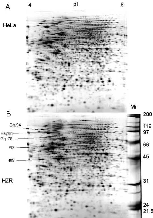
To probe the mechanisms responsible for zinc resistance of HZR cells, 2-DE has been
implemented to compare the HZR proteome with that of parental HeLa cells (Figure 1).
Lysates of four different cultures of each cell line were analyzed. The general pattern
displayed by all gels is quite similar, despite the addition of 200 µM zinc to the medium used
to grow HZR cells (Figure 1). This indicates that such gels are highly reproducible and that
only few proteins have changed intensities or positions between the two cell lines. Therefore
these differences are probably not due to a general and massive effect of zinc overload in
HZR cells, and they are likely to give insight into the mechanism responsible for the
phenotype of HZR cells. More detailed data were obtained with pI gradients between 5.5 and
7.5. Figure 2 shows silver-staining of representative gels obtained after separation of 500 µg
protein samples from HeLa and HZR cells, respectively. Spots of different intensities or
absent in one of the two gels were excised and subjected to trypsin digestion, MALDI-TOF
mass spectra measurements, and database searching. Table 1 summarizes the proteins, the
concentrations of which change between HeLa and HZR cells. This list includes a
co-chaperone, stress induced phosphoprotein-1 or Hsp70/Hsp90 organizing protein (Hop),
enzymes or subunits involved in oxidoreductase activities, thioredoxin reductase, flavoprotein
subunit of succinate dehydrogenase, 4-hydroxyphenyl-pyruvate dioxygenase, and carbonic
anhydrase II.
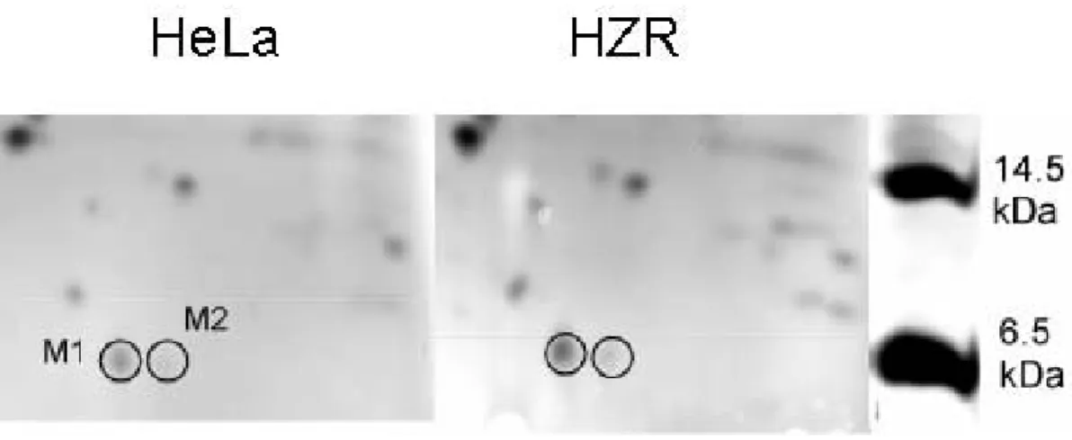
As Chimienti et al. [9] previously pointed out, metallothioneins largely contribute to zinc
adaptation of HZR cells. In an attempt to visualize metallothionein up-regulation by 2-DE,
higher concentration gels were applied in the second dimension. Two spots of apparent 6.5
kDa mass were detected. The protein labeled M1 is of higher intensity in gels of HZR extracts
Indeed, identification with mass spectrometry revealed that M1 did not correspond to
metallothionein but to ubiquitin. M2 is the ubiquitin-like molecule NEDD8, the function of
which includes regulation of the cell cycle, but its intensity is weak and it does not vary
between the two cell lines under study (Figure 3).
The role of the proteins listed in Table 1 in the cellular response to divalent metal stress has
been probed in the following.
3.2 ER stress and sensitivity to cadmium toxicity
Stress-induced phosphoprotein 1 (Hop) is a 60-kDa co-chaperone that mediates the
association of the heat shock proteins Hsp70 and Hsp90 [25]. The function of Hop has been
best characterized in in vitro systems examining the assembly of the progesterone receptor
[26] and glucocorticoid receptor [27] into hormone-binding competent hetero-complexes [28].
In previous investigations, cadmium has been reported to interfere with protein folding,
leading to accumulation of misfolded proteins in ER [29], and HZR cells have been shown to
sustain cadmium concentrations almost as high as zinc ones over 24 hours without increased
lethality (Rousselet et al. in preparation). To estimate the endoplasmic reticulum stress that
cells studied herein may experience under cadmium exposure, two different drugs were
applied in addition to the toxic metal. The sesquiterpene lactone, thapsigargin, inhibits
sarco-endoplasmic reticulum Ca2+-ATPase (SERCA) and consequently depletes the intracellular
calcium stores [30] and tunicamycin inhibits N-acetylglucosamine transferases, so preventing
glycosylation of newly synthesized glycoproteins.
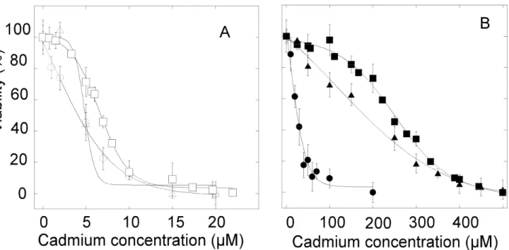
Viability tests were performed with the MTT assay in the presence of cadmium acetate for
HeLa and HZR cells with 50 nM of thapsigargin or tunicamycin. These concentrations of
inhibitors were chosen because they minimally decrease viability to the same extent (less than
ca. 20%) for both cell lines. After 24 hours of exposure, the half-maximal lethal
50 nM tunicamycin (Figure 4A). These values are not significantly different than those
measured in the absence of thapsigargin or tunicamycin (Rousselet et al. in preparation),
indicating that these concentrations of ER stressors do not sensitize HeLa cells to the
cadmium insult.
In the case of HZR cells, replacement of the 200 µM zinc sulfate by varying cadmium
concentrations for 24 hours and using the same concentration of thapsigargin as for HeLa
cells indicated that these cells were far more sensitive to cadmium than without thapsigargin
(Figure 4B). Cadmium half-maximal lethal dose measured after 24 hours of exposure was 25
µM with 50 nM thapsigargin. This value is ten times smaller than in the absence of
thapsigargin. Exposure of HZR cells to 50 nM tunicamycin (Figure 4B) led to a cadmium
half-maximal lethal dose of 165 µM, but this increased sensitivity to cadmium with
tunicamycin is not as large as with thapsigargin. Therefore, inhibition of SERCA by
thapsigargin and subsequent disruption of calcium homeostasis appears as a more potent
stress that enhances cadmium toxicity than inhibition of glycosylation. These experiments
indicate that increasing ER stress decreases cadmium resistance toward cadmium, in a very
sensitive way for HZR cells treated with thapsigargin.
Since HZR cells showed a strong sensitivity to the presence of 50 nM thapsigargin, the
metal content of cells under the conditions implemented to assess viability in the presence of
cadmium was measured by ICP-OES. Thapsigargin lowered cadmium accumulation in HeLa
cells, but not in HZR cells, at least in the cadmium concentration range that remained
accessible with 50 nM of the drug. It follows that the loss of viability of HZR cells is not
directly linked to intra-cellular cadmium in the presence of thapsigargin.
3.3 Proteasomal degradation and sensitivity to cadmium toxicity
As shown in Figure 3, ubiquitin was found to be up-regulated in HZR cells maintained in 200
proteins for removal by the proteasome is one of the major role of this small protein [13].
Up-regulation of ubiquitin in HZR cells under standard conditions, i.e. with 200 µM zinc, may
reveal enhanced protein turnover through the proteasome pathway.
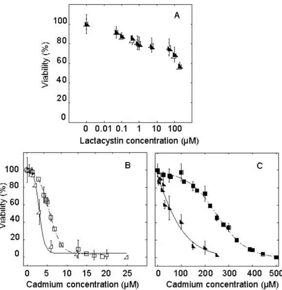
This pathway was probed by viability tests implementing the MTT assay on HeLa and
HZR cells with increasing concentrations of lactacystin [32] for 24 hours (Figure 5A). A
proteasomal inhibitor concentration (2.5 µM) leaving about 80% of the cells under study
viable after 24 hours of exposure was chosen in the following experiments. Half-maximal
lethal concentration after 24 hour-exposure to cadmium and 2.5 µM lactacystin was 3 µM for
HeLa cells (Figure 5B), as compared to 7 µM for cells exposed to cadmium alone (p value
<0.001). In the case of HZR cells, the cadmium half-maximal lethal dose was 75 µM after 24
hours of exposure to 2.5 µM lactacystine (Figure 5C). As in the case of HeLa cells, this value
is significantly smaller (p value <0.001) than that measured in the absence of lactacystin.
Qualitatively similar results were obtained by replacing the proteasomal irreversible inhibitor
lactacystin by the reversible inhibitor MG132. These data highlight the involvement of the
ubiquitin-proteasome pathway in the cellular response against cadmium toxicity in both HeLa
and HZR cells.
3.4 Involvement of 4-hydroxyphenylpyruvate dioxygenase activity in zinc adaptation and cadmium toxicity for HZR cells
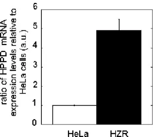
The 2-DE data revealed up-regulation of 4-hydroxyphenylpyruvate dioxygenase (HPPD) in
HZR cells as compared to HeLa cells (Figures 1 and 2). We have thus evaluated expression of
the HPPD gene in both cell lines: relative levels of HPPD transcripts were measured by
RT-PCR under standard growth conditions and compared to three different housekeeping genes.
The expression of the HPPD gene was found to be about five times higher in HZR than in
HeLa cells (Figure 6). This value agrees with the concentration ratio estimated for the
mainly by increased transcription or mRNA stabilization as compared to HeLa cells. The
increased concentration of HPPD in HZR cells does not seem to be triggered by the high zinc
concentration in which these cells are maintained. Indeed, HeLa cells exposed to zinc before
analysis by 2-DE did not display higher HPPD concentrations than HeLa cells kept in a
medium with a normal zinc concentration. However, the reversibility of the HZR phenotype
[4] was mirrored by the decrease of HPPD in HZR cells kept without excess of zinc for 3
days. Therefore, increased HPPD is associated with the HZR phenotype characterized by
resistance to both high zinc and cadmium concentrations, rather than with up-regulation
induced by short-term exposure to zinc.
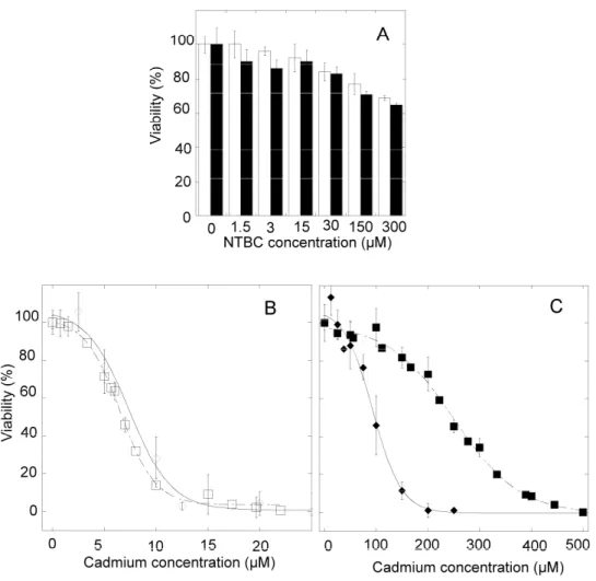
In order to know if the HPPD activity contributes to cadmium management in the presently
studied cells, a specific inhibitor of the enzyme, 2-nitro-4-(trifluoromethyl)
benzoyl-1,3-cyclohexanedione (NTBC), was used [33, 34]. HeLa and HZR cells were treated with
different concentrations of NTBC for 24 hours. Up to 300 µM of NTBC, viability slowly
decreased in a similar dose-dependent way for both cell lines (Figure 7A). The NTBC
concentration of 5 µM minimally affected viability. It was added with cadmium acetate for 24
hours to measure the effect of the inhibitor on the cellular sensitivity to cadmium. For HeLa
cells (Figure 7B), the half-maximal lethal cadmium concentration was unchanged in the
presence of NTBC as compared to HeLa cells without NTBC (p value between data sets with
and without the inhibitor= 0.63), whereas LC50 was 90 µM of cadmium in the presence of 5
µM NTBC for HZR cells (Figure 7C). No significant changes of the intra-cellular
concentrations of cadmium were measured for cells exposed to the same extra-cellular
cadmium concentration, whether 5 µM of NTBC were added or not. Therefore HeLa cells did
not seem to be more sensitive to cadmium in the presence of NTBC, but viability of HZR
cells in the presence of NTBC varied with the concentration of the inhibitor, thus showing
that NTBC sensitizes HZR cells for cadmium insult in a dose-dependent way.
3.5 Sensitivity of HZR cells to combined HPPD inhibition and cadmium treatments after long-term zinc removal
The above experiments were carried out by cadmium substitution of zinc in the HZR growth
medium for 24 hours. However the zinc and cadmium resistance phenotype of HZR cells is
reversible upon zinc withdrawal [9]. When zinc is removed from the growth medium for one
week, not only the resistance against zinc decreases [9], but also the resistance against
cadmium collapses to reach a LC50 of 40 µM for 24 hours of exposure to cadmium (Rousselet
et al. in preparation). These long-term zinc-depleted HZR cells were also treated with 5 µM or
10 µM NTBC in addition to cadmium for 24 hours. Viabilities obtained were similar for both
NTBC concentrations giving LC50 values of 40 µM on average. Therefore resistance against
cadmium toxicity of long-term zinc-depleted HZR cells was greatly decreased as compared to
zinc-maintained HZR cells, and NTBC no longer had any effect on the half-maximal lethal
cadmium concentration for these cells, as was observed for HeLa cells (Figure 7B). These
experiments demonstrate that cadmium handling by HZR cells depends on HPPD activity.
Cadmium sensitivity increases when this activity is inhibited (Figure 7) and phenotypic
reversal by long-term zinc removal makes these cells insensitive to HPPD inhibition.
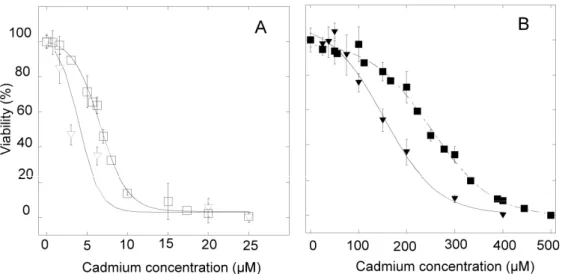
3.6 Combined effects of added tyrosine and HPPD inhibition on cadmium toxicity
The tyrosine degradation product, 4-hydroxyphenylpyruvate (HPP), is converted by HPPD to
homogentisate [35]. Since HZR cells over-produce HPPD, tyrosine catabolism may be more
active in these cells than in HeLa cells. Addition of tyrosine for 24 hours is not toxic for both
cell lines up to 6 mM (not shown). The growth medium of HeLa and HZR cells, which
already contains about 230 µM of this amino acid, was supplemented with sub-lethal 3 mM of
with increasing cadmium concentrations. They show a decrease of LC50 with 3 mM
L-tyrosine, which goes down to 5 µM for HeLa cells (Figure 8A) and 145 µM for HZR cells
(Figure 8B), instead of 7 µM and 250 µM, respectively, for cells exposed to cadmium alone
(Figure 8). Therefore, the burden of cadmium challenge is increased by the presence of
sub-lethal tyrosine concentrations for both cell lines. The combined effects of NTBC and tyrosine
on the sensitivity toward increasing cadmium concentration were also determined. The
cadmium LC50 obtained for HeLa cells in the presence of 3 mM tyrosine and 5 or 10 µM
NTBC did not change as compared to tyrosine alone. In the case of HZR cells, the same
experiments with both NTBC and tyrosine gave the same outcome, namely no significant
changes of LC50 for cadmium as compared to cells exposed to tyrosine alone.
The enzyme following HPPD in the tyrosine degradation pathway is homogentisate
dioxygenase (HGO), that is mainly responsible for homogentisate withdrawal in mammalian
cells. The activity of this enzyme in extracts of both HeLa and HZR cells has been measured.
As found for HPPD, HGO activity is ca. 5-fold higher in HZR cells (27 (SD 6) µM.min-1.(mg
of proteins)-1), as compared to HeLa cells (5.5 (SD 3.6) µM.min-1.(mg of proteins)-1).
Therefore, 4-hydroxyphenylpyruvate conversion and ring opening are both enhanced in HZR
cells as a result of adaptation to zinc.
Tyrosine has different biological roles in mammalian cells, as one of the component of
proteins, but also as the precursor of catecholamines, such as dopamine, thyroid hormones,
and melanin. The latter pigment has been shown to bind metals [36, 37], including zinc and
cadmium, and it has been repeatedly proposed to participate in heavy metal detoxification [38,
39]. Homogentisate is a precursor of secreted melanin pigments (pyomelanin) in some
microorganisms [40] and of plasma melanins in humans in cases of alkaptonuria with
inflammation and darkening of connective tissues in particular [41]. It is unknown at present
in mammalian cells. However, it is unlikely that these scavengers are in the form of
intracellular melanin, since the measured amount in HZR cells (range 21-110 ng/µg of total
proteins, 4 measurements) is not larger than in HeLa cells (range 45-180 ng/µg of total
proteins, 3 measurements). Therefore the increased activities of HPPD and HGO in HZR cells
do not favor melanin biosynthesis.
4. Discussion
In the present study adaptation to zinc overload developed by the HZR clone [9] was probed
at the proteomic level. A list of proteins was found to be up-regulated in these cells, with the
exception of carbonic anhydrase II that was decreased by a factor of 3 (Table 1). The
corresponding gene was shown to be up-regulated by zinc deficiency in rat esophageal
epithelia [42], in line with down-regulation upon zinc overload as noticed here. The biological
pathways and functions suggested by comparative studies between the HZR clone and
parental HeLa cells have been interrogated by use of specific inhibitors. Consequences on the
sensitivity to cadmium have been measured to gauge the involvement of these pathways in the
toxicity mechanism(s).
ER as a target of cadmium action in mammalian cells has been largely documented [43,
44]. The viability of HZR cells was far more sensitive than that of HeLa cells to the
simultaneous addition of the ER stressors tunicamycin or thapsigargin with cadmium. This
strongly suggests that the HZR cells, able to accommodate high intracellular zinc
concentrations [9], more heavily rely on ER functions than HeLa cells. However,
ER-associated chaperones, such as Grp94, PDI, and Grp78, or other components of the quality
control system of protein folding, such as Hsp90, do not show significant changes between
the two cell lines (Figure 1). It means that HZR cells do not display signs of generalized
Once cadmium is added to the cultures, perturbed protein biosynthesis does occur as
witnessed by the impairment of resistance against the toxic metal by small amounts of
proteasomal inhibitors. But, under these conditions, HeLa cells appear as sensitive as HZR
cells, since the decrease of viability is similar for the same inhibitor concentration. The
applied cadmium concentrations are quite different between the two cell lines, but it has been
found that viability and the intracellular concentrations of the metal are similarly related for
both HeLa and HZR cells (Rousselet et al. in preparation). Therefore, the increased amount of
ubiquitin in HZR cells does not seem to be needed for a more efficient degradation of
misfolded proteins via the proteasome pathway. It may rather be due to some other role of
ubiquitin, such as protein intracellular trafficking and other important biological functions
[45].
The only detected exception to the similar concentration of chaperones between the two
cell lines (Table 1) is Hop that is more abundant in HZR cells than in HeLa cells. Hop belongs
to the large group of co-chaperones which regulates the activity of heat shock proteins
through its tetratricopeptide repeat TPR1 and TPR2A domains [46]. It participates in the
maturation of the glucocorticoid receptor [47] and up-regulation of Hop has been
demonstrated in activated macrophages [48].
This single example among the several chaperones identified in 2-DE strongly suggests
that adaptation of HZR cells to zinc is a well-targeted phenomenon involving only a small
number of cellular components. Further experimental support for this statement is provided by
the more acute sensitivity of these cells to the combined effects of cadmium and thapsigargin
as compared to cadmium and tunicamycin. These two drugs target distinct activities, but they
eventually trigger the same cellular Unfolded Protein Response. The larger effect of
metal in HZR cells is more sensitive to adequate calcium intracellular distribution than to
proper processing of membrane and secreted proteins.
Besides Hop, another protein being unambiguously present at higher concentration in HZR
cells than in HeLa cells is the enzyme HPPD. It is an iron- and 2-oxo-glutarate-dependent
enzyme that catalyzes the complex conversion, including hydroxylation, decarboxylation and
rearrangement steps, of 4-hydroxyphenylpyruvate, the product of tyrosine aminotransferase,
to homogentisate [49, 50].
Missense mutations in the HPPD locus can cause two distinct genetic diseases, hereditary
type III Tyrosinemia and Hawkinsinuria [51]. Type I Tyrosinemia is due to
fumarylacetoacetate hydrolase deficiency [52], and, because symptoms of type III
Tyrosinemia and Hawkinsinuria [53] are less severe than those of type I Tyrosinemia, the
latter is treated by inhibition of HPPD with the 1,3 diketone NTBC [34], hence avoiding the
buildup of the deleterious metabolites fumarylacetoacetate and succinylacetone. These
metabolic and clinical data clearly indicate that defaults of tyrosine catabolism strongly
impact cellular fate.
HPPD is subjected to several post-translational processing events [35, 54], which may
explain the presence of at least two spots on 2D gels assigned to HPPD (Figure 2, Table 1),
but no variations between the relative intensities of these spots have been observed in our
experiments. The HPPD transcript is more abundant in the HZR than in the parental HeLa cell
line (Figure 6). Promoter analysis evidenced binding sites for several transcription factors,
including Sp1, CREB, and C/EBP [54]. The HPPD activity appeared more important in HZR
cells than in HeLa cells, since its inhibition selectively sensitized the former cells to cadmium.
However there is no association between HPPD activity and cadmium traffic since NTBC did
not change the amount of metal accumulated by cells. A modest impact of increased tyrosine
inactivation of HPPD should enhance cadmium toxicity in the presence of large amounts of
tyrosine. Instead, no additive effects of tyrosine and the HPPD inhibitor NTBC on the
viability curves with cadmium were detected. The NTBC dose-dependent decrease of HZR
cells viability in the presence of cadmium is thus not due to decreased tyrosine disposal.
The HPPD substrate, HPP, is the first compound produced in the tyrosine degradation
pathway. It has also been shown to be a substrate of phenylpyruvate tautomerase, which
produces the enol-form of HPP and is the same protein as macrophage migration inhibitory
factor (MIF) [55]. Whether increased depletion of HPP, as expected in HZR cells, influences
the cytokine, including growth-promoting [56], function of MIF is unknown. Indeed,
regulation of the balance between the two activities of this moonlighting protein is not
elucidated, and the physiological meaning of the enzymatic reaction has been questioned [57].
Yet, from what is known about regulation of enzymes involved in tyrosine turnover, including
HPPD [54], it is beyond doubt that signaling pathways regulate flow between the different
metabolic routes available for tyrosine. In this respect, disruption of calcium homeostasis by
thapsigargin in HZR cells, and its consequences on signaling pathways, strongly impacts the
resistance of these cells against cadmium (Figure 4B). MIF may also be involved in regulating
signaling pathways [58].
The proteomic approach described herein and complementary experiments have revealed
an unsuspected association between tyrosine catabolism and adaptation to zinc overload that
also contributes to the toxicity of cadmium in mammalian cells. Very recently [59],
up-regulation of HPPD has been observed in Arabidopsis thaliana upon exposure to cadmium, as
a means to increase vitamin E synthesis for which homogentisate is a precursor in plants.
Since this pathway is not present in animals, the association between HPPD activity and
Acknowledgements
This work was supported by a grant from the “Toxicologie Nucléaire et Environnementale”
program. Dr David King (Swedish Orphan International AB, Stockholm, Sweden) is
gratefully thanked for providing us with NTBC, as is Dr Pierre Richaud (CEA, IBEB, LB3M,
5. References
[1] Berg, J. M., Shi, Y., The galvanization of biology: a growing appreciation for the roles of zinc, Science 1996, 271, 1081-1085.
[2] Vallee, B. L., Falchuk, K. H., The biochemical basis of zinc physiology, Physiol Rev 1993, 73, 79-118.
[3] Chimienti, F., Seve, M., Richard, S., Mathieu, J., et al., Role of cellular zinc in
programmed cell death: temporal relationship between zinc depletion, activation of caspases, and cleavage of Sp family transcription factors, Biochem Pharmacol 2001, 62, 51-62.
[4] Truong-Tran, A. Q., Carter, J., Ruffin, R. E., Zalewski, P. D., The role of zinc in caspase activation and apoptotic cell death, Biometals 2001, 14, 315-330.
[5] Cousins, R. J., Liuzzi, J. P., Lichten, L. A., Mammalian zinc transport, trafficking, and signals, J Biol Chem 2006, 281, 24085-24089.
[6] Martelli, A., Rousselet, E., Dycke, C., Bouron, A., et al., Cadmium toxicity in animal cells by interference with essential metals, Biochimie 2006, 88, 1807-1814.
[7] Rooney, J. P., The role of thiols, dithiols, nutritional factors and interacting ligands in the toxicology of mercury, Toxicology 2007, 234, 145-156.
[8] Cigliano, S., Remondelli, P., Minichiello, L., Mellone, M. C., et al., Analysis of metal-regulated metallothionein and heat shock gene expression in HeLa-derived cadmium-resistant cells, Exp Cell Res 1996, 228, 173-180.
[9] Chimienti, F., Jourdan, E., Favier, A., Seve, M., Zinc resistance impairs sensitivity to oxidative stress in HeLa cells: protection through metallothioneins expression, Free Radic
Biol Med 2001, 31, 1179-1190.
[10] Sullivan, V. K., Burnett, F. R., Cousins, R. J., Metallothionein expression is increased in monocytes and erythrocytes of young men during zinc supplementation, J Nutr 1998, 128, 707-713.
[11] Beyersmann, D., Hechtenberg, S., Cadmium, gene regulation, and cellular signalling in mammalian cells, Toxicol Appl Pharmacol 1997, 144, 247-261.
[12] Rutkowski, D. T., Kaufman, R. J., A trip to the ER: coping with stress, Trends Cell Biol 2004, 14, 20-28.
[13] Meusser, B., Hirsch, C., Jarosch, E., Sommer, T., ERAD: the long road to destruction,
Nat Cell Biol 2005, 7, 766-772.
[14] Gorg, A., Boguth, G., Obermaier, C., Weiss, W., Two-dimensional electrophoresis of proteins in an immobilized pH 4-12 gradient, Electrophoresis 1998, 19, 1516-1519.
[15] Rabilloud, T., Valette, C., Lawrence, J. J., Sample application by in-gel rehydration improves the resolution of two-dimensional electrophoresis with immobilized pH gradients in the first dimension, Electrophoresis 1994, 15, 1552-1558.
[16] Rabilloud, T., Adessi, C., Giraudel, A., Lunardi, J., Improvement of the solubilization of proteins in two-dimensional electrophoresis with immobilized pH gradients, Electrophoresis 1997, 18, 307-316.
[17] Sinha, P., Poland, J., Schnolzer, M., Rabilloud, T., A new silver staining apparatus and procedure for matrix-assisted laser desorption/ionization-time of flight analysis of proteins after two-dimensional electrophoresis, Proteomics 2001, 1, 835-840.
[18] Neuhoff, V., Arold, N., Taube, D., Ehrhardt, W., Improved staining of proteins in polyacrylamide gels including isoelectric focusing gels with clear background at nanogram sensitivity using Coomassie Brilliant Blue G-250 and R-250, Electrophoresis 1988, 9, 255-262.
[19] Wagner, E., Luche, S., Penna, L., Chevallet, M., et al., A method for detection of overoxidation of cysteines: peroxiredoxins are oxidized in vivo at the active-site cysteine during oxidative stress, Biochem J 2002, 366, 777-785.
[20] Perkins, D. N., Pappin, D. J. C., Creasy, D. M., Cottrell, J. S., Probability-based protein identification by searching sequence databases using mass spectrometry data, Electrophoresis 1999, 20, 3551-3567.
[21] Carmichael, J., DeGraff, W. G., Gazdar, A. F., Minna, J. D., et al., Evaluation of a tetrazolium-based semiautomated colorimetric assay: assessment of chemosensitivity testing,
Cancer Res 1987, 47, 936-942.
[22] Martelli, A., Salin, B., Dycke, C., Louwagie, M., et al., Folding and turnover of human iron regulatory protein 1 depend on its subcellular localization, FEBS J 2007, 274, 1083-1092. [23] Schmidt, S. R., Muller, C. R., Kress, W., Murine liver homogentisate 1,2-dioxygenase. Purification to homogeneity and novel biochemical properties, Eur J Biochem 1995, 228, 425-430.
[24] Siegrist, W., Eberle, A. N., In situ melanin assay for MSH using mouse B16 melanoma cells in culture, Anal Biochem 1986, 159, 191-197.
[25] Chen, S., Smith, D. F., Hop as an adaptor in the heat shock protein 70 (Hsp70) and hsp90 chaperone machinery, J Biol Chem 1998, 273, 35194-35200.
[26] Kosano, H., Stensgard, B., Charlesworth, M. C., McMahon, N., et al., The assembly of progesterone receptor-hsp90 complexes using purified proteins, J Biol Chem 1998, 273,
[27] Dittmar, K. D., Pratt, W. B., Folding of the glucocorticoid receptor by the reconstituted Hsp90-based chaperone machinery. The initial hsp90.p60.hsp70-dependent step is sufficient for creating the steroid binding conformation, J Biol Chem 1997, 272, 13047-13054.
[28] Kimmins, S., MacRae, T. H., Maturation of steroid receptors: an example of functional cooperation among molecular chaperones and their associated proteins, Cell Stress
Chaperones 2000, 5, 76-86.
[29] Liu, F., Inageda, K., Nishitai, G., Matsuoka, M., Cadmium induces the expression of Grp78, an endoplasmic reticulum molecular chaperone, in LLC-PK1 renal epithelial cells,
Environ Health Perspect 2006, 114, 859-864.
[30] Inesi, G., Hua, S., Xu, C., Ma, H., et al., Studies of Ca2+ ATPase (SERCA) inhibition, J
Bioenerg Biomembr 2005, 37, 365-368.
[31] Herrmann, J., Lerman, L. O., Lerman, A., Ubiquitin and ubiquitin-like proteins in protein regulation, Circ Res 2007, 100, 1276-1291.
[32] Fenteany, G., Schreiber, S. L., Lactacystin, proteasome function, and cell fate, J Biol
Chem 1998, 273, 8545-8548.
[33] Kavana, M., Moran, G. R., Interaction of (4-hydroxyphenyl)pyruvate dioxygenase with the specific inhibitor 2-[2-nitro-4-(trifluoromethyl)benzoyl]-1,3-cyclohexanedione,
Biochemistry 2003, 42, 10238-10245.
[34] Lindstedt, S., Holme, E., Lock, E. A., Hjalmarson, O., et al., Treatment of hereditary tyrosinaemia type I by inhibition of 4-hydroxyphenylpyruvate dioxygenase, Lancet 1992, 340, 813-817.
[35] Neve, S., Aarenstrup, L., Tornehave, D., Rahbek-Nielsen, H., et al., Tissue distribution, intracellular localization and proteolytic processing of rat 4-hydroxyphenylpyruvate
dioxygenase, Cell Biol Int 2003, 27, 611-624.
[36] Szpoganicz, B., Gidanian, S., Kong, P., Farmer, P., Metal binding by melanins: studies of colloidal dihydroxyindole-melanin, and its complexation by Cu(II) and Zn(II) ions, J Inorg
Biochem 2002, 89, 45-53.
[37] Kokkinou, D., Kasper, H. U., Bartz-Schmidt, K. U., Schraermeyer, U., The pigmentation of human iris influences the uptake and storing of zinc, Pigment Cell Res 2004, 17, 515-518. [38] Nicolaus, B. J., A critical review of the function of neuromelanin and an attempt to provide a unified theory, Med Hypotheses 2005, 65, 791-796.
[39] Loumbourdis, N. S., Vogiatzis, A. K., Impact of cadmium on liver pigmentary system of the frog Rana ridibunda, Ecotoxicol Environ Saf 2002, 53, 52-58.
[40] Plonka, P. M., Grabacka, M., Melanin synthesis in microorganisms--biotechnological and medical aspects, Acta Biochim Pol 2006, 53, 429-443.
[41] Hegedus, Z. L., Nayak, U., Homogentisic acid and structurally related compounds as intermediates in plasma soluble melanin formation and in tissue toxicities, Arch Int Physiol
Biochim Biophys 1994, 102, 175-181.
[42] Liu, C. G., Zhang, L., Jiang, Y., Chatterjee, D., et al., Modulation of gene expression in precancerous rat esophagus by dietary zinc deficit and replenishment, Cancer Res 2005, 65, 7790-7799.
[43] Hiramatsu, N., Kasai, A., Du, S., Takeda, M., et al., Rapid, transient induction of ER stress in the liver and kidney after acute exposure to heavy metal: evidence from transgenic sensor mice, FEBS Lett 2007, 581, 2055-2059.
[44] Biagioli, M., Pifferi, S., Ragghianti, M., Bucci, S., et al., Endoplasmic reticulum stress and alteration in calcium homeostasis are involved in cadmium-induced apoptosis, Cell
Calcium 2007.
[45] Hurley, J. H., Lee, S., Prag, G., Ubiquitin-binding domains, Biochem J 2006, 399, 361-372.
[46] Scheufler, C., Brinker, A., Bourenkov, G., Pegoraro, S., et al., Structure of TPR domain-peptide complexes: critical elements in the assembly of the Hsp70-Hsp90 multichaperone machine, Cell 2000, 101, 199-210.
[47] Carrigan, P. E., Riggs, D. L., Chinkers, M., Smith, D. F., Functional comparison of human and Drosophila Hop reveals novel role in steroid receptor maturation, J Biol Chem 2005, 280, 8906-8911.
[48] Heine, H., Delude, R. L., Monks, B. G., Espevik, T., et al., Bacterial lipopolysaccharide induces expression of the stress response genes hop and H411, J Biol Chem 1999, 274, 21049-21055.
[49] Purpero, V. M., Moran, G. R., Catalytic, noncatalytic, and inhibitory phenomena: kinetic analysis of (4-hydroxyphenyl)pyruvate dioxygenase from Arabidopsis thaliana, Biochemistry 2006, 45, 6044-6055.
[50] Nakai, C., Nozaki, M., Hayaishi, O., Studies on a possible reaction intermediate of p-hydroxyphenylpyruvate dioxygenase, Biochem Biophys Res Commun 1975, 67, 590-595. [51] Tomoeda, K., Awata, H., Matsuura, T., Matsuda, I., et al., Mutations in the
4-hydroxyphenylpyruvic acid dioxygenase gene are responsible for tyrosinemia type III and hawkinsinuria, Mol Genet Metab 2000, 71, 506-510.
[52] Tanguay, R. M., Valet, J. P., Lescault, A., Duband, J. L., et al., Different molecular basis for fumarylacetoacetate hydrolase deficiency in the two clinical forms of hereditary
tyrosinemia (type I), Am J Hum Genet 1990, 47, 308-316.
[53] Scott, C. R., The genetic tyrosinemias, Am J Med Genet C Semin Med Genet 2006, 142, 121-126.
[54] Aarenstrup, L., Falch, A. M., Jakobsen, K. K., Neve, S., et al., Expression and post-translational modification of human 4-hydroxy-phenylpyruvate dioxygenase, Cell Biol Int 2002, 26, 615-625.
[55] Rosengren, E., Aman, P., Thelin, S., Hansson, C., et al., The macrophage migration inhibitory factor MIF is a phenylpyruvate tautomerase, FEBS Lett 1997, 417, 85-88. [56] Mitchell, R. A., Bucala, R., Tumor growth-promoting properties of macrophage migration inhibitory factor (MIF), Semin Cancer Biol 2000, 10, 359-366.
[57] Swope, M., Sun, H. W., Blake, P. R., Lolis, E., Direct link between cytokine activity and a catalytic site for macrophage migration inhibitory factor, EMBO J 1998, 17, 3534-3541. [58] Aeberli, D., Yang, Y., Mansell, A., Santos, L., et al., Endogenous macrophage migration inhibitory factor modulates glucocorticoid sensitivity in macrophages via effects on MAP kinase phosphatase-1 and p38 MAP kinase, FEBS Lett 2006, 580, 974-981.
[59] Collin, V. C., Eymery, F., Genty, B., Rey, P., et al., Vitamin E is essential for the
tolerance of Arabidopsis thaliana to metal-induced oxidative stress, Plant Cell Environ 2008,
Table 1. Identification of proteins differently present in HeLa and HZR cellsa.
a The labeled spots in Figure 2 are indicated with their identification using MALDI-TOF with
sequence coverage (% R). Swiss-Prot accession numbers, monomer masses and
isoelectro-focusing points obtained for each protein are also given. The average expression ratio
between HZR and HeLa cells estimated with the Delta2D software are indicated under E with
standard errors in brackets as described in Materials and Methods.
b the protein was not detected in HeLa cells
c the gels such as that in Figure 3 were not analyzed with the Delta2D software.
Spot Protein Accession MW / pI %R E (SE)
I 4-Hydroxyphenylpyruvate dioxygenase P32754 44775 / 6.50 44% 4.7 (1.5) II Thioredoxin reductase GRIM-12 Q9UES8 54580 / 6.36 21% b
III Stress-induced-Phosphoprotein 1 P31948 62599 / 6.40 8% 1.6 (0.3)
IV
Succinate dehydrogenase flavoprotein subunit, mitochondrial precursor
P31040 72645 / 7.06 29% 2.8 (0.3)
V Carbonic anhydrase II P00918 29097 / 6.86 44% 0.29 (0.09)
Figure 1. 2-DE of total cell extracts from HeLa (A) and HZR (B) cells. Two hundred
micrograms of proteins were separated on pH 4-8 linear immobilized pH gradients (horizontal
axis) and 10% continuous SDS gels (vertical axis). Molecular markers positions are indicated
at the right of the gel in (B). Detection was by silver staining. Some easily identified proteins,
mainly chaperones, are indicated in the bottom gel with PDI: protein disulfide isomerase and
Figure 2. Highly resolved part of the 2-DE gels of total HeLa (A) and HZR (B) cell extracts. Five
hundred micrograms of proteins were separated on pH 5.5-7.5 linear immobilized pH
gradients and 10% continuous SDS gels. Detection was by Coomassie Brillant Blue for
Figure 3. Low molecular weight part of 2-D electrophoretic gels of total HeLa (left) and HZR (right) cell extracts. Proteins were separated on pH 4-8 linear immobilized pH
Figure 4. Effects of ER stress on the viability of HeLa and HZR cells in the presence of cadmium. HeLa cells were stressed by cadmium alone (A open square), in the presence of 50
nM thapsigargin (A open circle), or in the presence of 50 nM tunicamycin (A open triangle),
24 hours before measuring viability. The same experiments were done for HZR cells (B) and
Figure 5. Proteasome involvement in the sensitivity of HeLa and HZR cells to cadmium toxicity. (A) Parental HeLa (open triangle) and HZR cells (filled triangle) were treated with
increasing concentrations of lactacystin in the growth medium for 24 hours and the percentage
of viable cells was measured and reported to that of non-exposed cells. (B) HeLa cells were
stressed by cadmium alone (open squares) or in the presence of 2.5 µM lactacystin (open
triangles) for 24 hours before measuring viability. (C) The same experiment as in (B) was
carried out with HZR cells without (filled squares) or with 2.5 µM lactacystin (filled
Figure 6. Real-time PCR of HPPD. Real-time quantitative, reverse transcriptase polymerase
chain reaction experiments were carried out with total RNA purified from HeLa (empty bars)
and HZR cells (filled bars). The results were calculated as a ratio to the set of reference genes
(RPLPo2, GAPDH and β-actin) and the value for HeLa cells was arbitrarily set to 1. Data are the mean and SD of two separate experiments.
Figure 7. HPPD inhibition and cadmium toxicity in HeLa and HZR cells. (A) Parental
HeLa (open bars) and HZR cells (filled bars) were treated with increasing concentrations of
NTBC in the growth medium for 24 hours and the viability was estimated by the MTT assay.
in 8 separate measurements. (B) Parental HeLa cells were stressed by cadmium alone (open
squares) or in the presence of 5 µM NTBC (open diamonds) 24 hours before measuring
viability. (C) The same experiments were carried out with HZR cells and represented with the
Figure 8. Tyrosine and cadmium toxicity in HeLa and HZR cells. (A) Parental HeLa cells
were stressed with cadmium alone (open squares) or in the presence of 3 mM tyrosine
(inversed open triangles) for 24 hours. Eight separate measurements with the MTT assay were
used to draw viability curves. (B) The same experiments were done with HZR cells and