HAL Id: tel-01685265
https://tel.archives-ouvertes.fr/tel-01685265
Submitted on 16 Jan 2018HAL is a multi-disciplinary open access archive for the deposit and dissemination of sci-entific research documents, whether they are pub-lished or not. The documents may come from teaching and research institutions in France or abroad, or from public or private research centers.
L’archive ouverte pluridisciplinaire HAL, est destinée au dépôt et à la diffusion de documents scientifiques de niveau recherche, publiés ou non, émanant des établissements d’enseignement et de recherche français ou étrangers, des laboratoires publics ou privés.
quantum dots
Lucia Mattera
To cite this version:
Lucia Mattera. Functionalization, bio-conjugation and toxicity studies of quantum dots. Organic chemistry. Université Grenoble Alpes, 2016. English. �NNT : 2016GREAV084�. �tel-01685265�
THÈSE
Pour obtenir le grade de
DOCTEUR DE LA COMMUNAUTÉ UNIVERSITÉ
GRENOBLE ALPES
Spécialité : CSV/Chimie-Biologie
Arrêté ministériel : 7 août 2006
Présentée par
Lucia MATTERA
Thèse dirigée par Peter REISS Et co-encadrée par David Djuradopréparée au sein du Laboratoire INAC/SPrAM UMR5819/LEMOH dans l'École Doctorale Chimie et Sciences du Vivant
Fonctionnalisation,
bio-conjugaison et études
toxicologiques de quantum dots
Thèse soutenue publiquement le 10 juin 2016 devant le jury composé de :
Prof. Frédérique LOISEAU
Professeur à l’Université Grenoble-Alpes, Présidente du jury
Dr. Michel de WAARD
Directeur de recherche INSERM, Rapporteur
Dr. Thomas PONS
Chargé de recherche INSERM, Rapporteur
Dr. Loïc CHARBONNIERE
Directeur de recherche CNRS, Examinateur
Prof. Niko HILDEBRANDT
Professeur à l’Université Paris Sud, Examinateur
Dr. David DJURADO
Directeur de recherche CNRS, Co-encadrant de thèse
Dr. Peter REISS
Chercheur au CEA Grenoble, Directeur de thèse
Dr. Claudia TORTIGLIONE
To my beloved parents and Massimo for their everlasting love and support….
Remerciements
A ce moment, ce sera que désormais je sens l'odeur des changements, fermant les yeux pour un moment, je revins mentalement le long voyage qui m'a conduit ici. Je me suis souvenu de toutes les années, je me suis souvenu des visages, des situations, les contre-charmes habituels, les appels téléphoniques désespérés, des sourires béats, le temps formidable que j’ai passé. Encore, le jour fantastique de ma soutenance, la présence de toutes les personnes qui sont restées jusqu’au but de la discussion. Les personnes de prêt et da loin qui m’ont suivi pendant ce voyage sont impressionnant et je n’aurai pas les paroles pour exprimer ma gratitude et mon remerciement à leur égard.
Je vais essayer de remercier tous en espérant de n’oublier personne, vous êtes beaucoup !!
Tout d’abord je souhaiterais commencer à adresser mes sentiments de gratitude à mon directeur de thèse, Peter Reiss pour m’avoir donné l’opportunité d’entreprendre ce voyage. Il a eu beaucoup de confiance en moi et j’ai eu la possibilité de m’améliorer et d’apprendre des domaines différents, de participer à des conférences nationales et internationales, formations, écoles thématiques, etc. Tout ça étaient des expériences formidables et uniques que je garderai toujours comme un don précieux dans ma vie. Peter, tu es formidable, c’était un plaisir et un honneur d’être étudiante dans ton laboratoire et ma gratitude est si grande. Merci aussi pour le temps précieux que tu as dédié à corriger mon manuscrit et pour tous les conseils.
Je remercie vivement mon co-encadrant David Djurado, il a eu aussi beaucoup de confiance en moi, David, merci pour les discussions pas seulement scientifique mais aussi de danse et de vie ;) et surtout pour tous le support soit moral soit scientifique, que tu m’as toujours donné et encore plus marqué dans le dernier période. Merci pour les remarques si précieux qui était de conseil fondamental pour l’écriture et pour avoir dédié aussi toi beaucoup de temps pour les corrections. Merci pour ta bonne humeur et pour la personne formidable que tu es !
Je tiens à remercier Michel de Waard et Thomas Pons d’avoir accepté d’être rapporteurs pour cette thèse et de leur intérêt marqué pour ce travail, d’avoir eu la patience de lire en détail mon manuscrit. Je suis honoré d'avoir été examiné par ce comité si prestigieux et je suis flattée pour ses appréciations sur mon travail. J’ai particulièrement appréciée l’intense et enrichissante discussion que nous avons eu lors de la soutenance. Merci beaucoup.
Mes remerciements vont également aux autres membres de mon jury Loïc Charbonnière et Niko Hildebrandt qui ont eu la gentillesse de faire partie aussi de mon comité de suivi de thèse durant ces trois années, et qui m’ont apporté de précieux conseils dans les directions à prendre sur ce sujet et pour l’enthousiasme que vous avez démontré pour mon travail. Merci beaucoup aussi à Frédérique Loiseau d’avoir accepté d’être présidente du jury.
Je tiens aussi à adresser mes plus sincères remerciements à Claudia Tortiglione (CNR, Pozzuoli Italy) pour le temps qu’elle m’a généreusement consacré toujours depuis que j’étais étudiante de master dans son laboratoire. Pour m’avoir donné l’opportunité aussi d’entreprendre ce voyage et pour m’avoir poussée à partir, pour l’amour, l’estime... Merci pour être venu à Grenoble et d’avoir faire partie de mon jury de thèse aussi, pour avoir accepté de collaborer alors que j’ai pu amplifier mon projet de recherche avec des données superbes de nanotoxicologie et pour le temps que tu m’as toujours dédiée. Je vais remercier également ta co-bureau Angela Tino pour m’avoir toujours stimulé d'améliorer et d'envisager tout dans une vision critique. Merci à vous deux pour tout, pour m’avoir encouragé, pour les conseils judicieux, pour votre disponibilité et l'intérêt et grâce à eux que j’ai pu aussi conquérir une nouvelle étape ici à Grenoble. Grazie infinite! Je leur souhaite beaucoup de bonheur et in bocca al lupo per tutto !
Mon travail de thèse a été accompli dans le cadre de l’ANR NanoFret, que je vais remercier pour avoir financier cette thèse et je voudrais remercier tous ceux qui en font partie : Shashi Bhuckory, David Wegner, Niko Hildebrandt (Nano Bio Photonics. Institut d’Electronique Fondamentale. Université Paris-Sud). Merci pour m’avoir accueillie dans votre laboratoire et pour le travail fait ensemble!), Loïc Charbonnière (IPHC, Strasbourg), Emmanuel Bois et Valerie Vallois (Cezanne ThermoFisher, Nîmes). Merci pour ce projet si passionnant, intéressant et multidisciplinaire, pour les réunions et pour votre professionnalité et bon humour. C’était un plaisir collaborer et travailler avec vous et je suis reconnaissante pour avoir eu l’occasion d’aborder différents domaines.
Un remercîment spécial en plus à Niko Hildebrandt et sa famille qui m’ont accueilli chez eux pendant mon séjour dans l’équipe NanoBio Photonics, pour les bons moments passés ensemble et en particulier la conférence à Paris « 30 years of colloidal quantum dots » pendant laquelle tu m’as pris photo avec presque tous les plus grands chercheurs (y compris toi et Peter ;) ) sur le QDs ☺ Je ne pourrais pas oublier tous ces bons moments passés ensemble et je vous remercie encore pour tous.
Un clin d’œil pour Shashi car on a traversée ensemble le gros de notre projet, merci pour tous le soutien aussi avant les conférences et pour tous nos discussions scientifiques et non-scientifiques ☺. Tu es un grand et bravo jeune chercheur. Grazie infinite Shashi….Je te souhaite le mieux pour le futur. A toi de jouer maintenant. ;)
Je tiens également à remercier toutes les personnes qui ont corrigé une partie de ma thèse, ainsi que toutes les autres personnes avec qui j’ai eu l’occasion de collaborer au cours de ma thèse.
Alors, un remerciement spécial au NanoBiomolecular group en Italie et tout particulier à Mariateresa Allocca et Alfredo Ambrosone pour la collaboration et l’échange avec le projet de nanotoxicologie. Siete fortissimi e vi auguro il meglio nella vita e nel futuro professionale. Un grande in bocca al lupo soprattutto a te Mariateresa ;). Un grande merci à les petit Hydra aussi :p.
Un vif remerciement à Adeline Tarantini et Marie Carrière pour le projet de nanotoxicologie aussi. Marie, merci pour ton sourire, ton soutien et tes conseils. Adeline c’était un grand plaisir travailler avec toi, merci pour tous le bon moment passé ensemble, notre discussion pendant les manips, la danse et pour le soutienne morale, tu es devenu une vraie amie. Je te souhaite tant bonne humeur pour la suite et bon courage pour tous ☺ Un grand merci aussi à ta
co-bureau Anne Vonkoschembahr pour tout le soutien aussi.
Un grand merci très spécial à Maria Moula-Karimdjy pour la collaboration avec son projet sur le sonde bimodale quantum dot/IRM qu’a enrichi mon manuscrit et pour le bon moment passé ensemble, pour ta gentillesse et bon humour et soutien unique et spéciale, spéciale comme toi. Il me manque déjà travailler en « causalité » avec toi :D. Garde toujours ton bon humour et la magnifique personne que tu es. Je t’adore.
Un remerciement spécial est obligatoire le faire à un autre collaborateur Fabio Annunziata. Grazie per tutto, pour les imagines TEM, pour le temps passée ensemble au travail et en dehors et pour m’avoir faire prouver l’expérience de l’escalade; e a proposito della scalata, non posso non ringraziare anche Luca De Trizio (IIT, Istituto Italiano di Tecnologia, Genova) per la collaborazione, per la scalata e per tutto….Grazie di cuore capo ☺ .
Je remercie Emile Rustique, pour m’avoir ouvert les portes de son laboratoire (NanoBio-LETI) lorsque j’en ai eu besoin pour les mesures du diamètre hydrodynamique et du potentiel zeta, et merci aussi à tous les components en particulier, à Arnaud Lemelle, Lisa Racine, Marie Escude et Mathieu Varche pour la patience et l’aide que vous m’avez toujours donnée quand je vous “coloniser” la machine pours les mesures. :D
Merci particulièrement aigu va à mes collègues de laboratoire qui m’ont accompagné lors de ce voyage, anciens (Olga Burchak, Franz Fuchs, Tim Senden (thanks for your excellent quantum dots), Axel Maurice, Aurélie Lefrançois, Ashley Gaulding, Luca Assumma, Jinhyung Park, Mohammad Jouni, Reback Matthew, Maria MendezMalaga) et presente, Louis Vaure, Clement Thomassé, Dorian Gaboriau, Florent Caffy (mon petit frère), Martina Sandroni, Christophe Linchenau, Maxime Godfroy, Cyril Aumaitre, Mathilde Bouchard, Maria Moulakarimdjy, Sebastiano Di Pietro (il grande chimico), Fanny Laporte, Jennifer Molloy pour avoir transformé les jours de laboratoire dans les moments de loisirs et de plaisir, mais surtout pour l'amitié que vous m'avez montrée. Ils m'ont encouragé dans les moments de désespoir et ils ont essayé par tous les moyens de me calmer! Merci aussi pour m’avoir appris la beauté de la langue française ☺. Grazie infinite vi adoro ragazzi !
Et je n’oublie pas tous les gens avec qui on a partagé quotidiennement les couloirs, les déjeuners et les pauses café. Merci Olga, tu as illuminé mes journées, tu es une femme et chercheur formidable. Nos discussions ont constitué une aide précieuse à mes yeux. Merci pour tous, gardes toujours ton bon humour et ton esprit unique et que donne toujours beaucoup de conforte à tout le monde qu’est à côté de toi. Grazie di cuore, ne pas changer, resta sempre la splendida persona che sei !
Un remerciement très très spécial à mon co-bureau depuis trois ans, Louis, pour m'avoir soutenu depuis toujours, pour la grande patience, l'intérêt et la disponibilité avec laquelle il m'a suivi au cours de ces année, pour l'aide dans le travail, avec le français, pour les précieux conseils et le soutien moral que aussi tous les composants de laboratoire ils ont jamais me faire manqué. Encore pour le truc rigolo que tous, en particulier avec Florent, mon petit frère, vous avez me faire croire tout le temps. C'était des moments formidables. Je vous adore.
Grazie mille Christope j’ai appris plein de choses en travaillant avec toi et j’ai beaucoup aimé toutes les nos discussions scientifiques et non, toujours accompagnées par une très bonne humeur. Grazie mille à ta femme aussi Jennifer, pour tous tes bons conseils et pour tous les bonnes moments passé ensemble. Grazie di cuore, vi auguro tutto il bene di questo mondo.
Un remerciement spécial est également adressé à Charlie Picot soleil moi ☺ pour m'avoir donné toujours beaucoup d'attention, pour m'avoir écouté et compris tous mon humeur et que, avec peu de mots, mais de grands gestes a toujours réussi à me remonter le moral. Grazie infinite ☺
Je remercie également tout le groupe de chercheurs que pendant la pause déjeuner et dans la vie du labo ont contribué à rendre les jours sans soucis et joyeux, en particulier, Frédéric Chandezon (notre grand Chef, merci pour votre chaleureux accueil), Jérôme Planès, Benjamin Grévin, Renaud Demadrille, Pascale Chenevier, Jean-Pierre Travers, Jérôme Faure-Vincent, Dmitry Aldakov, David Aradilla, Yann Kervella…..Merci pour le temps que vous m’avez accordé, pour tout ce que vous m’avez appris, pour votre gentillesse et votre soutien tant sur le plan matériel qu'humain, vos conseils et tous les très bons moments passés ensemble qui ont rendu ces trois années beau, serein et très agréables à vivre au quotidien.
Avec ces gens, j'ai partagé les plus beaux moments, mais aussi le plus dur de cette expérience, j'ai appris à les connaître et à l’aimer : je vous remercie pour tout. Vous êtes spécial, et grâce à tous vous je suis arrivée à la fin de ce voyage ... Je vous adore !
Enfin, je remercie tous ceux qui ont contribué à faire de ces années plus heureuses, partageant avec moi les joies mais aussi les petites déceptions en dehors du laboratoire.
Donc je remercie tous les amis de Grenoble, grâce à eux l'éloignement de la maison était parfois imperceptible, en particulier, Bastien, Carlos, François, Guillaume, Charles, Romuald, Monkrane, Johanna, Simone et i paesani: Stefano, Alessandra, Lisa, Dario. Vi voglio bene Guagliù’ !!..et tous mes colloques passée et présent en particulier Onintza (que comme une maman m’as pris à la main et m’aidée à faire mes premier pas dans un pays estranger) et Florent, pour leur patience et soutien de toujours. Tous les membres de mon association de tango ECOS DEL PLATA, en particulier mes prof. José Artigas et VerÓnica Cordero, et copine Narae Jung et Jeanne Alacoque.
J’ai un penser spécial pour toi, Narae, « mon petit grande angelo » qui par tes prières, tes encouragements, ton esprit formidable, unique et spécial m’a épaulé moralement tous les jours. Je t’adore. Un grand merci à Hugo aussi^^
Je remercie également tous mes oncles, cousins et cousines, en particulier Arianna que, avec une patience extrême, comme une sœur, a toujours réussi à me donner la force dans les heures les plus sombres. Je remercie en particulier Zia Anna et Zio Bernhard et Zio Nello pour leur amour énorme pour moi et pour tous les conseils que m’ont toujours donné. Je remercie tous mes amis pour me mettre en place dans les moments de nervosité, sans eux, tout serait plus difficile. I miei amici di sempre di Ischia, lontani ma vicini : Cristina, Paoluccio, Claudia, Terenz, Marisa et Maria, per avermi sempre sostenuta e mai criticata, hanno condiviso con me emozioni, ansie, successi e insuccessi. Vi voglio bene. Dans ce voyage, la présence permanente de chaque étape étaient mes parents, mon guide, mon père, le mythe de ma vie, ma mère, femme merveilleuse, ils m’ont donnée beaucoup de courage et elle a eu la force, malgré les difficultés, à être aussi présente lorsque de ma soutenance. Sans eux, je ne l'aurais pas fait même la moitié de ce que maintenant je vais célébrer, ils m'ont répété sans cesse au fil des ans de croire beaucoup en moi-même et m'ont poussée à faire face avec confiance tout ce qui se passerait sans jamais me faire peser tous qu’ils ont dû affronter pédant ces trois années. Grazie di cuore siete la mia vita. <3 Mio fratello, mia cognata e sua madre Anita che mi sono state sempre vicine e la mia dolce nipotina, Sara, sei la più bella del mondo. Ti amo amore di zia !.
Ringrazio inoltre “il mio angioletto” Sonia che veglia sempre su di me dall’alto e Sally che con la sua dolcezza dipinge i giorni bui con fantastici colori ; Gaetano Ponzano, caro amico scrittore e poeta, semplicemente grazie per la tua dolcezza e per il tuo fantastico spirito poetico!
Enfin, je remercie tous ceux avec qui j’ai partagé les petites étapes de ce long voyage, tous ceux qui pour un certain temps, ou bien plus qu’un certain temps, marchait à côté de moi et qui a été en mesure de me faire sentir des émotions fortes, chacune d’eux était un élément fondamental de ce puzzle.
Dulcis in fundo, Massimo, vita mia, je dis merci, merci d’être là, tout simplement. Ti amo!
A tutti voi un grazie di cuore....
Table of contents
1 General introduction ... 1
1.1 Colloidal semiconductor quantum dots (QDs) ... 1
1.1.1 Definition and photophysical properties of QDs ... 2
1.2 QDs in biotechnology ... 5
1.2.1 Surface functionalization ... 5
1.2.2 Bioconjugation strategies ... 8
1.2.3 QDs for in vitro diagnostics (IVD) ... 13
1.2.3.1 Homogeneous Tb-to-QD Förster Resonance Energy Transfer (FRET) immunoassays ... 13
1.2.3.2 Principle of FRET detection ... 15
1.2.3.3 FRET using QDs as energy donors or as acceptors ... 17
1.3 Cancer and nanomedicine: a brief introduction ... 18
1.4 Nanotoxicological aspects ... 19
1.5 Aim and motivation of the thesis ... 22
2 Phase transfer of QDs from organic solvent to aqueous medium ... 23
2.1 Introduction ... 23
2.2 Synthesis and properties of InPZnS@ZnSe/ZnS and CdSe@ZnS nanocrystals ... 27
2.3 Phase transfer and post-functionalization: synthesis of water dispersable, bifunctional QDs 29 2.4 Characterization of the obtained QDs ... 32
2.4.1 Optical characterization ... 32
2.4.1.1 Fluorescence quantum yields ... 34
2.4.2 Structural characterization ... 36
2.4.2.1 Hydrodynamic size and dispersibility of QDs ... 37
2.4.2.2 FTIR spectroscopy and gel electrophoresis ... 41
2.5 Conclusion ... 43
2.6 Experimental section ... 44
3 Application of QDs in biological detection and imaging... 50
3.1 Introduction ... 50
3.1.1 Antibodies and fragments of antibodies- structure and function ... 54
3.1.3 Photophysical characterization of the Tb-QD-AB conjugates ... 57
3.1.4 Homogeneous FRET immunoassays for PSA. ... 61
3.2 Dual modality probes by grafting of lanthanide complexes on the QD surface ... 66
3.2.1 Grafting of Gd complexes on InPZnS@ZnSe/ZnS nanocrystals and MRI studies ... 67
3.3 Grafting of Eu, Tb and Yb complexes on InPZnS@ZnSe/ZnS nanocrystals and optical studies: energy transfer vs. dual emission probes... 73
3.4 Conclusion ... 77
3.5 Experimental section ... 80
4 Nanotoxicology studies on QDs ... 85
4.1 Introduction ... 85
4.2 Toxicity studies of InPZnS@ZnSe/ZnS-and CdSe@ZnS nanocrystals ... 87
4.2.1 Quantum dot preparation ... 87
4.2.2 Hydra Vulgaris as a model system for nanotoxicology studies ... 89
4.2.3 In vivo and in vitro analyses to investigate interactions between semiconductor nanocrystals and Hydra ... 93
4.2.3.1 Hydra exposure to QDs: Impact on morphology, regeneration and reproductive capabilities ... 93
4.2.3.2 Hydra exposure to QDs: effects on the cellular level ... 97
4.2.3.3 Genotoxic effects ... 99
4.2.4 Cytotoxicity study using primary keratinocytes from human skin biopsies ... 101
4.3 Conclusion ... 102
4.4 Experimental section ... 104
5 Conclusion and perspectives ... 108
6 References ... 112
Abbreviations... 121
1
1
General introduction
1.1
Colloidal semiconductor quantum dots (QDs)
Over the last 30 years, following the pioneering work of Efros, Emikov, Brus and Henglein great advancements have been achieved in the field of colloidal nanocrystals (NCs) synthesis and engineering.1-4
While early studies focused primarily on CdS and CdSe based NCs and the study of their size-dependent optical properties, the field has now expanded to include various classes of materials with different types of core, shell or passivation chemistry for manifold applications, spanning form biology, optoelectronics to solar and thermal energy conversion.5 Fluorescent QDs are mainly characterized by their unique optical
properties, which make them appealing alternatives to conventional organic dyes in a number of applications, in particular in biological labeling and signaling.6 Organic dyes are characterized by asymmetric
emission spectra and narrow absorption spectra, which means that they can only be excited within a narrow window of wavelengths. QDs instead exhibit narrow, symmetrical and tunable emission spectra according to their size and composition. This allows a closer spacing of different probes without substantial spectral overlap. Furthermore, they also have broad absorption spectra, allowing the excitation of all colors of QDs simultaneously with a single excitation light source and the minimization of sample auto-fluorescence of biological background by choosing an appropriate excitation wavelength.7, 8 Moreover, they
exhibit excellent photostability compared to most organic dyes that suffer of photobleaching,6 as evidenced
in Figure 1-1. For all these reasons, a part of our work will be focus on the synthesis and functionalization of QDs in order to replace traditional luminescent probes in biological detection.
Another interesting asset of the QDs is their large surface area giving the possibility to introduce numerous additional functionalities by surface functionalization. The facile linking of multiple functionalities enables the production of multimodal diagnostic and therapeutic agents, including in addition to the possibility of fluorescence detection for example active species and their controlled release, contrast agents for other imaging modes like MRI, molecules for tumor targeting, cell penetration, and so on. These later advantages make QDs ideal candidates for biological sensing and imaging as demonstrated in different studies.9-11
For biological purpose, the efforts of researchers to develop QD as probe has focused in particular on the synthesis, solubilization and bioconjugation of highly luminescent and stable QDs, and this part we will discuss in section 1.2.
2 Figure 1-1. Images taken from Ref.6, 10, representing the unique photo-physical properties of QD probes. A) Narrow size-tunable light emission profile enables precise control over the probe color by varying the nanoparticles size. B) Photobleaching curves showing that QDs are several thousand time more photostable than organic dyes under the same condition, whereas their quick photobleaching limits accurate quantitative analysis. C) Comparison of mouse skin and QD emission spectra, demonstrating the capability of absorbing high-energy (UV-blue) light of QDs allow efficient separation of the QD signal over fluorescent background.
1.1.1 Definition and photophysical properties of QDs
Colloidal semiconductor NCs or QDs are crystalline particles of dimensions between 1 and 10 nm, i.e. in most cases significantly smaller than the exciton Bohr diameters of the associated semiconductors. In this case, the electron-hole pair (exciton) is “squeezed” in the particle and the optical properties of the QDs are depending on their size. The biggest particles experience the lowest spatial confinement hence the longest emission wavelength and vice versa. Beside the influence on the band gap, which is a function of the QD diameter, the quantum confinement effect also leads to the discretization of the electron and hole energy levels (Figure 1-2). The relative positions of the highest occupied state and lowest unoccupied state, equivalent to the highest occupied molecular orbital (HOMO) and lowest unoccupied molecular orbital (LUMO) for molecular dyes, and corresponding to the top of the valence band and the bottom of the conduction band of the bulk material, are determined by the size of the QD.
3 Figure 1-2. Quantum confinement effect: a) size-dependent emission of CdSe QDs under UV light b) Discretization of the valence
and conduction bands (VB, CB) into discrete energy levels. Absorption (Abs.) takes place of photons with larger energy than the band gap energy (Eg), fluorescence emission (Em.) occurs from the lowest excited level to the highest ground state level.12
One of the challenges in QD synthesis is that their optical properties can be dramatically affected by surface trap sites. Unpassivated surface atoms can act as recombination centers for photoexcited carriers and diminish or even extinct fluorescence emission. In order to passivate them and increase the fluorescence intensity of the QDs, the most widespread approach consists in coating them with shells of semiconductors of different nature (Figure 1-3).13 In such core/shell NCs the emissive core is surrounded by a shell of a few
atomic layers of a larger band gap semiconductor. The shell greatly improves the photoluminescence quantum yield (PL QY) as well as the photo- and chemical stability of the QDs, while the size of the core determines the emission wavelength of the QD.
Figure 1-3. Illustration of core, core/shell and core/shell/shell QDs.14
In this case, the band alignment of the core and shell materials is chosen in a way that the conduction band edge of the shell (the higher band gap material) is of higher energy than that the core (the lower band gap material), and the valence band edge of the shell has lower energy than that the core (type I alignment). Consequently, both electrons and holes are confined in the core. CdSe/ZnS and InP/ZnS core/shell NCs are typical examples. There are also other core/shell systems, in which one of the carriers is localized in the
4 core and the other one in the shell, namely in cases of staggered (type II) band alignment. CdSe/ZnTe and InP/CdSe are examples where the electron (hole) is confined in the core and the opposite carrier in the shell.15 Interestingly, the resulting band gap of such type II systems is narrower than the gap of each of the
constituting materials. On the other hand, generally an additional outer shell of a large band gap material is required as the carrier localized in the shell can easily be trapped on surface states. Such core/shell/shell systems find also widespread use in type I systems, the most intensively investigated being CdSe/CdS/ZnS. In this case, the intermediate shell serves as “lattice adapter”, i.e. its purpose is to reduce crystallographic strain at the interface between the core and outer shell material. Even though lattice mismatch is less a problem in 0D QDs than in 2D quantum wells, its value should not exceed 10% (like in CdSe/ZnS) and ideally is below 5% (like in CdSe/CdS and in CdS/ZnS).
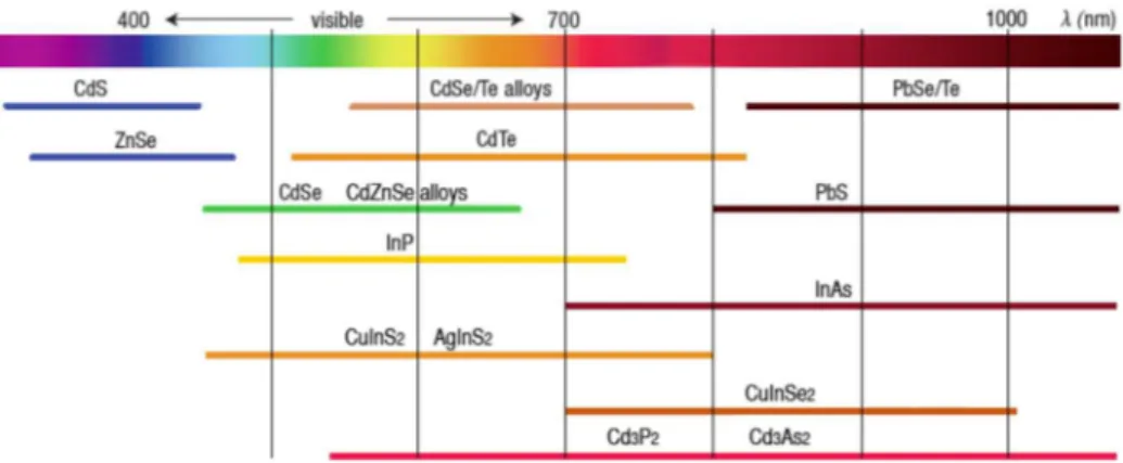
Nowadays it is possible to synthesize QDs that emit within the whole visible and NIR spectral regions by tuning the size and/or the composition of the particle. Figure 1-4 shows the emission ranges reported for different types of QDs. Nonetheless, to date most of the studies focus on cadmium chalcogenide NCs like CdSe, whose intrinsic toxicity restricts their wide-scale application. Indium phosphide (InP) is one of the most promising alternatives.16 It is a III-V semiconductor (bulk value of E
g: 1.35 eV) with a Bohr exciton radius of ∼ 10 nm.17 Its photoluminescence can be tuned from blue to the near infrared by varying the size
of the NCs and the very limited number of toxicological studies indicate a much lower intrinsic toxicity compared with CdSe.18, 19
Figure 1-4. PL emission ranges for the most studied types of semiconductor nanocrystals.20
Although the optical properties of InP-based NCs in terms of PL line width and QY are (still) inferior to the best reported Cd-containing QDs, significant progress in their chemical synthesis has been made in the past decade. While InP core NCs have a QY of less than 1%, values above 50% have been reported for InP/ZnS core@shell NCs.21, 22 Our team reported the single-step synthesis of InPZnS alloy NCs reaching a PL QY
5 enhance the emission efficacy.25 The best value reported today (85%) has been obtained by using a thin
GaP interfacial layer between the InZnP core and the ZnS shell.26
1.2
QDs in biotechnology
1.2.1 Surface functionalization
The use of semiconductor QDs as biological fluorescents probes requires that they (a) are water soluble, (b) present long-term colloidal stability without aggregation and precipitation, (c) offer reactive groups on their surface, and (d) maintain their photophysical properties when transferred into aqueous buffer. NC surface functionalization is a process consisting in the introduction of amphiphilic or hydrophilic organic molecules or macromolecules, peptides or other bioactive molecules on the surface of the QDs in order to induce water solubility, prevent their aggregation, enhance their resistance to oxidation, reduce their toxicity, optimize their behavior (e.g. circulation, uptake) in biological environment and allow their link with specific targets.27
High quality QDs are mostly synthesized in non-polar organic solvents; their hydrophobic surface must be converted to a hydrophilic one in order to solubilize them in aqueous buffer. This solubilization procedure of QDs in aqueous media while maintaining their emission properties and achieving high colloidal stability is a great challenge. It can be achieved by either (a) ligand exchange, a process primarily driven by mass-action in which the native hydrophobic ligand is substituted with bifunctional ligands or by (b)
encapsulation of the original hydrophobic QD within a heterofunctional amphiphilic coating. In the latter
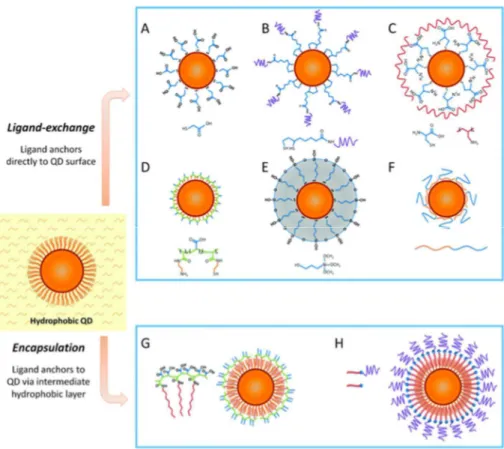
case the strategy is based on compounds capable of assembling with the QD surface via hydrophobic interactions and bearing amphiphilic moieties leading to the desired change of solubility (Figure 1-5). The hydrophobic domain allows for the encapsulation of QDs by a hydrophobic cavity whereas the hydrophilic domain enables the dispersion of QDs in aqueous solution. The coating molecules include phospholipids, bock copolymer, liposomes and appropriate precursors for generating a silica shell around the particles.10, 28
This solubilization strategy can easily be extended to insert additional functionalities on the QD surface. Another appealing feature of the encapsulation strategy is the conservation of the PL QY as the initial surface state is not modified.
6 Figure 1-5. Scheme illustrating different strategies for the water solubilization of hydrophobic QDs. A-F: Ligand-exchange
procedures; G-H: encapsulation procedures.10
In contrast, ligand exchange with bifunctional molecules containing an anchoring site for the QD surface and a hydrophilic site assuring dispersibility of the QDs in aqueous media is a method that radically changes the surface of the NCs. By consequence this strategy bears a high risk of diminishing the PL QY through the generation of surface trap states. These can trap charge carriers despite a protective inorganic shell because the latter is generally only a few atomic layers thick and therefore the escape of the carriers to surface states, e.g. via tunnelling, is still possible. On the other hand, the ligand exchange strategy has a strong potential for providing QDs that are smaller in size than the encapsulation technique, which can be of importance for some applications (see below). It also offers the possibility of coupling biological entities.29
Most procedures for ligand exchange use bifunctional thiol-based molecules for the incubation with the hydrophobic QDs, which are usually coated with fatty acid or trioctylphosphine oxide (TOPO) ligands.30
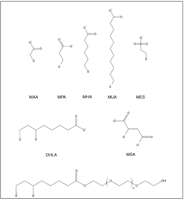
Thiols can strongly interact with ZnS, which is the most widely applied shell material for QDs. Common examples of such ligands are thioalkyl acids containing a carboxylate terminal group such as mercaptoacetic acid (MAA), mercaptopropionic acid (MPA), or mercaptoundecanoic acid (MUA). In their thiolate form these molecules interact strongly with the ZnS surface of the QDs, exposing the polar carboxylate group to the surrounding solution and imparting aqueous solubility (Figure 1-6).31
7 Figure 1-6. Commonly used thioalkyl acid ligands for aqueous solubilization of QDs: mercaptoacetic acid (MAA), mercaptopropionic
acid (MPA), mercaptohexanoic acid (MHA), mercaptoundecanoic acid (MUA), dihydrolipoic acid (DHLA), mercaptosuccinic acid (MSA), mercapthoethane sulfonate (MES) and DHLA appended with poly(ethylene glycol) (PEG).31
In summary, two major methods for engineering the surface chemistry exist in view of the use of QDs in biological media. Encapsulation approaches are very efficient but they imply a large increase in the hydrodynamic diameter of the QDs, from less than 10 nm to 15-25 nm in most cases. Maintaining a small hydrodynamic diameter is crucial for some biological applications. This is particularly true for those relying on Förster resonance energy transfer (FRET) because the FRET efficiency is strongly dependent on the distance between the donor and the acceptor (cf. 1.2.3.1). Therefore, we focused in this work on the aqueous phase transfer of QDs by means of ligand exchange with small zwitteronic molecules that offer several advantages for bioapplications (cf. Chapter 2).
8
1.2.2 Bioconjugation strategies
As discussed in section 1.1, most organic dyes generally used as fluorescent probes suffer from photobleaching, a low brightness above background fluorescence, a wide overlap of the absorption and the emission spectra of different dye molecules, and so on.7, 32 These shortcomings severely limit the use of
organic dyes for detection of rare events or multiplexed imaging and analysis. In contrast, QDs are characterized by bright, stable fluorescence and, hence, are particularly interesting as tools for biological imaging and diagnostics.33 Moreover, all visible and NIR emitting QDs can be excited at the same
wavelength in the UV/blue, which can be very far from their respective emission bands, removing the need of several excitation sources and filtering systems. Despite these advantages QDs issues related to the reproducibility, stability and toxicity of QD-bioconjugates still negatively impact their widespread utilization in biotechnologies.34
The first proof-of-principle applications of QDs in biological imaging have been reported by Bruchez et al. and Chan et al. in 1998.7, 8 Since then the optical properties of QDs have been greatly exploited in a variety
of cell imaging experiments, in vivo imaging, fluoroimmunoassays, DNA sequencing, and other types of bioconjugation.35 Different conjugation protocols have been developed to specifically bind biomolecules to
QDs, such as peptides, lipids, polysaccharides or nucleic acids, creating divers bio-non-bio interactions. Given the versatility of the QD surface for bioconjugation, the possibility of applications is enormous. For example, QDs can be labeled with tumor-targeting antibodies and traced with fluorescence imaging techniques or can be employed in tracking cancer cells in metastasis. However, the efficiency of a fluorescence based probe in biomedical imaging highly depends on the fate of the photons propagating in and out of the living tissue and for diagnostics the stability of the probe in biological serum. Due to the large sizes of antibodies (ca. 10 nm in length for IgG), AB conjugation to QDs is a challenging task (see section 1.2).
Among the large number of bioconjugation strategies we only discuss the most widely used in the following.36 Among them, amine-reactive chemistry using N-hydroxysuccinimid (NHS-) esters,
carboxyl-reactive chemistry using carbodiimide (1-ethyl-3-(3-dimethylaminopropyl)carbodiimide – EDC crosslinking for coupling with an amine-containing compound), and sulfhydryl-reactive chemistry using maleic acid imides are the most prominent. Since a heterobifunctional crosslinker has different reactive groups on either end of the molecule, each side can be directed specifically toward different functional groups on proteins or other molecules. Therefore, heterobifunctional reagents are commonly used to crosslink proteins and other molecules in a two- or three-step process that limits the degree of polymerization often obtained using homobifunctional crosslinkers.36 The most popular heterobifunctional reagents are those
9 an active ester, most often an NHS ester, while the sulfhydryl-reactive group may be one of several different functional groups. Succinimidyl 4-(N-maleimidomethyl)cyclohexane-1-carboxylate (SMCC) is a hetero-bifunctional reagent of utmost utility in protein conjugation processes. The NHS ester end of the reagent can react with a primary amine group to form stable amine bonds. The maleimide end of SMCC is specific for the coupling to sulfhydryls when the reaction pH is in the range of 6.5-7.5. However, this compound suffers from a cross-bridge that is both water-insoluble and immunogenic. Redesigning this crosslinker to have a PEG cross-bridge provides enhanced water solubility for modified proteins or other molecules as well as displaying very low immunogenicity. Moreover, a PEG group used as a cross-bridge in a heterobifunctional reagent to prepare immunogen conjugates will result in non-immunogenic modifications on the carrier protein and thus no antibody production against the polyether linker.37
Figure 1-7. Principles of nanoparticles bioconjugation. A) Amine-reactive chemistry: primary amines on the antibody are modified
with N-hydroxysuccinimide (NHS) esters that conjugate in solution with activated QDs (functionalized with 4-formylbenzamide). B) Sulfhydryl-reactive chemistry: maleimide-functionalized QDs conjugate with antibodies via in situ reduced disulfide bonds.38
In this work we have focused our attention on the conjugation of maleimide-functionalized QDs to antibody sulfhydryl groups and therefore we review some strategies using the sulfhydryl chemistry in the following. Min et al. demonstrated a method to efficiently capture and quantify circulating tumor cells (CTCs) using Anti-EpCAM antibody-conjugated QDs.39 The QD-attached CTCs are isolated using anti-IgG-modified
magnetic beads. The antibody was thiolated using succinimidyl-S-acetylthioacetate (SATA) and the QDs were coated with DSPE-PEG 2000 and DSPE-PEG2000-amine (Figure 1-8). Anti-EpCAM antibody was successful conjugated using a heterobifunctional linker, sulfo-SMCC.
10 Figure 1-8. Construction of anti-EpCAM antibody-conjugated quantum dots (anti-EpCAM-QDs) as primary nanoparticles. DSPE-PEG
2000-methoxy and DSPE-PEG 2000-amine were grafted onto the surface of the QDs and the resulting QDs and antibodies were conjugated using a sulfo-SMCC linker.39
In another example of sulfhydryl coupling chemistry, the conjugation of anti-interleukin-10 antibodies to CdSe/ZnS QDs by means of a SMCC was demonstrated (cf. Figure 1-9).40 Interleukin-10 molecules
participate in the inter-cellular communication. A commercially available conjugation kit from Invitrogen was used to perform the attachment of the antibodies to the QDs.
Figure 1-9. Conjugation of CdSe/ZnS QD PEG amide to thiol-containing anti-interleukin-10 antibodies using SMCC as linker.40
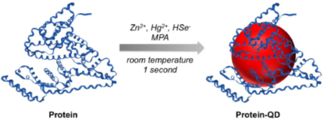
Recently, a new strategy for the preparation of near-infrared (NIR) emitting protein-functionalized QDs at room temperature was presented (Figure 1-10).41 Zn
xHg1-xSe QDs were synthetized in aqueous solution and directly protein-functionalized as the protein molecules and small hydrophilic thiols (e.g. mercaptopropionic acid) are both used simultaneously as ligands during the reaction. According to the authors, protein molecules will bind to the QDs through the coordination of amino acid residues with Zn2+ ions on QD surface and the small thiol-containing MPA molecules would serve as additional surface ligands.
11 Figure 1-10. One-step synthesis of protein-functionalized NIR emitting ZnxHg1-xSe QDs using Zn2+, Hg2+, and HSe- ions as precursors
in aqueous solution and MPA and the protein as ligands.41
The instructive review article of Montenegro et al. summarizes strategies of controlled bioconjugation of nanoparticles with special emphasize on the questions of how to precisely adjust the number of biomolecules per NP precisely and how to attach ABs on NPs in an oriented way (cf. Figure 1-11).42
Controlling the AB orientation on the NC surface is of crucial importance to ensure its optimal interaction with antigens.
Figure 1-11. Schematic representation of strategies AB conjugation on NCS and for controlling their orientation.42 The different methods are discussed in the text.
Figure 1-11a shows the electrostatic adsorption between the antibody and the surface of the NP. It is based on the electrostatic interactions, eventually enforced by hydrophobic, hydrogen binding, and van der Waals attractive forces. Using this method causes denaturation of the immobilized ABs, thus yielding poor reproducibility.43, 44 Strategy b) is less straightforward as it involves the covalent binding of ABs on the NC
surface, which needs generally to be preceded by the insertion of functional groups on the NP surface. It also relies on the use of chemical linkers/crosslinkers, and/or chemical modification of the AB. Method c)
12 involves binding through sugar moieties of the AB, which guarantees an oriented immobilization on the NCs’ surface. Carbohydrate chains on the Fc region of the AB are mildly oxidized to CHO (reactive aldehydes) and can be directly reacted with primary amines on the NC surface or to NCs that have been activated with hydrazide groups. Strategy d) uses appropriate crosslinking biomolecules to obtain oriented immobilization of ABs. These biomolecules are directly coupled on the NP surface. Finally, method e) combines ionic adsorption plus covalent binding to promote oriented immobilization. To achieve this, bifunctional NPs are prepared containing both ionizable groups and reactive groups at their surface.
In summary, different strategies based on sulfhydryl chemistry using SMCC or EDC/NHS crosslinkers for bioconjugation of proteins to NCs have been developed over the last years. In this study we are focusing on the development of compact QD-AB conjugates. As mentioned we will use the maleimide/sulfhydryl bioconjugation strategy. With the goal to further reduce the size of our probes we will apply antibody fragments instead of full ABs. The use of fragmented ABs is also advantageous because their conjugates display less interference with various Fc binding proteins, less immunogenicity (due to lack of the Fc region), and lower nonspecific binding to surfaces or membranes. Enzymatic digests of IgG can result in two particularly useful fragments called Fab and F(ab’)2, prepared by action of papain and pepsin, respectively (Figure 1-12).
Figure 1-12. Antibody hypervariable regions. Schematized drawing of how the three hypervariable regions in each light and heavy
13
1.2.3 QDs for in vitro diagnostics (IVD)
One of the most popular biotechnological applications with high potential for the use of QDs is in vitro diagnostics (IVD). A good IVD agent should be highly luminescent, stable, capable of bioconjugation and yield a very high sensitivity, i.e. a very low detection limit (LOD). Among the large number of different immunoassays in clinical diagnosis, we will focus here on those based on FRET, and in particular time-gated Tb-to-QD FRET. Indeed, a rapid, sensitive and specific immunoassay for protein markers in whole blood or plasma would largely improve the early diagnosis as well as monitoring therapy and disease progression. Important parameters for the immunoassay are that it must be fast, simple of use and inexpensive allowing for uncomplicated diagnosis and better treatment. Homogeneous assays based on FRET are an ideal basis to meet the challenging requirements of IVD. They do not require any washing or separation steps (homogeneous), the fast solution-phase kinetics allow short incubation times and time-resolved detection permits nearly background-free measurements. Moreover, the ratiometric format (luminescence detection of FRET from a donor to an acceptor) offers an instantaneous suppression of sample or measurement fluctuations resulting in an extremely good reproducibility. Since the first proof-of-concept study in 2005,46
which used biotin-streptavidin as biological binging model, there have been many applications that used Ln-to-QD FRET for versatile, multiplexed, and sensitive bioanalysis.
1.2.3.1 Homogeneous Tb-to-QD Förster Resonance Energy Transfer (FRET) immunoassays
Homogeneous FRET sandwich immunoassays, which use donors and acceptors labeled with antibodies that bind to different epitopes of a biomarker, are a smart solution to perform the rapid and separation-free biomarker detection. Since several years Ln-donor-based time-gated FRET immunoassays using organic dyes as acceptors have been applied in commercial diagnostic kits. Indeed, this technology can also be found on many commercial fluorescence plate readers for biological and biochemical analysis. Geißler et al. achieved picomolar detection limits for five different lung cancer tumor markers by combining 15 different biomolecules (10 antibodies interacting with 5 tumor markers) in a 5-fold multiplexed FRET immunoassay using Tb-to-dye time-gated FRET.47 Nonetheless spectral crosstalk in such multiplex assays using dyes is
unavoidable and therefore computational treatment of the data is required. Initial studies on biotin-streptavidin binding systems showed the advantages of Tb-to-QD FRET over Tb-to-dye FRET concerning multiplexing and sensitivity.48 However, the implementation of Tb-to-QD FRET in immunoassays has been
limited, due to the much larger sizes of the antibodies, compared to biotin-streptavidin and the difficulty to create reproducibly stable and highly luminescent QD-AB conjugates. Only very recently, different
14 examples have demonstrated the efficient use of this system. Wegner and collaborators used TG-FRET immunoassays against prostate specific antigen (PSA). Figure 1-13 shows the principle of the QD-based homogeneous FRET immunoassay that explored all different combinations of conjugates. It showed that the combination of Tb-IgG conjugates and QD-F(ab) conjugates provided the best sensitivity compared to the other possible combinations of donor and acceptor antibody conjugates.11 In other words, the best
immunoassay systems combining maximum sensitivity (minimum LOD), minimum antibody modification (no IgG reduction for the Tb conjugates), and maximum separation efficiency was the (Tb-IgG)+(QD-F(ab)) system. LODs down to 1.6 ng/mL in 50 µL serum samples demonstrated the relevance of these assays for clinical diagnostics. It should be noted that these Tb-to-QD FRET immunoassays used a commercial QD-antibody conjugation kit from eBioscience, which is not available anymore. We will see in Chapter 3 that we developed a strategy for the synthesis of much more compact QD-AB conjugates which allowed to achieve a further twofold improvement of the LOD.
Figure 1-13. Top: Tb-AB conjugates; bottom: QD-AB conjugates. Each containing different primary antibodies against PSA.11
Very recently, Bhuckory et al. investigated currently commercially available QDs (Qdot-ITK from Thermo Fisher – Life Technologies), which were conjugated to free sulfhydryls of reduced anti-PSA IgG antibodies using sulfo-EMCS crosslinkers (Figure 1-14). Tb-complexes were conjugated to anti-PSA IgG antibodies (specific to a different PSA epitope than the F(ab)) using amino-reactive chemistry. The performance of the resulting FRET immunoassay will be discussed in comparison with our results in Chapter 3.
15 Figure 1-14. Scheme showing the reduction of IgG antibodies followed by the sulfhydryl coupling reaction with
maleimide-functionalized QDs.49
1.2.3.2 Principle of FRET detection
Förster Resonance Energy Transfer is the non-radiative transfer of energy between a donor and an acceptor, forming the FRET pair, via a dipole-dipole coupling mechanism.50 The distance between the donor
and the acceptor molecules is typically in the range of 1-10 nm, in extraordinary conditions FRET at larger distances up to 20 nm has been reported. Ln-to-QD FRET allows for a complete suppression of the emission of photoexcited QDs by using time-gated PL detection, i.e. by using a pulsed excitation source and applying a delay (in the microsecond range) between excitation and detection. This is possible due to the large differences in the excited state lifetimes of both fluorophores, nanoseconds in the case of QDs, milliseconds in the case of Ln complexes. As a result, the observed QD PL is a pure FRET signal that is generated by sensitization from the Ln. This acceptor-background-free FRET has some influence on the FRET efficiency (ηFRET, eq.3), which is generally defined by the donor-acceptor distance (r) and the Förster radius (or Förster distance) R0. The Förster radius (R0) is the distance between donor and acceptor where the energy transfer is 50% efficient (Figure 1-15). R0 can be calculated using the spectral overlap integral of
donor luminescence and acceptor absorption, as defined by eq. 1.
(1)
where K2 is the orientation factor between the two dipole moments, Φ
D is the donor luminescence
quantum yield, n is the refractive index of the solvent, NA is Avogadro’s number, and J(λ) is the spectral
overlap integral defined by eq. 2.
(2)
J is dependent on the acceptor molar absorptivity (ԑ) and the donor area-normalized emission spectrum (FD). ) ( 128 ) 10 ln( 9 4 5 2 6 0
λ
π
κ
J N n R A r D Φ =λ
λ
λ
ε
λ
λ
F
d
J
(
)
=
∫
D(
)
(
)
416 Finally, the FRET efficiency ηFRET can be calculated using eq. 3, displaying the characteristic r –6 distance dependence:
(3)
where τ are the decay times of the donor in absence (subscript “D”) and in presence (subscript “DA”) of the acceptor. The sensitivity of ηFRET to the D-A distance is shown in Figure 1-15.
Figure 1-15. Left: Visualization of the spectral overlap integral J(λ) between the donor D and acceptor A. Right: Relationship
between the Förster radius and the FRET efficiency where the FRET efficiency is inversely proportional to the sixth power of the D-A distance.
In this Figure it becomes clear that the largest dynamic range lies in a region between 0.5 R0 < r < 2.0 R0.
Beyond this region, FRET is either too efficient (100%) or negligible (0%), making it insensitive to distance changes, which is important for applications relying on the use of FRET as so-called “molecular ruler”. After donor excitation, FRET is in competition with radiative and non-radiative deactivation of the donor. Upon acceptor excitation via FRET, it can again return to its energetic ground state by radiative or non-radiative transitions (cf. Figure 1-16).
6 6 0 6 0 1 r R R D DA FRET = −
τ
= +τ
η
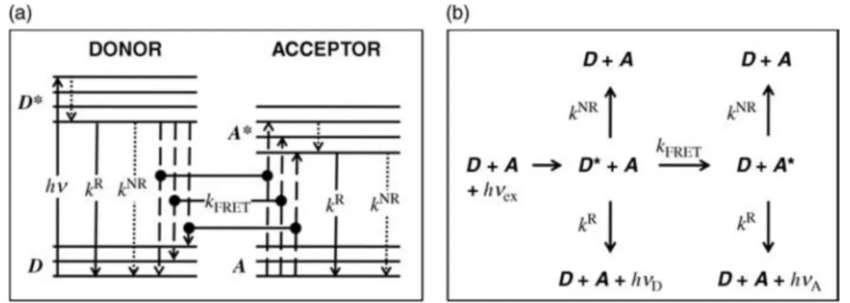
17 Figure 1-16. Principle of donor / acceptor interaction in FRET. (a) Simplified energy-level diagram (Jablonski diagram) representing
the excitation of the donor (hv) from an electronic ground state (D) to an excited state (D*), subsequent inner relaxation (dotted arrow), followed by radiative decay (kR), non-radiative decay (kNR) or FRET (k
FRET). For FRET occurring from D to A, the difference between the respective energy levels needs to be equal (resonance condition). After FRET, the acceptor is in an excited state (A*), followed by radiative or non-radiative decay to its ground state (A). (b) Summary of the different decay pathways after donor excitation (hvex).
1.2.3.3 FRET using QDs as energy donors or as acceptors
QDs can be used as both FRET donors and acceptors. The main advantages of the QDs used as donor are 1) their size tunability, which allows to adjust a spectral overlap with almost any acceptor, 2) their broad absorption spectra which allow excitation at almost any wavelength, 3) the attachment of several acceptors to the large surface of QDs, which allows an increase of the FRET efficiency.
Using QDs as acceptors there is efficient direct excitation of the QDs at almost any wavelength used for donor excitation, due to their broad absorption spectrum. This will lead to inefficient FRET, as a result of the small ratio of excited Tb donors to ground state QD acceptors. To avoid this problem, the use of long-lived luminescent lanthanide complexes (LLCs) as donors, pulsed excitation and time-gated detection is the best solution (cf. Figure 1-17). Indeed, several microseconds after the excitation pulse, all QDs have decayed back to their ground states, whereas most of the LLCs have remained in their excited states leading to efficient FRET. Moreover, due to the large spectral overlap between Ln emission and QD absorption, these FRET systems result in very long Förster distances of >10 nm.51
18 Figure 1-17. Scheme of the FRET pair composed of Tb donors and QD acceptors indicating the ranges of excited state lifetimes.
1.3
Cancer and nanomedicine: a brief introduction
Nanomedicine is a cutting-edge area of biomedical research that exploits the application of nanotechnology to medical science. It involves the design and development of novel nanostructured material that, once engineered, promise a profound impact in prevention, diagnosis, and treatment of several diseases.52
Cancer, due to an abnormal accumulation of cells, is one of the diseases that have the strongest impact on the population. It is estimated that 80% of cancer related deaths are due to metastases. Indeed, morbidity and mortality associated with tumors mostly result from the invasion of adjacent and distant tissue given rise to metastases. This dismal scenario is primarily due to the fact that most patients are diagnosed when the cancer has reached an advanced stage, which often is the result of a lack of specific symptoms and limitation in diagnostics that allow the disease to elude detection during its formative stage. Thus, it is clear that further progress needs a deeper understanding of tumor initiation and progression and methods for monitoring tumor development. Tumor cells gain advantages in initial growth through dysfunction of growth factor responses that are mediated by receptor proteins expressed on cell membranes.53 Thus,
methods to detect, identify and quantify cell surface proteins or marker proteins in a sensible way may potentially facilitate reliable early-stage cancer diagnostics. We have already addressed this topic in section 1.2.2, and we highlighted that one of the promising methods in this context is based on FRET-immunoassays, to directly quantify the biomarker concentration in serum samples. Among the myriad of biomarkers, in this thesis we use the prostate specific antigen (PSA) as biomarker specific for prostate cancer. This cancer is common, a frequent cause of cancer death and, has surpassed lung cancer as the most common cancer in men.54 PSA is a glycoprotein and is expressed by both normal and neoplastic
prostate tissue. The measurement of its absolute value in serum is useful for determining early stage of prostate cancer. The American Cancer society systematically reviews the literature assessing PSA performance.54 This analysis estimated the sensitivity of a PSA cut-off level of 4.0 ng/mL was 21% for
19 detecting any prostate cancer and 51% for detecting high-grade cancers (Gleason ≥8). Using a cut-off value of 3.0 ng/mL increased these sensitivities to 32 and 68%, respectively. Thus, we note that a lower PSA cut-off value is highly desirable, because a fraction of men with PSA levels below 4 ng/mL were found to have prostate cancer.55
This improvement of test sensitivity by lowering the PSA cut-off value is however normally accompanied by a reduced test specificity, leading to far more false-positive test and unnecessary biopsies.56 To avoid this problem time-gated FRET immunoassays have proven a great potential on the way towards systems for real-time in vitro diagnostics. The commercial KRYPTOR compact plus plate reader is a fully automated, closed laboratory analysis system that can be perform various analyses in random-access operations using TRACE (Time Resolved Amplified Cryptate Emission) Technology. Cezanne/Thermo Fisher (partner of my PhD project) employs TRACE technology in the scope of an exclusive license to develop innovative in vitro diagnostic reagents.
1.4
Nanotoxicological aspects
In this Introduction we have shown that QDs offer great opportunities for bioapplications. On the other hand, the use of NCs in real-life biological imaging, detection and diagnostics is still very limited. One major drawback which severely limits the potential for clinical translation of QDs is the toxicity concern of commonly used II-VI semiconductors, such as CdSe and CdTe QDs. These semiconductor QDs are easily disintegrated in biological systems if their surfaces are not carefully coated with inert protective shells, biocompatible polymers, and biomolecules. The release of Cd2+ ions as a result of degradation of the coatings that surround the NC triggers severe toxicity due to the fact that Cd2+ can penetrate the cell using calcium channels and saturate them. It also reduces the availability of antioxidant factors and thus increases the concentration of reactive oxygen species (ROS).57 Therefore, over the past years, the
emphasis has shifted toward the synthesis of non-Cadmium based QDs for bioapplications. In this direction, QDs made up of III-V semiconductors, such as indium phosphide (InP), have drawn considerable attention. It is hard to give a single explanation for the toxicity of QDs, which appears to depend very much on the chemical composition of the core (or core/shell structure) and the surface, which represents the key interface interaction with biological components. The small size, combined with very high surface:volume ratio, makes NPs very reactive compared to larger particles. In general, particles covered with thick polymer coatings are less damaging to the cells,58 while the elimination of this protective coating (for example,
through irradiation, low pH, lysosomal or metabolic degradation) can induce cell damage and death.59 Two
20 of metal ions from the core of the NC, the second one is the formation of reactive molecules, such as reactive oxygen species (ROS).
Figure 1-18. ROS produced by QDs can cause damage to organelles. In addition, the precipitation of QDs on the cell surface even
without the entry into the cell can damage the function and eventually lead to cell death.60
Indeed, NPs often escape the endosomal trafficking and continue their journey into the cytoplasm, in cell organelles, or into the nucleus, affecting the cells’ structural or functional integrity. NPs can further induce genotoxic effects either by direct interaction with the genetic material or by indirect action through reactive oxygen species or ions released from the NP core. QDs act as active redox nanoparticles (electron donors)61 and can generate highly reactive radicals with or without exposure to light.62 At high
concentrations, ROS can cause damage to cellular proteins, lipids, DNA and carbohydrates, causing apoptosis or necrosis.
Toxicological studies of cadmium-free QDs are very scarce in the literature but suggest that InP-based QDs minimize the toxic effects observed with their CdSe-based counterparts. This feature is of course an essential prerequisite for the use of such NCs in the biomedical field. Nanotoxicology studies conducted both in vitro and in vivo on Drosophila melanogaster show that CdSe/ZnS QDs are more toxic than InP/ZnS QDs despite the similar physicochemical properties and equivalent localization in the cell.63 The effect is
due to the release of core metal ions indicating that the indium toxicity is virtually zero compared to that of cadmium. Other findings in mice confirm that InP/ZnS QDs do not show toxicity in vivo during the evaluation period (84 days), suggesting that these NPs are suitable for use in biological systems.64
Concomitant with the increased use of QDs in consumer products such as sun creams, textile fibers, TV screens and other types of displays, solar cells, etc. there is a strong need to increase the investigation of
21 potential toxic effects due to unintentional release or contamination of the environment through powder/water waste-streams.65-68 Furthermore, an urgent evaluation of QD toxicity on human beings is
strongly needed.
Actually, most nanotoxicity studies have focused on in vitro investigations, representing over-simplification of bio/non-bio interactions and not taking in account the real biological complexity and systemic networking of whole animals; hence, in vivo tests are central to achieve an accurate estimation of nanotoxicity in living organisms.69 The European directives encourage experiments on model systems to
minimize testing on vertebrate animals. We will address this point in Chapter 4 where we present recent studies using a small invertebrate animal, the freshwater polyp Hydra vulgaris, and in parallel preliminary studies on keratinocytes from human skin biopsies.
22
1.5
Aim and motivation of the thesis
The work presented in this PhD thesis is part of the ANR project NanoFRET and our main task was the development of stable, highly luminescent and FRET-compatible QD-antibody conjugates. Such immuno-QDs are together with the AB-labeled Tb donor integral part of FRET immunoassays and of potential interest for the project’s industrial partner Cezanne-Thermo Fisher. One of the main motivations of the NanoFRET project is to fill the current lack of stable, functional, highly luminescent and small QD-antibody conjugates that provide a high binding capacity and yield efficient FRET, generating highly sensitive immunoassays.
In my thesis I first focus on the development of a QD functionalization strategy yielding stable, compact and highly luminescent NCs to be conjugated with antibodies and used as FRET acceptor in immunoassays. We will present this strategy in Chapter 2, keeping in mind all the main characteristics required. In Chapter 3 the binding of fragmented antibodies on this nanoprobe as well as the evaluation in FRET immunoassays against PSA will be addressed. In addition, we extend our studies to systems relying on the grafting of paramagnetic (Gd) or luminescent (Tb, Eu, Yb) lanthanide complexes on the QD surface with the goal to design multimodal probes.
Chapter 4 aims at assessing the toxicological impact of the different InP- and CdSe-based QDs used in our study. The main interest of these experiments is the direct comparison of the different QD materials, while using an identical surface chemistry. We have tested them in different biological systems, ranging from in
vitro cultured human cells to in vivo animal models, Hydra polyps and keratinocytes from human skin
biopsies, respectively.
Each chapter contains its corresponding experimental section in the end.
The manuscript is completed by a Conclusion and Perspectives section, a list of abbreviations and a list of my scientific publications / communications.