HAL Id: hal-01870357
https://hal.umontpellier.fr/hal-01870357
Submitted on 13 Nov 2018
HAL is a multi-disciplinary open access
archive for the deposit and dissemination of
sci-entific research documents, whether they are
pub-lished or not. The documents may come from
teaching and research institutions in France or
abroad, or from public or private research centers.
L’archive ouverte pluridisciplinaire HAL, est
destinée au dépôt et à la diffusion de documents
scientifiques de niveau recherche, publiés ou non,
émanant des établissements d’enseignement et de
recherche français ou étrangers, des laboratoires
publics ou privés.
Study
Sandra Martin, Borja Rodríguez-Herreros, Jared Nielsen, Clara Moreau,
Claudia Modenato, Anne Maillard, Aurélie Pain, Sonia Richetin, Aia Jønch,
Abid Qureshi, et al.
To cite this version:
Sandra Martin, Borja Rodríguez-Herreros, Jared Nielsen, Clara Moreau, Claudia Modenato,
et al..
Quantifying the Effects of 16p11.2 Copy Number Variants on Brain Structure:
A
Multisite Genetic-First Study.
Biological Psychiatry, Elsevier, 2018, 84 (4), pp.253 - 264.
Quantifying the Effects of 16p11.2 Copy
Number Variants on Brain Structure:
A Multisite Genetic-First Study
Sandra Martin-Brevet, Borja Rodríguez-Herreros, Jared A. Nielsen, Clara Moreau,
Claudia Modenato, Anne M. Maillard, Aurélie Pain, Sonia Richetin, Aia E. Jønch,
Abid Y. Qureshi, Nicole R. Zürcher, Philippe Conus, 16p11.2 European Consortium, Simons
Variation in Individuals Project (VIP) Consortium, Wendy K. Chung, Elliott H. Sherr,
John E. Spiro, Ferath Kherif, Jacques S. Beckmann, Nouchine Hadjikhani, Alexandre Reymond,
Randy L. Buckner, Bogdan Draganski, and Sébastien Jacquemont
ABSTRACT
BACKGROUND: 16p11.2 breakpoint 4 to 5 copy number variants (CNVs) increase the risk for developing autism spectrum disorder, schizophrenia, and language and cognitive impairment. In this multisite study, we aimed to quantify the effect of 16p11.2 CNVs on brain structure.
METHODS: Using voxel- and surface-based brain morphometric methods, we analyzed structural magnetic resonance imaging collected at seven sites from 78 individuals with a deletion, 71 individuals with a duplication, and 212 individuals without a CNV.
RESULTS: Beyond the 16p11.2-related mirror effect on global brain morphometry, we observe regional mirror differences in the insula (deletion . control . duplication). Other regions are preferentially affected by either the deletion or the duplication: the calcarine cortex and transverse temporal gyrus (deletion . control; Cohen’s d . 1), the superior and middle temporal gyri (deletion , control; Cohen’s d , 21), and the caudate and hippocampus (control . duplication; 20.5 . Cohen’s d . 21). Measures of cognition, language, and social responsiveness and the presence of psychiatric diagnoses do not influence these results.
CONCLUSIONS: The global and regional effects on brain morphometry due to 16p11.2 CNVs generalize across site, computational method, age, and sex. Effect sizes on neuroimaging and cognitive traits are comparable. Findings partially overlap with results of meta-analyses performed across psychiatric disorders. However, the lack of correlation between morphometric and clinical measures suggests that CNV-associated brain changes contribute to clinical manifestations but require additional factors for the development of the disorder. These findings highlight the power of genetic risk factors as a complement to studying groups defined by behavioral criteria. Keywords: 16p11.2, Autism spectrum disorder, Copy number variant, Genetics, Imaging, Neurodevelopmental disorders
https://doi.org/10.1016/j.biopsych.2018.02.1176
Autism spectrum disorder (ASD) and related neurodevelopmental
disorders are defined behaviorally and characterized by a
signif-icant clinical and etiologic heterogeneity. As a consequence, investigating ASD under the assumption of an underlying
homo-geneous condition has resulted in controversial findings in the
field of neuroimaging(1). Increased brain growth early in
devel-opment(2–4)and alterations of many regional brain volumes(5)
have been implicated in ASD, but results have proven difficult
to replicate(1,6–8).
To mitigate some of these issues, cohorts of individuals with shared genetic risk factors have been assembled to minimize the
noise introduced by etiologic and biological heterogeneity (9).
Such a“genetic-first” study design provides the opportunity to
investigate a given neurodevelopmental risk (and associated mechanism) shared by individuals who carry the same genetic etiology irrespective of the psychiatric diagnosis.
Copy number variants (CNVs) at the 16p11.2 (breakpoints
4–5, 29.6–30.2 Mb-hg19)(10)are among the most frequent risk
factors for neurodevelopmental and psychiatric conditions. There is a similar 10-fold enrichment of deletions and
dupli-cations in ASD cohorts (11,12), and both CNVs have large
effects on IQ (Z scores of 1.5 and 0.8, respectively) and Social Responsiveness Scale (SRS) (Z scores of 1 and 2, respectively) (10,13–15). However, there are phenotypic differences be-tween both CNVs: the 10-fold enrichment in schizophrenia
cohorts (16,17) is only observed for duplications, and only
deletions affect measures of language by 1.5 Z scores (18).
Previous studies demonstrated“mirror” effects of both CNVs
on head circumference and body mass index(13,19).
Neuro-imaging studies reported gene-dosage effects on global brain
metrics(20,21). However, large global effects and sample size
limited the interpretation of the regional analyses, any estimate of effect size, and the generalizability of study results across different ascertainments.
In the current study, we aimed at quantifying the effects of 16p11.2 deletions and duplications on brain structure. We also examined the generalizability of our results across cohorts, scanning sites, sex, and a broad age range. Finally, we aimed
at understanding the influence of clinical ascertainment. In
particular, we asked whether language, social responsiveness, IQ, or the presence of psychiatric disorders may impact any of
the findings. To this end, we analyzed structural magnetic
resonance imaging (MRI) performed at seven sites from two international cohorts of 16p11.2 CNV carriers, familial control subjects, and unrelated control subjects. Voxel- and surface-based methods were performed in parallel on 361 partici-pants, including 307 individuals not previously analyzed at the regional level, using whole-brain statistical methods.
METHODS AND MATERIALS Participants
Data were acquired in two different cohorts in North America and Europe. Enrollment in the Simons Variation in Individuals
Project(22)included referral by clinical genetic centers or
web-based networks, or active online registration of families, while in the European 16p11.2 consortium the families were directly recruited by the referring physician.
Carriers were ascertained regardless of clinical diagnoses or age. The CNV carriers were either probands (n = 76) referred to the genetic clinic for the investigation of neurodevelopmental and psychiatric disorders, or their relatives (parents [n = 49], siblings [n = 14], and other relatives [n = 10]). Familial control subjects were relatives who do not carry a 16p11.2 CNV.
All families participated in a larger phenotyping project, as
previously reported(10,13,20–22). Trained neuropsychologists
performed all cognitive and behavioral assessments, including tests of overall cognitive functioning (nonverbal IQ [NVIQ])
(23–27)and phonological skills (standard score of the nonword
repetition) (28,29). Participants also completed a broad
screening measure of social impairment, the SRS(30).
Expe-rienced, licensed clinicians provided clinical DSM-5 diagnoses
(31), using all information obtained during the research
evalu-ation. NVIQ scores and psychiatric diagnoses were available for all participants. SRS total score was available for 77% of the participants (72 of 78 deletion carriers, 57 of 71 duplication carriers, and 149 of 212 control subjects), and phonological measures for 43% of the participants (56 of 78 deletion car-riers, 19 of 71 duplication carcar-riers, and 81 of 212 control subjects). Full description of cognitive and psychiatric
assessment is available in the Supplemental Methods and
Materials.
We analyzed data from 78 16p11.2 (breakpoints 4–5)
dele-tion carriers, 71 duplicadele-tion carriers, 72 familial control sub-jects, and 140 unrelated control subsub-jects, including data not previously analyzed at the regional level on 64 deletion carriers,
54 duplication carriers, 51 familial control subjects, and 138 unrelated control subjects. The latter were selected among volunteers from the general population who had neither a major DSM-5 diagnosis nor a relative with a neuro-developmental disorder.
The study was approved by the institutional review boards of each consortium. Signed informed consent was obtained from the participants or legal representatives. Full description
of participants is available inTable 1,Supplemental Table S1,
and theSupplemental Methods and Materials.
MRI Data Acquisition and Processing
The MRI data included T1-weighted (T1w) anatomical images acquired at seven sites using different 3T whole-body scan-ners: Philips Achieva (Philips Healthcare, Andover, MA) and Siemens Prisma Syngo and TIM Trio (Siemens Corp., Erlangen, Germany). Four sites used multiecho sequences for 264 par-ticipants (52 deletion carriers, 51 duplication carriers, 21 fa-milial control subjects, and 140 unrelated control subjects), and three sites used single-echo sequences for 97 participants (26 deletion carriers, 20 duplication carriers, and 51 familial control subjects). Thirty-four scans were excluded from the analysis based on standardized visual inspection, which
identified significant artifacts potentially compromising the
accurate tissue classification and boundary detection (details
inSupplemental Methods and Materials).
Surface-Based Morphometry. In FreeSurfer 4.5.0 (http:// surfer.nmr.mgh.harvard.edu), each participant’s T1w image was registered to a custom hybrid template consisting of 48 subjects (12 deletion children, 12 noncarrier children, 12
dupli-cation adults, and 12 noncarrier adults)(21). Then, we used
FreeSurfer’s volumetric(32)and surface-based(33)algorithms
with default settings. We estimated the total intracranial volume
(eTIV)(34), global brain measures, cortical thickness, and
sur-face area. The cortical thickness and sursur-face area maps were resampled in fsaverage5 space and spatially smoothed with a Gaussian kernel of 8-mm full width at half maximum.
Voxel-Based Morphometry. In parallel we processed
subjects’ T1w data within the computational anatomy
frame-work of SPM12 (http://www.fil.ion.ac.uk/spm). T1w images
were classified in different brain tissue classes using the
“unified segmentation”(35)and an enhanced set of brain
tis-sue priors (36). Aiming at optimal spatial registration, we
applied the diffeomorphic registration algorithm DARTEL(37)
followed by a Gaussian spatial smoothing with 8-mm full width at half maximum. Of note, total intracranial volume computed by SPM is referred to as TIV.
Regions of interest were extracted using maximum
proba-bility tissue labels (http://www.neuromorphometrics.com)
within SPM12 using data from the OASIS project (http://www.
oasis-brains.org).
All MRI scanning parameters and processing are detailed in theSupplemental Methods and Materials.
Data Analysis
Our whole-brain voxel-based morphometry (VBM)(38)analysis
gray matter (GM) volume differences within the general linear
model framework of SPM12(39). SPM t maps were generated
with a voxel-level threshold of p , .05 after familywise error correction for multiple comparisons over the whole GM volume
using Gaussian random field theory (40). We generated
Cohen’s d maps from familywise error–corrected t scores to
show the unbiased magnitude of the effects (Supplemental
Methods and Materials).
Surface-based analyses tested regional differences in cortical thickness and surface area using linear models. For each vertex in the cerebral cortex surface mesh, we ran a multiple regression analysis. The vertexwise results were cor-rected for multiple comparisons at a false discovery rate of q , .05(41,42)(Supplemental Methods and Materials).
The main effects of linear and quadratic expansions of age, sex, MRI site, and NVIQ were included as additional variables.
The cubic expansion of age did not show any significant effect
and was subsequently removed from all analyses. In an attempt to increase the power of our analyses we controlled for the effect of the seven scanning sites by introducing them as a random factor in a linear mixed model. This approach did not change the obtained results.
Z scores for global brain metrics were obtained in CNV car-riers and familial control subjects using the adjusted measures from unrelated control subjects as the reference population.
Regional analyses were also corrected for the SPM estimate of TIV, the mean cortical thickness, or the total cortical surface area. In addition to the linear effect of gene dosage, we investigated the quadratic term to identify nonreciprocal ef-fects of both CNVs. Post hoc analyses comparing deletion carriers and control subjects as well as duplication carriers and
control subjects identified regions predominantly altered by
each CNV.
We analyzed the interaction of the genetic groups with the regressors (age, sex, MRI site, NVIQ) as well as the MRI pa-rameters (single-echo vs. multiecho) and three other clinical variables: SRS, phonological processing (nonword repetition),
and psychiatric diagnoses. NVIQ did not show any significant
effect and was removed from the analyses on the whole dataset. Dice index was computed to estimate the overlap between 16p11.2-related alterations and statistical maps ob-tained from a large cross-disorder neuroimaging meta-analysis (http://anima.fz-juelich.de) (43). We computed the rate of overlap between both maps. Finally, to motivate future
hy-potheses we relied on the Neurosynth database (http://
neurosynth.org) to meta-analytically decode the functional association of the structural alterations observed in the gene dosage analyses of the 16p11.2 CNV carriers. All these
ana-lyses are detailed in theSupplemental Methods and Materials.
Linear models on global metrics and regions of interest were
performed in R, version 3.2.5 (http://www.r-project.org; R
Project for Statistical Computing, Vienna, Austria), and voxel-and surface-based analyses in MATLAB 2016b (The Math-Works, Inc., Natick, MA).
RESULTS Demographics
We analyzed 78 deletion carriers, 71 duplication carriers, and
212 familial and unrelated control subjects (Table 1,
Table 1. Po pulation Cha racter istics Deletion Control Duplication EU (n = 25) SVIP (n = 53) EU (n = 83) SVIP (n = 129) EU (n = 23) SVIP (n = 48) Age, Years, Mean (SD), Range [Minimum –Maximum] 21.2 (14) [6.3 –53.6 a , b] 13.8 (9.5) [6.7 –48 c] 28.5 (12.6) [7.8 –62.5 b] 2 4 (14.5) [6.1 –63.4] 32.5 (13.3) [9.8 –58.1] 30.1 (15.4) [6.3 –63.1] Male/Female, n 14/11 30/23 56/27 71/58 13/10 25/23 HC Z Score n =2 3 n =5 2 n =4 6 n =2 7 n =2 2 n =4 8 Mean (SD) 0.28 (1.23) a , b 1.34 (1.2) 2 0.21 (1.3) c 0.32 (1) c 2 0.64 (1.9) 2 1.08 (1.4) NVIQ, Mean (SD) 81 (13) b 89 (14) 107 (16) c 103 (12) c 78 (18) b 89 (20) SRS Raw Total Score n =2 0 n =5 2 n =2 5 n = 124 n =1 1 n =4 6 Mean (SD) 62 (32) 70 (37) 33 (17) c 19 (13) c 84 (40) b 57 (38) Phonological Skills n =9 n =4 7 n =3 n =7 8 n =3 n =1 6 Mean (SD) 4.8 (1.5) 5.5 (2.4) 12.3 (2.1) 8.4 (2.2) c 10.7 (1.5) 6.25 (2.2) Phonology skills were evaluated in SVIP with the comprehensive test of phonological processing, nonword repetition subtest (29) ; and in Europe with the developmental neuropsychological assessment, nonword repetition task (28) . Mean standard scores are shown. Low sample size did not allow statistics on phonological skills of the European (EU) cohort. HC, head circumference; NVIQ, nonverbal IQ; SRS, Social Responsiveness Scale; SVIP, Simons Variation in Individuals Project cohort. aSigni fi cantly different between copy number variant carrier groups in the same cohort; analysis of covariance, post hoc comparison, p , .05 Bonferroni corrected. bSigni ficantly different from the same genetic group in the other cohort; analysis of covariance, post hoc comparison, p , .05 Bonferroni corrected. cSigni fi cantly different from all the genetic groups in the same cohort; analysis of covariance, post hoc comparison, p , .05 Bonferroni corrected.
Supplemental Table S1), including new data on 138 individuals and data on 307 individuals not previously analyzed with whole-brain statistical methods. Age ranges from 6 to 63 years. Deletion carriers and control subjects from the Simons Variation in Individuals Project cohort are younger than the same groups in the European cohort, and deletion carriers overall are younger than the other groups. There is no
sig-nificant difference in sex ratio across genetic groups and
cohorts. Mean NVIQ is 81 and 89 in deletion carriers, and 78 and 89 in duplication carriers for the European and Simons Variation in Individuals Project cohorts, respectively. Ninety percent of the deletion carriers, 69% of the duplication carriers, and 25% of familial control subjects meet criteria for at least one psychiatric diagnosis. Twelve categories of di-agnoses are recorded across the CNV carrier groups, including ASD in 13% of deletion carriers and 11% of
duplication carriers (Table 2).
Global Brain Metrics
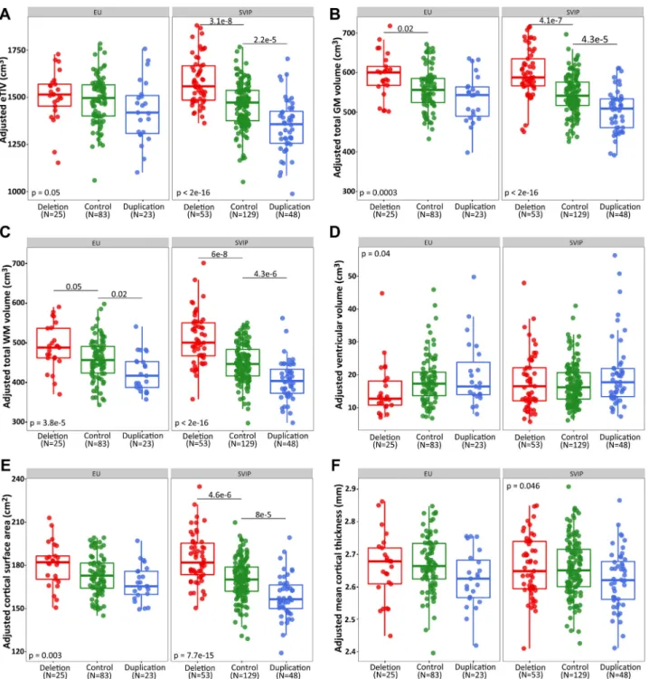
Head circumference Z scores (Table 1) and eTIV (Figure 1A)
correlate negatively with the number of genomic copies of the 16p11.2 locus in both cohorts. Both GM and white matter total
volumes contribute to this effect on eTIV (Figure 1B, C). The
effect sizes on global brain metrics are up to 1 Z score for the
deletion and approximately 20.4 Z score for the duplication
(Supplemental Table S2). FreeSurfer and SPM estimates of
TIV, GM, and white matter are comparable across groups,
cohorts, and MRI parameters (Supplemental Figure S1). Gene
dosage preferentially affects cortical surface area and not
thickness (Figure 1E, F). Of note, age-related thinning of
cortical thickness is not significantly different between genetic
groups (Supplemental Figure S2).
Regional Brain Differences Related to the 16p11.2 CNVs
In both cohorts, the whole-brain VBM analysis shows a negative relationship between the number of genomic copies at the 16p11.2 locus and the volume of several brain
regions. Alterations with an effect size .1 Cohen’s
d (detected with a conservative power of 74.4% for
family-wise error–corrected p , .05) include the bilateral anterior
and posterior insula, transverse temporal gyrus, and
cal-carine cortex (Figure 2A, Supplemental Table S3). Regions
with smaller volumes in deletion carriers compared with control subjects and duplication carriers include the bilateral precentral gyrus and middle and superior temporal gyri. Altered regions with smaller effect sizes are detailed in Supplemental Table S3.
There is a high degree of overlap between VBM findings
with large effects and regional cortical surface area alterations, namely the insula, transverse temporal gyrus, and calcarine cortex (negative gene dosage), as well as the precentral gyrus
Table 2. DSM-5 Diagnoses
Deletion Familial Control Subjects Duplication EU (n = 25) SVIP (n = 53) EU (n = 45) SVIP (n = 27) EU (n = 23) SVIP (n = 48)
Neurodevelopmental Disorders 3 5 – – 4 6 Intellectual Disability
Communication disorder 16 53 – 2 – 3 Autism spectrum disorder – 10 – 1 2 6 Attention-deficit/hyperactivity disorder 2 12 2 4 1 7 Specific learning disorder 1 14 1 – 4 4 Motor disorder, tic disorder – 26 – 1 1 9 Schizophrenia Spectrum and Other Psychotic Disorders – – – – 1 – Bipolar and Related Disorders – – – – 1 – Depressive Disorders 2 3 7 1 5 9 Anxiety Disorders 2 7 – 3 9 13 Obsessive-Compulsive and Related Disorders 1 1 – – 1 2 Trauma and Stressor-Related Disorders 1 – – – – 2 Elimination Disorders 5 14 – 2 1 2 Disruptive, Impulse-Control, and Conduct Disorders 1 5 – – – – Substance-Related and Addictive Disorders 3 – – – 1 – Feeding and Eating Disorders – – 2 – – – Other Conditions That May Be a Focus of Clinical Attention – 9 – – – 9
From the DSM-5(31).
A total of 20 of 25 European (EU) cohort deletion carriers (80%) had at least one psychiatric diagnosis: 11 had one diagnosis and 9 had several diagnoses (between two andfive); 17 of 23 EU cohort duplication carriers (74%) had at least one psychiatric diagnosis: 7 had one diagnosis and 10 had several diagnoses (two or three); 9 of 45 familial control subjects (20%) had at least one psychiatric diagnosis: 6 had one diagnosis and 3 had two diagnoses. In the Simons Variation in Individuals Project (SVIP) dataset, 50 of 53 deletion carriers (94.3%) had at least one psychiatric diagnosis: 6 had one diagnosis and 44 had several diagnoses (between two and eight); 32 of 48 SVIP duplication carriers (66.6%) had at least one psychiatric diagnosis: 10 had one diagnosis and 22 had several diagnoses (between two andfive); 9 of 27 familial control subjects (33%) had at least one psychiatric diagnosis: 4 had one diagnosis and 5 had two diagnoses. In both cohorts, unrelated control subjects without psychiatric diagnosis were recruited.
and superior and middle temporal gyri (positive gene dosage). Regions with smaller effect size and no overlap are shown in Figure 3; Supplemental Figure S3A, C; and Supplemental
Table S4. Cortical thickness, on the other hand, shows little
overlap with the VBM results (Figure 3; Supplemental
Figure S3B,D; andSupplemental Table S5).
Figure 1. Effects of gene dosage on global brain measures in the European (EU) and Simons Variation in Individuals Project (SVIP) cohorts. Boxplots of (A)
estimated total intracranial volume (eTIV), (B) gray matter (GM) volume, (C) white matter (WM) volume, (D) ventricular volume, (E) cortical surface area, and (F) mean cortical thickness in each genetic group separately for the EU and SVIP cohorts. Gene dosage effect is estimated with a linear model using the number of 16p11.2 genomic copies (1, 2, or 3), and including linear and quadratic expansions of age, sex, nonverbal IQ, and magnetic resonance imaging site asfixed covariates. In each box, the bold line corresponds to the median. The bottom and top of the box show the 25th (quartile 1 [Q1]) and 75th (quartile 3 [Q3]) percentiles, respectively. The upper whisker ends at the largest observed data value within the span from Q3 to Q31 1.5 3 the interquartile range (Q3 2 Q1), and lower whisker ends at the smallest observed data value within the span for Q1 to Q12 (1.5 3 interquartile range). Circles that exceed whiskers are outliers. Post hoc comparisons show Bonferroni-corrected p values.
These regional results are not influenced by subjects’ age, sex, cohort, MRI site, or MRI protocol (multiecho vs. single-echo): None of the variables shows an interaction with genetic
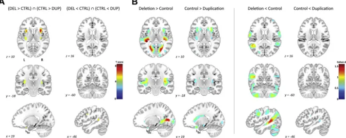
groups (Figure 2B, Supplemental Figures S4 and S5A). In
particular, a subgroup of participants who underwent both multi- and single-echo protocols presents the same alterations (Supplemental Figure S5B). NVIQ does not show any main effect on regional brain structure and was removed as a co-variate for the subsequent analyses. Given the above obser-vations, we pooled all data.
Relationship Between Total Brain Volume and Regional Differences
We examined the contribution of global differences to regional alterations. There was no relationship between global metrics
and any of the aforementioned large effect regional findings,
even after adding GM volume as a covariate in the VBM ana-lyses. We then tested for correlations between eTIV and the
raw or adjusted volumes of some significant regions
(Supplemental Figure S6). This demonstrates that small, average, or large brains contribute equally to the regional ef-fects of 16p11.2 CNVs.
Mirror Effects Versus Differential Contribution of CNVs to Regional Differences
To differentiate reciprocal from nonreciprocal effects driven by either the deletion or the duplication carriers, we compared the linear and quadratic effects of gene dosage. The nonreciprocal
effects of the 16p11.2 deletion and duplication identified by the
quadratic term are detailed inSupplemental Figure S7. Post
hoc analyses show that the deletion preferentially impacts the volume and surface area of the calcarine cortex and the
Figure 2. Effects of gene dosage on regional gray matter volume in the Europe (EU) and Simons Variation in Individuals Project (SVIP) cohorts. (A) Left
panels (deletion. duplication) show voxel-based whole-brain maps, with the volumes of regions showing a negative relationship with the number of 16p11.2 genomic copies. Right panels (deletion, duplication) present the volumes of regions showing a positive relationship with the number of 16p11.2 genomic copies. (B) Negative and positive gene dosage effects on gray matter volume following a leave-one-out approach by systematically removing one of the magnetic resonance imaging sites. All the analyses are controlled for linear and quadratic expansions of age, sex, magnetic resonance imaging site, total intracranial volume, and nonverbal IQ. Results significant at a threshold of p , .05 familywise error corrected for multiple comparisons are displayed in standard Montreal Neurological Institute space. Color bars represent Cohen’s d. L, left; R, right.
transverse temporal gyrus (deletion. control) and the superior
and middle temporal gyri (deletion , control), with absolute
effect size .j1j Cohen’s d. The duplication carriers do not
show any neuroanatomical differences with effect size .j1j
Cohen’s d. We observe GM volume changes in the caudate
and hippocampus with Cohen’s d between j0.5j and j1j
(duplication , control) (Figure 4B, Supplemental Table S6).
Differences with smaller effect sizes or identified only by one of
the analytical methods such as alterations in the cerebellum,
precentral gyrus, and cingulate are detailed in the
L Lateral L Medial R Lateral R Medial L Lateral L Medial R Lateral R Medial VBM Only Surface Area Only
A
B
Figure 3. Overlap between voxel-based and surface-based results for cortical alterations associated with gene dosage. The relationship between gene
dosage and the morphometric features was compared in the pooled sample (n = 361). The voxel-based and surface-based statistical maps are thresholded at the multiple comparisons–corrected p value and then projected on the cortical surface mesh. Regions with effect size $1 Cohen’s d and overlapping between voxel-based and surface-based analyses are (A) the bilateral insula, transverse temporal gyrus, calcarine cortex and (B) the precentral, superior and middle temporal gyri. L, left; R, right; VBM, voxel-based morphometry.
Figure 4. Differential and overlapping contribution of deletion and duplication to the regional gray matter volume differences. (A) Results of voxel-based
whole-brain maps from the conjunction analysis of both negative (deletion. control AND control . duplication) and positive (deletion , control AND con-trol, duplication) gene dosage. The main mirror pattern is the insula. (B) Results of voxel-based whole-brain maps showing the effect size in regions with larger volume in deletion carriers compared with control subjects (deletion. control), in control subjects compared with duplication carriers (control . duplication), in regions with smaller volume in deletion carriers compared with control subjects (deletion, control), and in control subjects compared with duplication carriers (control, duplication). Results significant at a voxel-level threshold of p , .05 familywise error corrected for multiple comparisons are displayed in standard Montreal Neurological Institute space. Color bars represent t scores for panel (A) and Cohen’s d for panel (B). CTRL, control individuals; DEL, deletion carriers; DUP, duplication carriers; L, left; R, right.
Supplemental Table S6andSupplemental Figures S8A–Dand S9C–F.
The reciprocal mirror effects of the 16p11.2 deletion and duplication are restricted to the bilateral insula. The post hoc conjunction analysis shows that the deletion is associated with an increase of the volume and surface area of the insula, and the duplication is associated with a decrease of this region (Figure 4A, Supplemental Table S6). We do not observe reciprocal effects of gene dosage for cortical thickness
mea-surements (Supplemental Figure S9A,B).
Relationship With Psychiatric Diagnosis and Cognitive Traits
Because the 16p11.2 locus is associated with more than one
psychiatric diagnosis, we quantified the overlap of our findings
with a large, cross-disorder neuroimaging meta-analysis [http://anima.fz-juelich.de (43)]. We observe that the 16p11.2-related VBM map overlaps 33% of the meta-analytic map (Dice index): 46% for the cluster including the left insula, 28% for the right insula, and none for the dorsal anterior cingulate cortex.
We used Neurosynth to meta-analytically decode the psy-chological terms most closely associated with the main
anatomical clusters identified in the VBM analysis.Supplemental
Table S7 illustrates the domains most associated with each cluster. The transverse, superior, and middle temporal gyri (re-gions predominantly affected in deletion carriers) show top as-sociations with language, phonology, and auditory terms. The anterior insula and caudate (alterations found in duplication carriers) are associated with terms such as reward, pain, and
executive function (Supplemental Figure S10). Recognizing such
inverse inferences can provide hypotheses for future studies but are unable to support strong conclusions.
However, the measures of NVIQ, SRS, and phonological processing measured in participants do not show main effects or interact with the gene dosage effects. The presence of low general intelligence (NVIQ), language impairment (measured by phonological processing), or poor social skills (SRS), or the presence and number of comorbid psychiatric diagnoses,
does not change any of the neuroanatomicalfindings
associ-ated with the 16p11.2 deletion or duplication.
Ascertainment and Additional Factors Contributing to Changes in Brain Structure
We tested whether ascertaining carriers for
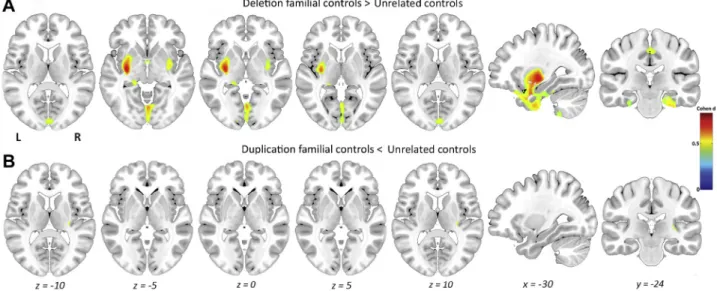
neuro-developmental symptoms could bias our results. Because clinical ascertainment may enrich as well for additional neu-rodevelopmental factors present in CNV carriers and their families, we investigated potential brain alterations in the family members who do not carry a 16p11.2 CNV. Comparing control subjects from deletion families (n = 51) and unrelated control subjects shows changes in volume and thickness with medium
effect size (.0.5 Cohen’s d) of the left posterior insula and right
lingual gyrus; changes of volume also include the putamen and
hippocampus (Figure 5,Supplemental Figure S14E). No effect
was found for the cortical surface area (Supplemental
Figure S13E).
However, comparing deletion or duplication carriers with fa-milial or unrelated control subjects does not change any of the
global (Supplemental Table S2andSupplemental Figure S11) or
regional findings reported above (Supplemental Figures S12,
S13A–D, andS14A–D).
DISCUSSION
This large, multisite dataset combines new and previously published data to expand our understanding of the neuroan-atomical differences associated with 16p11.2 deletions and
Figure 5. Contribution of familial control subjects to the regional gene dosage-dependent gray matter volume differences. Results of voxel-based
whole-brain maps showing (A) regions with larger volume in control subjects from deletion families (n = 51) compared with unrelated control subjects (n = 140); and (B) regions with smaller volume in control subjects from duplication families (n = 21) compared with unrelated control subjects (n = 140). Results significant at a voxel-level threshold of p , .05 familywise error corrected for multiple comparisons are displayed in standard Montreal Neurological Institute space. Color bars represent effect size (Cohen’s d). L, left; R, right.
duplications. The effect of the reciprocal CNVs on brain structure is generalizable across heterogeneously ascertained
cohorts and remains significant beyond differences in MRI
scanners, imaging protocols, analysis with two complementary computational methods, sex, age, and presence and number of comorbid psychiatric diagnoses. We extend previous neu-roimaging studies by characterizing the reciprocal and differ-ential effect of deletions and duplications on brain structure. While 16p11.2 deletions and duplications impact reciprocally bilateral insula [a gateway for sensory interoception,
self-recognition, and emotional awareness (44)], differences in
other brain areas are predominantly associated with either CNV.
Recent publications have questioned the reliability of neu-roimaging studies that are prone to both type I and II errors
(45). Our results provide robust estimates for CNV effect sizes
on brain structure. Our sample size is adequate to detect the large effects associated with both CNVs, greatly reducing the
probability of spurious findings. In imaging genetics, it has
often been assumed that genetic variants may have larger effects on imaging phenotypes than on clinical traits or
psy-chiatric risk(45). Our study shows, however, that the effect size
of CNVs on brain structure is similar to their effect previously
published for cognitive and behavioral traits(13,18). The effect
of the deletion is approximately twice that of the duplication for global and regional brain volumes as well as clinical traits (such
as IQ loss)(13).
The brain regions showing gene dosage effects are implicated in phonology, language, reward, and executive function networks. These are diverse functions that are each complex and heterogeneous. Nonetheless, the associations raise hypotheses for future studies. Similarly, the spatial
overlap between ourfindings and the meta-analytical results
performed across all Axis I psychiatric diagnoses from the
DSM-IV-TR(43)may provide clues to pathological patterns
underlying the risk for psychiatric diagnoses conferred by 16p11.2 CNVs.
The effects of CNVs on brain structure are not changed by ascertainment for either neurodevelopmental or psychiatric symptoms. Differences in IQ, language ability, or social responsiveness or the presence and number of psychiatric
diagnoses do not influence any of the findings. We have
pre-viously reported a similar observation for cognition showing that the 16p11.2 deletion is associated with a decrease in IQ of 25 points regardless of whether carriers have intellectual
dis-abilities or intelligence in the normal range(13).
This observation is consistent with an additive model
un-derlying psychiatric disorders (46). Under this assumption,
brain alterations associated with CNVs contribute to, but do not necessarily correlate with, a psychiatric diagnosis because additional brain alterations or other factors are required for the onset of the disorder. This is in agreement with studies demonstrating that GM changes in the superior temporal gy-rus, insula, and cingulate are observed in individuals both diagnosed with psychosis and at high risk for developing
psychosis(47).
Contrasting familial and unrelated control subjects reveals regional differences partially overlapping with the 16p11.2 gene dosage alterations. Of note, these alterations involve cortical thickness as opposed to CNV-related cortical surface
changes. This may suggest the presence of additional factors in these families ascertained in the neurodevelopmental clinic. Assortative mating in families (in particular when the CNV is inherited) may also contribute to an increase of risk
factors(48).
We are not implying that our findings are specific to the
16p11.2 locus. Differences in global and local GM volumes as well as surface and thickness have been observed in similar regions in 22q11.2 deletion carriers, another large-effect-size genetic risk factor for psychiatric conditions
(49–51). They are also reminiscent of decreased regional
volumes in brain areas associated with emotion and face processing demonstrated in individuals with a 7q11.23
deletion(52,53). It is still unclear whether these shared
al-terations in brain structure relate to similar changes in tis-sue properties and underlying molecular mechanisms, but they may suggest neuroanatomical convergence across different genetic risk factors. This is illustrated by a study of
several genetically modified mouse models of ASD and
in-tellectual disability showing that their regional neuroana-tomical alterations can be grouped in three different
clusters(6).
Limitations
The broad age range of our dataset (6–63 years of age) is a
potential limitation. However, we did notfind any interaction
between age and effects of gene dosage. The global and
regional alterations remain unchanged in age-specific
sub-groups, with the caveat of a significant decrease in power
(Supplemental Figures S2 and S4A). The developmental onset of global and regional differences in 16p11.2 CNV carriers remains unknown, but the insula, striatum, and thal-amus are also altered in a 7-day-old 16p11.2 deletion mouse
model (54,55), suggesting an early developmental effect.
However, specific anatomical effects are difficult to interpret
between humans and mouse models. The multisite data represents another limitation and can introduce false-positive findings. However, investigating the impact of sites using main as well as random effects did not identify any biases introduced by the different scanners: this means that the effect of the CNV may be more important than the noise introduced by the multiple MRI sites. Finally, the missing clinical data may limit our power to detect correlations be-tween the brain morphometric measurements and the cognitive and clinical data.
The strong results of this multisite genetic-first
neuro-imaging study provide a robust characterization of 16p11.2 deletion and duplication effects on neuroanatomy. The deletion and duplication of the same genetic interval may affect brain regions in opposing ways, but other structures are preferentially altered by one of the two CNVs. The morphometric effect sizes are comparable to those previ-ously recorded on cognitive traits. Results are generalizable across sites, computational methods, age, sex, and ascer-tainment for psychiatric or neurodevelopmental disorders. This suggests that these brain alterations are related to the risk conferred by the CNVs rather than the clinical manifes-tations observed in carriers. This highlights the relevance of studying genetic risk factors as a complement to groups
defined on the basis of behavioral criteria. Future longitudinal studies are required to establish the onset of these alterations.
ACKNOWLEDGMENTS AND DISCLOSURES
This work was supported by Simons Foundation Grant Nos. SFARI219193 (to RLB) and SFARI274424 (to AR); a Bursary Professor fellowship of the Swiss National Science Foundation (SNSF) (to SJ); SNSF Grant No. 31003A160203 (to AR); SNSF Sinergia Grant No. CRSII33-133044 (to AR); SNSF National Centre of Competence in Research Synapsy and project Grant No. 32003B_159780 (to BD); Foundation Parkinson Switzerland and Foundation Synapsis (to BD); European Union Seventh Framework Pro-gramme (FP7/2015-2018) Grant No. 604102 (Human Brain Project) (to BD); the Roger De Spoelberch and Partridge Foundations; a Canada Research Chair in neurodevelopmental disorders (to SJ), and a chair from the Jeanne et Jean Louis Levesque Foundation (to SJ). This research was enabled by support provided by Calcul Quebec (http://www.calculquebec.ca) and Compute Canada (http://www.computecanada.ca).
We thank all of the families at the participating Simons Variation in In-dividuals Project (VIP) sites, as well as the Simons VIP Consortium. We appreciate obtaining access to imaging and phenotypic data on SFARI Base. Approved researchers can obtain the Simons VIP population dataset described in this study by applying at https://base.sfari.org. Statistical support was provided by data scientist Steven Worthington, at the Institute for Quantitative Social Science, Harvard University. We are grateful to all families who participated in the 16p11.2 European Consortium.
The authors report no biomedicalfinancial interests or potential conflicts of interest.
ARTICLE INFORMATION
From the Service of Medical Genetics (SM-B, BR-H, CMod, AMM, AP, SR, JSB, SJ), Laboratoire de Recherche en Neuroimagerie (SM-B, CMod, FK, BD), Département des neurosciences cliniques, Centre Cantonal Autisme (AMM, AP), Service of General Psychiatry (PC), Department of Psychiatry, Centre Hospitalier Universitaire Vaudois and University of Lausanne; and Center for Integrative Genomics (AR), University of Lausanne, Lausanne, Switzerland; CHU Sainte-Justine Research Center (BR-H, CMor, AEJ, SJ), Université de Montréal, Montréal, Quebec, Canada; Department of Psy-chology (JAN, RLB) and Center for Brain Science (JAN, AYQ, RLB) Harvard University, Cambridge; and Department of Psychiatry (JAN, RLB), Massa-chusetts General Hospital; and Athinoula A. Martinos Center for Biomedical Imaging (NRZ, NH, RLB), Department of Radiology, Massachusetts General Hospital, Harvard Medical School, Boston, Massachusetts; Department of Clinical Genetics (AEJ), Odense University Hospital; and Human Genetics (AEJ), Department of Clinical Research, University of Southern Denmark, Odense, Denmark; Department of Neurology (AYQ), University of Kansas Medical Center, Kansas City, KS; Simons Foundation (WKC, JES); and Departments of Pediatrics and Medicine (WKC), Columbia University, New York, New York; Department of Neurology (EHS), Department of Pediatrics, and Weill Institute for Neurosciences, University of California, San Fran-cisco, California; Gillberg Neuropsychiatry Centre (NH), University of Goth-enburg, GothGoth-enburg, Sweden; and the Department of Neurology, Max Planck Institute for Human Cognitive and Brain Sciences (BD), Leipzig, Germany.
Contributors to the Simons VIP Consortium include the following: Hanalore Alupay, BS, Benjamin Aaronson, BS, Sean Ackerman, MD, Katy Ankenman, MSW, Ayesha Anwar, BA, Constance Atwell, PhD, Alexandra Bowe, BA, Arthur L. Beaudet, MD, Marta Benedetti, PhD, Jessica Berg, MS, Jeffrey Berman, PhD, Leandra N. Berry, PhD, Audrey L. Bibb, MS, Lisa Blaskey, PhD, Jonathan Brennan, PhD, Christie M. Brewton, BS, Randy Buckner, PhD, Polina Bukshpun, BA, Jordan Burko, BA, Phil Cali, EdS, Bettina Cerban, BA, Yishin Chang, MS, Maxwell Cheong, BE, MS, Vivian Chow, BA, Zili Chu, PhD, Darina Chudnovskaya, BS, Lauren Cornew, PhD, Corby Dale, PhD, John Dell, BS, Allison G. Dempsey, PhD, Trent Deschamps, BS, Rachel Earl, BA, James Edgar, PhD, Jenna Elgin, BS, Jennifer Endre Olson, PsyD, Yolanda L Evans, MA, Anne Findlay, MA,
Gerald D Fischbach, MD, Charlie Fisk, BS, Brieana Fregeau, BA, Bill Gaetz, PhD, Leah Gaetz, MSW, BSW, BA, Silvia Garza, BA, Jennifer Gerdts, PhD, Orit Glenn, MD, Sarah E Gobuty, MS, CGC, Rachel Golembski, BS, Marion Greenup, MPH, MEd, Kory Heiken, BA, Katherine Hines, BA, Leighton Hinkley, PhD, Frank I. Jackson, BS, Julian Jenkins III, PhD, Rita J. Jeremy, PhD, Kelly Johnson, PhD, Stephen M. Kanne, PhD, Sudha Kessler, MD, Sarah Y. Khan, BA, Matthew Ku, BS, Emily Kuschner, PhD, Anna L. Laak-man, MEd, Peter Lam, BS, Morgan W. Lasala, BA, Hana Lee, MPH, Kevin LaGuerre, MS, Susan Levy, MD, Alyss Lian Cavanagh, MA, Ashlie V. Llo-rens, BS, Katherine Loftus Campe, MEd, Tracy L. Luks, PhD, Elysa J. Marco, MD, Stephen Martin, BS, Alastair J. Martin, PhD, Gabriela Marzano, HS, Christina Masson, BFA, Kathleen E. McGovern, BS, Rebecca McNally Keehn, PhD, David T. Miller, MD, PhD, Fiona K. Miller, PhD, Timothy J. Moss, MD, PhD, Rebecca Murray, BA, Srikantan S. Nagarajan, PhD, Kerri P. Nowell, MA, Julia Owen, PhD, Andrea M. Paal, MS, Alan Packer, PhD, Patricia Z. Page, MS, Brianna M. Paul, PhD, Alana Peters, BS, Danica Peterson, MPH, Annapurna Poduri, PhD, Nicholas J. Pojman, BS, Ken Porche, MS, Monica B. Proud, MD, Saba Qasmieh, BA, Melissa B. Ramocki, MD, PhD, Beau Reilly, PhD, Timothy P. L. Roberts, PhD, Dennis Shaw, MD, Tuhin Sinha, PhD, Bethanny Smith-Packard, MS, CGC, Anne Snow Gal-lagher, PhD, Vivek Swarnakar, PhD, Tony Thieu, BA, MS, Christina Tri-antafallou, PhD, Roger Vaughan, PhD, Mari Wakahiro, MSW, Arianne Wallace, PhD, Tracey Ward, BS, Julia Wenegrat, MA, and Anne Wolken, BS. Members of the European 16p11.2 Consortium include the following: Addor Marie-Claude, Service de génétique médicale, Centre Hospitalier Universitaire Vaudois, Lausanne University, Switzerland; Andrieux Joris, Institut de Génétique Médicale, CHRU de Lille, Hopital Jeanne de Flandre, France; Arveiler Benoît, Service de génétique médicale, CHU de Bordeaux-GH Pellegrin, France; Baujat Geneviève, Service de Génétique Médicale, CHU Paris - Hôpital Necker-Enfants Malades, France; Sloan-Béna Frédérique, Service de médecine génétFrédérique, Hôpitaux Universitaires de Genève -HUG, Switzerland; Belfiore Marco, Service de génétique médicale, Centre Hospitalier Universitaire Vaudois, Lausanne University, Switzerland; Bon-neau Dominique, Service de génétique médicale, CHU d’Angers, France; Bouquillon Sonia, Institut de Génétique Médicale, Hopital Jeanne de Flan-dre, Lille, France; Boute Odile, Hôpital Jeanne de FlanFlan-dre, CHRU de Lille, Lille, France; Brusco Alfredo, Genetica Medica, Dipartimento di Scienze Mediche, Università di Torino, Italy; Busa Tiffany, Département de génétique médicale, CHU de Marseille, Hôpital de la Timone, France; Caberg Jean-Hubert, Centre de génétique humaine, CHU de Liège, Belgique; Campion Dominique, Service de psychiatrie, Centre hospitalier de Rouvray, Sotteville lès Rouen, France; Colombert Vanessa, Service de génétique médicale, Centre Hospitalier Bretagne Atlantique CH Chubert- Vannes, France; Cor-dier Marie-Pierre, Service de génétique clinique, CHU de Lyon, Hospices Civils de Lyon, France; David Albert, Service de Génétique Médicale, CHU de Nantes, Hôtel Dieu, France; Debray François-Guillaume, Service de Génétique Humaine, CHU Sart Tilman - Liège, Belgique; Delrue Marie-Ange, Service de génétique médicale, CHU de Bordeaux, Hôpital Pellegrin, France; Doco-Fenzy Martine, Service de Génétique et Biologie de la Reproduction, CHU de Reims, Hôpital Maison Blanche, France; Dunkhase-Heinl Ulrike, Department of Pediatrics, Aabenraa Hospital, Sonderjylland, Denmark; Edery Patrick, Service de génétique clinique, CHU de Lyon, Hospices Civils de Lyon, France; Fagerberg Christina, Department of Clin-ical Genetics, Odense University hospital, Denmark; Faivre Laurence, Centre de génétique, Hôpital d’Enfants, CHU Dijon Bourgogne - Hôpital François Mitterrand, France; Forzano Francesca, Ambulatorio di Genetica Medica, Ospedali Galliera di Genova, Italy and Clinical Genetics Department, 7th Floor Borough Wing, Guy’s Hospital, Guy’s & St Thomas’ NHS Foundation Trust, Great Maze Pond, London SE1 9RT, UK; Genevieve David, Département de Génétique Médicale, Maladies Rares et Médecine Per-sonnalisée, service de génétique clinique, Université Montpellier, Unité Inserm U1183, CHU Montpellier, Montpellier, France; Gérard Marion, Ser-vice de Génétique, CHU de Caen, Hôpital Clémenceau, France; Giachino Daniela, Genetica Medica, Dipartimento di Scienze Cliniche e Biologiche, Università di Torino, Italy; Guichet Agnès, Service de génétique, CHU d’Angers, France; Guillin Olivier, Service de psychiatrie, Centre hospitalier du Rouvray, Sotteville lès Rouen, France; Héron Delphine, Ser-vice de Génétique clinique, CHU Paris-GH La Pitié Salpêtrière-Charles Foix - Hôpital Pitié-Salpêtrière, France; Isidor Bertrand, Service de
Génétique Médicale, CHU de Nantes, Hôtel Dieu, France; Jacquette Aurélia, Service de Génétique clinique, CHU Paris-GH La Pitié Salpêtrière-Charles Foix - Hôpital Pitié-Salpêtrière, France; Jaillard Sylvie, Service de Génétique Moléculaire et Génomique – Pôle biologie, CHU de Rennes, Hôpital Pontchaillou, France; Journel Hubert, Service de génétique médicale, Centre Hospitalier Bretagne Atlantique CH Chubert- Vannes, France; Keren Boris, Centre de Génétique Moléculaire et Chromosomique, CHU Paris-GH La Pitié Salpêtrière-Charles Foix - Hôpital Pitié-Salpêtrière, France; Lacombe Didier, Service de génétique médicale, CHU de Bordeaux-GH Pellegrin, France; Lebon Sébastien, Pediatric Neurology Unit, Department of Pediatrics, Lausanne University Hospital, Lausanne, Switzerland; Le Caignec Cédric, Service de Génétique Médicale - Institut de Biologie, CHU de Nantes, France; Lemaître Marie-Pierre, Service de Neuropédiatrie, Centre Hospitalier Régional Universitaire de Lille, France; Lespinasse James, Ser-vice génétique médicale et oncogénétique, Hotel Dieu, Chambéry, France; Mathieu-Dramart Michèle, Service de Génétique Clinique, CHU Amiens Picardie, France; Mercier Sandra, Service de Génétique Médicale, CHU de Nantes, Hôtel Dieu, France; Mignot Cyril, Service de Génétique clinique, CHU Paris-GH La Pitié Salpêtrière-Charles Foix - Hôpital Pitié-Salpêtrière, France; Missirian Chantal, Département de génétique médicale, CHU de Marseille, Hôpital de la Timone, France; Petit Florence, Service de génétique clinique Guy Fontaine, Hôpital Jeanne de Flandre, CHRU de Lille, France; Pilekær Sørensen Kristina, Department of Clinical Genetics, Odense Uni-versity Hospital, Denmark; Pinson Lucile, Département de Génétique Méd-icale, Maladies Rares et Médecine Personnalisée, service de génétique clinique, Université Montpellier, Unité Inserm U1183, CHU Montpellier, Montpellier, France; Plessis Ghislaine, Service de Génétique, CHU de Caen, Hôpital Clémenceau, France; Prieur Fabienne, Service de génétique clin-ique, CHU de Saint-Etienne - Hôpital Nord, France; Rooryck-Thambo Caroline, Laboratoire de génétique moléculaire, CHU de Bordeaux-GH Pellegrin, France; Rossi Massimiliano, Service de génétique clinique, CHU de Lyon, Hospices Civils de Lyon, France; Sanlaville Damien, Laboratoire de Cytogénétique Constitutionnelle, CHU de Lyon, Hospices Civils de Lyon, France ; Schlott Kristiansen Britta, Department of Clinical Genetics, Odense University Hospital, Denmark; Schluth-Bolard Caroline, Laboratoire de Cytogénétique Constitutionnelle, CHU de Lyon, Hospices Civils de Lyon, France; Till Marianne, Service de génétique clinique, CHU de Lyon, Hos-pices Civils de Lyon, France; Van Haelst Mieke, Department of Genetics, University Medical Center Utrecht, Holland; Van Maldergem Lionel, Centre de Génétique humaine, CHRU de Besançon - Hôpital Saint-Jacques, France.
SM-B, BR-H, and JAN contributed equally to this work as jointfirst au-thors. BD and SJ contributed equally to this work as joint senior auau-thors.
Address correspondence to Sébastien Jacquemont, M.D., Service of Medical Genetics, Lausanne University Hospital and University of Lausanne, CH-1015 Lausanne, Switzerland; and Department of Pediatrics, Division of Medical Genetics, Centre Hospitalier Universitaire Sainte-Justine Research Center, Montreal, QC H3T 1C5, Canada; E-mail:sebastien.jacquemont@
umontreal.ca.
Received Oct 18, 2017; revised Feb 1, 2018; accepted Feb 24, 2018. Supplementary material cited in this article is available online athttps://
doi.org/10.1016/j.biopsych.2018.02.1176.
REFERENCES
1. Pua EPK, Bowden SC, Seal ML (2017): Autism spectrum disorders:
Neuroimagingfindings from systematic reviews. Res Autism Spectr
Disord 34:28–33.
2. Sparks BF, Friedman SD, Shaw DW, Aylward EH, Echelard D,
Artru AA, et al. (2002): Brain structural abnormalities in young children
with autism spectrum disorder. Neurology 59:184–192.
3. Courchesne E, Pierce K, Schumann CM, Redcay E, Buckwalter JA,
Kennedy DP, Morgan J (2007): Mapping early brain development in
autism. Neuron 56:399–413.
4. Hazlett HC, Gu H, Munsell BC, Kim SH, Styner M, Wolff JJ, et al.
(2017): Early brain development in infants at high risk for autism
spectrum disorder. Nature 542:348–351.
5. Stanfield AC, McIntosh AM, Spencer MD, Philip R, Gaur S, Lawrie SM
(2008): Towards a neuroanatomy of autism: A systematic review and
meta-analysis of structural magnetic resonance imaging studies. Eur
Psychiatry 23:289–299.
6. Ellegood J, Anagnostou E, Babineau BA, Crawley JN, Lin L,
Genestine M, et al. (2014): Clustering autism: using neuroanatomical differences in 26 mouse models to gain insight into the heterogeneity.
Mol Psychiatry 20:118–125.
7. Amaral DG (2011): The promise and the pitfalls of autism research:
An introductory note for new autism researchers. Brain Res
1380:3–9.
8. Amaral DG, Schumann CM, Nordahl CW (2008): Neuroanatomy of
autism. Trends Neurosci 31:137–145.
9. Franke B, Ripke S, Anttila V, Hibar DP, van Hulzen KJE, Smoller JW,
et al. (2016): Genetic influences on schizophrenia and subcortical brain volumes: Large-scale proof of concept. Nat Neurosci 19:420–
431.
10. Zufferey F, Sherr EH, Beckmann ND, Hanson E, Maillard AM,
Hippolyte L, et al. (2012): A 600 kb deletion syndrome at 16p11.2 leads to energy imbalance and neuropsychiatric disorders. J Med Genet
49:660–668.
11. Weiss LA, Shen Y, Korn JM, Arking DE, Miller DT, Fossdal R, et al.
(2008): Association between microdeletion and microduplication at
16p11.2 and autism. N Engl J Med 358:667–675.
12. Sanders SJ, He X, Willsey AJ, Ercan-Sencicek AG, Samocha KE,
Cicek AE, et al. (2015): Insights into autism spectrum disorder genomic architecture and biology from 71 risk loci. Neuron
87:1215–1233.
13. D’Angelo D, Lebon S, Chen Q, Martin-Brevet S, Snyder LG, Hippolyte L,
et al. (2016): Defining the effect of the 16p11.2 duplication on cognition,
behavior, and medical comorbidities. JAMA Psychiatry 73:20–11.
14. Hanson E, Bernier R, Porche K, Jackson FI, Goin-Kochel RP,
Snyder LG, et al. (2015): The Cognitive and behavioral phenotype of the 16p11.2 deletion in a clinically ascertained population. Biol
Psy-chiatry 77:785–793.
15. Moreno-De-Luca A, Evans DW, Boomer KB, Hanson E, Bernier R,
Goin-Kochel RP, et al. (2015): The role of parental cognitive, behav-ioral, and motor profiles in clinical variability in individuals with
chro-mosome 16p11.2 deletions. JAMA Psychiatry 72:119–8.
16. McCarthy SE, Makarov V, Kirov G, Addington AM, McClellan J,
Yoon S, et al. (2009): Microduplications of 16p11.2 are associated with
schizophrenia. Nat Genet 41:1223–1227.
17. Marshall CR, Howrigan DP, Merico D, Thiruvahindrapuram B, Wu W,
Greer DS, et al. (2016): Contribution of copy number variants to schizophrenia from a genome-wide study of 41,321 subjects. Nat
Genet 49:27–35.
18. Hippolyte L, Maillard AM, Rodriguez-Herreros B, Pain A,
Martin-Brevet S, Ferrari C, et al. (2016): The number of genomic copies at the 16p11.2 locus modulates language, verbal memory, and inhibition.
Biol Psychiatry 80:129–139.
19. Jacquemont S, Reymond A, Zufferey F, Harewood L, Walters RG,
Kutalik Z, et al. (2011): Mirror extreme BMI phenotypes associated with
gene dosage at the chromosome 16p11.2 locus. Nature 478:97–102.
20. Maillard AM, Ruef A, Pizzagalli F, Migliavacca E, Hippolyte L, Adaszewski S,
et al. (2014): The 16p11.2 locus modulates brain structures common to
autism, schizophrenia and obesity. Mol Psychiatry 20:140–147.
21. Qureshi AY, Mueller S, Snyder AZ, Mukherjee P, Berman JI,
Roberts TPL, et al. (2014): Opposing brain differences in 16p11.2
deletion and duplication carriers. J Neurosci 34:11199–11211.
22. Simons VIP Consortium (2012): Simons Variation in Individuals
Project (Simons VIP): A genetics-first approach to studying autism
spectrum and related neurodevelopmental disorders. Neuron
73:1063–1067.
23. Wechsler D (2004): WPPSI-III Echelle d’intelligence pour la période
pré-scolaire et primaire: Troisiéme édition. Paris: ECPA, Les Editions
du Centre de Psychologie Appliquée.
24. Wechsler D (2005): WISC-IV Échelle d’intelligence de Wechsler pour
enfants: WISC-IV. Paris: ECPA, Les Editions du Centre de Psychologie
Appliquée.
25. Wechsler D (2008): WAIS III Echelle d’intelligence pour adultes. Paris:
26. Wechsler D (1999): Wechsler Abbreviated Scale of Intelligence. San
Antonio, TX: The Psychological Corporation.
27. Elliott CD (2006): Differential Abilities Scale–2nd Edition (DAS-II). San
Antonio, TX: The Psychological Corporation.
28. Korkman M, Kemp SL, Kirk U (2008): Nepsy, Bilan
Neuro-psychologique de l’enfant: Manuel. Paris: ECPA, les Éditions du
Centre de Psychologie Appliquée.
29. Wagner RK, Torgesen JK, Rashotte CA (1999): Comprehensive Test of
Phonological Processes (CTOPP). Austin, TX: Pro-Ed.
30. Constantino JN (2002): The Social Responsiveness Scale. Los
Angeles: Western Psychological Services.
31. American Psychiatric Association (2013): Diagnostic and Statistical
Manual of Mental Disorders, 5th ed. Washington, DC: American
Psy-chiatric Association.
32. Fischl B, Salat DH, Busa E, Albert M, Dieterich M, Haselgrove C, et al.
(2002): Whole brain segmentation: Neurotechnique automated labeling
of neuroanatomical structures in the human brain. Neuron 33:341–355.
33. Fischl B, Dale AM (2000): Measuring the thickness of the human
ce-rebral cortex from magnetic resonance images. Proc Natl Acad Sci U
S A 97:11050–11055.
34. Buckner RL, Head D, Parker J, Fotenos AF, Marcus D, Morris JC,
Snyder AZ (2004): A unified approach for morphometric and functional
data analysis in young, old, and demented adults using automated atlas-based head size normalization: Reliability and validation against manual
measurement of total intracranial volume. Neuroimage 23:724–738.
35. Ashburner J, Friston KJ (2005): Unified segmentation. Neuroimage
26:839–851.
36. Lorio S, Fresard S, Adaszewski S, Kherif F, Chowdhury R,
Frackowiak RS, et al. (2016): New tissue priors for improved
auto-mated classification of subcortical brain structures on MRI.
Neuro-image 130:157–166.
37. Ashburner J (2007): A fast diffeomorphic image registration algorithm.
Neuroimage 38:95–113.
38. Ashburner J, Friston KJ (2000): Voxel-based morphometry—The
methods. Neuroimage 11:805–821.
39. Friston KJ, Holmes AP, Worsley KJ, Poline JP, Frith CD,
Frackowiak RSJ (1994): Statistical parametric maps in functional
im-aging: A general linear approach. Hum Brain Mapp 2:189–210.
40. Worsley KJ (2005): An improved theoretical P value for SPMs based on
discrete local maxima. Neuroimage 28:1056–1062.
41. Genovese CR, Lazar NA, Nichols T (2002): Thresholding of statistical
maps in functional neuroimaging using the false discovery rate.
Neu-roimage 15:870–878.
42. Toro R, Perron M, Pike B, Richer L, Veillette S, Pausova Z, Paus T
(2008): Brain size and folding of the human cerebral cortex. Cereb
Cortex 18:2352–2357.
43. Goodkind M, Eickhoff SB, Oathes DJ, Jiang Y, Chang A,
Jones-Hagata LB, et al. (2015): Identification of a common neurobiological
substrate for mental illness. JAMA Psychiatry 72:305–323.
44. Namkung H, Kim S-H, Sawa A (2017): The insula: An underestimated
brain area in clinical neuroscience, psychiatry, and neurology. Trends
Neurosci 40:200–207.
45. Carter CS, Bearden CE, Bullmore ET, Geschwind DH, Glahn DC,
Gur RE, et al. (2017): Enhancing the informativeness and replicability of
imaging genomics studies. Biol Psychiatry 82:157–164.
46. Weiner DJ, Wigdor EM, Ripke S, Walters RK, Kosmicki JA, Grove J,
et al. (2017): Polygenic transmission disequilibrium confirms that common and rare variation act additively to create risk for autism
spectrum disorders. Nat Genet 450. 86–50.
47. Fusar-Poli P, Radua J, McGuire P, Borgwardt S (2012):
Neuroana-tomical maps of psychosis onset: Voxel-wise meta-analysis of
antipsychotic-naive VBM studies. Schizophr Bull 38:1297–1307.
48. Nordsletten AE, Larsson H, Crowley JJ, Almqvist C, Lichtenstein P,
Mataix-Cols D (2016): Patterns of nonrandom mating within and
across 11 major psychiatric disorders. JAMA Psychiatry 73. 354–2.
49. Chow EWC, Ho A, Wei C, Voormolen EHJ, Crawley AP, Bassett AS
(2011): Association of schizophrenia in 22q11.2 deletion syndrome and gray matter volumetric deficits in the superior temporal gyrus. Am
J Psychiatry 168:522–529.
50. Jalbrzikowski M, Jonas R, Senturk D, Patel A, Chow C, Green MF,
Bearden CE (2013): Structural abnormalities in cortical volume, thick-ness, and surface area in 22q11.2 microdeletion syndrome:
Relation-ship with psychotic symptoms. Neuroimage Clin 3:405–415.
51. Schmitt JE, Vandekar S, Yi J, Calkins ME, Ruparel K, Roalf DR, et al.
(2015): Aberrant cortical morphometry in the 22q11.2 deletion
syn-drome. Biol Psychiatry 78:135–143.
52. Meyer-Lindenberg A, Mervis CB, Berman KF (2006): Neural
mecha-nisms in Williams syndrome: A unique window to genetic influences on
cognition and behaviour. Nat Rev Neurosci 7:380–393.
53. Reiss AL (2004): An experiment of nature: Brain anatomy parallels
cognition and behavior in Williams syndrome. J Neurosci 24:5009–5015.
54. Portmann T, Yang M, Mao R, Panagiotakos G, Ellegood J, Dolen G, et al.
(2014): Behavioral abnormalities and circuit defects in the basal ganglia of
a mouse model of 16p11.2 deletion syndrome. Cell Rep 7:1077–1092.
55. Ellegood J, Crawley JN (2015): Behavioral and neuroanatomical


