HAL Id: inserm-00324973
https://www.hal.inserm.fr/inserm-00324973
Submitted on 6 Oct 2008HAL is a multi-disciplinary open access archive for the deposit and dissemination of sci-entific research documents, whether they are pub-lished or not. The documents may come from teaching and research institutions in France or abroad, or from public or private research centers.
L’archive ouverte pluridisciplinaire HAL, est destinée au dépôt et à la diffusion de documents scientifiques de niveau recherche, publiés ou non, émanant des établissements d’enseignement et de recherche français ou étrangers, des laboratoires publics ou privés.
A new role for complement C3: regulation of antigen
processing through an inhibitory activity.
Christian Villiers, François Cretin, Nicole Lefebvre, Patrice Marche,
Marie-Bernadette Villiers
To cite this version:
Christian Villiers, François Cretin, Nicole Lefebvre, Patrice Marche, Marie-Bernadette Villiers. A new role for complement C3: regulation of antigen processing through an inhibitory activity.. Molecular Immunology, Elsevier, 2008, 45 (13), pp.3509-16. �10.1016/j.molimm.2008.05.015�. �inserm-00324973�
A NEW ROLE FOR COMPLEMENT C3: REGULATION OF ANTIGEN
PROCESSING THROUGH AN INHIBITORY ACTIVITY
Christian L. Villiers
a,b, François Cretin
b,c,d, Nicole Lefebvre
a,b, Patrice N. Marche
a,b,
Marie-Bernadette Villiers
a,b,*a
Inserm, U823, Analytic Immunology of Chronic Pathologies, Grenoble, France
b
University J. Fourier, Grenoble, France
c
Commissariat à l’Energie Atomique,DSV –iRTSV, BBSI, Grenoble, France
d
CNRS, UMR5092, Grenoble, France
Abstract
Increasing evidence underlines the involvement of complement component C3 in the establishment of acquired immunity which appears to play a complex role and to act at different levels. As antigen proteolysis by antigen presenting cells is a key event in the control of antigen presentation efficiency, and consequently in the quality of the immune response, we investigated whether C3 could modulate this step. Our results demonstrate for the first time that C3 can interfere with antigen proteolysis: i) proteolysis of tetanus toxin (TT) by the lysosomal fraction from a human monocytic cell line (U937) is impaired in the presence of C3, ii) this effect is C3-specific and involves the C3c fragment of the protein, iii) C3c is effective even after disulfide disruption, but none of its three constitutive peptides is individually accountable for this inhibitory effect, iv) the target protease(s) exhibit(s) a serine-protease activity. The physiological relevance of our results is demonstrated by experiments showing a subcellular colocalisation of TT and C3 after their uptake by U937 and the reduction of TT proteolysis once internalised together with C3.
These results highlight a novel role for C3 that broadens its capacity to modulate acquired immune response.
Keywords: Complement; C3; Antigen presentation; Macrophages; Proteolysis
1. Introduction
The involvement of the complement system, a key component of innate immunity, in the establishment of the adaptative immune response is now well-documented (reviewed in references (Carroll, 2004; Walport, 2001)). Among the components of the complement cascade, C3 plays a unique role, due to its different characteristics: not only is it the most abundant complement protein in serum, but it is also the meeting point of the three activation pathways of the complement cascade. Moreover, C3 belongs to the α2-macroglobulin (α2-M) family that contains an intrachain thioester bond (Dodds and Law, 1988). The breakdown of this bond upon activation of C3 to C3b results in a transient capacity for the molecule to form a covalent bond with nucleophilic acceptors such as amino
Abbreviations : α2-M, alpha2-macroglobulin; Ag, antigen; DFP, di-isopropyl fluorophosphate; LF,
lysosomal fraction; SBTI, soybean trypsin inhibitor; TT, tetanus toxin.
*
corresponding author at : INSERM, U823, équipe 8, Institut Albert Bonniot, Rond point de
la Chantourne, F-38706 LA TRONCHE Cedex, France.
Ph: + 33 4 76 54 94 33; Fax: + 33 4 76 54 94 14
or hydroxyl groups (Law et al., 1979; Sim et al., 1981). Finally, C3b can undergo successive rounds of cleavage leading to different fragments (iC3b, C3dg), each of them being able to interact specifically with receptors expressed by most immune cells (Villiers and Villiers, 2003; Villiers et al., 2004). These characteristics confer a variety of different properties to C3 and its fragments, including acting as bifunctional ligands between antigens (Ag) and cells (Villiers et al., 2003; Villiers et al., 1996; Weiss et al., 1989), as signalling effectors (Cherukuri et al., 2001; Fearon and Carter, 1995; Wagner et al., 2006), or as anaphylatoxins (Hawlisch et al., 2004; Schulman et al., 1988; Takafuji et al., 1994). The role of C3 in acquired immune response modulation has first been demonstrated “in vivo”, using complement-depleted or -deficient animals (Bottger et al., 1986; Ochs et al., 1983; Pepys, 1974). Increasing evidence from “in vivo” as well as “in vitro” experiments has raised since, revealing the complexity of this modulatory effect which operates at different levels, including cytokine release, T cell proliferation, regulatory T cell development and B cell activation/differentiation (reviewed in references (Carroll, 2004; Morgan et al., 2005; Wagner and Hansch, 2006)). The mechanisms involved are not yet fully understood and are still under investigation.
Previous work using C3b-TT complexes demonstrated the influence of C3b on Ag presentation at the cellular level. Ag recognition by T lymphocytes requires Ag processing by antigen presenting cell (APC), which involves limited proteolysis, loading of the antigenic peptides onto MHC-II molecules and presentation of the MHC-II – peptide complexes to the cell surface. We demonstrated previously that C3b fixation on TT results in increased Ag uptake by APC (Rey-Millet et al., 1994; Villiers et al., 1996), improved antigenic peptide binding on MHC class II molecules (Serra et al., 1997), modified intracellular trafficking (Perrin-Cocon et al., 2004; Rey-Millet et al., 1994) and delayed Ag proteolysis in subcellular compartments (Jacquier-Sarlin et al., 1995). To gain new insight into this last point, we assessed here whether C3b per se, affects the activity of lysosomal proteases involved in Ag processing, using TT as model Ag. Experiments were performed with monocytic cells (U937) as i) they have higher protease content than B cells or dendritic cells (Delamarre et al., 2005) and ii) they are known to secrete larger amount of C3 at sites of inflammation than other APC (Einstein et al., 1977). Our results constitute the first evidence that C3b can indeed inhibit lysosomal proteases. This inhibitory activity is specific of C3b and is supported by the C3c fragment. The presence of the three C3c polypeptides is required, although their disulfide linkage is not imperative. The use of known protease inhibitors suggests that the target-protease(s) of C3b belong(s) to the serine-protease family. Finally, experiments using living cells demonstrate the physiological relevance of these results.
2. Materials and methods
2.1. Proteins
Purified TT was generously supplied by Dr J.R. Cartier (Institut Mérieux, France)
and was further purified as described previously (Villiers et al., 1991). Trypsin
(L-tosylphenylalanyl chloromethane-treated), soybean trypsin inhibitor (SBTI), ovalbumin,
BSA, and hemoglobin were from Sigma Aldrich Co., St Louis, Mo., USA. Elastase was from
Roche Diagnostic, Meylan, France. α2-M was a kind gift from Dr J.-J. Feige (IRTSV/APV,
Grenoble, France). Immunoglobulin G2a was produced by a hybridoma derived from
BALB/c.
2.2. Purification of C3 and its fragments
C3 was purified from outdated human citrated plasma obtained from the
Etablissement Français du Sang (Grenoble, France) according to (Al Salihi et al., 1982). C3b
was prepared by incubating purified C3 with 0.1% (w/w) trypsin at 23°C for 1 h and the
reaction was stopped by adding 0.2% (w/w) SBTI and incubating at 37°C for 15 min. C3c
and C3de were obtained by incubating C3 with elastase (100/5, w/w) in PBS during 18 h at
37°C. The two fragments were further purified using a Sephadex G-100 superfine (2.6 x 90)
column equilibrated in 150 mM NaCl, 20 mM Tris-HCl, pH 7.2, at a flow rate of 8 ml/h. The
three polypeptides constituting C3c (α1, α2 and β) were obtained after reduction of the
molecule (1h at 37°C in the presence of 50 mM dithiothreitol) followed by alkylation (30min
at 37°C in the presence of 137 mM iodoacetamide), and purification by reverse phase-HPLC
using a Vydac C4 column (Interchim, Montluçon, France). The polypeptides were acidified
using trifluoroacetic acid 0.1% (v/v), loaded onto the column at 0.25ml/min and eluted using
a linear acetonitril gradient (0-80%, v/v). Absorbance was monitored at 215 nm. The
concentration of purified proteins was estimated from absorbance at 280 nm, using E1%, 1
cm = 11.5 for TT (Villiers et al., 1993), 9.7 for C3 and C3b (Tack and Prahl, 1976), 10.0 for
C3c, 13.34 for C3de and for the fragments (α1: 4.57, α2: 11.23, and β: 7.97), values were
calculated using the ExPASy facilities.
2.3. Protein labelling
2.3.1 Radioactivity: Proteins were labelled with Na
125I using the iodogen method
(Salacinski et al., 1981).
125I (3.7GBq/ml) was from GE-Healthcare (France) and the Iodogen
iodination reagent was from Pierce Chemical Co. (Interchim, Montluçon, France).
Free iodine was eliminated by filtration through a Sephadex G50-fine column
(GE-Healthcare, France). Specific radioactivity was 170 Bq/µg.
2.3.2. Fluorescence: Proteins were labelled with Cy2 (TT) and Cy3 (C3)
(GE-Healthcare, France) as recommended by the manufacturer.
2.4. Cells and culture condition
U937 cells (Sundström and Nilsson, 1976) were obtained from the American Type
Culture Collection (Manassas, VA, USA) and maintained in culture at 0.5 x 10
6cells/ml in
RPMI-1640 (Invitrogen, France), supplemented with 2 mM L-glutamine and 10%
heat-inactivated (56°C, 30min) foetal calf serum. Cultures were routinely passaged every 3 days
and cells were used 24 h after the last passage.
2.5. Subcellular fractionation
Cells (70 x 10
6cells/ml in 250 mM sucrose, 1 mM EDTA, 1 mM hepes, pH 7.2) were
disrupted and fractionated on a Percoll gradient as previously described (Villiers et al., 1996).
LF was characterized as previously described (Perrin-Cocon et al., 2004) and stored at -80°C
until use.
2.6. Analysis of TT proteolysis
2.6.1. by lysosomal fraction:
125I-TT (5 µg) was incubated in 50 mM Na acetate (50
µl) for 18 h at 37°C, in the presence of LF (equivalent to 2.5 x 10
6cells) and of different
proteins (5 µg). The mixture was analyzed on SDS-PAGE (10-15% acrylamide) in non
reducing conditions and the radioactivity associated with the protein bands was quantified
using a Phosphorimager (GE-Healthcare, France). TT proteolysis was assumed to be the
reduction in the 150 kDa band (= intact TT) radioactivity compared to the total radioactivity
present on the gel.
2.6.2. by U937 cells: 10 x 10
6cells were incubated at 37°C for different times in the
presence of
125I-TT (0.1mg/ml) alone or with C3b (0.1mg/ml). Then, cells were washed three
times in PBS and disrupted using a “cell-disrupter” (1 shot-0.75 kW model, Constant systems
Ltd., Warwick, U.K.) with a pressure of 350 bar. Two successive centrifugations were
performed first to remove intact cells and membranes (3min at 1,500g) and then to
concentrate the subcellular compartments (10min at 10,000g). The pellet was resuspended in
50µl of denaturing buffer (8M urea, 2% SDS, 0.2M tris, pH 8.0) and analysed on SDS-PAGE
(7.5% acrylamide).
2.7. Confocal microscopy
Cells in 100 µl culture medium (10
7/ml) were incubated in the presence of fluorescent
TT and C3b (0.1 mg/ml each) at 37°C for 2h. Cells were washed three times in PBS and
allowed to adhere on poly-L-lysine-coated lamellas for 40 min at room temperature. Lamellas
were washed three times in PBS and cells were fixed with paraformaldehyde 3% (w/v) in
PBS, 10min at room temperature. Samples were mounted in Dako fluorescent mounting
medium (Beckman Coulter, Roissy CDG, France), sealed, and stored at 4 °C in the dark.
Fixed cells were imaged at room temperature using the inverted confocal and two-photon
laser-scanning microscope (LSM 510 NLO, Carl Zeiss) equipped with a 40×/1.3
Plan-Neofluar oil immersion objective. The pinhole adjustment to 1 Airy unit resulted in less than
0.7 μm optical slice at 488 nm excitation wavelength. The image plane was chosen to be near
the equator of the cell body and nucleus. No significant photobleaching was induced during
image acquisition in either detection channel.
3. Results
3.1. Inhibitory effect of C3 upon tetanus toxin (TT) proteolysis by lysosomal fraction (LF)
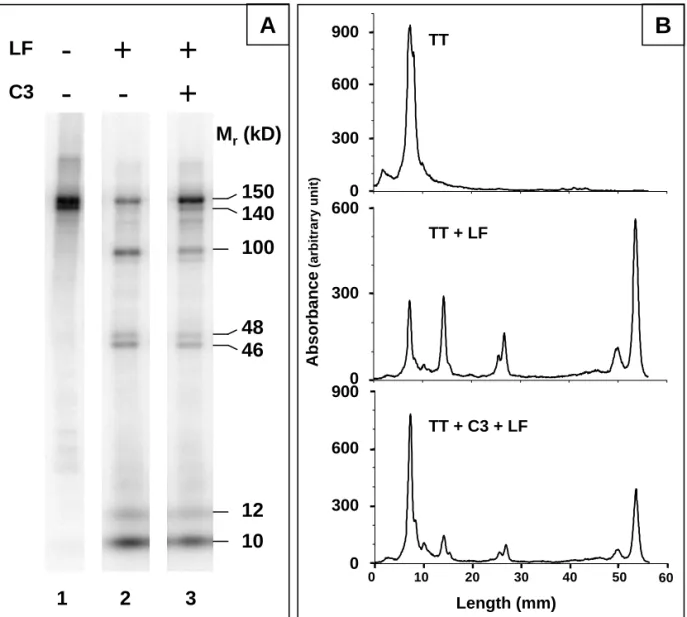
In order to assess whether C3 could affect some proteolytic processes, 125I-TT proteolysis by the LF of U937 cells was analyzed in the presence or absence of C3, as described in the Materials and Methods section. As shown in Fig.1, TT is present as two close bands (Mr = 150,000 and 140,000) on
SDS-PAGE under non-reducing conditions (lane 1) and is partially proteolyzed by LF (lane 2), as indicated by the appearance of three new major bands (Mr = 100,000, 48,000 and 46,000) together
with small fragments (Mr < 15,000) migrating with the dye front, and the parallel decrease in band
intensity corresponding to intact TT. An additional, fainter, band featuring an apparent Mr = 120,000
can also be detected. When C3 is added to the mixture prior to incubation of TT with LF, a similar electrophoretical pattern is observed, although the amount of non-proteolyzed TT remains higher than in the absence of C3 (lane 3): C3 does therefore not modify the proteolytic fragments generated by LF, but rather at least partially inhibits its proteolytic activity.
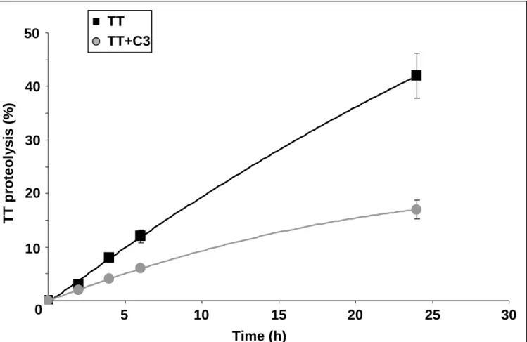
The inhibitory effect of C3 on TT proteolysis by LF was confirmed by a kinetic analysis: As shown in Fig.2, in these conditions, only 15% of input TT is proteolyzed after a 24 h incubation in the presence of C3 against 45% in its absence.
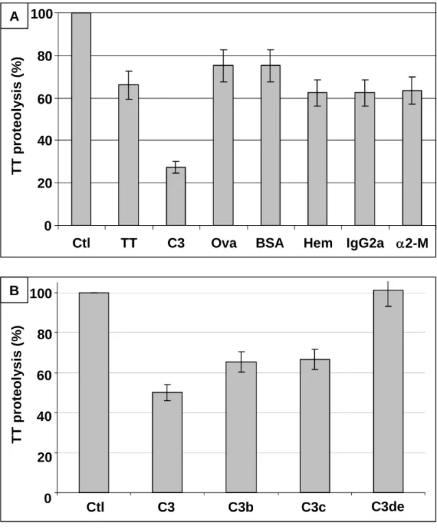
3.2. Specificity of the C3 inhibitory effect
To assess the specificity of the observed C3 inhibitory effect, 125I-TT was incubated with LF
in the presence of various unlabelled proteins (TT=150kDa, C3=185kDa, ovalbumine (Ova)=43kDa, BSA=67kDa, hemoglobine (Hem)=68kDa, IgG2a=150kDa, α2-M=4x180kDa) and its proteolysis was quantified after SDS-PAGE. An assay including only 125I-TT and LF was used as reference (100%
proteolysis). As shown in Fig.3A, proteolysis is substantially inhibited only in the presence of C3; the other proteins tested, by contrast, only slightly decrease TT proteolysis by LF. Surprisingly, this is also the case of α2-M, which belongs to the same family as C3 (thioester-containing proteins). The observed inhibitory effect is therefore specific to C3. The mild proteolysis inhibitory effect featured by all the tested proteins with the exception of C3 is probably due to the fact that these molecules are all potential substrates for proteases, and therefore potential 125I-TT competitors for LF proteolysis.
Therefore, in the following experiments, the reference assays will always include unlabelled TT (5µg) in order to eliminate the slight inhibition due to this phenomenon and to take in account only the effect due to C3.
3.3. Identification of the C3 fragment involved in proteolysis inhibition
Under physiological conditions, C3 undergoes successive proteolytic cleavages leading to various fragments, namely C3b, which is subsequently cleaved into iC3b (Mr = 175,000 and 173 000
respectively), then into C3c (Mr = 135,000) and C3de (Mr = 40,000) (Villiers and Villiers, 2003). In
order to identify the C3 fragment involved in proteolysis inhibition, 125I-TT was incubated with LF in
the presence of each of the different C3 fragments and analyzed using SDS-PAGE. As shown in Fig.3B, TT degradation by lysosomal proteases is impaired in the presence of C3, C3b and C3c fragments (40-50% inhibition), but this effect is less significant in the presence of C3de (80%). Therefore, the observed proteolysis inhibitory effect of C3 is related to the C3c fragment. As evidenced using SDS-PAGE (Fig.4A), the C3c fragment (apparent Mr = 135,000 in non reducing
conditions) is made of three disulfide-linked polypeptides: the C3 β chain (Mr = 75,000) and the two
α chain-derived polypeptides (α1, Mr = 40,000 and α2, Mr = 27,000). We wondered whether the C3c
inhibitory effect required the integrity of the molecule. To that aim, C3c was further incubated with trypsin (trypsin/C3c = 1/100, w/w, 2h at 37°C): further cleavage of β, α1 and α2 was obtained, leading
to three other polypeptides (β*, Mr = 58,000, α1*, Mr = 34,000 and α2*, Mr = 23,000). 125I-TT
proteolysis by LF in the presence or absence of trypsin-treated C3c was no longer inhibited by C3c after trypsin digestion (Fig.4B), testifying to the need for the integrity of this fragment. As i) proteolytic activities are present in LF and ii) proteolyzed C3c does not retain any inhibitory effect, we investigated whether the C3 fragment is degraded upon incubation in the presence of LF. C3c is fairly resistant to proteolysis by lysosomal proteases (12.5% degradation upon a 18h incubation, data not shown), which is consistent with its inhibiting activity upon TT degradation by LF.
3.4. Identification of the C3c domains involved in proteolysis inhibition
To localize the proteolysis inhibitory activity within C3c, the protein was reduced and we purified its three polypeptide constituents (β, α1 and α2) using HPLC (Fig.5A). Each polypeptide was
then tested individually for its capacity to inhibit the proteolytic activity of LF. As shown in Fig.5B, whereas reduction of C3c by itself does not abolish its inhibitory effect, neither fragment is alone sufficient to impair TT proteolysis; in other words, the inhibitory effect of C3c, although independent of disulfide bridge integrity, cannot be attributable to only one of its three constitutive peptides.
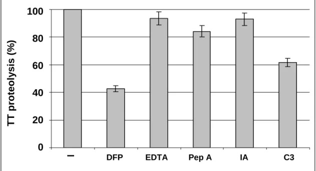
3.5. Characterisation of the protease family inhibited by C3
The protease(s) involved in TT proteolysis by LF was (were) characterized by incubating TT and LF in the presence of different inhibitors: DFP, EDTA, pepstatin A, iodoacetic acid . As illustrated in Fig.6, proteolysis of TT is mainly due to DFP-sensitive protease(s) rather than metallo-, acid- or thiol-proteases, as it is affected only slightly by EDTA, pepstatin A or iodoacetic acid (7-15% inhibition), whereas DFP strongly decreases TT proteolysis (58%), to an extent similar to C3 (40%). Moreover, the TT fragments which accumulate upon TT + LF incubation in the presence of DFP are similar to those observed in the presence of C3 whereas the presence of the other inhibitors leads to a different accumulation of fragments (data not shown). These results suggest that the target-protease(s) for C3 belong(s) to the serine-protease family.
3.6. Colocalisation of TT and C3 upon cellular internalisation
To determine if our observations made in a cell free system are physiologically relevant, we have first analysed the localisation of TT and C3b after their uptake by living cells (C3b was used instead of C3 in order to allow C3b internalisation via complement receptors). To do so, U937 were allowed to internalise Cy2- TT and Cy3-C3b at 37°C before being examined by confocal microscopy.
As it can be seen in Fig.7, some intracellular compartments contain only C3b. On the contrary and although TT is less efficiently internalised, it always colocalise with C3b as there is always an overlapping of green with red signals (yellow in merged images). One observes this phenomenon whatever the duration of the incubation, from 20min to 2h.
3.7. Inhibitory effect of C3 upon TT proteolysis in U937
As we demonstrated the inhibitory activity of C3 upon TT proteolysis by LF and the colocalisation of these two proteins after U937 internalisation, we wondered whether C3 could also influence TT proteolysis in living cells. After allowing U937 to internalise 125I-TT alone or with C3b
for different times at 37°C, we analysed TT proteolysis on SDS-PAGE. The quantification of the peak corresponding to intact TT indicates that C3b interferes with intracellular proteolytic activities in living cells as the amount of unproteolysed TT is doubled in presence of C3b, at least during the first 40min after internalisation (Fig. 8).
4. Discussion
The influence of complement component C3 on Ag presentation has been highlighted since several years. Indeed, it has been demonstrated that C3b modulates Ag processing and presentation by different APC upon covalent binding to Ag (Jacquier-Sarlin et al., 1995; Rey-Millet et al., 1994). Using macrophages from C3-deficient mice, Zhou et al. (Zhou et al., 2006) have presented evidence that C3 also modulates the potency of APC to stimulate the T-cell response, suggesting a potential effect of free C3 on immune reponse. As peptide generation through Ag degradation is a key event for both Ag presentation and T cell stimulation, we wondered whether free C3 could modulate Ag proteolysis in APC. To be able to correlate our present work with previous ones, we used the same Ag model (TT).
Our results demonstrate that proteolysis of TT by the LF from a human monocytic cell line (U937) was impaired by C3. This effect was quite specific of C3, as α2-M, another protease inhibitor belonging to the same intramolecular thioester bond-containing family (Armstrong and Quigley, 1999), did not affect TT proteolysis. The specificity of C3 inhibitory effect is strengthened by the fact that all other proteins tested were without effect although they are added at same or greater number of molecules compared to C3, depending on their MW. We further identified C3c as being responsible for inhibiting TT proteolysis by LF. It is noteworthy that C3c retained its proteolysis inhibitory activity even after disulfide disruption, although neither of its three constitutive peptides could individually be accountable for it. This observation could be related to the intrinsic structure of C3c: C3c consists of several domains, one of which is formed by parts of both the β- and α-chains (Janssen et al., 2005). This β/α intertwined domain, which is not stabilized by a disulfide bridge and is thereby preserved even after reduction of the molecule, which is confirmed by the impossibility of separating the three polypeptides by gel filtration at neutral pH (data not shown). Thus, it is likely that this β/α intertwined domain could be involved in the observed inhibitory effect of C3c.
Previous studies have illustrated the role of C3 in Ag presentation (Jacquier-Sarlin et al., 1995; Rey-Millet et al., 1994; Santoro et al., 1994), and particularly in Ag degradation: indeed, Jacquier-Sarlin et al. reported limited TT proteolysis by B cells in the presence of C3b (Jacquier-Jacquier-Sarlin et al., 1995). Furthermore, antigenic peptides production and loading on MHC II molecules are modified upon covalent binding of C3b to the Ag (Cretin et al., 2007; Serra et al., 1997). It has been shown that the TT-C3b interaction is fairly stable in endosomes, whereas it is gradually disrupted in lysosomes (Rey-Millet et al., 1994). Moreover, LF contains proteolytic activities leading to the generation of C3c-like fragments (Rey-Millet et al., 1993). These results, together with our present work, enable us to suggest a mechanism by which C3b binding to Ag may modulate Ag presentation: after their uptake by APC, Ag-C3b complexes are delivered to lysosomes, where C3c fragments are generated upon lysosomal proteolytic activities. These fragments then interfere with the lysosomal proteases and at least partially inhibit their activity.
This hypothesis is strengthened by our experiments using living cells. First, analysis by confocal microscopy of U937 having internalised TT and C3 showed that C3 is always found in subcellular
compartments containing TT, which makes possible their interaction. The great efficiency of C3 uptake compared to TT is probably due to the presence of C3 receptors at the surface of U937 (CD11b, CD11c, (Lopez-Rodriguez and Corbi, 1997)). Thus, whereas TT is internalised only via pinocytosis and phagocytosis, C3 could enter the cells using both receptor –independent (colocalisation with TT) and receptor -dependent pathway which may lead in this case to a different subcellular localisation (C3 alone) as shown previously on B cells (Perrin-Cocon et al., 2004). Second, the analysis of the intracellular proteolysis of TT by living APC such as U937 demonstrates that C3 is able to exert its inhibitory effect in physiological conditions. This effect decreases with time, but persists at least during the first hour after internalisation. It has been demonstrated that modulation of Ag proteolysis may interferes with Ag presentation, either by increasing or by decreasing production of antigenic peptides suitable for MHC binding (Delamarre et al., 2006; Polla et al., 2007).
The proteases involved in Ag degradation and the mechanisms that control their activities are not yet fully understood and differ according to the Ag and the cells involved (Burster et al., 2005). Cathepsins are often associated with Ag proteolysis (Hewitt et al., 1997; Hsieh et al., 2002; Ruckrich et al., 2006; Shi et al., 1999), but other proteases such as asparagine endopeptidase (AEP) also play an important role (Manoury et al., 1998). In our hands, C3c inhibited TT proteolysis in a way similar to DFP, indicating that the target protease(s) belongs to the serine-protease family; this conclusion is strengthened by the fact that all other protease inhibitors tested had less effect on TT proteolysis. A previous study by Rey-Millet et al. (Rey-Millet et al., 1993) pointed out the presence of a serine-protease activity within LF, but its involvement in Ag processing had not been established. Most attempts to identify the proteases accounting for intracellular Ag proteolysis are usually indirect, consisting in the analysis of Ag presentation to T cells in the presence of different inhibitors. Ag degradation has either a creative or a destructive effect, mainly depending on MHC restriction (Puri and Factorovich, 1988; Takahashi et al., 1989). Nevertheless, inhibition of Ag degradation is often related to increased Ag presentation, as demonstrated using protease inhibitors (Vidard et al., 1991), antigen resistant to degradation (Delamarre et al., 2006) or modulation of intravacuolar pH (Savina et al., 2006). Indeed, reduced intracellular proteolysis avoids the destruction of T cell epitopes, favoring the production of peptide-MHC classs II complexes. This is particularly important in cells such as macrophages which possess elevated degradation capacity due to their high protease content. Here, we provide direct evidence for the control of Ag proteolysis by C3c, which could be related to the previously described C3-mediated enhancement of Ag presentation and, more generally, of the establishment of a specific immune response. C3 is therefore directly involved in Ag degradation by APC through at least two distinct mechanisms: i) C3b modulates Ag trafficking within the APC (Perrin-Cocon et al., 2004) thereby potentially affecting Ag processing through modified protease activities located within the intracellular compartments, and ii) C3 directly inhibits Ag proteolysis by lysosomal proteases, as illustrated by our results. The C3 present in APC could be of extracellular origin, via C3b binding to exogenous Ag, as well as of intracellular origin, since C3 is synthesized by various APC such as dendritic cells (Peng et al., 2006) or macrophages (Fischer et al., 1998). Whether endogenous C3 could influence Ag proteolysis in the same way as exogenous C3 or not remains to be established. Nevertheless, the relevance of the role of C3 in Ag presentation “in vivo” has been highlighted by Zhou et al.(Zhou et al., 2006), who demonstrated that stimulation of alloreactive T cells is impaired in C3-deficient mice.
The capacity of C3c to control lysosomal degradation could provide an explanation as to why induction of an increased Ab response in mice requires injection of three C3d molecules bound to Ag (Dempsey et al., 1996) versus a single molecule of C3b (Villiers et al., 1999). Indeed, only C3b allows local production of C3c which could control Ag proteolysis favouring Ag presentation, while Ag uptake can be ensured by both C3b and C3d.
In conclusion, our results, by revealing a novel role for C3, supplement the integrative view on the modulatory role of C3 in acquired immune response, and especially on Ag processing: C3 acts at different levels such as Ag uptake (Villiers et al., 1996), intracellular Ag trafficking and targeting (Perrin-Cocon et al., 2004), proteolytic machinery of Ag processing (these results), Ag peptide editing (Cretin et al., 2007) and loading on MHC II molecules (Serra et al., 1997), all of these increasing Ag presentation efficiency (Jacquier-Sarlin et al., 1995) and thus the quality of the acquired immune response (Dempsey et al., 1996; Villiers et al., 1999).
References
Al Salihi, A., Ripoche, J., Pruvost, L., Fontaine, M., 1982. Purification of complement components by hydrophobic affinity chromatography on phenyl-sepharose: purification of human C5. FEBS Lett 150, 238-242.
Armstrong, P. B., Quigley, J. P., 1999. Alpha2-macroglobulin: an evolutionarily conserved arm of the innate immune system. Dev Comp Immunol 23, 375-390.
Bottger, E. C., Metzger, S., Bitter-Suermann, D., Stevenson, G., Kleindienst, S., Burger, R., 1986. Impaired humoral immune response in complement C3-deficient guinea pigs: absence of secondary antibody response. Eur J Immunol 16, 1231-1235.
Burster, T., Beck, A., Tolosa, E., Schnorrer, P., Weissert, R., Reich, M., Kraus, M., Kalbacher, H., Haring, H. U., Weber, E., Overkleeft, H., Driessen, C., 2005. Differential processing of autoantigens in lysosomes from human monocyte-derived and peripheral blood dendritic cells. J Immunol 175, 5940-5949.
Carroll, M. C., 2004. The complement system in regulation of adaptive immunity. Nat Immunol 5, 981-986.
Cherukuri, A., Cheng, P. C., Sohn, H. W., Pierce, S. K., 2001. The CD19/CD21 complex functions to prolong B cell antigen receptor signaling from lipid rafts. Immunity 14, 169-179.
Cretin, F. C., Serra, V. A., Villiers, M. B., Laharie, A. M., Marche, P. N., Gabert, F. M., 2007. C3b complexation diversifies naturally processed T cell epitopes. Mol Immunol 44, 2893-2899. Delamarre, L., Couture, R., Mellman, I., Trombetta, E. S., 2006. Enhancing immunogenicity by
limiting susceptibility to lysosomal proteolysis. J Exp Med 203, 2049-2055.
Delamarre, L., Pack, M., Chang, H., Mellman, I., Trombetta, E. S., 2005. Differential lysosomal proteolysis in antigen-presenting cells determines antigen fate. Science 307, 1630-1634. Dempsey, P. W., Allison, M. E., Akkaraju, S., Goodnow, C. C., Fearon, D. T., 1996. C3d of
complement as a molecular adjuvant: bridging innate and acquired immunity. Science 271, 348-350.
Dodds, A. W., Law, S. K., 1988. Structural basis of the binding specificity of the thioester-containing proteins, C4, C3 and alpha-2-macroglobulin. Complement 5, 89-97.
Einstein, L. P., Hansen, P. J., Ballow, M., Davis, A. E., 3rd, Davis, J. S. t., Alper, C. A., Rosen, F. S., Colten, H. R., 1977. Biosynthesis of the third component of complement (C3) in vitro by monocytes from both normal and homozygous C3-deficient humans. J Clin Invest 60, 963-969.
Fearon, D. T., Carter, R. H., 1995. The CD19/CR2/TAPA-1 complex of B lymphocytes: linking natural to acquired immunity. Annu Rev Immunol 13, 127-149.
Fischer, M. B., Ma, M., Hsu, N. C., Carroll, M. C., 1998. Local synthesis of C3 within the splenic lymphoid compartment can reconstitute the impaired immune response in C3-deficient mice. J Immunol 160, 2619-2625.
Hawlisch, H., Wills-Karp, M., Karp, C. L., Kohl, J., 2004. The anaphylatoxins bridge innate and adaptive immune responses in allergic asthma. Mol Immunol 41, 123-131.
Hewitt, E. W., Treumann, A., Morrice, N., Tatnell, P. J., Kay, J., Watts, C., 1997. Natural processing sites for human cathepsin E and cathepsin D in tetanus toxin: implications for T cell epitope generation. J Immunol 159, 4693-4699.
Hsieh, C. S., deRoos, P., Honey, K., Beers, C., Rudensky, A. Y., 2002. A role for cathepsin L and cathepsin S in peptide generation for MHC class II presentation. J Immunol 168, 2618-2625. Jacquier-Sarlin, M. R., Gabert, F. M., Villiers, M. B., Colomb, M. G., 1995. Modulation of antigen
processing and presentation by covalently linked complement C3b fragment. Immunology 84, 164-170.
Janssen, B. J., Huizinga, E. G., Raaijmakers, H. C., Roos, A., Daha, M. R., Nilsson-Ekdahl, K., Nilsson, B., Gros, P., 2005. Structures of complement component C3 provide insights into the function and evolution of immunity. Nature 437, 505-511.
Law, S. K., Lichtenberg, N. A., Levine, R. P., 1979. Evidence for an ester linkage between the labile binding site of C3b and receptive surfaces. J Immunol 123, 1388-1394.
Lopez-Rodriguez, C., Corbi, A. L., 1997. PU.1 negatively regulates the CD11c integrin gene promoter through recognition of the major transcriptional start site. Eur J Immunol 27, 1843-1847. Manoury, B., Hewitt, E. W., Morrice, N., Dando, P. M., Barrett, A. J., Watts, C., 1998. An asparaginyl
endopeptidase processes a microbial antigen for class II MHC presentation. Nature 396, 695-699.
Morgan, B. P., Marchbank, K. J., Longhi, M. P., Harris, C. L., Gallimore, A. M., 2005. Complement: central to innate immunity and bridging to adaptive responses. Immunol Lett 97, 171-179. Ochs, H. D., Wedgwood, R. J., Frank, M. M., Heller, S. R., Hosea, S. W., 1983. The role of
complement in the induction of antibody responses. Clin Exp Immunol 53, 208-216.
Peng, Q., Li, K., Patel, H., Sacks, S. H., Zhou, W., 2006. Dendritic cell synthesis of C3 is required for full T cell activation and development of a Th1 phenotype. J Immunol 176, 3330-3341. Pepys, M. B., 1974. Role of complement in induction of antibody production in vivo. Effect of cobra
factor and other C3-reactive agents on thymus-dependent and thymus-independent antibody responses. J Exp Med 140, 126-145.
Perrin-Cocon, L. A., Villiers, C. L., Salamero, J., Gabert, F., Marche, P. N., 2004. B cell receptors and complement receptors target the antigen to distinct intracellular compartments. J Immunol 172, 3564-3572.
Polla, B. S., Gabert, F., Peyrusse, B. M., Jacquier-Sarlin, M. R., 2007. Increased proteolysis of diphtheria toxin by human monocytes after heat shock: a subsidiary role for heat-shock protein 70 in antigen processing. Immunology 120, 230-241.
Puri, J., Factorovich, Y., 1988. Selective inhibition of antigen presentation to cloned T cells by protease inhibitors. J Immunol 141, 3313-3317.
Rey-Millet, C. A., Chesne, S., Colomb, M. G., 1993. Associated complement C3b. Towards an understanding of its intracellular modifications. Mol Immunol 30, 855-864.
Rey-Millet, C. A., Villiers, C. L., Gabert, F. M., Chesne, S., Colomb, M. G., 1994. C3b covalently associated to tetanus toxin modulates TT processing and presentation by U937 cells. Mol Immunol 31, 1321-1327.
Ruckrich, T., Brandenburg, J., Cansier, A., Muller, M., Stevanovic, S., Schilling, K., Wiederanders, B., Beck, A., Melms, A., Reich, M., Driessen, C., Kalbacher, H., 2006. Specificity of human cathepsin S determined by processing of peptide substrates and MHC class II-associated invariant chain. Biol Chem 387, 1503-1511.
Salacinski, P. R. P., McLean, C., Sykes, J. E. C., Clement-Jones, V. V., Lowry, P. J., 1981. Iodination of proteins, glycoproteins and peptides using a solid phase oxidising agent,
1,3,4,6-tetrachloro-3,6 diphenyl glycoluril (Iodogen). Anal Biochem 117, 136-146.
Santoro, L., Drouet, C., Reboul, A., Mach, J. P., Colomb, M. G., 1994. Covalent binding of C3b to monoclonal antibodies selectively up-regulates heavy chain epitope recognition by T cells. Eur J Immunol 24, 1620-1626.
Savina, A., Jancic, C., Hugues, S., Guermonprez, P., Vargas, P., Moura, I. C., Lennon-Dumenil, A. M., Seabra, M. C., Raposo, G., Amigorena, S., 2006. NOX2 controls phagosomal pH to regulate antigen processing during crosspresentation by dendritic cells. Cell 126, 205-218. Schulman, E. S., Post, T. J., Henson, P. M., Giclas, P. C., 1988. Differential effects of the complement
peptides, C5a and C5a des Arg on human basophil and lung mast cell histamine release. J Clin Invest 81, 918-923.
Serra, V. A., Cretin, F., Pepin, E., Gabert, F. M., Marche, P. N., 1997. Complement C3b fragment covalently linked to tetanus toxin increases lysosomal sodium dodecyl sulfate-stable HLA-DR dimer production. Eur J Immunol 27, 2673-2679.
Shi, G. P., Villadangos, J. A., Dranoff, G., Small, C., Gu, L., Haley, K. J., Riese, R., Ploegh, H. L., Chapman, H. A., 1999. Cathepsin S required for normal MHC class II peptide loading and germinal center development. Immunity 10, 197-206.
Sim, R. B., Twose, T. M., Paterson, D. S., Sim, E., 1981. The covalent-binding reaction of complement component C3. Biochem J 193, 115-127.
Sundström, G., Nilsson, K., 1976. Establishment and characterization of a human histiocytic cell-line (U937). Int J Cancer 17, 565-577.
Tack, B. D., Prahl, J. W., 1976. Third component of human complement: purification from plasma and physicochemical characterization. Biochemistry 15, 4513-4521.
Takafuji, S., Tadokoro, K., Ito, K., Dahinden, C. A., 1994. Degranulation from human eosinophils stimulated with C3a and C5a. Int Arch Allergy Immunol 104 Suppl 1, 27-29.
Takahashi, H., Cease, K. B., Berzofsky, J. A., 1989. Identification of proteases that process distinct epitopes on the same protein. J Immunol 142, 2221-2229.
Vidard, L., Rock, K. L., Benacerraf, B., 1991. The generation of immunogenic peptides can be selectively increased or decreased by proteolytic enzyme inhibitors. J Immunol 147, 1786-1791.
Villiers, C. L., Villiers, M. B., 2003. Modifications of the properties of C3 complement protein after proteolytic cleavage. Recent Res. Devel. Biochem. 4, 685-696.
Villiers, M. B., Gabert, F. M., Jacquier, M. R., Villiers, C. L., Colomb, M. G., 1993. Involvement of the Zn-binding region of tetanus toxin in B and T recognition. Influence of Zn fixation. Mol Immunol 30, 129-136.
Villiers, M. B., Marche, P. N., Villiers, C. L., 2003. Improvement of long-lasting response and antibody affinity by the complexation of antigen with complement C3b. Int Immunol 15, 91-95.
Villiers, M. B., Perrin-Cocon, L., Marche, P. N., Villiers, C. L., 2004. Complement receptors and B lymphocytes. Crit Rev Immunol 24, 465-478.
Villiers, M. B., Villiers, C. L., Jacquier-Sarlin, M. R., Gabert, F. M., Journet, A. M., Colomb, M. G., 1996. Covalent binding of C3b to tetanus toxin: influence on uptake/internalization of antigen by antigen-specific and non-specific B cells. Immunology 89, 348-355.
Villiers, M. B., Villiers, C. L., Laharie, A. M., Marche, P. N., 1999. Amplification of the antibody response by C3b complexed to antigen through an ester link. J Immunol 162, 3647-3652. Villiers, M. B., Villiers, C. L., Wright, J. F., Maison, C. M., Colomb, M. G., 1991. Formation of
covalent C3b-tetanus toxin complexes: a tool for the in vitro study of antigen presentation. Scand J Immunol 34, 585-595.
Wagner, C., Hansch, G. M., 2006. Receptors for complement C3 on T-lymphocytes: relics of evolution or functional molecules? Mol Immunol 43, 22-30.
Wagner, C., Ochmann, C., Schoels, M., Giese, T., Stegmaier, S., Richter, R., Hug, F., Hansch, G. M., 2006. The complement receptor 1, CR1 (CD35), mediates inhibitory signals in human T-lymphocytes. Mol Immunol 43, 643-651.
Walport, M. J., 2001. Complement. Second of two parts. N Engl J Med 344, 1140-1144.
Weiss, L., Fischer, E., Haeffner-Cavaillon, N., Jouvin, M. H., Appay, M. D., Bariety, J., Kazatchkine, M., 1989. The human C3b receptor (CR1). Adv Nephrol Necker Hosp 18, 249-269.
Zhou, W., Patel, H., Li, K., Peng, Q., Villiers, M. B., Sacks, S. H., 2006. Macrophages from C3-deficient mice have impaired potency to stimulate alloreactive T cells. Blood 107, 2461-2469.
Figure Captions
Fig. 1. TT proteolysis by LF. 125I-TT (5 µg) was incubated for 18 h at 37°C in the presence or absence
of LF from U937 cells (2.5 x 10⁶ cells) and of C3 (5 µg). The mixture was analyzed by SDS-PAGE
under non reducing conditions (A). The radioactivity associated with the protein bands was quantified using a phosphorimager (B).
Fig. 2. Kinetic analysis of TT proteolysis by LF in the presence or absence of C3. 125I-TT (5 µg) was
incubated at 37°C for different times in the presence of LF from U937 cells (2.5 x 10⁶ cells) and of C3
(5 µg). TT proteolysis was analyzed by SDS-PAGE and quantified as described in Materials and Methods. Results are given as means ±SD from at least three separate experiments.
Fig. 3. Specificity of the C3 inhibitory effect and identification of the C3 fragments involved. 125I-TT (5 µg) was incubated for 18 h at 37°C in the presence of LF from U937 cells (2.5 x 10⁶ cells) and of 5 µg of various proteins: A) TT, C3, ovalbumine (Ova), BSA, hemoglobine (Hem), IgG2a, α2-M and B) C3 fragments. 125I-TT proteolysis was analyzed by SDS-PAGE and quantified as described in
Materials and Methods. (A) 100% proteolysis was determined using an assay including only 125I-TT +
LF (Ctl). (B) 100% proteolysis was determined using an assay including 125I-TT + LF and unlabelled
TT (Ctl). Results are given as means ±SD from at least three separate experiments.
Fig. 4. Loss of the C3 inhibitory effect after C3c cleavage. (A) SDS-PAGE analysis of C3c (5 µg) in non reducing (NR) and reducing (R) conditions before and after cleavage by trypsin (see Materials and Methods). (B) TT proteolysis by LF in the presence of intact or trypsin-proteolyzed C3c. 125I-TT (5 µg) was incubated for 18 h at 37°C in the presence of LF from U937 cells (2.5 x 10⁶ cells) and C3c (5 µg) treated or not by trypsin. TT proteolysis was analyzed by SDS-PAGE and quantified as described in Materials and Methods. 100% proteolysis was determined using an assay including 125I-TT + LF
and unlabelled TT. Results are given as means ±SD from at least three separate experiments.
Fig. 5. Identification of C3c domains involved in proteolysis inhibition. After reduction and alkylation of C3c, the different polypeptides were purified by reverse phase-HPLC on a Vydac C4 column (see Materials and Methods). (A) Elution profile. (B) TT proteolysis by LF in the presence of reduced or non reduced C3c or each of its constitutive peptides. 125I-TT (5 µg) was incubated for 18 h at 37°C in
the presence of LF from U937 cells (2.5 x 10⁶ cells) and C3c or its peptides (5 µg). TT proteolysis was
analyzed by SDS-PAGE and quantified as described in Materials and Methods. 100% proteolysis was determined using an assay including 125I-TT + LF and unlabelled TT (Ctl). Results are given as means ±SD from at least three separate experiments.
Fig. 6. Characterization of the lysosomal protease family involved in TT proteolysis. 125I-TT (5 µg)
was incubated for 18 h at 37°C in the presence of LF from U937 cells (2.5 x 10⁶ cells) and C3 (5 µg)
or different protease inhibitors (DFP, EDTA, iodoacetic acid (5mM) or pepstatin A (1.5µM)). TT proteolysis was analyzed by SDS-PAGE and quantified as described in Materials and Methods. 100% proteolysis was determined using an assay including 125I-TT + LF and unlabelled TT (Ctl). Results are
given as means ±SD from at least three separate experiments.
Fig. 7. Localisation of internalised TT and C3b by confocal microscopy. U937 were incubated at 37°C for 2h in the presence of Cy3-C3b and Cy2-TT. Merged images of red (C3b) and green (TT)
fluorescence are shown.
Fig. 8. TT proteolysis by U937 in the presence or absence of C3b. 125I-TT (100µg) was internalised by U937 (10 x 106 cells) at 37°C for the indicated times in the presence or not of C3b (100µg). After cell
disruption, samples were treated as described in Materials and Methods and analysed by SDS-PAGE. Residual intact TT was quantified after 20, 40 and 60min incubation as described in Materials and Methods. Results are given as means ±SD from at least three separate experiments.