HAL Id: hal-02321357
https://hal.sorbonne-universite.fr/hal-02321357
Submitted on 21 Oct 2019
HAL is a multi-disciplinary open access
archive for the deposit and dissemination of
sci-entific research documents, whether they are
pub-lished or not. The documents may come from
teaching and research institutions in France or
abroad, or from public or private research centers.
L’archive ouverte pluridisciplinaire HAL, est
destinée au dépôt et à la diffusion de documents
scientifiques de niveau recherche, publiés ou non,
émanant des établissements d’enseignement et de
recherche français ou étrangers, des laboratoires
publics ou privés.
optogenetically engineered photoreceptors
Marcela Garita-Hernandez, Maruša Lampič, Antoine Chaffiol, Laure Guibbal,
Fiona Routet, Tiago Santos-Ferreira, Sylvia Gasparini, Oliver Borsch,
Giuliana Gagliardi, Sacha Reichman, et al.
To cite this version:
Marcela Garita-Hernandez, Maruša Lampič, Antoine Chaffiol, Laure Guibbal, Fiona Routet, et al..
Restoration of visual function by transplantation of optogenetically engineered photoreceptors. Nature
Communications, Nature Publishing Group, 2019, 10 (1), pp.4524. �10.1038/s41467-019-12330-2�.
�hal-02321357�
Restoration of visual function by transplantation of
optogenetically engineered photoreceptors
Marcela Garita-Hernandez
1,6
, Maru
ša Lampič
1,6
, Antoine Chaf
fiol
1
, Laure Guibbal
1
, Fiona Routet
1
,
Tiago Santos-Ferreira
2
, Sylvia Gasparini
2
, Oliver Borsch
2
, Giuliana Gagliardi
1
, Sacha Reichman
1
,
Serge Picaud
1
, José-Alain Sahel
1,3,4
, Olivier Goureau
1
, Marius Ader
2
, Deniz Dalkara
1,7
& Jens Duebel
1,5,7
A major challenge in the treatment of retinal degenerative diseases, with the transplantation
of replacement photoreceptors, is the difficulty in inducing the grafted cells to grow and
maintain light sensitive outer segments in the host retina, which depends on proper
inter-action with the underlying retinal pigment epithelium (RPE). Here, for an RPE-independent
treatment approach, we introduce a hyperpolarizing microbial opsin into photoreceptor
precursors from newborn mice, and transplant them into blind mice lacking the
photo-receptor layer. These optogenetically-transformed photophoto-receptors are light responsive and
their transplantation leads to the recovery of visual function, as shown by ganglion cell
recordings and behavioral tests. Subsequently, we generate cone photoreceptors from human
induced pluripotent stem cells, expressing the chloride pump Jaws. After transplantation into
blind mice, we observe light-driven responses at the photoreceptor and ganglion cell levels.
These results demonstrate that structural and functional retinal repair is possible by
com-bining stem cell therapy and optogenetics.
https://doi.org/10.1038/s41467-019-12330-2
OPEN
1Sorbonne Université, Institut de la Vision, INSERM, CNRS, 75012 Paris, France.2CRTD/Center for Regenerative Therapies Dresden, CMCB, TU Dresden,
Dresden, Germany.3CHNO des Quinze−Vingts, DHU Sight Restore, INSERM-DGOS CIC 1423, Paris, France.4Department of Ophthalmology, The
University of Pittsburgh School of Medicine, Pittsburgh, USA.5Department of Ophthalmology, University Medical Center Göttingen, Göttingen, Germany.
6These authors contributed equally: Marcela Garita-Hernandez, Maru
ša Lampič.7These authors jointly supervised this work: Deniz Dalkara, Jens Duebel.
Correspondence and requests for materials should be addressed to D.D. (email:deniz.dalkara@gmail.com) or to J.D. (email:jens.duebel@gmail.com)
123456789
C
ell replacement therapy offers hope for the treatment of
late-stage retinal degeneration, when the outer retinal
photoreceptor layer is lost
1–3. However, a remaining
obstacle of photoreceptor replacement is that transplanted cells
have to develop into functional photoreceptors with light
sensi-tive outer segments (OS). Indeed, in mouse models of severe
degeneration, the formation of light-sensitive OS by transplanted
photoreceptors has been difficult to achieve
4–6. Recent studies,
using retinal sheet transplantation lead to major improvements in
terms of OS formation and light sensitivity
7,8. Despite these
promising results, a major problem has not yet been solved:
photoreceptors need tight interaction with the retinal pigment
epithelium (RPE) in order to maintain their structure and
func-tion via continuous disc shedding and renewal
9. Since in retinal
degenerative diseases the RPE is often also compromised
9,10, the
probability that transplanted photoreceptors stay sensitive to light
is very low
11,12. To tackle this problem, we introduce optogenetic
light sensors into photoreceptors, derived from the developing
mouse retina as well as from human induced pluripotent stem
cells (hiPSCs), and transplant them into mouse models of severe
retinal degeneration. The key point of our approach is that these
optogenetically-transformed photoreceptors stay functional based
on the activity of the microbial opsin, even in the absence of
properly formed OS and without the support from the RPE.
Results
Neonatal mouse-derived NpHR photoreceptor precursors. For
optogenetic transformation of mouse photoreceptors, eyes of
newborn wild-type mice at post-natal day (P) 2 were injected with
an adeno-associated viral (AAV) vector encoding enhanced
Natronomonas pharaonis halorhodopsin eNpHR2.0 (NpHR)
13under the control of the rhodopsin promoter
(AAV-Rho-NpHR-YFP) (Fig.
1
a and Supplementary Fig. 1). At P4, photoreceptor
precursors were sorted by magnetic activated cell sorting (MACS)
using the photoreceptor specific cell surface marker CD73
14,15.
The harvested cells were transplanted via sub-retinal injections
into two blind mouse models of late-stage retinal degeneration
(Cpfl1/Rho
−/−mice
16aged 9 to 18 weeks and C3H rd/rd (rd1)
mice
17aged 4 to 11 weeks; see Supplementary Table 1 for a
complete overview of mouse ages). At these ages, the vast
majority of outer nuclear layer (ONL) cells were lost in host mice
(Fig.
1
b, e). Cpfl1/Rho
−/−mice are left with 2–3 rows of
photo-receptors at the age of 9 weeks, and a single row of photophoto-receptors
by 10–12 weeks of age. These mice are born with non-functional
rods and cones
16. Rd1 mice loose photoreceptor OS and only a
single row of cone cell bodies in the ONL remains by 3 weeks
after birth
18. Four weeks after transplantation, we investigated the
morphology of the transplanted donor cells and their ability to
integrate into the host retina. In both mouse models, we found
NpHR-positive donor cells in close contact to cell bodies of rod
bipolar cells, but none of the transplanted cells displayed correctly
formed OS (Fig.
1
c, d, f, g). Transplanted cells expressed the
synaptic marker Synaptophysin (Supplementary Fig. 2)
suggest-ing synapse formation between donor photoreceptors and the
downstream neurons. We quantified the number of YFP
+cells in
the subretinal space transplanted with donor-derived
NpHR-expressing rod precursors and found substantial numbers of cells
to survive at four weeks post transplantation (Supplementary
Fig. 3). Next, we assessed potential material transfer between
transplanted cells and remaining photoreceptors by
fluorescence
in situ hybridization with Y chromosome-specific probe (Y
chromosome FISH). NpHR-expressing rod precursors derived
from male P4 mice were injected into female Cpfl1/Rho
−/−mice
at 9 weeks of age, and imaged after 4 weeks using structured
illumination microscopy (Fig.
1
h). Y chromosome
+/YPF
+cells
(transplanted donor cells) and Y chromosome
−/YFP
+cells
(endogenous photoreceptor that underwent material transfer)
were quantified. 90% of YFP
+cells co-stained with Y
chromo-some probe, leaving only very few cells exclusively YFP
+. This
could either be due to an artifact or very rare events of
cyto-plasmic exchange among donor and host photoreceptors
(Fig.
1
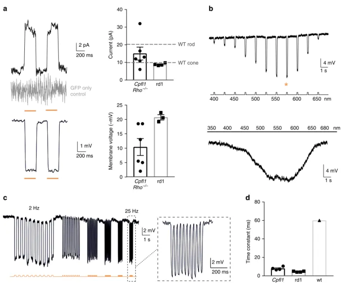
h, i). We then tested if we can elicit light responses from
these NpHR-positive donor cells in the absence of functional OS.
Two-photon targeted patch-clamp recordings revealed robust
responses to orange light pulses (580 nm, 10
16photons cm
−2s
−1)
(Fig.
2
b and Supplementary Fig. 4). There were no measurable
light-evoked currents in transplanted photoreceptors expressing
GFP only, which is consistent with the
finding that the
trans-planted cells lacked their light sensitive OS. Stimulation at
dif-ferent wavelengths showed a spectral sensitivity matching the
action spectrum of NpHR (Fig.
2
b). To measure the temporal
properties of NpHR-positive photoreceptors, we recorded
pho-tocurrents using light pulses at increasing frequencies, and we
observed that they could follow up to 25 Hz (Fig.
2
c and
Sup-plementary Fig. 4). Although, frequencies above 10 Hz are
filtered
out by the bipolar cells, the ability of optogenetically engineered
photoreceptors to respond to light in a faster than natural pace
implies that retinal ganglion cells (RGCs) receiving signal from
these cells should follow high-frequency stimulation in a similar
manner to normal retina
19. The rise constants were significantly
faster compared to photocurrents of wild-type mice (Fig.
2
d).
Both, from the spectral (peak current at 580 nm) and the
tem-poral (Tau
ON< 10 ms) response properties we concluded that the
photocurrents were driven by the introduced NpHR (Fig.
2
a–d
and Supplementary Fig. 4).
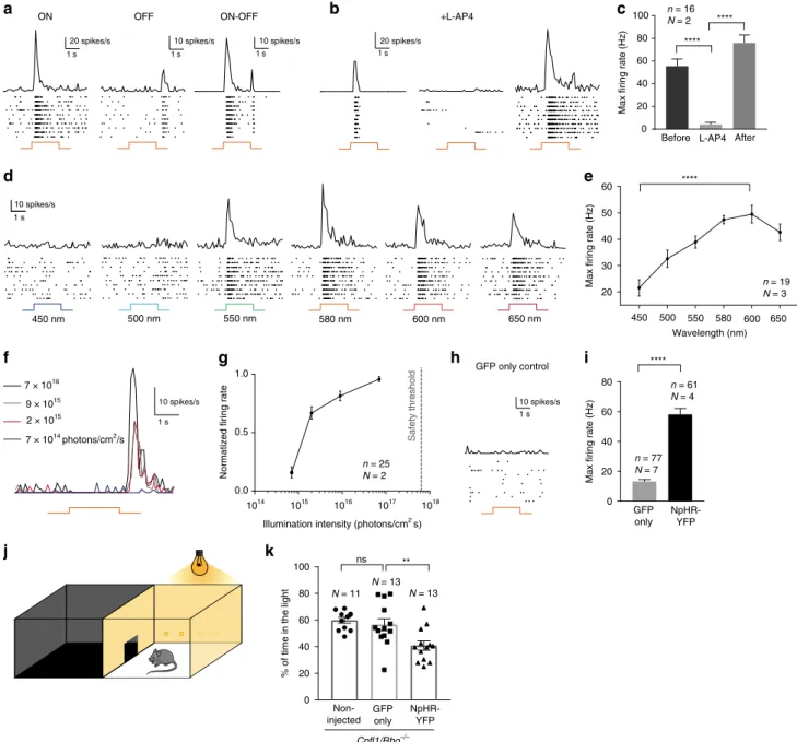
Connectivity and signal transmission to host neurons. Next, we
investigated if the signals from transplanted photoreceptors are
transmitted to RGCs, the output neurons of the retina. By using
extracellular spike recordings, we measured ON- and OFF-light
responses in RGCs. These results demonstrate that
NpHR-induced signals are transmitted to the retinal output neurons via
ON- and OFF-pathways suggesting that the transplanted
photo-receptors can form functional synaptic connections with the inner
retinal neurons (Fig.
3
a and Supplementary Fig. 5), which was
supported by histological analysis (Supplementary Fig. 2).
Recordings performed under pharmacological block of
photo-receptor input to ON-bipolar cells (50 μM L-AP4) showed
complete abolition of ON light responses, which recovered after
20 min of L-AP4-washout. These control experiments confirmed
that
light-induced
signals
were
indeed
transmitted
via
photoreceptor-to-bipolar cell synapses (Fig.
3
b, c). By stimulating
treated retinas at different wavelengths we determined the
spec-tral sensitivity of the light responses, which peaked at
580–600 nm, reflecting the action spectrum of NpHR (Fig.
3
d, e).
To assess the light intensities required to trigger spike responses,
we used light pulses (580 nm) at different intensities. Importantly,
the intensities required to evoke light responses were well below
the safety limit for optical radiation in the human eye
20,21(Fig.
3
f,
g and Supplementary Fig. 5). We did not observe measurable light
responses in retinae from age-matched control-mice, where
photoreceptor precursors expressing only GFP were transplanted
(Fig.
3
h, i and Supplementary Fig. 5). Lastly, to test whether the
behaviour of treated mice could be modulated by light, we used
the light/dark box test
22employing high intensity orange light
(Fig.
3
j). Treated Cpfl1/Rho
−/−mice displayed robust light
avoidance behaviour (40.7 ± 3.5% of time in the illuminated
compartment), compared to non-injected (59.8 ± 2.2%) mice and
mice transplanted with photoreceptor precursors expressing GFP
(56.6 ± 4.5%) (Fig.
3
k).
Generation of hiPSC-derived Jaws-expressing photoreceptors.
To evaluate the translatability of our approach to human subjects,
we asked if it is possible to replace the mouse donor cells with
optogenetically-transformed hiPSCs (Fig.
4
a). To do so, we
first
optimized a previous protocol of differentiation based on the
self-generation of 3D neural-retina-like structures
23. Using this
sys-tem, we generated cone-enriched retinal organoids, expressing the
pan-photoreceptor markers Cone Rod Homeobox (CRX) and
recoverin (RCVRN) alongside the cone-specific marker cone
arrestin (CAR) (Fig.
4
b–f and Supplementary Fig. 6). Contrary to
nocturnal rodents, cone photoreceptors are responsible for high
acuity daylight vision in humans, and are therefore the preferred
choice for transplantation. To render these immature cones light
sensitive, we used the hyperpolarizing chloride pump Jaws, a
red-shifted cruxhalorhodopsin, Jaws, derived from Haloarcula
(Halobacterium) salinarum and engineered to result in red
light–induced photocurrents three times those of earlier
silen-cers
24. Jaws was chosen for iPSC experiments based on its
enhanced expression level and improved membrane trafficking in
human tissue, compared to NpHR
24–26. By using an AAV vector,
Cl–
Optogenetic transformation Transplantation
a
Newborn wt mice
Donor source
Blind mice
Untreated Post transplantation
b
OPL INL GCL GCLc
d
GFP PKCa DAPIf
e
GFP PKCa DAPIg
GFP PKCa DAPIGFP PKCa DAPI
GFP PKCa DAPI P110 GFP PKCa DAPI
SRS OPL INL GCL OPL INL P70 OPL GCL P70 INL OPL INL OPL INL Cpfl1/Rho –/– rd1 SRS SRS SRS SRS SRS P110
h
GFP Y chromosome DAPI i Cpfl1/Rho –/– P90 GCL INL SRS Positive Negative 0 20 40 60 80 100 Y-chromosome staining % of GFP+ cellsFig. 1 Transplanted photoreceptor precursors, expressing NpHR, integrate into the retina of blind mice. a Eyes of wild-type mice at P2 were injected with AAV-Rho-NpHR-YFP. Two days later, retinas were dissected and photoreceptor precursors sorted out. These cells were transplanted via sub-retinal
injections into blind mice.b–g Immunofluorescence analysis on vertical sections of Cpfl1/Rho−/−(b–d) and rd1 (e–g) retinas. b Age-matched
non-transplantedCpfl1/Rho−/−retina.c, dCpfl1/Rho−/−retina transplanted with NpHR-photoreceptors showing NpHR-YFP+cells (stained with anti-GFP
antibody, green) located on top of host PKCα bipolar cells (red). e Age-matched non-transplanted rd1 retina. f, g Rd1 retina transplanted with
NpHR-photoreceptors.h, i Y chromosome FISH. h A retinal section showing Y chromosome labelling (magenta) and immunohistochemistry staining for GFP
(green) with DAPI counterstaining (white) 4 weeks after transplantation of NpHR-expressing rods from male donors into a femaleCpfl1/Rho−/−mouse
(P60 at the time of transplantation).i Quantification of NpHR-expressing cells containing Y chromosome from five individual experimental retinas (N = 5).
The vast majority of NpHR-YFP+cells (stained with anti-GFP antibody) also contained a Y chromosome (90.9 ± 1.2%), proving that they originate from
donor mice Values are mean ± SEM with corresponding data points overlaid. Error bars are SEM. Source data are provided as a Source Datafile. Scale bars
are 25μm. SRS—subretinal space, OPL—outer plexiform layer, INL—inner nuclear layer, GCL—ganglion cell layer, P—postnatal day
WT rod
a
b
c
d
d
1 s 4 mV GFP only control WT cone 400 450 500 550 600 650 nm 350 400 450 500 550 600 650 680 nm 4 mV 1 s 2 pA 200 ms 200 ms 1 mV 200 ms 2 mV 2 mV 1 s 2 Hz 25 Hz*
rd1 0 5 10 15 20 25 Membrane voltage (–mV) rd1 0 10 20 30 40 Current (pA) Cpfl1 Rho–/– rd1 wt 0 20 40 60 80 Time constant (ms) Cpfl1 Rho–/– Cpfl1 Rho–/–Fig. 2 Transplanted NpHR-expressing photoreceptor precursors respond to light. Light response characteristics from cells recorded by whole-cell
patch-clamp technique in treatedCpfl1/Rho−/−mice. The resting membrane potential (RMP) of transplanted photoreceptors in the dark (at 0 current) for the
recordings presented in thefigure was −36 ± 1.5 mV. a Left, light-evoked responses of NpHR- photoreceptors stimulated with two consecutive flashes (top,
current response; bottom, voltage response), absence of the response in GFP only expressing photoreceptor shown in grey. Right, comparison of response amplitudes. Mean photocurrent peak (top) and mean peak voltage response (bottom). Mean values observed in wild-type rods and cones are indicated
with a dashed line58.b Representative action spectrum from a NpHR photoreceptor stimulated at different wavelengths. Top, stimuli ranging from 400 nm
to 650 nm, separated by 25 nm steps. Maximal voltage responses were obtained at 575 nm (denoted with an orange star). Bottom, continuous‘rainbow’
stimulation between 350 and 680 nm.c Temporal properties: Modulation of NpHR-induced voltage responses at increasing stimulation frequencies from 2
to 25 Hz.d Comparison of rise time constants in the two models and in wild-type cones. In all panels: Light stimulations were performed at 8.7 × 10¹⁶
photons cm−2s−1and 590 nm, if not stated otherwise. The timing and duration of stimulation is depicted with underlying orange lines (for 590 nm stimuli;
a, c) or with a black line with associated wavelengths noted below (b).n = number of cells. Values are mean ± SEM with corresponding data points
encoding Jaws-GFP under the control of CAR promoter, we
delivered the microbial opsin to the hiPSC-derived cone
photo-receptors (Fig.
4
g, h). Single cell recordings from optogenetically
transformed cones in retinal organoids revealed solid light
responses, matching the response properties of Jaws, while
recordings from hiPSC-derived cones, expressing GFP only,
showed no light responses (Fig.
4
i–l). Additionally, monolayer
cultures of these human cones expressing Jaws, maintained their
ability to strongly respond to light after dissociation of the retinal
organoids (Supplementary Fig. 7). These results collectively
Epifluo 1 s 20 spikes/s 1 s 10 spikes/s 10 spikes/s 1 s 20 spikes/s 1 s
a
h
d
+L-AP4 ON OFF ON-OFF GFP only control 1 s 10 spikes/sj
k
10 spikes/s 1 s 450 nm 500 nm 550 nm 580 nm 600 nm 650 nmb
c
****e
7 × 10¹⁴ photons/cm2/sf
2 × 10¹⁵ 9 × 10¹⁵ 7 × 10¹⁶ 1014 1015 1016 1017 1018 0.0 0.5 1.0Illumination intensity (photons/cm2 s)
Safety threshold
g
i
n = 19 N = 3 n = 16 N = 2 n = 25 N = 2 ** ns N = 13 N = 13 N = 11 0 20 40 60 80 100 **** **** **** n = 77 N = 7 n = 61 N = 4 10 spikes/s 1 s Non-injected GFP only NpHR-YFP GFP only NpHR-YFPBefore L-AP4 After
450 500 550 580 600 650
Max firing rate (Hz)
Wavelength (nm)
Normatized firing rate Max firing rate (Hz)
% of time in the light
Cpfl1/Rho–/– 0 20 40 60 80
Max firing rate (Hz)
20 30 40 50 60 0 20 40 60 80 100
Fig. 3 NpHR-triggered responses are transmitted to RGCs and induce light avoidance behaviour. a–i Averaged spike responses obtained from multi-electrode
array (MEA) recordings shown as PSTH and raster plots recorded in transplantedCpfl1/Rho−/−mice (stimulation: 580 nm, 7 × 10¹⁶photons cm−2s−1).
a Representative traces from three RGCs responding either with an ON-, OFF-, or ON/OFF-response pattern. b Representative traces from a cell before, during
ON bipolar cell blockade, and after wash-out, andc quantification of maximum firing rates for these conditions. d Representative responses to wavelengths
ranging from 450 nm to 650 nm.e Quantification of RGC action spectrum (shown for OFF responses). The cells reach their peak firing rate at 580 nm (ON
responses, data not shown) and 600 nm (OFF responses).f PSTHs of a single RGC responding to stimuli of increasing intensities (from 7 × 10¹⁴to 7 × 10¹⁶
photons cm−2s−1).g Intensity curve. The dashed line indicates the maximum light intensity allowed in the human eye at 590 nm20,21.h Unresponsive cell from
a control retina transplanted with GFP only-expressing photoreceptors.i Maximumfiring rate in mice treated with GFP only photoreceptors versus mice treated
with NpHR-photoreceptors (shown for ON responses).j Schematic representation of the dark/light box test. k Percentage of time spent in the light
compartment for: non-treatedCpfl1/Rho−/−mice,Cpfl1/Rho−/−mice treated with GFP only photoreceptors, andCpfl1/Rho−/−mice treated with
NpHR-photoreceptors (illumination: 590 nm, 2.11 × 10¹⁵photons cm−2s−1). In all panels: The timing and duration of stimulation is depicted with underlying orange
lines (for 580 nm stimuli;a, b, f, h) or with a black line with associated wavelengths noted below (d).N = number of retinas, n = number of cells. Values are
mean ± SEM. Corresponding data points are overlaid in (k). Error bars are SEM. Statistical significance assessed using Mann–Whitney Student’s test (**p < 0.01;
demonstrate the possibility to induce robust optogenetic light
responses in photoreceptors derived from hiPSCs in the absence
of light sensitive OS.
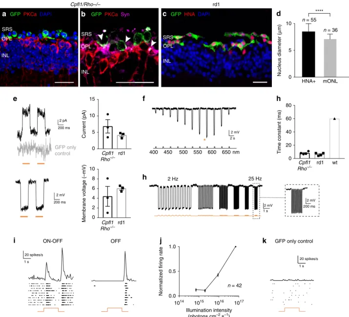
Transplantation of hiPSC-derived Jaws-expressing PRs. In
order to transplant Jaws-positive photoreceptors, we dissociated
the retinal organoids and injected the cell suspension subretinally
into the blind hosts (Cpfl1/Rho
−/−, age 10 to 15 weeks; rd1, age 4
to 5 weeks). In both Cpfl1/Rho
−/−and rd1 mice, we observed
Jaws-expressing donor cells in close proximity to the host INL
several weeks after transplantation (Fig.
5
a–c). Due to recent
concerns about material transfer in photoreceptor
transplanta-tion
27–29, we stained cryosections from the transplanted retinas
with the human nuclear antigen (HNA) and we examined the size
of the transplanted cells (HNA
+) in relation to the chromatin
structure and diameter of host cells (Fig.
5
d). HNA stained cell
counts confirmed that only a very small portion (5%) of the
GFP
+labeled cells could potentially be endogenous mouse cells
that underwent material transfer (HNA
−/GFP
+) (Supplementary
Cl–
Blind mice Retinal organoids
a
Fold change relative to day 35
0.1 1 10 100 1000 10,000 100,000 ** *** D35 D70 ARR3 D35 D70 RCVRN 200 ms 10 pA 10 pA 5 s
k
GFP only controlb
f
400 450 500 550 600 650 590 nm 2 Hz 30 Hz 1 s 1 mVl
590 nmj
Donor source Optogenetic transformation Transplantation
Confluent iPSC Feeder free
Xeno free
Optogenetic transformation via AAV infection
Dissociation of retinal organoids and transplantation
into blind mice
D70 D42 D28 D0 Mechanical isolation of retinal organoids 2D culture 3D culture hiPSCs CRX CAR DAPI
d
e
c
AAV2-7m8-mCAR-JAWS-GFP CRX CAR DAPIg
h
GFP CAR DAPIi
n = 10 N = 8 n = 10 N = 8 n = 10 N = 5 n = 10 N = 6 nmFig. 8). Both the HNA staining and nuclei comparison confirmed
the human identity of transplanted cells in close proximity of the
host INL. The transplanted GFP
+cells were RCVRN positive
(Supplementary Fig. 8) and located next to PKCα-positive bipolar
cells (Fig.
5
a). They expressed the synaptic marker Synaptophysin
in close apposition to the bipolar cell dendrites (Fig.
5
b),
sug-gesting that the human cells form synaptic connections with the
host bipolar cells. The transplanted cells displayed robust
Jaws-induced photocurrents by patch clamp, demonstrating the
func-tionality of the microbial opsin in the host environment (Fig.
5
e).
The measured photocurrents peaked at 575 nm and showed fast
kinetics (Tau
ON< 10 ms) (Fig.
5
f–h), reflecting the response
properties of Jaws. At the ganglion cell level, we observed
ON-and OFF responses from different ganglion cell types, which
shows that Jaws-driven signals from transplanted photoreceptors
were transmitted via second order neurons (Supplementary
Fig. 9) to ON and OFF ganglion cells (Fig.
5
i and Supplementary
Fig. 10). The light intensity requirements were again below the
safety threshold for the human retina
20,21(Fig.
5
j and
Supple-mentary Fig. 10). After transplantation of control human donor
cells, expressing GFP only, no light responses were detected
(Fig.
4
k and Supplementary Fig. 10), as expected in absence of
OS-like structures.
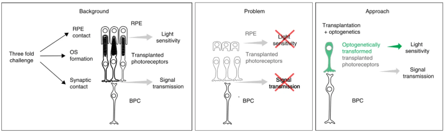
Discussion
Transplantation of healthy photoreceptors holds great promise to
restore vision in patients with outer retinal degeneration. This
approach has received significant attention over the past years as
it can restore vision independently from the cause of
photo-receptor cell loss
30. Significant progress has been made in the
generation
23,26,31–35, purification
36,37and transplantation of
photoreceptors
3,7,35,36,38–40from hiPSCs. However,
photo-receptor replacement faces a three-fold challenge: transplanted
cells need to develop (1) synaptic contact to bipolar cells for
signal transmission, (2) functional photoreceptor OS, and (3)
tight contact to RPE cells to maintain OS light-sensitivity (Fig.
6
).
This makes photoreceptor transplantation complex and
challen-ging. Recent studies have shown that the recipient environment is
of great importance for successful integration and survival of
transplanted photoreceptor cells. In animals with severely
degenerated ONL, transplanted photoreceptor precursors derived
from postnatal mouse retina
4–6or from hiPSCs
35failed to
develop normal OS structure and establish correct OS polarity
with respect to host RPE. The RPE cells are indispensable for OS
renewal as they phagocytose the shed OS discs. Moreover, they
re-isomerize the chromophore all-trans-retinal into 11-cis-retinal.
Thus, in the absence of intimate contact with the RPE
photo-receptors cannot maintain their light sensitivity
41.
For an OS and RPE-independent treatment approach, we
introduced a hyperpolarizing microbial opsin into photoreceptors
derived from either neo-natal mouse retinas or from human retinal
organoids
derived
from
iPSCs.
We
transplanted
these
optogenetically-transformed photoreceptors into blind mice
lack-ing the photoreceptor layer. We have shown that these cells can
mediate visual function, as demonstrated by a battery of tests from
RGC recordings to behavioural tests. The paradigm that
trans-planted photoreceptors migrate and structurally integrate into the
ONL of the recipient has been challenged recently by several
groups
27–29providing strong evidence that cytoplasmic material
transfer occurs between transplanted cells, residing in the
sub-retinal space, and remaining photoreceptor cells of the host. In
these experiments, however, late-stage degeneration animals were
used to model patients with advanced disease, thus there are only
few remaining photoreceptors, minimizing the potential
con-tribution of material transfer
42. To distinguish between potential
fusion events and structural integration of donor photoreceptors,
we performed Y chromosome FISH and HNA staining in the
Cpfl1/Rho
−/−model where some remaining cells were visible in
earlier transplantation time-points. Our Y chromosome FISH
experiments revealed a very limited number of events of potential
material transfer (<10%). In our blind rd1 mice, only sparse
population of cones and no rods remain after 36 days of age
18. We
confirmed this observation in our control animals, obviating the
possibility of material transfer from the transplanted
NpHR-expressing mouse progenitors to remnant ONL cells of the host.
Moreover, NpHR-positive cells that were attached to the host INL
visibly show rod nuclear morphology, indicating that these are
indeed donor cells and not remaining cones. As for the
trans-plantation of Jaws-expressing hiPSCs, histological analysis using a
human-specific nuclear marker (HNA) in transplanted mice,
confirmed that the vast majority (95%) of GFP-expressing cells
were HNA positive. This result along with the measured nuclei size
confirmed the human origin of the transplanted cells, ruling out
material exchange between human donor photoreceptors and
mouse host cells. Although these do not fully rule out that material
transfer may contribute to the improved functional responses, we
have observed that the level of functional improvement is
inde-pendent of the host age at time of transplantation, further
sup-porting the optogenetically transformed photoreceptors are the
major source of functional light responses (Supplementary Fig. 11).
Moreover, material transfer is rare between human donor and
mouse host photoreceptors
35,36(Fig.
5
d and Supplementary Fig. 8),
arguing against a significant contribution of material transfer to the
observed functional improvements.
Lastly, any possible rescue effect mediated by remaining host
photoreceptors is expected to be very minor as our control groups
Fig. 4 Jaws-expressing photoreceptors, derived from hiPSCs, are sensitive to light. a Human iPSCs were differentiated towards retinal organoids and were infected with AAV-mCar-Jaws-GFP. After further maturation, cells were dissociated and iPSC-derived photoreceptors were transplanted into blind mice.b Schematic diagram of the differentiation and viral transformation of retinal organoids. c Bright-field image of a retinal organoid at D30 of differentiation.
d, e Characterization of a representative retinal organoid at D70, depicting a thick layer of photoreceptors immunoreactive for CRX (green) and CAR (red).
f Real-time qRT-PCR analysis of photoreceptor specific markers CAR (ARR3) and RCVRN. N = number of biological replicates, n = number of organoids.
Values are mean ± SEM. Error bars are SEM. Statistical significance assessed using Mann–Whitney Student’s test (**p < 0,01; ***p < 0,001). Source data
are provided as a Source Datafile. g Live GFP fluorescence observed at D54 (12 days post infection). h A single cone photoreceptor stained with GFP
(green) and CAR (red) at D70.i Brightfield/epifluorescence image of a GFP+cell patched inside a retinal organoid at D70 of differentiation. Scale bars are
100μm (c, d, g, i) and 25 μm (e, h). j–l Patch-clamp data from Jaws-cones within organoids. The resting membrane potential (RMP) of Jaws-expressing
photoreceptors in the dark (at 0 current) for the recordings presented in thefigure was -41,7 ± 3,9 mV. Source data are provided as a Source Data file.
Stimulation at 590 nm if not stated otherwise.j Photocurrent responses after stimulation with two consecutiveflashes at 3.5 × 10¹⁷photons cm−2s−1,
absence of response in GFP only expressing cones is shown in grey.k Photocurrent action spectrum corresponding to a Jaws-cone stimulated at
wavelengths ranging from 400 to 650 nm. Maximal responses were obtained at 575 nm (at 8.7 × 10¹⁶photons cm−2s−1).l Modulation of Jaws-induced
voltage responses at increasing stimulation frequencies from 2 to 30 Hz. The timing and duration of stimulation is depicted with underlying orange lines
transplanted at the same ages with wild-type donor-derived
photoreceptor precursors or hiPSC-derived photoreceptors
expressing GFP only, never showed any detectable functional
responses. This confirms that any possible rescue effect on
remaining host photoreceptors cannot be a result of the
trans-plantation itself and suggests that the functional outcomes are a
direct consequence of the presence of an optogenetic protein
expressed in the transplanted photoreceptors.
In conclusion, by using immature photoreceptors equipped with a
microbial opsin, we went beyond the current limitations of
opto-genetic gene therapy approaches. Optoopto-genetic approaches
com-monly target bipolar cells or RGCs that are viable targets in late
h
f
GFP only control OFF ON-OFF 20 spikes/s GFP only controlh
i
j
k
1014 1015 1016 1017 0.0 0.5 1.0Normatized firing rate
*
2 s 400 450 500 550 600 650 nm Illumination intensity (photons cm–2 s–1) 2 pA 200 ms 2 mV 200 ms 2 mV 1 s 2 mV 2 mV 200 ms 2 Hz 25 Hz 1 s 20 spikes/s 1 s rd1e
n = 42 Cpfl1/Rho–/–GFP PKCa DAPI GFP PKCa Syn
a
b
SRS OPL INL SRS INL OPL SRS OPL INL GFP HNA DAPIc
mONL 0 5 10 **** Nucleus diameter ( µ m) HNA+ n = 55 n = 36 Cpfl1 Rho–/– rd1 Cpfl1 Rho–/– rd1 0 5 10 15 Current (pA) 0 2 4 6 8 10 Membrane voltage (–mV) Cpfl1 Rho–/– rd1 wt 0 20 40 60 80 Time constant (ms)d
Fig. 5 hiPSC-derived photoreceptors display Jaws-induced light responses that are transmitted to RGCs. Immunofluorescence analysis of
Jaws-cone-treatedCpfl1/Rho−/−(a, b) and rd1 (c) retinas. a Transplanted cells (green) overlie host PKCα bipolar cells (red), DAPI counterstaining (blue). b GFP, PKCα
and synaptophysin staining. Arrows point to synaptic connections.c GFP+Jaws-cones co-express HNA. Scale bars are 20µm. SRS—subretinal space,
OPL—outer plexiform layer, INL—inner nuclear layer. d Measurement of nuclear size of HNA+cells, transplanted in rd1 mice, and ONL cells of a wild-type
mouse.e–h Patch-clamp data from Jaws-cones after transplantation into blind mice. The RMP of Jaws-photoreceptors at 0 current was −40.8 ± 5.2 mV.
Stimulation at 590 nm if not stated otherwise.e Left, representative photocurrents (top) and voltage hyperpolarization (bottom) after stimulation with two
consecutiveflashes, absence of the response in GFP only cones shown in grey. Right, comparison of response amplitudes of Jaws-cones in different models
(top, mean photocurrent peak; bottom, mean voltage peak).f Voltage action spectrum corresponding to a Jaws-expressing cell stimulated at wavelengths
from 400 to 650 nm. Maximal responses were obtained at 575 nm (orange star).g Temporal properties: Jaws-induced hyperpolarization at increasing
stimulation frequencies from 2 to 25 Hz.h Comparison of response rise time constant between Jaws-cones transplanted inCpfl1/Rho−/−and rd1 models,
and wild-type cones.i–k Averaged spike responses obtained from MEA recordings shown as PSTH and raster plots from a transplanted Cpfl1/Rho−/−
mouse.i Representative examples of ON/OFF and OFF-responding RGCs (stimulation: 580 nm, 7 × 10¹⁶photons cm−2s−1).j Intensity curve. k Recording
from a control retina transplanted with GFP only cones. In all panels: Stimulations are depicted with underlying orange lines (for 580–590 nm stimuli;
e, g, i, k) or with a black line with associated wavelengths noted below (f).n = number of cells. Values are mean ± SEM. Corresponding data points are
overlaid in (e, h). Error bars are SEM. Statistical significance assessed using Mann–Whitney Student’s test (****p < 0,0001). Source data are provided as a
stages of retinal degenerative diseases such as retinitis pigmentosa or
age-related macular degeneration. Unfortunately, conferring light
sensitivity to cells downstream from photoreceptors, bypasses the
important information processing normally conducted by the inner
retinal circuitry. Photoreceptor-directed optogenetic therapy that
aims to rescue the function of remaining dormant cones harnesses
the information processing of the inner retina allowing the recovery
of complex visual responses such as lateral inhibition and directional
selectivity in previously blind mice
43, but this strategy can only be
useful in patients with remaining cones which represent a minor
portion of late-stage retinitis pigmentosa patients
44. Here, we use the
synergy of cell replacement and optogenetic therapy that allows the
restoration of retinal structure with stem cell derivatives and visual
function with microbial opsins. In a future perspective,
optogen-etically engineered hiPSC-derived cones could serve as donor cells
for photoreceptor transplantation in late-stage retinal degeneration.
In patients, degenerative diseases of the retina such as retinitis
pigmentosa, age-related macular degeneration, and Leber congenital
amaurosis, often manifest RPE degeneration along with
photo-receptor degeneration, especially in their late stages
10,45–47. Our
approach bodes well for applications in such patients who can only
obtain limited benefit from transplantation of photoreceptors in the
absence of chromophore replenishment from their dystrophic RPE.
Methods
Animals. Wild-type C57BL/6 mice (Janvier Laboratories) were used as a source of photoreceptor precursor donor cells. The following two models are both models of late-stage degeneration and were used as cell recipients. Cone photoreceptor
function loss 1/rhodopsin-deficient double-mutant Cpfl1/Rho−/−mice16were
provided by Marius Ader and rederived by Charles River Laboratory. The line was
the result of crossing Cone photoreceptor function loss 1 (Cpfl1) mice48with
rhodopsin knock-out mice (Rho−/−)49. The outcome were mice with no functional
photoreceptors starting from eye opening and with the ONL degenerating to one
row of cell bodies by 10 to 12 weeks16. Retinal degeneration 1 (rd1) mice (C3Hrd/
rd)17were provided by Thierry Leveillard. The retina in these mice degenerates to a
single row of cones by 3 weeks of age18,50.
All mice were housed under a 12-h light-dark cycle with free access to food and water. We have complied with all relevant ethical regulations for animal testing and research. All animal experiments and procedures were approved by
the local animal experimentation ethics committee (Le Comité d’Ethique pour
l’Expérimentation Animale Charles Darwin) and were carried out according to institutional guidelines in adherence with the National Institutes of Health guide for the care and use of laboratory animals as well as the Directive 2010/63/EU of the European Parliament.
AAV production. Recombinant AAVs were produced using the triple-transfection method on HEK293 cells (ATCC CRL-1573), harvested 24–72 h post transfection
and purified by iodixanol gradient ultracentrifugation51. The 40% iodixanol
fraction was collected after a 90 min spin at 354000 g. Concentration and buffer exchange were performed against PBS containing 0.001% Pluronic. AAV vector stocks titers were then determined based on real-time quantitative PCR titration
method using ITR primers52and SYBR Green (Thermo Fischer Scientific).
AAV-infection of photoreceptor precursors. Wild-type mice (C57BL/6J) at P2 were anesthetized on ice. Eyelids were cut and 1 μl of AAV9 2YF carrying eNpHR
gene under the control of human rhodopsin promoter and fused to thefluorescent
reporter eYFP (AAV9 2YF hRho-eNpHR-eYFP), or of AAV9 2YF hRho-GFP in the case of GFP only-expressing controls, was injected bilaterally using an ultrafine 34-gauge Hamilton syringe.
MACS with CD73. Two days following the AAV injections in P2 mice, at P4, retinas were isolated from the injected wild-type mice and cells were enriched using
CD73 cell surface marker before transplantation14,15. Briefly, retinas were
dis-sociated, pelleted by centrifugation (5 min at 300 g), resuspended in 500 μL MACS buffer (phosphate-buffered saline [PBS; pH 7.2], 0.5% BSA, 2 mM EDTA) and incubated with 10 μg/mL rat anti-mouse CD73 antibody (BD Biosciences, 550738) for 5 minutes at 4 °C. After washing in MACS buffer, cells were centrifuged for 5 minutes at 300 g. The cell pellet was resuspended in 480 μL MACS buffer and 120 μL goat anti-rat IgG magnetic beads (Miltenyi Biotec, 130-048-501). The suspension was incubated for 15 min at 4 °C followed by a washing step with MACS buffer and centrifugation. Before magnetic separation, the cells were
resuspended in MACS buffer andfiltered through a 30-μm pre-separation filter.
The cell suspensions were applied onto a LS columnfixed to a MACS separator.
The column was rinsed with 3 × 3 mL MACS buffer and theflow through was
collected (CD73 negative cells). The column was removed from the magnet and placed in a new collection tube. The CD73-positive fraction was eluted by loading 5 mL MACS buffer and immediately applying the plunger supplied with the column. The cells were then counted and concentrated to about 200,000 cells/μl. Maintenance of hiPSC culture. All experiments were carried out using hiPSC-2
cell line, previously established from human dermalfibroblasts from an 8-year-old
boy (gift from P. Rustin, INSERM U676, Paris) by co-transfecting
OriP/EBNA1-based epi-somal vectors pEP4EO2SEN2K (3μg), pEP4EO2SET2K (3 μg) and
pCEP4-M2L (2μg) (Addgene) via nucleofection (Nucleofector 4D, V4XP,
withDT-130 program; Lonza)31, and recently adapted to feeder-free conditions23.
Cells were kept at 37 °C, under 5% CO2/95% air atmosphere, and 20% Oxygen
tension and 80-85% of humidity. Colonies were cultured with Essential 8™ medium
(Thermo Fisher Scientific) in culture dishes coated with truncated recombinant
human Vitronectin and passaged once a week23.
Generation of retinal organoids from human iPS cells. Human iPSC were differentiated towards retinal organoids following an optimized protocol based on
the one published by Reichman et al.23. Briefly,hiPSC-2 cell line was expanded to
80% confluence in Essential 8™ medium were switched in Essential 6™ medium
(Thermo Fisher Scientific). After 3 days, cells were moved to the Proneural medium (Supplementary Table 2). The medium was changed every 2–3 days. After 4 weeks of differentiation, neural retina-like structures grew out of the cultures and were mechanically isolated. Pigmented parts, giving rise to RPE were carefully removed. The extended 3D culture in Maturation medium (Table S1) allowed the RPE Transplanted photoreceptors BPC RPE contact Synaptic contact Light sensitivity Signal transmission Three fold challenge BPC Approach BPC Light sensitivity Signal transmission Background iss Signg nsm na gn Signal transmission Light sensitivity Signal transmission OS
formation Transplantedphotoreceptors
RPE Problem Optogenetically transformed transplanted photoreceptors Transplantation + optogenetics
Fig. 6 Schematic illustrating the three-fold challenge in photoreceptor cell replacement. In order to provide visual improvement, transplanted
photoreceptors need to form functional OS, retain in close contact to the RPE to maintain light sensitivity, and develop synaptic connection to host bipolar cells for signal transmission. After transplantation into animals with severely degenerated ONL, photoreceptors fail to develop normal OS structure and establish correct polarity with respect to host RPE. In addition, in retinal degeneration, the RPE is often compromised alongside photoreceptors. All this undermines the success of photoreceptor replacement. We therefore introduced a hyperpolarizing microbial opsin into the photoreceptors before
transplantation, developing an OS- and RPE-independent approach for vision restoration in late-stage retinal degeneration. RPE—retinal pigment
formation of retinal organoids. Addition of 10 ng/ml Fibroblast growth factor 2 (FGF2, Preprotech) at this point favoured the growth of retinal organoids and the
commitment towards retinal neurons instead of RPE lineage53. In order to promote
the commitment of retinal progenitors towards photoreceptors, we specifically blocked Notch signalling for a week starting at day 42 of differentiation using the
gamma secretase inhibitor DAPT (10 μM, Selleckchem)54. Floating organoids were
cultured in 6 well-plates (10 organoids per well) and medium was changed every 2 days. Supplementary Table 2 summarizes the formulations for the different media used.
Infection of retinal organoids with AAV expressing Jaws. Introduction of Jaws
optogene was done by one single infection at day 42 at a 5 × 10¹⁰vg per organoid.
Retinal organoids were infected with an AAV with an engineered capsid,
AAV2-7m855carrying Jaws gene under the control of mouse cone arrestin promoter and
fused to thefluorescent reporter GFP (AAV2-7m8-mCAR-Jaws-GFP). For GFP
only-expressing controls, an infection with AAV2-7m8-mCAR-GFP was carried out in the same manner as mentioned above.
Monolayer cultures of dissociated cells. After removal of any pigmented tissue, 70-day old retinal organoids were collected and washed three times in Ringer solution (Supplementary Table 2) before dissociation with two units of pre-activated papain at
28.7μ/mg (Worthington) in Ringer solution for 25 min at 37 °C. Once a
homo-geneous cell suspension was obtained after pipetting up and down, papain was deactivated with Proneural medium (Supplementary Table 2). Cells were centrifuged and resuspended in pre-warmed Proneural medium. Dissociated retinal cells were plated onto coverslips coated with human recombinant 30 μg/cm² Laminin
(Sigma-Aldrich) and 150 μg/cm² Poly-L-Ornithine in 24 well-plates56. Monolayers
were incubated at 37 °C in a standard 5% CO2/95% air incubator and medium was changed every 2 days for the next 15–20 days, before immunostaining.
Preparation of cells for transplantation. At day 70 of differentiation retinal organoids were dissociated using papain as described above to obtain a single cell suspension in Proneural medium (Supplementary Table 2). Cell suspension was filtered through a 30 μm mesh (Miltenyi Biotec) to remove residual aggregates. After counting, cells were centrifuged and resuspended in Proneural medium at a concentration of 300,000 cells/μl.
RNA isolation and real-time RT-qPCR. Total RNA isolation was performed using a NucleoSpin RNA XS kit (Macherey-Nagel), according to the manufacturer’s instructions. RNA concentration and purity were determined using a NanoDrop ND-1000 Spectrophotometer (Thermo Fisher Scientific).
Reverse transcription was carried out with 250 ng of total RNA using the QuantiTect retrotranscription kit (Qiagen). Quantitative PCR (qPCR) reactions were performed using Taqman Array Fast plates and Taqman Gene expression master mix (Thermo Fisher Scientific) in an Applied Biosystems real-time PCR machine (7500 Fast System). All samples were normalized against a housekeeping
gene (18S) and the gene expression was determined based on theΔΔCT method.
Average values were obtained from at least four biological replicates. The primer sets and MGB probes (Thermo Fisher Scientific) labelled with FAM for
amplification are listed in Supplementary Table 3.
Transplantation procedure. Mice were sedated by intraperitoneal injection of ketamine (50 mg/kg) and xyazine (10 mg/kg) and the pupils were dilated with tropicamide drops. The mice were placed onto a heating pad to maintain the temperature at 37 °C. A drop of Lubrithal eye gel (Dechra) was used to keep the eyes hydrated during the surgery. A small glass slip was put on the eye to enable visualization through the Leica Alcon ophthalmic microscope while a syringe with a blunt, 34-gauge needle was inserted tangentially through the conjunctiva and sclera. One microlitre of cell suspension including 200,000–300,000 cells was injected between the retina and RPE, into the subretinal space, creating a bullous retinal detachment. Injections were performed bilaterally. Mice were placed into a warm chamber after the surgery until their awakening.
Tissue preparation and immunostaining. 70-day old organoids were washed in
PBS andfixed in 4% paraformaldehyde for 10 min at 4 °C before they were
incubated overnight in 30% sucrose (Sigma-Aldrich) in PBS. Organoids were embedded in gelatin blocks (7.5% gelatin (Sigma-Aldrich), 10% sucrose in PBS)
and frozen using isopentane at−50 °C.
At least 4 weeks after transplantation, mice were sacrificed by CO2inhalation
followed by a cervical dislocation. The eyeballs were removed,fixed in 4%
paraformaldehyde for 30 minutes at room temperature (RT) and incubated overnight at 4 °C in PBS containing 30% (w/v) sucrose (Sigma-Aldrich). The eyes were then dissected to obtain only the back of the eye with the retina and the RPE. The samples were embedded in gelatin blocks (7.5% gelatin (Sigma-Aldrich), 10%
sucrose in PBS), frozen with liquid nitrogen and stored at−80 °C.
Ten micrometres of thick sections were obtained using a Cryostat Microm and
mounted on Super Frost Ultra Plus® slides (Menzel Gläser). Cryosections were
washed in PBS (5 min, RT) and then permeabilised in PBS containing 0.5 % Triton
X-100 during 1 h at RT. Blocking was done with PBS containing 0.2% gelatin, 0.25% Triton X-100 for 30 min at RT and incubation with primary antibodies was performed overnight at 4 °C. We used the following primary antibodies for immunostaining: hCAR (1:20,000; gift from Cheryl Craft), CRX (1:5000; Abnova, H00001406-M02), GFP (1:500; Abcam, ab13970), HNA (1:200; Millipore,
MAB4383), Ki67 (1:200; BD Pharmagen, 550609), PKCα (1:100; Santa Cruz,
sc-208), RCVRN (1:5000-1:2000; Millipore, AB5585), Synaptophysin (1:200; Sigma, SAB4502906). The antibodies used are also listed in Supplementary Table 4. After incubation with primary antibodies, sections were washed with PBS containing
0.25% Tween20 and incubated withfluorochrome-conjugated secondary antibodies
(1:500; Thermo Fisher Scientific) for 1 h at RT. After successive washing in PBS-Tween20, nuclei were counterstained with DAPI (4′-6′-diamino-2-phenylindole, dilactate; Invitrogen-Molecular Probe, Eugene, OR) at a 1:1000 dilution. Samples were further washed in PBS and dehydrated with 100% ethanol before mounting
usingfluoromount Vectashield (Vector Laboratories).
FISH for Y chromosome detection. For combined chromosomalfluorescence
in situ hybridization (Y chromosome FISH) and immunohistochemistry, retinas from
female Cpfl1/Rho−/−mice transplanted with male donor-derived rod precursors
(N= 5) were collected 4 weeks post-surgery, fixed for 1 h at 4 °C with freshly prepared
4% paraformaldehyde (Merck Millipore), incubated in 30% sucrose overnight, fol-lowed by cryopreservation. After embedding and freezing in OCT medium, cryo-sections of 12 μm were rehydrated with 10 mM sodium citrate buffer pH 6, antigen retrieval performed (80 °C, 25 min). Sections were washed in PBS for 5 min and incubated with a primary antibody against GFP (1:500; Abcam, ab13970) overnight at RT, followed by incubation with secondary antibody conjugated to AlexaFluor 488 (1:1000; Jackson Immunoresearch, 103545155) overnight at RT. Next, slides were post-fixed in 2% PFA for 10 min, pre-treated with 50% formamide for 1 h at RT, then hybridization of the XMP Y orange probe (Metasystems, D-1421-050-OR) to the Y chromosome was performed. To allow the probe to penetrate the tissue, samples were incubated for 3 h at 45 °C in a HybEZ II oven. Then, samples were transferred to a hot block at 80 °C for 5 min, to denature DNA. Afterwards, probes were hybridized with DNA for 2 days at 37 °C. Posthybridization consisted of 3 × 15 min washes with 2× SCC at 37 °C and 2 × 5 min stringency washes with 0.1× SCC at 60 °C. Finally, sections were counterstained with DAPI (1:15,000; Sigma). The samples were imaged
and quantified using structured illumination microscopy (SIM; ApoTome, Zeiss).
For information on antibodies used, see Supplementary Table 4.
Quantification of YFP+cells after transplantation. Tranplanted host eyes
(N= 6) were processed and cryosectioned as described for the Y chromosome FISH
experiment, and subsequently stained for GFP (1:500; Abcam, ab13970) and
pho-toreceptor specific marker RCVRN (1:5000; Millipore, AB5585), followed by
sec-ondary antibody staining (1:1000; Jackson Immunoresearch) Every fourth serial section from whole experimental retinas was used to quantify the total amount of
YFP+photoreceptors. Cells were counted from images obtained with the
Nano-Zoomer microscope (Hamamatsu Photonics). Following these cell counts, the resulting value was multiplied by four to estimate the total amount of labelled cells per retina.
For information on antibodies used, see Supplementary Table 4.
Nuclear size measurements. Measurements of the nuclear size were performed with FIJI software (NIH) on immunostained sections of rd1 transplanted retinas and compared with the values in wild-type mice.
Image acquisition. Immunofluorescence was observed using a Leica DM6000
microscope (Leica microsystems) equipped with a CCD CoolSNAP-HQ camera (Roper Scientific) or using an inverted or upright laser scanning confocal micro-scope (FV1000, Olympus) with 405, 488, 515 and 635 nm pulsing lasers. The images were acquired sequentially with the step size optimized based on the Nyquist–Shannon theorem. The analysis was conducted in FIJI (NIH). Images were put into a stack, Z-sections were projected on a 2D plane using the MAX intensity setting in the software’s Z-project feature, and the individual channels were merged.
Images of Y chromosome labelled retinas were acquired using SIM (ApoTome,
Zeiss). Samples stained to perform quantification of surviving YFP+
photoreceptors were imaged with the NanoZoomer microscope (Hamamatsu Photonics).
Light stimulation of NpHR-positive, Jaws-positive, and control cells. Light-triggered responses were measured in donor cells before transplantation—in vivo
in AAV-injected wild-type donor mice at P12 for NpHR+, and in retinal organoids
and monolayer cultures from dissociated organoids for Jaws+ cells. In order to measure light responses we used a monochromatic light source (Polychrome V,
TILL photonics). After patching the cells wefirst stimulated them with a pair of
590 nm full-field light pulses. Then the activity spectrum was measured by using
lightflashes ranging from 400 to 650 nm (separated by 25 nm steps). Finally we
generated light pulses at different frequencies ranging between 2 and 30 Hz in order determine the temporal response properties of NpHR and Jaws in AAV-transduced cells. Stimulation and analysis were performed using custom-written
software in Matlab (Mathworks) and Labview (National Instruments). We used
light intensities ranging between 1 × 10¹⁶and 3.2 × 10¹⁷photons cm−2s−1.
Live two-photon imaging and patch-clamp recordings. Donor mouse retina (P12), retinal organoids or monolayer cultures from dissociated organoids were placed in the recording chamber of the microscope at 36 °C in oxygenated (95%
O2/5% CO2) Ames medium (Sigma-Aldrich) during the whole experiment.
Transplanted mice were sacrificed by CO2inhalation followed by quick cervical
dislocation, and eyeballs were removed. Retinae from Cpfl1/Rho−/−or rd1 mice
were isolated in oxygenated (95% O2/5% CO2) Ames medium and whole mount
retinas with ganglion cell side down were placed in the recording chamber of the microscope at 36 °C for the duration of the experiment for both live two-photon imaging and electrophysiology.
A custom-made two-photon microscope equipped with a 25x water immersion objective (XLPlanN-25 × -W-MP/NA1.05, Olympus) equipped with a pulsed femto-second laser (InSight™ DeepSee™ - Newport Corporation) were used for
imaging and targeting AAV-transducedfluorescent photoreceptor cells (eYFP+or
GFP+cells). Two-photon images were acquired using the excitation laser at a
wavelength of 930 nm. Images were processed offline using ImageJ (NIH). A CCD camera (Hamamatsu Corp.) was also used to visualize the donor cells or the retina under infrared light.
For patch-clamp recordings, AAV-transducedfluorescent cells were targeted
with a patch electrode under visual guidance using the reporter tag’s fluorescence. Whole-cell recordings were obtained using the Axon Multiclamp 700B amplifier (Molecular Device Cellular Neurosciences). Patch electrodes were made from borosilicate glass (BF100-50-10, Sutter Instrument) pulled to 7–10 MΩ and filled
with 115 mM K Gluconate, 10 mM KCl, 1 mM MgCl2, 0.5 mM CaCl2, 1.5 mM
EGTA, 10 mM HEPES, and 4 mM ATP-Na2 (pH 7.2). Photocurrents were
recorded while voltage-clamping cells at a potential of−40 mV. Some cells were
also recorded in current-clamp (zero) configuration, hence allowing us to monitor the membrane potential during light stimulations.
A monochromatic light source (Polychrome V, TILL photonics) was used to stimulate cells during electrophysiological experiments and hence record photocurrents or changes in cells membrane potential. First, in order to measure
the activity spectrum of NpHR and Jaws, we used 300 ms lightflashes ranging from
650 to 400 nm (25 nm steps; interstimulus interval 1.5 s) at a constant light
intensity of 1.2 × 10¹⁶photons cm−2s−1. Then this light source was used at a
constant wavelength of 590 nm to generate light pulses at different frequencies (ranging from 2 to 30 Hz) in order determine the temporal response properties of optogenetic proteins used. Stimuli were generated using custom-written software in LabVIEW (National Instruments) and output light intensities were calibrated using
a spectrophotometer (USB2000+, Ocean Optics).
Multi-electrode array recordings and data analysis. The mice were euthanized, the retinas isolated, cut each in two pieces and placed in Ames medium bubbled
with 95% O2and 5% CO2. Each piece was mounted separately on a cellulose
membrane soaked overnight in poly-L-lysin and gently pressed against a 60-μm electrode spacing 252 channel multi-electrode array chip (256MEA60/10iR, Multi Channel Systems) with RGCs facing the electrodes. The piece remained perfused
with oxygenated Ames medium at 34 °C throughout the experiment. Fullfield light
stimuli were applied with a Polychrome V monochromator (TILL Photonics) driven by a STG2008 stimulus generator (Multichannel Systems) using custom written stimuli in MC_Stimulus II (MC_Stimulus II Version 3.4.4, Multichannel Systems).
The basic stimulus pattern applied was 10 repetitions of 2-s stimuli of 580 nm
light (close to excitation maximum for NpHR and Jaws) and intensity of 7 × 10¹⁶
photons cm−2s−1, with 10 s intervals. To assess temporal dynamics of responding
cells, stimuli ranging from 1 ms to 2 s were played to the retina. Action spectrum of optogenetic protein-expressing cells was examined by playing sets of stimuli of different wavelengths (450 nm, 500 nm, 550 nm, 580 nm, 600 nm, 650 nm; 10 stimuli of 2 s with 10 s intervals for each wavelength). To determine sensitivity
of responding cells, stimuli of lower intensities were also used (1 × 10¹⁴, 7 × 10¹⁴,
2 × 10¹⁵and 9 × 10¹⁵photons cm−2s−1). During the experiments aiming to show
that the light responses are really coming from the ONL, we perfused the tissue
with L-AP4 (50μM) for at least 20 min before the recordings in order to block
input from photoreceptors to ON bipolar cells. This was followed by at least 15 min rinse with Ames medium and another set of light stimulation to observe whether the response returned.
Data were acquired using the MC_Rack software (MC_Rack v4.5, Multi Channel Systems). RGC responses were amplified and sampled at 20 kHz. Data was
thenfiltered with a 200 Hz high pass filter and individual channels were spike
sorted using template matching and cluster grouping based on principal component analysis of the waveforms in Spike2 software v.7 (Cambridge Electronic Design Ltd). The raster plots and peristimulus time histogram data (bin size of 10 ms) were constructed in MATLAB using custom scripts from spike-sorted channels and further processed in Adobe Illustrator CS4 (Adobe Systems) for presentation.
Maximumfiring rate for each responding cell was measured in the 2 s after the
onset (for ON-responding cells) or 2 s after the offset (for OFF-responding cells) of
the stimulus. The number of cells and mice that were used for quantitative analysis are stated in Figure legends. Error bars were calculated over cells.
Light/dark box. For light-avoidance behaviour, we used a custom-made dark-light
box22,57of dimensions 36 cm × 20 cm × 18 cm, divided longitudinally into two
equal sized compartments with a non-transparent wall with a 7 cm × 5 cm hole in the middle. The light compartment was equipped with eight 590 nm LEDs (Cree XP-E, amber, Lumitronix) 3 cm from the bottom of the box. A light intensity of
2.05 × 10¹⁶photons cm−2s−1was used for all the experiments. The mice were
habituated in the dark for at least 2 h prior the testing. Each mouse was introduced into the light compartment and was left in the box for at least 5 min before the start of illumination. The lights were turned on when the mouse was in the light compartment and were left on for at least 5 min. The behaviour of the mice was recorded with a camera and subsequently analyzed manually by recording the times spent in each compartment after the start of illumination, and using the Smart Vision Tracking Software (Harvard Apparatus). The mouse’s head was used to define the compartment it occupied.
Statistical analyses. Data was analyzed with GraphPad Prism and it was expressed as mean ± standard error of mean (SEM). Comparisons between
values were analyzed using unpaired two-tailed non-parametric Mann––Whitney
Student’s test. A level of p < 0.05 was considered significant. The labels used were: *p < 0.05, **p < 0.01, ***p < 0.001 and ****p < 0.0001.
Reporting summary. Further information on research design is available in the Nature Research Reporting Summary linked to this article.
Data availability
The data that support thefindings of this study are available from the corresponding author upon reasonable request. The source data underlying Figs.1i,2a, d,3c, e, g, i, k,4f,
5d, e, h, j, resting membrane potential values (RPM), and Supplementary Figs. 3c, 6b, e, j and 8c are provided as a Source Datafile.
Code availability
The MATLAB codes that were used to represent MEA raster and PSTH data are available from the corresponding author upon reasonable request.
Received: 6 November 2018 Accepted: 28 August 2019
References
1. Jayakody, S. A., Gonzalez-Cordero, A., Ali, R. R. & Pearson, R. A. Cellular
strategies for retinal repair by photoreceptor replacement. Prog. Retin. Eye Res. 46, 31–66 (2015).
2. West, E. L., Pearson, R. A., MacLaren, R. E., Sowden, J. C. & Ali, R. R.
Cell transplantation strategies for retinal repair. Prog. Brain Res. 175, 3–21 (2009).
3. Santos-Ferreira, T. F., Borsch, O. & Ader, M. Rebuilding the missing Part-A
review on photoreceptor transplantation. Front. Syst. Neurosci. 10, 105 (2017).
4. Eberle, D. et al. Outer segment formation of transplanted photoreceptor
precursor cells. PloS ONE 7, e46305 (2012).
5. Barber, A. C. et al. Repair of the degenerate retina by photoreceptor
transplantation. Proc. Natl Acad. Sci. USA 110, 354–359 (2013).
6. Singh, M. S. et al. Reversal of end-stage retinal degeneration and restoration of
visual function by photoreceptor transplantation. Proc. Natl Acad. Sci. USA 110, 1101–1106 (2013).
7. Mandai, M. et al. iPSC-derived retina transplants improve vision in rd1
end-stage retinal-degeneration mice. Stem Cell Rep. 8, 1112–1113 (2017).
8. Iraha, S. et al. Establishment of immunodeficient retinal degeneration model
mice and functional maturation of human ESC-derived retinal sheets after transplantation. Stem Cell Rep. 10, 1059–1074 (2018).
9. Strauss, O. The retinal pigment epithelium in visual function. Physiological
Rev. 85, 845–881 (2005).
10. Wright, A. F., Chakarova, C. F., Abd El-Aziz, M. M. & Bhattacharya, S. S. Photoreceptor degeneration: genetic and mechanistic dissection of a complex trait. Nat. Rev. Genet. 11, 273–284 (2010).
11. Milam, A. H., Li, Z. Y. & Fariss, R. N. Histopathology of the human retina in retinitis pigmentosa. Prog. Retin. Eye Res. 17, 175–205 (1998).
12. Chiba, C. The retinal pigment epithelium: an important player of retinal disorders and regeneration. Exp. Eye Res. 123, 107–114 (2014).
13. Gradinaru, V., Thompson, K. R. & Deisseroth, K. eNpHR: a Natronomonas halorhodopsin enhanced for optogenetic applications. Brain Cell Biol. 36, 129–139 (2008).