HAL Id: inserm-01800381
https://www.hal.inserm.fr/inserm-01800381
Submitted on 25 May 2018
HAL is a multi-disciplinary open access
archive for the deposit and dissemination of
sci-entific research documents, whether they are
pub-lished or not. The documents may come from
teaching and research institutions in France or
abroad, or from public or private research centers.
L’archive ouverte pluridisciplinaire HAL, est
destinée au dépôt et à la diffusion de documents
scientifiques de niveau recherche, publiés ou non,
émanant des établissements d’enseignement et de
recherche français ou étrangers, des laboratoires
publics ou privés.
Histoplasmosis: An oral malignancy-like clinical picture
Tomasz Chroboczek, Julie Dufour, Alain Renaux, Christine Aznar, Magalie
Demar, Pierre Couppié, Antoine Adenis
To cite this version:
Tomasz Chroboczek, Julie Dufour, Alain Renaux, Christine Aznar, Magalie Demar, et al..
Histo-plasmosis: An oral malignancy-like clinical picture. Medical Mycology Case Reports, [Amsterdam] :
Elsevier, 2018, 19, pp.45 - 48. �10.1016/j.mmcr.2017.11.001�. �inserm-01800381�
Contents lists available atScienceDirect
Medical Mycology Case Reports
journal homepage:www.elsevier.com/locate/mmcr
Histoplasmosis: An oral malignancy-like clinical picture
Tomasz Chroboczek
a, Julie Dufour
a, Alain Renaux
b, Christine Aznar
c,d, Magalie Demar
c,d,
Pierre Couppie
a,d, Antoine Adenis
d,e,⁎aService de Dermatologie Vénérologie, CH de Cayenne, Cayenne, France bService d′Oto-Rhino-Laryngologie, CH de Cayenne, Cayenne, France
cLaboratoire Hospitalo-Universitaire de Parasitologie-Mycologie, CH de Cayenne, Cayenne, France dEcosystèmes Amazoniens et Pathologies Tropicales, EA3593, Université de Guyane, Cayenne, France eCentre d′Investigation Clinique Antilles-Guyane, Inserm CIC1424, CH de Cayenne, Cayenne, France
A R T I C L E I N F O
Keywords: Histoplasma capsulatum Histoplasmosis HIV Oral Differential diagnosisA B S T R A C T
HIV-associated histoplasmosis is mainly misdiagnosed for granulomatous diseases, such as tuberculosis. Nonetheless, malignancy-like lesions have been reported sporadically in HIV-infected patients. Although the main reported lesions are erosive or ulcerated, here a rare case of oral tumor is reported. This case raises the awareness of this presentation, and the importance of accurate identification in the laboratory. Performing systematic specific stains for fungal elements and culture on tissue samples ensures accurate differential diag-nosis.
1. Introduction
Histoplasma capsulatum is responsible for histoplasmosis, a deep systemic mycosis with worldwide distribution that can affect both im-munocompetent and immunocompromised individuals [1]. Histo-plasmosis is mainly an airborne disease. Disruption of soils con-taminated by H. capsulatum aerosolizes microconidia which are eventually inhaled by a host[1].
Although it is mostly asymptomatic and spontaneously resolutive in immunocompetent persons, histoplasmosis is symptomatic and dis-seminated in 95% of HIV-infected patients with a CD4 count less than 200/mm³ [2]. In endemic areas, the pathophysicological mechanism that leads to disease remains debated (new infection or endogenous reactivation)[3].
During HIV infection, the evolution is highly variable, from extreme latency to fulminant forms[2]. The most frequent presentation is sub-acute, with symptoms evolving for over one or two months. The clinical presentation is misleading, with non-specific symptoms. Fever, fatigue and weight loss are almost constant. The presence of respiratory signs, and/or digestive signs, and/or superficial lymphadenopathies is fre-quent. Neurological, oral and cutaneous involvements are less frefre-quent. For 10–20% of patients, the presentation is fulminant, similar to septic shock and rapidly fatal[4].
The diagnosis of histoplasmosis requires pathological investigations that are often invasive (bone marrow sample or tissue biopsy). Moreover, it is necessary that clinicians suspect the diagnosis when
facing certain clinical situations, in order to ask for specific fungal in-vestigations[4]. Among these are the direct examination of a fresh sample and fungal culture which remains the gold standard method despite its low sensitivity and the slow growth rate of the fungus in culture (up to six weeks)[1,5]. Among non-culture-based diagnostic tests, serology is not very contributive in HIV-infected patients and molecular tools are not commercialized yet. However, robust enzyme immunoassays (EIAs) on urine, blood or bronchoalveolar lavage are being commercialized[6]. Recent developments in monoclonal antigen detection in urine and blood are promising since the most sensitive EIA developed to date has only been available in the USA [7]. Standard biological examinations are not specific but may orient clinicians.
While waiting for a mycological gold standard-based diagnosis, when clinical suspicion is high, clinicians have two options for treat-ment initiation according to disease severity: intravenous liposomal amphotericin B (L-AmB) for severe cases or oral itraconazole for non-severe cases[8]. Although L-Amb has shown its fungicidal activity and its efficacy on survival, it is nephrotoxic[9]. L-AmB or alternative lipid formulation of AmB at the same dosage are safer and may be preferred to Deoxycholate-AmB. Itraconazole is fungistatic and exposes to drug-drug interactions if the treatment of potential coinfections and or an-tiretroviral therapy need to be introduced at the same time. This is compounded by a reduced bioavailability during HIV infection, which makes reaching the recommended effective blood levels long and dif-ficult[1].
Despite numerous publications on HIV-associated histoplasmosis,
https://doi.org/10.1016/j.mmcr.2017.11.001
Received 7 December 2016; Received in revised form 12 September 2017; Accepted 24 November 2017
⁎Corresponding author at: Centre d'investigation clinique Antilles-Guyane, Centre Hospitalier de Cayenne, Av. Desflamboyants, BP6006, 97306 Cayenne CEDEX, France E-mail address:antoine.adenis@gmail.com(A. Adenis).
2211-7539/ © 2017 The Author(s). Published by Elsevier B.V. on behalf of International Society for Human and Animal Mycology. This is an open access article under the CC BY-NC-ND license (http://creativecommons.org/licenses/BY-NC-ND/4.0/).
the true burden of HIV-associated histoplasmosis remains unknown in endemic areas[1,10]. Nevertheless the disease may rank among the top causes of AIDS-related deaths in Latin America and probably accounts for thousands of deaths every year while being undiagnosed[11]. The disease has been stated a neglected disease because, even in endemic areas, physicians awareness seems insufficient and a few laboratories are able to perform the diagnosis[6,10]. With no access to diagnostic tools or adapted antifungal therapy, one of the main issues is that his-toplasmosis is probably often misdiagnosed for another disease, and therefore physicians do not ask for a fungal diagnosis and do not treat it with antifungals. Reported since the discovery of histoplasmosis, the main differential diagnosis of HIV-associated histoplasmosis is tu-berculosis[12]. Malignancy-like clinical presentations also represent a challenging differential diagnosis requiring specific stainings and cul-ture when performing a tumor biopsy in endemic or non-endemic areas. Regarding malignancy-like lesions of the mucous membrane caused by H. capsulatum gastrointestinal and oral involvements have been re-ported in non-HIV and in HIV-infected patients[13–17].
Here we report a rare case of oral HIV-associated histoplasmosis because it is important to reinforce awareness of physicians and dentists on fungal infections when facing a malignancy-like oral lesion.
2. Case
A 52 year-old man presented at the emergency department for fever and fatigue. He was a homeless man originating from Haïti and hab been living in French Guiana for over ten years. He was known for an advanced HIV infection diagnosed 7 years before when the patient presented with concomitant pulmonary tuberculosis and pneumocys-tosis. At the time of HIV infection diagnosis, the CD4 lymphocyte count and HIV viral load were at 57/mm3 and over 6 log10, respectively. The patient received six weeks of anti-tuberculosis therapy and cotrimox-azole for pneumocystosis beforefleeing the hospital and interrupting medical follow up. Other past medical events were chronic obstructive pulmonary disease due to smoking, a negative hepatitis C serology, serological evidence of a past toxoplasmosis infection (IgM antibody negative and IgG antibody positive) and serological evidence of a re-solved hepatitis B (HBs antigen negative, HBc antibody and HBs anti-body positive).
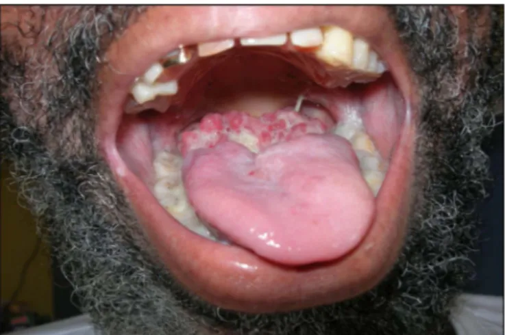
Upon admission in the emergency department, the patient reported anorexia and a 15 kg weight loss over a period of a few months, and a recent dysphonia. There was no dyspnea. Fever was 38.5 °C, and there was a large burgeoning and necrotic ulceration of the base of the tongue and the anterior part of the soft palate pillars, with a whitish coating (Fig. 1). There were no lymphadenopathies, and the rest of the ex-amination was without particularities.
Biologically, lactate deshydrogenase (LDH) was elevated at 210 IU/
l, albumin concentration was 26.7 mg/l, ferritine was 482 mg/l, C-re-active Protein (CRP) was 45 mg/l, creatinine awast 49 μmol/l, and liver enzymes were normal. The hematological results showed platelets at 215,000/mm3
, hemoglobin at 9.3 g/dl, and white blood cells at 4 400/ mm3. The CD4 lymphocyte count was 40/mm3 with a HIV viral load of 5.5 log10. The search for acid-fast bacilli was negative on 3 sputum samples (direct examination and culture) and in the blood cultures.
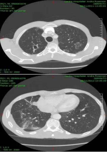
Radiologically, the chest Xray showed an interstitial syndrome suggestive of histoplasmosis with sequellae of the past tuberculosis (Fig. 2). Computed tomography showed an ulcerated lesion of the tongue and the oropharyngeal region (Fig. 3), right jugulo-carotidian lymphadenopathies, an aspect of pulmonary miliary,fibrotic sequelae associated with a localized bronchial dilatation of the right lung, and
Fig. 1. Oral examination upon hospital admission.
Fig. 2. Chest X-Ray upon hospital admission.
Fig. 3. Computed tomography of the pharyngeal region upon hospital admission (sagittal section).
T. Chroboczek et al. Medical Mycology Case Reports 19 (2018) 45–48
finally disseminated centrolobular emphysema (Fig. 4).
Multiple biopsies of the base of the tongue and of the amygdala lodges were performed. Pathological examination showed poly-morphous granulomatous inflammation rich with histiocyte nests and lymphocytes, as well as the presence of intra-histiocytic yeasts of H. capsulatum (Fig. 5). The Acid-Fast Bacilli (AFB) smears were negative. Cytological examination of tissue smears and fungal culture at 25 °C
confirmed the presence of H. capsulatum (Fig. 6).
Bronchial fibroscopy with bronchoalveolar lavage was also per-formed showing white secretions on the right side, and areas of an-thracosis. Samples microbiological analyses were all negative, notably regarding Koch Bacilli (smear and culture) and histoplasmosis (direct examination and culture).
Histoplasmosis immunodiffusion serology and RT-PCR, available on site, were not performed. Antigen detection for histoplasmosis was not available and not realized.
The final diagnosis was disseminated histoplasmosis, associated with oral candidiasis, in an HIV patient with severe immunosuppres-sion. The patient was treated with itraconazole at a dose of 100 mg, 3 tablets morning and evening for 3 consecutive days, followed by 2 ta-blets morning and evening daily. The initial evolution was favorable with a marked reduction of the lesions after one week of treatment (Fig. 7). Complete healing with ad integrum restitution of the oral cavity was obtained after 6 months of itraconazole 400 mg daily in association with antiretroviral therapy.
3. Discussion
Oral presentations are commonly described in the literature among immunocompetent or immunosuppressed individuals, with highly variable frequencies (< 10% to 60%)[15]. Nevertheless, they are not frequent in areas where physician awareness, good access to mycology Fig. 4. Computed tomography of the thoracic region upon hospital admission (horizontal
sections).
Fig. 5. LEFT: Tongue biopsy with Histoplasma capsulatum yeasts colored in fuchsia (Periodic-Acid-Schiff, ×40), RIGHT: Tongue biopsy with Histoplasma capsulatum yeasts colored in black (Gommori-Grocott, silver staining, ×20).
Fig. 6. Tissue smears with cells of the base of the tongue, showing intra cellular yeasts of Histoplasma Capsulatum (May-Grumwald-Giemsa, ×100).
Fig. 7. Oral examination showing a marked decreased of the lesion after one week of antifungal therapy with itraconazole.
laboratories and antifungal therapy converge to an early diagnosis and treatment[15].
HIV-associated histoplasmosis is thefirst AIDS-defining condition in French Guiana, where HIV prevalence exceeds 1% since the 1990's [18]. In the past ten years we observed a sharp decline in mortality together with a stable number of new cases[18]. As for mortality we observed a sharp decline in the incidence trends for muco-cutaneous presentations of HIV-associated histoplasmosis [15]. It used to re-present a large proportion of diagnosed cases since it wasfirst described by dermatologists looking for leishmaniasis [15]. To date, cutaneous presentations are scarce but oral mucosal presentations although rare have remained stable over time (unpublished data).
It is commonly described that oral involvement occurs at the late stage of histoplasmosis and HIV infection[15]. Regarding HIV infec-tion, in almost all published series, the CD4 count at the time of oral histoplasmosis diagnosis was < 70/mm3 [15]. Regarding histo-plasmosis infection, oral involvement is often associated with involve-ment of another organ in the context of a progressive disseminated form of the disease [15]. However, authors have considered potential pri-mary oral lesions, which may heal spontaneously [13]. Although the presence of H. capsulatum couldn’t be documented at the pulmonary level in our patient, the literature describes a frequent association be-tween oral and pulmonary forms of histoplasmosis[13,15]. Hence, in HIV-infected patients, oral involvement seems to represent the late expression of the progressive spread of H. capsulatum infection through the reticuloendothelial system.
Oral mucosal manifestations are mainly represented by erosive or plaque-like lesions, ulcerations or ulcero-vegetative or nodular-ulcera-tive lesions. Localizations are mainly the lips, palate, gingiva, tongue and pharynx[13,15,16]. Any chronic oropharyngeal lesions, solitary or not, requires performing biopsies so that pathologists can rule out malignancies and microbiologists can rule out infectious diseases. If malignancies are the most common etiologies, notably squamous cell carcinoma, infectious diseases such as tuberculosis, syphilis, cytome-galovirus, paracoccidioidiomycosis, histoplasmosis or talaromycosis must be evoked by physicians facing an oral mucosal lesion in HIV-infected individuals[13,15,16].
Certain diagnoses rely on direct examination and fungal culture together with the required confirmatory culture (i.e. conversion of the mold cultured into the yeast phase of H. capsulatum)[1]. In the absence of fungal culture, there is a consensus that diagnostic may rely only on histology performed with specific stains for fungal elements (Periodic-acid Schiff and Gomori-Grocott methenamine silver)[1,5]. Still con-fusion with C. glabrata, T. marneffei, Leishmania, Trypanosoma and other staining artifacts may persist if the clinical picture and the past medical history are not joined to the request[18]. In our report, although no confirmatory subculture was performed, we identified the mold phase of H. capsulatum from clinical samples and we feel confident with the diagnostic of histoplasmosis because of the local epidemiology and the complete healing after several months of itraconazole.
Oral presentations of HIV-associated histoplasmosis are not asso-ciated with a poor prognosis since mortality at one month was not shown to be higher than other presentations of the disease[15]. Hence, greater mortality at six months or more may be linked to the patients’ immune deficiency. Indeed, the vital prognosis of these patients seems to be linked to the level of immunosuppression, and not to the late stage of H. capsulatum infection. Despite oral presentations occurring in pa-tients at the very late HIV stages with low CD4 counts, introducing oral itraconazole seems to be a good strategy in the absence of organ failure or concomitant opportunistic infections. But, physicians must pay at-tention to drug-drug interactions when introducing antiretroviral therapy (notably efavirenz)[1].
This report highlights the importance of including infectious dis-eases, notably fungal diseases such as histoplasmosis as a potential differential diagnosis when facing an oral mucosal lesion resembling a tumor. The capacity of physicians to suspect the diagnosis, to prescribe
specific fungal investigations, and to start promptly antifungal therapy are key elements in patient care. In addition, physicians should not differ initiation of antiretroviral therapy since the patient prognosis is at risk considering the advanced stage of HIV infection[1].
Acknowledgements
We acknowledge an Investissement d'Avenir grant of the Agence Nationale de la Recherche (CEBA ANR-10-LABX-25-01). AA and TC draft the manuscript.
TC, JD, AR, CA and PC managed the patient, the diagnosis and re-vise the manuscript.
Conflict of interest There are none. Ethical form
Authors declared that they have obtained a written and signed consent from the patient to use his medicalfiles in the context of bio-medical research. Plus, a specific oral consent to take pictures and publish the case report was obtained from the patient.
References
[1] A.A. Adenis, C. Aznar, P. Couppie, Histoplasmosis in HIV-infected patients: a review of new developments and remaining Gaps, Curr. Trop. Med. Rep. 1 (2014) 119–128. [2] C.A. Kauffman, Histoplasmosis: a clinical and laboratory update, Clin. Microbiol Rev. 20
(1) (2007) 115–132.
[3] M. Hanf, A. Adenis, P. Couppie, B. Carme, M. Nacher, HIV-associated histoplasmosis in French Guiana: recent infection or reactivation? AIDS 24 (11) (2010) 1777–1778. [4] J. Wheat, Endemic mycoses in AIDS: a clinical review, Clin. Microbiol. Rev. 8 (1) (1995)
146–159.
[5] B. De Pauw, T.J. Walsh, J.P. Donnelly, D.A. Stevens, J.E. Edwards, T. Calandra, et al., Revised definitions of invasive fungal disease from the European Organization for Research and Treatment of Cancer/invasive Fungal Infections Cooperative Group and the National Institute of Allergy and Infectious Diseases Mycoses Study Group (EORTC/MSG) Consensus Group, Clin. Infect. Dis. 46 (12) (2008) 1813–1821.
[6] D.R. Falci, E.R. Hoffmann, D.D. Paskulin, A.C. Pasqualotto, Progressive disseminated histoplasmosis: a systematic review on the performance of non-culture-based diagnostic tests, Braz. J. Infect. Dis. (2016).
[7] P. Couppie, C. Aznar, B. Carme, M. Nacher, American histoplasmosis in developing countries with a special focus on patients with HIV: diagnosis, treatment, and prognosis, Curr. Opin. Infect. Dis. 19 (5) (2006) 443–449.
[8] L.J. Wheat, A.G. Freifeld, M.B. Kleiman, J.W. Baddley, D.S. McKinsey, J.E. Loyd, et al., Clinical practice guidelines for the management of patients with histoplasmosis: 2007 update by the Infectious Diseases Society of America, Clin. Infect. Dis. 45 (7) (2007) 807–825.
[9] P.C. Johnson, L.J. Wheat, G.A. Cloud, M. Goldman, D. Lancaster, D.M. Bamberger, et al., Safety and efficacy of liposomal amphotericin B compared with conventional ampho-tericin B for induction therapy of histoplasmosis in patients with AIDS, Ann. Int. Med. 137 (2) (2002) 105–109.
[10] M. Nacher, A. Adenis, S. Mc Donald, M. Do Socorro Mendonca Gomes, S. Singh, I. Lopes Lima, et al., Disseminated histoplasmosis in HIV-infected patients in South America: a neglected killer continues on its rampage, PLoS Negl. Trop. Dis. 7 (11) (2013) e2319. [11] neglected histoplasmosis in Latin America G, Disseminated histoplasmosis in Central and
South America, the invisible elephant: the lethal blind spot of international health or-ganizations, AIDS 30 (2) (2016) 167–170.
[12] A. Adenis, M. Nacher, M. Hanf, C. Basurko, J. Dufour, F. Huber, et al., Tuberculosis and Histoplasmosis among Human Immunodeficiency Virus-Infected Patients: a Comparative Study, Am. J. Trop. Med. Hyg. (2014).
[13] I.P. Klein, M.A.T. Martins, M.D. Martins, V.C. Carrard, Diagnosis of HIV infection on the basis of histoplasmosis-related oral ulceration, Spec. Care Dent. 36 (2) (2016) 99–103. [14] S.L. Hernández, S.A. López de Blanc, R.H. Sambuelli, H. Roland, C. Cornelli, V. Lattanzi,
et al., Oral histoplasmosis associated with HIV infection: a comparative study, J. Oral. Pathol. Med. 33 (8) (2004) 445–450.
[15] P. Couppie, E. Clyti, M. Nacher, C. Aznar, D. Sainte-Marie, B. Carme, et al., Acquired immunodeficiency syndrome-related oral and/or cutaneous histoplasmosis: a descriptive and comparative study of 21 cases in French Guiana, Int. J. Dermatol. 41 (9) (2002) 571–576.
[16] V.S. Antonello, V.F. Zaltron, M. Vial, Oliveira FMd, Severo LC. Oropharyngeal histo-plasmosis: report of eleven cases and review of the literature, Rev. da Soc. Bras. De. Med. Trop. 44 (2011) 26–29.
[17] L.A. Weed, E.M. Parkhill, The diagnosis of histoplasmosis in ulcerative disease of the mouth and pharynx, Am. J. Clin. Pathol. 18 (2) (1948) 130.
[18] A. Adenis, M. Nacher, M. Hanf, V. Vantilcke, R. Boukhari, D. Blachet, et al., HIV-asso-ciated histoplasmosis early mortality and incidence trends: from neglect to priority, PLoS Negl. Trop. Dis. 8 (8) (2014) e3100.
T. Chroboczek et al. Medical Mycology Case Reports 19 (2018) 45–48