Transapical aortic
‘valve-in-valve’ procedure for degenerated
stented bioprosthesis
Enrico Ferrari*
Cardiovascular Surgery Department, Centre Hôpitalier Universitaire Vaudois (CHUV), University Hospital of Lausanne, CH-1011 Lausanne, Switzerland
* Corresponding author. Cardiovascular Surgery Department, Centre Hôpitalier Universitaire Vaudois (CHUV), 46 rue du Bugnon, CH-1011 Lausanne, Switzerland. Tel: +41-79-3101386; fax: +41-21-3142278; e-mail: [email protected] (E. Ferrari).
Received 24 May 2011; received in revised form 5 July 2011; accepted 7 July 2011
Summary
Standard surgical aortic valve replacement with a biological prosthesis remains the treatment of choice for low- and mid-risk elderly patients (traditionally >65 years of age) suffering from severe symptomatic aortic valve stenosis or insufficiency, and for young patients with formal contraindications to long-lasting anticoagulation. Unfortunately, despite the fact that several technical improvements have noticeably improved the resistance of pericardial and bovine bioprostheses to leaflet calcifications and ruptures, the risk of early valve failure with rapid degeneration still exists, especially for patients under haemodialysis and for patients <60 years of age at the time of surgery. Until now, redo open heart surgery under cardiopulmonary bypass and on cardioplegic arrest was the only available thera-peutic option in case of bioprosthesis degeneration, but it carried a higher surgical risk when elderly patients with severe concomitant comorbidities were concerned. Since a few years, the advent of new transcatheter aortic valve procedures has opened new horizons in cardiac surgery and, in particular, the possibility of implanting stented valves within the degenerated stented bioprosthesis, the so-called‘valve-in-valve’ (VinV) concept, has become a clinical practice in experienced cardiac centres. The VinV procedure represents a minimally invasive approach dedicated to high-risk redo patients, and published preliminary reports have shown a success rate of 100% with absence of significant valvular leaks, acceptable transvalvular gradients and low complication rate. However, this procedure is not riskless and the most important concerns are about the size mismatch and the right positioning within the degenerated bioprosthesis. In this article, we review the limited available literature about VinV procedures, underline important technical details for the positioning and provide guidelines to prevent valve–prosthesis mismatch comparing the three sizes of the only commercially available transapical device, the Edwards Sapien™, with the inner diameter of three of the most commonly used stented bioprostheses.
Keywords:Transapical aortic valve implantation• Valve-in-valve procedure • High-risk patients • Redo cardiac surgery • Degenerated aortic bioprosthesis
INTRODUCTION
Aortic valve stenosis (AS) is the most frequent valvular heart disease in developed countries, and affects the elderly population [1, 2]. Aortic valve replacement (AVR) with cardiopulmonary bypass, cardioplegic arrest and aortic cross-clamping through a median sternotomy, an upper sternotomy or a right mini-thoracotomy, represents, for the time being, the treatment of choice for severe AS, and provides good operative outcomes and long-term results [3,4]. Patients also affected by regurgitant aortic valves or by aortic endocarditis are eligible for standard AVR and, following the standard international guidelines, all patients over the age of 65 years at the time of surgery, or younger patients with contraindications to the long-lasting anticoagulation therapy, are ideal candidates for the implantation of a biological prosthesis. In particular, stented bioprostheses, both pericardial and porcine, do not require anticoagulation, are easy to be implanted with a standardized and reproducible surgical technique, have excellent haemodynamic performances and, thanks to the improved treatments (anticalcification) and construction, have an increased longevity [5–8]. However, despite all attempts to decrease
the incidence of leaflet calcifications and structural failure, early degeneration can occur (especially in young patients and in patients under haemodialysis) and, nowadays, the treatment of choice for the replacement of a malfunctioning bioprosthesis is a cardiac reoperation with a mortality rate that lies below 5% in the latest series [9–13]. Unfortunately, despite the fact that the redo itself is not an independent risk factor for re-AVR, redo valve surgery in the elderly high-risk patient with severe comorbidities is still related to a higher operative risk with increased hospital mor-tality and postoperative complication rate [13–15].
Thus, the transcatheter aortic valve procedure plays a key role, and the possibility of implanting stent-valves into failed stented bioprostheses, the‘valve-in-valve’ (VinV) concept, represents an alternative for redo high-risk patients [16–19]. As regards to the transapical access for aortic VinV procedures, we are observing a burden in the number of performed cases, and experienced centres employ this technique routinely for selected cases. Moreover, a few VinV case reports and limited series have appeared in the literature showing a good outcome with low transvalvular gradients, no major leaks and few postoperative complications [18]. Nevertheless, the risk of valve–prosthesis
© The Author 2011. Published by Oxford University Press on behalf of the European Association for Cardio-Thoracic Surgery. All rights reserved.
AD UL T C ARDIA C
European Journal of Cardio-Thoracic Surgery 41 (2012) 485–490
REVIEW
mismatch still exists [20]. In this article, we underline important details for transapical VinV in stented bioprostheses, we expose the review of clinical results and haemodynamic parameters and, in order to avoid the mismatch, we suggest guidelines for the sizing comparing the transapical Sapien™ platform with three common stented bioprostheses.
TECHNICAL ASPECTS
Edwards Sapien™
The only available transapical stent-valve is the Sapien™ (Edwards Lifesciences, Irvine, CA, USA) (Fig. 1A), a balloon-expandable stent with an inner bovine pericardial valve. It is available in two sizes, 23 and 26 mm, and is inserted using the Ascendra™ delivery system. Recently, a new XT generation (Fig.1B), with the Ascendra II delivery system, was launched with some innovations such as the cobalt–chromium stent, a smaller delivery system (22F and 24F), a semi-closed leaflet profile and a bigger 29 mm size (for transapical).
Patients selection
Symptomatic patients with degenerated bioprostheses present-ing with severe comorbidities are candidates for transapical aortic VinV (high-risk). The logistic EuroSCORE and the STS score calculate the predicted mortality, and the inclusion and exclu-sion criteria are similar to those proposed for standard TAVI [21,
22]. However, due to the fact that during VinV procedures in stented bioprostheses thefixation of the valve is guaranteed by radial forces applying against the rigid ring (unlike in standard TAVI where heavy calcifications of the annulus and valve are required forfixation), not only stenosis but also intra-prosthetic
incompetence due to leaflet ruptures or tears is treatable with this approach. Bioprosthetic endocarditis remains a formal con-traindication because the infected bioprosthetic leaflets are not removed during the procedure. The presence of a concomitant mitral prosthesis seems not to interfere with aortic VinV, and candidates for VinV procedures require neither specific preo-perative exams nor cardiac imaging and do not even require an injected cardiac scan to measure the annulus, given that the size of the valve is pre-determined by the size of the bioprosthesis.
Sizing (valve–prosthesis match)
During the implantation of a stent-valve within a stented bio-prosthesis, there is a risk of severe mismatch, creating either a relevant transvalvular gradient, when the orifice of the bio-prosthesis is too small compared with the stent-valve diameter, or a stent-valve embolization when the stent-valve is too small compared with the inner size of the prosthesis.
In order to identify which stent-valvefits perfectly into differ-ent sizes of a given stdiffer-ented aortic bioprosthesis, we measured the internal diameter of three common aortic bioprostheses, from the labelled size of 21–25 mm, using the Hegar cervical dilators (ranging from 15 to 27 mm). Then, we suggested which of the sizes of the currently available Sapien™ valve is the most indicated for aortic VinV when a degenerated St Jude Medical Trifecta™, Sorin Biomedica Mitroflow™ or Edwards Perimount™ Magna Ease is in place (Table1).
In order to simplify the decision-making process, we can state that all 23 mm Sapien™ stent-valves implanted into the 21 mm size stented bioprosthesis are at risk for high postoperative trans-valvular gradient (expected gradient >30 mmHg in clinical prac-tice) [18, 20]. Thus, we suggest careful consideration of this option only for inoperable elderly patients with limited body surface areas (<1.8 m2). Concerning the 23 and 25 mm sizes, the measured inner diameters can easily accept the 23 mm Sapien™. In the end, the 27 mm Sorin Mitroflow™ can accept a 23 mm Sapien™, whereas the 27 mm Trifecta™ and Perimount™ require the implantation of a 26 mm Sapien™ valve.
Imaging
TAVI requires high-quality imaging based on echocardiography and angiography. However, during VinV procedures, angiogra-phies are almost no longer necessary and the procedure can be performed under transoesophageal echocardiographic and fluoroscopic control without contrast. Effectively, the positioning of the fluoroscopic machine on a plane perpendicular to the aortic valve is very easy and does not require repeated angiogra-phies, given that the ring of the bioprosthesis is radiopaque (Fig.2A). Moreover, during stent-valve positioning, the ring acts as a landmark and, again, angiographies are not necessary (see the next section) (Fig.2B). In regard to postoperative control, the transoesophageal echocardiogram can confirm good valve pla-cement and function within the diseased bioprosthesis: if the Sapien™ is well positioned, peri-prosthetic leaks will not appear as the stent-valve expands into a prosthetic cylinder without rel-evant burden calcifications (usually, degenerations and tears appear in the prosthetic leaflets). Fluoroscopy can show the cir-cumferential stent-valve deployment whereas an angiography can confirm coronary patency (Fig.2C and D).
Figure 1:(A) the Edwards Sapien™ valve; (B) the new Sapien™ XT generation available in three sizes for transapical applications: 23, 26 and 29 mm.
In conclusion, VinV does not require high doses of contrast and can be proposed for patients suffering from chronic renal failure [23].
Positioning
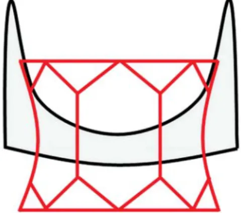
According to the experience obtained by thefirst VinV implan-ters, we suggest keeping the lower margin of the Sapien™ 2–3 mm below the radiopaque margin of the ring (Fig. 2D). Using this stratagem, the lateral shape of the Sapien™ remains rec-tangular or, at least, with the proximal diameter a bit smaller than the distal diameter (inverted trunk pyramid): in this way, valve function is preserved without risk of stenosis or malfunc-tioning. If, on the contrary, the stent-valve is positioned too low, the resulting lateral clepsydra shape can modify the Sapien™ geometry with the risk of stenosis and early degeneration. To better describe VinV stent-valve positioning, two drawings in Figs3and4explain this mechanism in standard Primount™ and Mitroflow™ valves.
Implantation
Stent-valve implantation follows, basically, the same rules of standard TAVI. Nevertheless, there is a general consensus in not performing valvuloplasty before, because of a potential risk of calcium embolization from the degenerated bioprosthesis.
RESULTS
Table2summarizes clinical and haemodynamic data from pub-lished aortic VinV series with 38 successful transapical procedures performed in 38 patients with degenerated stented bioprostheses [24–28]. During our personal clinical experience, we performed six aortic VinV procedures and, despite the limited experience, we can confirm that this technique has acceptable postoperative results. Haemodynamically, there were no leaks and the measured mean gradient was 18 mmHg. All patients were rapidly Table 1: Measured inner diameter of three commonly used aortic bioprostheses with the corresponding suggested Sapien™ size
Labelled size (mm)
Inner diameter from industry (mm)
Measured inner diameter (mm)
Suggested Sapien™ size (mm)
Sorin Biomedica Mitroflow™ 21 17.3 17 23a
23 19 19 23
25 21 20 23
27 22.9 22 23
Edwards Perimount™ Magna Ease 21 20 18 23a
23 22 21 23
25 24 22 23
27 26 24 26
St Jude Medical Trifecta™ 21 18.3 18 23a
23 20.3 20 23
25 22.1 22 23
27 24.1 24 26
aThe suggested Sapien™ size will create high transvalvular gradients because of the too small inner diameter of the bioprosthesis. This option must be
carefully considered only for inoperable patients.
Figure 2:Fluoroscopic images from a VinV case: (A) thefluoroscopic machine is positioned perpendicular to the ring of the bioprosthesis; (B) valve posi-tioning and implantation are facilitated by the presence of the ring and do not require angiographies; (C) angiographic control; (D)final result.
AD UL T C ARDIA C
extubated, they all left the intensive care unit at postoperative Day 1 and there were no relevant complications. In one case, we treated a patient with a degenerated 21 mm bioprosthesis and, as expected, we measured a high transvalvular peak gradient of 35 mmHg. The patient, an 86-year-old lady with a EuroSCORE of 51% and a porcelain aorta, was considered inoperable, and the VinV procedure was the only available option: in spite of the high gradient, she left our department without signs of cardiac decom-pensation. In another similar case with a 21 mm size bioprosth-esis, Seiffertet al. [27] also implanted a 23 mm Sapien™ valve, with a transvalvular gradient of 35 mmHg and a good outcome [27], whereas Silvaet al. [20] explanted the Sapien™ and the 21 mm Hankock bioprosthesis 1 year after VinV for progressive dys-pnoea and a mean gradient of 43 mmHg. Following these find-ings, we do not suggest aortic VinV in the 21 mm bioprosthesis.
Our clinical results are in line with the published literature and the procedural success rate is 100% in all centres, confirming that valve positioning and implantation are feasible. However, despite these good operative results, one patient at extreme sur-gical risk (EuroSCORE >80%) died within 30 days from low cardiac output, and this event confirms the high-risk profile of this subgroup of patients [27].
Concerning the valve sizing, among a total number of 38 Sapien™ valves, 36 were 23 mm and 4 were 26 mm. This trend confirms our findings during the measurement of three com-mercial bioprostheses: the 23 mm Sapien™ fits within the 21 (risk of high gradients), 23 and 25 mm tested bioprostheses, and
also into the 27 mm Sorin Mitroflow™, whereas the 26 mm Sapien™ fits into the 27 mm Perimount™ and Trifecta™.
However, in these first reports, the 26 mm Sapien™ was also employed in one 25 mm Edwards Perimount™, in one 25 mm CE Porcine and in two 25 mm Medtronic Hancock™, suggesting that the larger inner diameter of the 25 mm bioprosthesis can accept both the 23 and 26 mm stent-valves without risk of high gradients or embolization.
DISCUSSION
Results from limited transapical aortic VinV series suggest that this technique, dedicated to high-risk patients, guarantees accep-table transvalvular gradients in 23 and 25 mm degenerated bio-prostheses with absence of relevant leaks and complications. VinV in the 21 mm bioprosthesis creates high gradients and should be considered only in inoperable patients. Thus, a few topics must be underlined in order to standardize the technique and facilitate the decision-making process for the sizing.
(i) The procedure does not require a specific preoperative cardiac imaging to measure the aortic annulus because the stent-valve sizing is determined by the inner diameter of the previously implanted bioprosthesis. We have personally measured the inner diameter of three commonly used bio-prostheses, and we can say that our data do not differ from data given by the industry except for the Edwards Perimount™ Magna Ease where the given diameters are overestimated by 2 mm. Thus, a CT scan can be useful when a doubt exists about the real internal diameter of a bioprosthesis.
Figure 4:The schematic positioning of a Sapien™ stent-valve within a Sorin Biomedica Mitroflow™ aortic bioprosthesis.
Figure 3: The schematic positioning of a Sapien™ within an Edwards Perimount™ aortic bioprosthesis.
(ii) Once the inner diameter is determined, we suggest identify-ing the ideal stent-valve size thatfits into the bioprosthesis. The 23 mm Sapien™ valve seems to be the most usable size because it fits into the mostly used stented bioprostheses: the 23 mm and the 25 mm. The 21 mm size bioprosthesis can also be treated by VinV, but high gradients are expected and, then, we strongly encourage the implantation of a 23 mm or a larger bioprosthesis during standard AVRs because it will allow further VinV options.
(iii) During the procedure, the ring of the bioprosthesis is useful forfluoroscopy orientation and valve positioning. VinV does not require high doses of contrast; it may even be per-formed without angiographies and can be perper-formed in patients with chronic renal failure.
(iv) There is a consensus among expert implanters to not perform valvuloplasty before stent-valve implantation (risk of embolization).
(v) The stent-valve is implanted within the stented bioprosthesis with the lower margin 2–3 mm below the margin of the ring. This positioning guarantees correct valve functioning [28]. (vi) Concerning the durability of aortic VinV, we do not yet have
mid-/long-term results because only a few patients have a follow-up longer than 1 year.
Another concept that should be taken into consideration is the possibility, as long as big sizes are a guarantee (>23 mm), of implanting biological bioprostheses in patients younger than 65 years of age: in fact, the risk of early degeneration can be com-pensated by the absence of long-lasting anticoagulation and by VinV options. However, bigger clinical series and mid-/long-term results are necessary before changing the clinical practice.
CONCLUSION
This limited clinical experience confirms that transapical VinV procedures for degenerated stented bioprostheses do not require a specific cardiac imaging (with limited contrast injec-tions) and guarantee good results with acceptable gradients (excepting for the 21 mm bioprosthesis) and no major leaks. Concerning the sizing, the 23 mm Sapien™ seems to be the most useful stent-valve because it fits within the most widely used stented bioprostheses: the 23 mm and the 25 mm. In view of all of these facts, we recommend implanting a large bioprosthesis (equal or superior to 23 mm diameter) during standard AVRs in order to prevent size mismatch in case of VinV.
Conflict of interest: none declared.
REFERENCES
[1] Nkomo VT, Gardin JM, Skelton TN, Gottdiener JS, Scott CG, Enriquez-Sarano M. Burden of valvular heart diseases: a population-based study. Lancet 2006;368:1005–11.
[2] Lester SJ, Heilbron B, Gin K, Dodek A, Jue J. The natural history and rate of progression of aortic stenosis. Chest 1998;113:1109–14.
[3] Bonow RO, Carabello BA, Kanu C, de Leon AC Jr, Faxon DP, Freed MD et al. ACC/AHA 2006 guidelines for the management of patients with valvular heart disease: a report of the American College of Cardiology/ American Heart Association Task Force on Practice Guidelines (writing committee to revise the 1998 Guidelines for the Management of Patients with Valvular Heart Disease): developed in collaboration with the Society of Cardiovascular Anesthesiologists: endorsed by the Society for Cardiovascular Angiography and Interventions and the Society of Thoracic Surgeons. Circulation 2006;114:e84–231.
Table 2: Transapical aortic‘VinV’ procedures for degenerated stented bioprostheses: review
Authors Degenerated bioprosthesis Sapien™ size Success rate (%) Mean transvalvular gradients (mmHg)
Leaks 30-day mortality (%)
Ferrariet al. [18] 1× Mitroflow 21 mm 6× 23 mm 100 18 No 0 2× Mitroflow 23 mm
1× Mitroflow 25 mm 1× Perimount 23 mm 1× Perimount 25 mm
Marotoet al. [24] 2× Hancock 25 mm. 2× 23 mm 100 15 2× Grade 1 0 Webbet al. [25] 1× Perimount 21 mm 7× 23 mm 100 20 2× Grade 1 0
3× Perimount 23 mm 1× 26 mm 2× Perimount 25 mm
1× Mosaic 21 mm 1× Ionescu Shiley 21 mm
Pasicet al. [26] 2× Hancock 21 mm 9× 23 mm 100 19 No 0 3× Hancock 23 mm
1× Mosaic 23 mm 2× Perimount 21 mm 1× Perimount 23 mm
Seiffertet al. [27] 1× Biocor 23 mm 4× 23 mm 100 19 No 25 (one extreme-risk patient died from low cardiac output) 1× Hancock 21 mm
1× Hancock 23 mm 1× Hancock 25 mm
Kempfertet al. [28] 3× Perimount 21 mm 6× 23 mm 100 11 2× Grade 1 0 2× Hancock 25 mm 3× 26 mm 1× Mosaic 21 mm 1× Epic 21 mm 1× Mitroflow 23 mm 1× CE porcine 25 mm AD UL T C ARDIA C
[4] Ferrari E, Tozzi P, Hurni M, Ruchat P, Stumpe F, von Segesser LK. Primary isolated aortic valve surgery in octogenarians. Eur J Cardiothorac Surg 2010;38:128–33.
[5] McClure RS, Narayanasamy N, Wiegerinck E, Lipsitz S, Maloney A, Byrne JG et al. Late outcomes for aortic valve replacement with the Carpentier–Edwards pericardial bioprosthesis: up to 17-year follow-up in 1,000 patients. Ann Thorac Surg 2010;89:1410–6.
[6] David TE, Armstrong S, Maganti M. Hancock II bioprosthesis for aortic valve replacement: the gold standard of bioprosthetic valves durability? Ann Thorac Surg 2010;90:775–81.
[7] Chan V, Kulik A, Tran A, Hendry P, Masters R, Mesana TG et al. Long-term clinical and hemodynamic performance of the Hancock II versus the Perimount aortic bioprostheses. Circulation 2010;122:S10–6. [8] ISTHMUS Investigators. The Italian study on the Mitroflow postoperative
results (ISTHMUS): a 20-year, multicentre evaluation of Mitroflow peri-cardial bioprosthesis. Eur J Cardiothorac Surg 2011;39:18–26.
[9] Chikwe J, Filsoufi F, Carpentier AF. Prosthetic valve selection for middle-aged patients with aortic stenosis. Nat Rev Cardiol 2010;7:711–9. [10] Jaussaud N, Gariboldi V, Giorgi R, Grisoli D, Chalvignac V, Thuny Fet al. Risk of reoperation for aortic bioprosthesis dysfunction. J Heart Valve Dis 2009;18:256–61.
[11] Leontyev S, Borger MA, Davierwala P, Walther T, Lehmann S, Kempfert J et al. Redo aortic valve surgery: early and late outcomes. Ann Thorac Surg 2011;91:1120–6.
[12] Christiansen S, Schmid M, Autschbach R. Perioperative risk of redo aortic valve replacement. Ann Thorac Cardiovasc Surg 2009;15:105–10. [13] Caus T, Albertini JN, Chi Y, Collart F, Monties JR, Mesana T. Multiple
valve replacement increases the risk of reoperation for structural degeneration of bioprostheses. J Heart Valve Dis 1999;8:376–83. [14] Davierwala PM, Borger MA, David TE, Rao V, Maganti M, Yau TM.
Reoperation is not an independent predictor of mortality during aortic valve surgery. J Thorac Cardiovasc Surg 2006;131:329–35.
[15] Drews T, Pasic M, Buz S, Unbehaun A, Dreysse S, Kukucka M et al. Transapical aortic valve implantation after previous heart surgery. Eur J Cardiothorac Surg 2011;39:625–30.
[16] Walther T, Kempfert J, Borger MA, Fassl J, Falk V, Blumenstein Jet al. Human minimally invasive off-pump valve-in-a-valve implantation. Ann Thorac Surg 2008;85:1072–3.
[17] Walther T, Falk V, Dewey T, Kempfert J, Emrich F, Pfannmüller Bet al. Valve-in-a-valve concept for transcatheter minimally invasive repeat xenograft implantation. J Am Coll Cardiol 2007;50:56–60.
[18] Ferrari E, Marcucci C, Sulzer C, von Segesser LK. Which available transa-pical transcatheter valve fits into degenerated aortic bioprostheses? Interact Cardiovasc Thorac Surg 2010;11:83–5.
[19] Azadani AN, Jaussaud N, Matthews PB, Chuter TA, Ge L, Guy TSet al. Aortic valve-in-valve implantation: impact of transcatheter–bioprosthesis size mismatch. J Heart Valve Dis 2009;18:367–73.
[20] Silva D, Stripling JH, Hansen L, Riess FC. Aortic valve replacement after transapical valve-in-valve implantation. Ann Thorac Surg 2011;91: e5–7.
[21] Vahanian A, Alfieri OR, Al-Attar N, Antunes MJ, Bax J, Cormier B et al. Transcatheter valve implantation for patients with aortic stenosis: a pos-ition statement from the European Association of Cardio-Thoracic Surgery (EACTS) and the European Society of Cardiology (ESC), in collab-oration with the European Association of Percutaneous Cardiovascular Interventions (EAPCI). Eur J Cardiothorac Surg 2008;34:1–8.
[22] Malaisrie CS, Tuday E, Lapin B, Wang E, Lee R, McGee EC et al. Transcatheter aortic valve implantation decreases the rate of unoperated aortic stenosis. Eur J Cardiothorac Surg 2011;40:43–8.
[23] Van Linden A, Kempfert J, Rastan AJ, Holzhey D, Blumenstein J, Schuler G et al. Risk of acute kidney injury after minimally invasive transapical aortic valve implantation in 270 patients. Eur J Cardiothorac Surg 2011; 39:835–42.
[24] Maroto LC, Rodríguez JE, Cobiella J, Marcos P. Transapical off-pump aortic valve-in-a-valve implantation in two elderly patients with a degenerated porcine bioprosthesis. Eur J Cardiothorac Surg 2010;37: 738–40.
[25] Webb JG, Wood DA, Ye J, Gurvitch R, Masson JB, Rodés-Cabau Jet al. Transcatheter valve-in-valve implantation for failed bioprosthetic heart valves. Circulation 2010;121:1848–57.
[26] Pasic M, Unbehaun A, Dreysse S, Buz S, Drews T, Kukucka M et al. Transapical aortic valve implantation after previous aortic valve replace-ment: clinical proof of the‘valve-in-valve’ concept. J Thorac Cardiovasc Surg 2011;142:270–7.
[27] Seiffert M, Franzen O, Conradi L, Baldus S, Schirmer J, Meinertz Tet al. Series of transcatheter valve-in-valve implantations in high-risk patients with degenerated bioprostheses in aortic and mitral position. Catheter Cardiovasc Interv 2010;76:608–15.
[28] Kempfert J, Van Linden A, Linke A, Borger MA, Rastan A, Mukherjee C et al. Transapical off-pump valve-in-valve implantation in patients with degenerated aortic xenografts. Ann Thorac Surg 2010;89: 1934–41.

![Table 2 summarizes clinical and haemodynamic data from pub- pub-lished aortic VinV series with 38 successful transapical procedures performed in 38 patients with degenerated stented bioprostheses [24 – 28]](https://thumb-eu.123doks.com/thumbv2/123doknet/14927642.664277/3.918.86.837.135.349/summarizes-haemodynamic-successful-transapical-procedures-performed-degenerated-bioprostheses.webp)