CT-guided methylene-blue labelling before thoracoscopic
resection of pulmonary nodules
Riccardo E. Vandoni
a, Jean-Franc¸ois Cuttat
a, Ste´phane Wicky
b, Michel Suter
a ,*
aDepartment of Surgery, Centre Hospitalier Universitaire Vaudois, 1011 Lausanne, Switzerland b
Department of Radiology, Centre Hospitalier Universitaire Vaudois, Lausanne, Switzerland Received 13 October 1997; revised version received 5 April 1998; accepted 12 May 1998
Abstract
Objective: Evaluation of the efficiency of our technique of methylene-blue labelling of pulmonary nodules to facilitate thoracoscopic
recognition and excision. Design: Patients with a peripheral pulmonary nodule smaller than 2.5 cm and not in contact with the visceral pleura were included. Under tomodensitometric guidance, the nodules were labelled with methylene-blue within hours before thoracoscopic wedge resection. If frozen section revealed a primary bronchial carcinoma, thoracotomy and classical resection were performed during the same anesthesia. Results: Between July 1992 and August 1996, 54 nodules were removed in 51 patients. Labelling was performed between 75 and 270 min before surgery and was complicated in 13 patients (25.4%) by a small pneumothorax without any clinical consequence. Labelling allowed successful thoracoscopic recognition of 50 nodules (92%) and thoracoscopic wedge resection was possible in all but one cases (91%). Five patients (9%) required thoracotomy. Histology showed a benign lesion in 22 cases, a primary lung carcinoma in 17 and a metastases in 15. Twenty of the 22 benign nodules (91%) were removed without thoracotomy. According to the protocol, 13 patients with a primary lung tumour underwent lobectomy during the same session. There was no mortality nor morbidity amongst patients who had thoracoscopy only. Conclusions: Our technique of labelling peripheral pulmonary nodules with methylene-blue is very effective and is not associated with any relevant complication. Thoracoscopic excision and diagnosis is possible in more than 90% of the cases. We therefore recommend this simple, low-cost and reliable technique for nodules not in contact with the visceral pleura before thoracoscopic wedge resection.1998 Elsevier Science B.V. All rights reserved
Keywords: Thoracoscopy; Pulmonary nodule; Methylene-blue
1. Introduction
A solitary pulmonary nodule is defined as a single intra-pulmonary spherical lesion, surrounded by normal lung tis-sue, without accompanying enlarged lymph node [1]. It is not uncommon, as radiographic surveys of adults show a nodule in 0.1-0.2% of cases. About 150 000 new nodules are diagnosed every year in the US. Up to 40% of these nodules are malignant, making their removal for diagnostic and ther-apeutic purposes of first importance [1,2].
Bronchoscopic or transthoracic biopsy do not achieve a high diagnostic yield, especially when no malignant cells are found [1–5]. In the past years, thoracoscopic resection has demonstrated a much higher specificity and sensibility,
the challenge being the localization of the nodule at the time of operation [2–9]. Nodules that are very superficial or in contact with the visceral pleura are seen easily but, because thoracoscopic vision is two-dimensional and because palpa-tion is limited to only one finger and to a limited area, deeper nodules in the outer third of the lung parenchyma may be challenging to identify, especially if their size is small. The aim of this study is to present our results with a method using CT-guided diluted methylene-blue labelling of the nodules before thoracoscopic resection.
A solitary pulmonary nodule must be considered malig-nant unless proven otherwise. Bronchoscopy is very often negative in patients with peripheral nodules. Transthoracic CT-guided needle biopsy is helpful if malignant cells are shown, but is able to specifically diagnose only 15% of all benign lesions [1,2,5,8,10]. All the malignant lesions must be removed, and 85% of the benign lesions need resection
1010-7940/98/$19.00 1998 Elsevier Science B.V. All rights reserved P I I S 1 0 1 0 - 7 9 4 0 ( 9 8 ) 0 0 1 6 0 - 2
* Corresponding author. Tel.: +41 021 3142351; fax: +41 024 4688623; e-mail: [email protected]
before a definitive diagnosis can be made. It is therefore our policy, in the absence of suspect mediastinal lymphadeno-pathy (mediastinal node.1 cm on CT), to perform thoraco-scopic resection of peripheral pulmonary nodules of less than 2.5 cm in diameter as measured on CT, and located in the outer third of the lung parenchyma, as an initial diag-nostic measure unless there is a contraindication.
2. Material and methods
The patients whose nodule was in contact with the visc-eral pleura were operated on directly. The patients with a peripheral pulmonary nodule not in direct contact with the visceral pleura were included in this study. Bronchoscopy or CT-guided needle biopsy were not performed routinely. Patients with severe chronic obstructive pulmonary disease, or coagulation disorders were excluded. In patients with a peripheral lung nodule and mediastinal lymphadenopathy, tissue diagnosis was obtained via mediastinoscopy. Patients
with a suspected metastasis from a not locally controlled primary tumour were also excluded. Although there is still controversy about the best approach for resection of lung metastases, we elected thoracoscopy for patients with a solitary peripheral nodule and a history of malignancy if complete CT with 3-mm slices showed only one nodule. In this setting, a new pulmonary nodule is not necessarily a metastasis, and tissue diagnosis must be obtained as for the remaining cases.
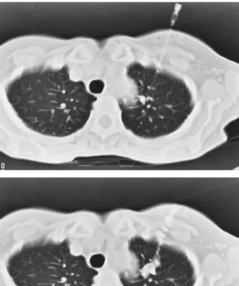
After CT identification, a 25 G needle was introduced into the nodule percutaneously. The puncture point was chosen so that the distance between the nodule and the skin was as short as possible, and considering the location of the nodule in relation to the ribs and scapula. After CT control of the position of the needle tip (Fig. 1A), a solution of 0.7 ml of methylene-blue and 0.3 ml of non-ionic contrast mean was injected into the nodule and along the needle tract (lung, pleura and chest wall) during withdrawal of the nee-dle. In this way, an external (skin) and internal (pleura) mark would be present at the time of thoracoscopy.
Com-Fig. 1. O.D., 37 years. History of renal cell carcinoma with left nephrectomy 5 years ago. CT-guided labelling of a single peripheral pulmonary nodule in the left upper lobe. (A) CT control after placement of the needle into the nodule. (B) Control after injection of the solution of methylene-blue with contrast. Histology after successful thoracoscopic removal confirmed the metastatic nature of the nodule.
puterized tomodensitometry control of the labelling was performed with high resolution thin cuts of the thorax in expirium (Fig. 1B). Presence or absence of pneumothorax was noted at that time. Delay between labelling and thora-coscopy was kept as short as possible.
Patients were then transferred to the operating theatre and were given general anaesthesia with a double-lumen endo-tracheal tube and placed in the lateral decubitus position. The first 10-mm trocar was inserted along the mid-axillary line once access to the pleural cavity had been obtained by finger dissection, at a level depending on the location of the nodule. The mid-axillary line was chosen for the optic to allow for an optimal working angle between the two other ports. A 7-mm trocar was used to introduce a Babcock for-ceps and a 12-mm trocar was used for the introduction of the endostapler (Endo-GIA,USSC). Whenever possible, these ports were placed after identification of the nodule and in relation to its position. Methylene-blue stain was identified on the parietal and visceral pleura (Fig. 2). The nodule was then localized and resected with the endostapler. The depth of the wedge - the inclination of the stapler - was estimated based on the depth of the nodule on CT, in order to include a
safety margin of between 5 and 10 mm. Once the wedge resection was completed, the nodule was placed in a plastic bag to avoid contact with the chest wall, and removed from the thoracic cavity through the largest port hole. After care-ful macroscopic examination of the resected specimen for completeness of the excision, the nodule was sent for frozen section examination. If primary bronchial carcinoma was diagnosed, open lobectomy was performed immediately unless there was a contraindication. We did not perform video-assisted thoracoscopic lobectomy in this series. If the nodule could not be found at thoracoscopy, a small thoracotomy was performed.
Statistical analysis was performed using the Student t-test or the x2-test with Yates correction as appropriate.
3. Results
Among 132 patients referred to our department with a solitary pulmonary nodule between July 1992 and August 1996, 51 were included in this series with a total of 54 nodules (48 unilateral and three bilateral) labelled and
Fig. 2. J.H., 54 years. Thoracoscopic view before removal of the lesion: a blue spot is clearly visible on the parietal as well as on the visceral pleura. Histology showed a hamartoma.
removed. The remaining patients had either thoracoscopic resection without labelling when the nodule was in contact with the visceral pleura, or thoracotomy when the nodule was too deeply located. There were 36 males and 15 females, with an age ranging from 27 to 78 years (mean 53.4 years). The nodules were localized as shown in Table 1. Bronchoscopy was performed in 24 of these patients and was negative in all cases. Only six patients had preoperative negative CT-guided needle biopsy before referral. In none of the patients was the diagnosis made preoperatively, although in 15 who had had resection of a primary malig-nancy, lung metastasis was suspected. The distance from the centre of the nodule to the nearest pleural surface ranged from 12 to 45 mm (mean 18 mm) as measured on the CT images.
Labelling of the nodule was possible in all the cases, with a positive CT control of nodule and needle tract injection. We did not observe any major complication, but two patients had cough during injection and 13 developed a small and asymptomatic pneumothorax. Delay between labelling and operation ranged from 75 to 270 min (mean 162 min). Unilateral thoracoscopy lasted between 30 and 150 min (mean 51 min) and the three bilateral procedures between 120 and 180 min.
Thoracoscopic identification and complete wedge resec-tion of the nodule was successful for 49 nodules. The labelled area was overlying the nodule in all cases, and its size did not depend on the size of the lesion, as the same amount of contrast and methylene-blue was injected in all cases. It never exceeded 3 cm on the visceral pleura. We did not notice that time elapsed between labelling and thoraco-scopy affected the size of the labelled area, but it did affect the density of the coloration. The resection was complete with a tissue margin of at least 5 mm in all cases. In four patients, no blue coloration was visible. In three of these, the long delay between labelling and operation (270, 260 and 190 min) was probably responsible for failure, which we consider to be due to diffusion of methylene-blue. In the remaining case, delay was short (115 min) and failure could not be explained. As dye was still present in the skin, we hypothesized that most of the dye was injected in the thor-acic wall. In one case the coloration was easily recognized but the nodule was too deep (45 mm) for a thoracoscopic wedge resection. Overall, thoracoscopic resection failed for any reason, and thoracotomy was necessary for wedge resection in five patients. In the remaining patients, all of
whom had successful thoracoscopic wedge resection, 11 had thoracotomy and open lobectomy after frozen section showed primary lung cancer. Two of the five patients who were primarily converted also underwent lobectomy for the same reason. Lobectomy, therefore, was performed in a total of 13 patients.
For those patients in whom only thoracoscopic wedge resection of the nodule was performed, the mean duration of chest tube drainage was 2.8 days (1-11) and the mean post-operative stay was 5.2 days (1-13).
Table 2 displays the detailed nature of all the nodules. Twenty-two of them (40%) were benign. Fifteen of the 32 malignant lesions were metastases and 17 primary lung tumours. As stated, and despite negative margins on frozen section, 13 patients with the latter underwent lobectomy and lymph nodes dissection through formal thoracotomy during the same anesthesia. This is in accordance to the rule that lobectomy achieves a higher rate of local control than wedge resection for T1 bronchial carcinoma. According to the TNM classification of lung cancer, ten patients were staged as T1NO after lobectomy, and three as T1N1. No patient had N2 disease. In three patients, lobectomy was not performed because of a poor respiratory function. The last patient, with a diagnosis of lymphoma, underwent intrave-nous chemotherapy. Overall, thoracotomy was obviated in 20 out of 22 patients with benign disease, in the 15 patients with a single metastases, in one patient with lymphoma, and in three patients with lung cancer who could not tolerate lobectomy.
Analysis showed no statistically significant relation between failure of thoracoscopic localization and the size of the nodule, its depth in relation to the visceral pleura and the delay between labelling and thoracoscopy. There was, Table 1
Localization of the nodules
Upper right lobe 14
Lower left lobe 14
Lower right lobe 12
Upper left lobe 8
Lingula 3
Middle right lobe 3
Table 2
Histopathological diagnosis
Benign nodule 22
Nodule without tumour 9
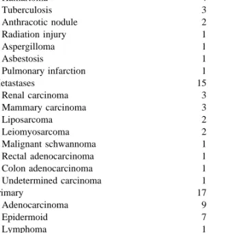
Hamartoma 4 Tuberculosis 3 Anthracotic nodule 2 Radiation injury 1 Aspergilloma 1 Asbestosis 1 Pulmonary infarction 1 Metastases 15 Renal carcinoma 3 Mammary carcinoma 3 Liposarcoma 2 Leiomyosarcoma 2 Malignant schwannoma 1 Rectal adenocarcinoma 1 Colon adenocarcinoma 1 Undetermined carcinoma 1 Primary 17 Adenocarcinoma 9 Epidermoid 7 Lymphoma 1
nevertheless, a trend for a greater depth (P=0.24) and a longer delay (P=0.20) in patients with failed thoracoscopic resection.
Three of our patients died 27, 50, and 90 days after thor-acoscopy, of causes unrelated to the procedure (heart infarc-tion, end-stage liver failure due to cirrhosis, and end-stage metastatic carcinoma with bone metastases, but without recurrence in the lungs. The 30 days mortality was 1.9% and the in-hospital mortality 5.8%.
4. Discussion
Thoracoscopic resection of pulmonary nodules has been performed widely in the recent years. Many authors con-sider this approach as the first choice for peripheral unde-termined pulmonary nodules because the sensitivity of other diagnostic tools is relatively poor. At bronchoscopy, the lesion is virtually never seen. Lavage for cytology, however, is sometimes useful. For small nodules needle biopsy or fine needle aspiration do not achieve a diagnosis in more than 50-70% of the cases. A malignant lesion cannot be ruled out because no tumour cell is found at cytology, unless a spe-cific benign diagnosis, e.g. tuberculoma, can be made. This is possible in only about 15% of the cases [1,2,5,8,10].
Pulmonary nodules are frequently subpleural and there-fore easy to be recognized at thoracoscopy. Deeper lesions in the outer third of the parenchyma are more difficult to detect, because no direct finger palpation is usually possible. Non palpable nodules in the breast or microcalification foci are located with a hook before biopsy. In a similar manner, peripheral pulmonary nodules can be labelled preopera-tively to facilitate their recognition and thereby their resec-tion when thoracoscopy is performed.
Several techniques of localization of deep pulmonary nodules have been described alone or in association, includ-ing the use of a metallic hook wire such as those used for nodules in the breast, peroperative endoechography and methylene-blue staining under fluoroscopic guidance [3,5–9]. Peroperative finger palpation through one of the trocar holes is possible, although limited to the underlying parenchyma and provided the lung parenchyma surrounding the nodule is normal [2].
Insertion of a guide wire into the nodule has been exten-sively used for localization of breast cancers. Since displa-cement of the hook during reposition of the patient in the operating room or during lung deflation is not infrequent, this technique has been associated to methylene-blue stain-ing of the lung surface [3,7]. Complications consist mainly in pneumothorax and haemorrhage [3]. The presence of gas may limit the use of echography. Atelectatic or deflated lung, however, has a consistence similar to that of liver, so that echographic images are interpretable. Not only can tumour margins be defined, but also its relations to vessels and bronchioles [3,8].
Although we have no experience with the aforementioned
methods, we believe that our technique is very effective for the localization of pulmonary nodules. It is easy to perform and minimally invasive, as no foreign body is left in the patient. A good collaboration between the radiologist and the surgeon is necessary and a high-quality CT-scanner must be located nearby the operating theatre. Provided the time between labelling and operation is kept as short as possible, ideally below 120-150 min, the blue coloration of either the parietal or visceral pleura is clearly visible. A technique using a solution of methylene-blue, contrast mean and collagen has been described, which should allow the stain to remain visible for several hours [11]. The camera should be placed in order to allow a good triangulation between the two other ports, which should be inserted after identification of the nodule if possible. One port placed through the marked skin overlying the nodule can be help-ful, because the port hole can be used for finger palpation. In most cases, however, intrathoracic handling of the lung and instruments is facilitated when the ports are placed a few centimetres away from the working area.
In our series only four nodules (7.4%) could not be detected because of lack of coloration. Delay between label-ling and operation was particularly long (270, 260, 190 min) in three cases. We do not have any explanation for the last one, in which CT control after labelling showed the same nice image as that shown on Fig. 1. As the skin was still coloured when thoracoscopy was performed, we assumed that most of the dye had been injected into the thoracic wall. We could not find any data in the literature regarding diffu-sion of methylene-blue after intraparenchymal injection in the lung. In the remaining cases (92.6%) labelling lead to positive identification. We did not observe any side effect related to the mixture injected, nor did we notice any major complication. Thirteen patients developed a small but totally asymptomatic pneumothorax.
Overall thoracoscopic excision of peripheral pulmonary nodules after methylene-blue labelling under CT-guidance avoided thoracotomy in 35 patients (64%) and more speci-fically in 20 of the 22 patients (91%) who had a benign nodule. It must be emphasized, however, that the nodule must strictly be located in the outer third of the lung par-enchyma on CT. In one of our patients with failed thoraco-scopic resection, part of the nodule was in the outer third, but its centre was 45 mm deep, and the stapler could not be closed because the parenchyma was too thick. Wedge resec-tion was performed at thoracotomy with a GIA-90 stapler. The emergence of new staplers with a wider opening could be helpful in such a situation.
Frozen-section histological examination allows the diag-nosis to be established immediately during the operation. This is important, because definitive treatment including lobectomy and lymph-nodes dissection can then be done during the same anaesthesia, in a manner similar to that used for breast cancer surgery. The fact that three patients eventually had pathological T1N1 stage is in accordance with the results after thoracotomy: positive N1 nodes
some-times are not suspected on the basis of the preoperative CT-scan.
Thoracoscopic resection of lung metastases is still a mat-ter of controversy, and no study has clearly demonstrated one method (unilateral thoracotomy, bilateral thoracotomy, thoracoscopy, sternotomy) to be superior to the other, nor that resection of metastases that are not visualized on CT prolongs survival. Many thoracic surgeons assume that thor-acotomy is mandatory in the setting of pulmonary metas-tases, because it allows palpation of the entire lung and resection of small metastases undetected by CT. On the other hand, a new solitary pulmonary nodule in a patient with a history of malignancy is not necessarily a metastasis. In such a situation, we usually obtain a tissue diagnosis. If the lesion is confirmed to be a secondary lesion, and the resection margins are 5 mm or more, follow-up of the patient with sequential CT-scans is undertaken, and thora-cotomy is performed only if new lesions appear. If the resection margins are too small, we convert to thoracotomy. It is true that palpation can disclose a higher number of metastasis that is expected according to the CT-scan. Whether resection of occult metastases affects survival, however, is still a matter of debate, which is beyond the scope of this paper in which we focus on a labelling tech-nique. We consider that close follow-up and elective reo-peration after resection of a single metastasis is acceptable. Among our patients, only two developed isolated recurrent metastases in the controlateral lung. Four developed recur-rent multiple lung metastases, but also synchronous extra-pulmonary lesions. Three developed only extraextra-pulmonary secondaries. Four were lost from follow-up, and two are free of disease after 18 and 30 months. Indication for thoraco-scopic resection of lung metastases should nevertheless be careful and limited to patients with a small solitary periph-eral lesion, as in all our patients.
5. Conclusions
For peripheral pulmonary nodules not in close relation
with the visceral pleura for which thoracoscopic resection is foreseen, we recommend preoperative labelling with diluted methylene-blue under CT-guidance to outline the lesion and facilitate its recognition during thoracoscopy. Side effects are minimal and success is the rule provided the time between labelling and operation is kept within a maximum of 3 h.
References
[1] Shulkin AN. Management of the indeterminate solitary pulmonary nodule: a pulmonologist’s view. Ann Thorac Surg 1993;56:743– 744.
[2] Mack MJ, Hazelrigg SR, Landreneau RJ, Acuff TE. Thoracoscopy for the diagnosis of the inderteminate solitary pulmonary nodule. Ann Thorac Surg 1993;56:825–832.
[3] Shennib H. Intraoperative techniques for pulmonary nodules. Ann Thorac Surg 1993;56:745–748.
[4] Wagner RB. Percutaneous thransthoracic needle biopsy of the lung. Letter to the editor. Ann Thorac Surg 1995;60:233.
[5] Wassmer FA, Cuttat JF. Inte´reˆt de la thoracoscopie diagnostique et the´rapeutique. Me´d Hyg 1993;51:2074–2077.
[6] Kerrigan DC, Spence PA, Crittenden MD, Tripp MD. Methylene-blue guidance for simplified resection of a lung lesion. Ann Thorac Surg 1992;53:163–164.
[7] Mack MJ, Gordon MJ, Postma TW, Berger MS, Aronoff RJ, Acuff TE, Ryan WH. Percutaneous localization of pulmonary nodules for thoracoscopic lung resection. Ann Thorac Surg 1992;53:1123– 1124.
[8] Shennib H, Bret P. Intraoperative transthoracic ultrasonographic localization of occult lung lesions. Ann Thorac Surg 1993;55:767– 769.
[9] Wicky S, Mayor B, Cuttat JF, Schnyder P. CT-guided localizations of pulmonary nodules with methylene-blue injections for thoraco-scopic resections. Chest 1994;106:1326–1328.
[10] Dowling RD, Ferson PF, Landreneau RJ. Thorascopic resection of pulmonary metastases. Chest 1992;102:1450–1454.
[11] Nomori H, Horio H. Colored collagen is a long-lasting point marker for small pulmonary nodules in thoracoscopic operations. Ann Thorac Surg 1996;61:1070–1073.