© The Author [2013]. Published by Oxford University Press [on behalf of the Society for Experimental Biology]. All rights reserved. For permissions, please email: [email protected]
ReseaRch papeR
Pathogen and Circadian Controlled 1 (PCC1) regulates polar
lipid content, ABA-related responses, and pathogen defence
in Arabidopsis thaliana
Ricardo Mir1, M. Luisa Hernández2, Eliane Abou-Mansour3, José Manuel Martínez-Rivas2, Félix Mauch3,
Jean-Pierre Métraux3 and José León1,*
1 Instituto de Biología Molecular y Celular de Plantas (CSIC-Universidad Politécnica de Valencia), Ciudad Politécnica de la Innovación, Edificio 8E, Avda. Ingeniero Fausto Elio s/n, 46022 Valencia, Spain
2 Instituto de la Grasa (CSIC), Avda. Padre García Tejero 4, 41012-Sevilla, Spain 3 Department of Biology, University of Fribourg, Fribourg, Switzerland
*To whom correspondence should be addressed. E-mail: [email protected]
Received 17 April 2013; Revised 17 May 2013; Accepted 20 May 2013
Abstract
Pathogen and Circadian Controlled 1 (PCC1) was previously characterized as a regulator of defence against patho-gens and stress-activated transition to flowering. Plants expressing an RNA interference construct for the PCC1 gene (iPCC1 plants) showed a pleiotropic phenotype. They were hypersensitive to abscisic acid (ABA) as shown by reduced germination potential and seedling establishment, as well as reduced stomatal aperture and main root length in ABA-supplemented media. In addition, iPCC1 plants displayed alterations in polar lipid contents and their corresponding fatty acids. Importantly, a significant reduction in the content of phosphatidylinositol (PI) was observed in iPCC1 leaves when compared with wild-type plants. A trend in reduced levels of 18:0 and increased levels of 18:2 and particularly 18:3 was also detected in several classes of polar lipids. The enhanced ABA-mediated responses and the reduced content of PI might be responsible for iPCC1 plants displaying a complex pattern of defence against pathogens of different lifestyles. iPCC1 plants were more susceptible to the hemi-biotrophic oomycete pathogen Phytophthora
brassicae and more resistant to the necrotrophic fungal pathogen Botrytis cinerea compared with wild-type plants.
Key words: ABA, Arabidopsis, defence, lipids, transcriptome.
Introduction
Plant growth is controlled by endogenous factors as well as through diverse responses to environmental conditions or to biotic interactions. Under photoperiodic growing conditions, the circadian clock machinery controls numerous processes including leaf movement, daily vegetative growth, seed dor-mancy break, transition to flowering, flower bud opening, stomatal aperture, and sensitivity to light and stress environ-mental cues (Adams and Carré, 2011). Although light control exerted on those processes shares some common components, significant changes in the respective mechanisms have evolved in each case (Jackson, 2009). For instance, increasing recent evidence suggests that many responses to stress are actually
controlled by the circadian clock, including phytopathogenic interactions (Roden and Ingle, 2009; W. Wang et al., 2011), abiotic stress (Sánchez et al., 2011), and wounding (Morker and Roberts, 2011). Moreover, regulation by the circadian clock has also been linked to basal defence against pathogens through salicylic acid (SA)- and phosphate-related responses (G.Y. Wang et al., 2011). PCC1 was originally identified as a pathogen-responsive gene with a circadian clock-regulated pattern of expression (Sauerbrunn and Schlaich, 2004). PCC1 was further characterized as a potential SA-induced activa-tor of flowering under UV light-stressed conditions (Segarra et al., 2010). Moreover, RNA interference (RNAi) transgenic
lines with strong down-regulation of PCC1 gene expression (iPCC1 lines) displayed a late flowering phenotype under non-stressed long-day conditions (Segarra et al., 2010). PCC1 codes for a small 81 amino acid protein. Its function and mode of action have remained elusive despite its potential involve-ment in SA-activated defence responses to pathogens and activation of flowering. PCC1 has a cysteine-rich C-terminus (CYSTM) domain proposed to function as a potential site for membrane anchoring and dimerization (Venancio and Aravind, 2010). Here it is reported that the reduced expres-sion of the PCC1 gene correlates with the appearance of sev-eral phenotypes in seeds and leaves, including abscisic acid (ABA)-regulated processes such as seed germination, seedling establishment, and stomatal closure, as well as alterations in the content and composition of polar lipids. Concomitantly, a complex pattern of defence responses to different pathogens was observed in plants with strongly reduced PCC1 expres-sion, suggesting that PCC1 plays important regulatory roles in controlling metabolic and hormonal responses as well as the execution of defence strategies.
Materials and methods
Plant materials and growing conditionsTransgenic lines expressing RNAi constructs for the PCC1 gene were previously described (Segarra et al., 2010). Seeds were surface sterilized with 30% bleach and 0.01% Tween-20, washed extensively with miliQ sterile water, and sown in Murashige and Skoog (MS) medium supplemented with 0.8% agar and 1% sucrose. After 3 d of stratification at 4 ºC on MS-containing Petri dishes, seeds were transferred to a growth chamber under white fluorescent light (flu-ence rate of 70 μmol m–2 s–1) with a 16 h light/8 h dark photoperiod and a controlled temperature of 19–23 ºC. Alternatively, stratified seeds were sown on soil and cultivated in environmentally controlled growth chambers under long days (16 h light/8 h dark) or short days (8 h light/16 h dark), as indicated. Plants were grown under a 12 h light/12 h dark photoperiod for pathogen-related experiments.
Generation of pPCC1::GUS transgenic lines
A 1.1 kb fragment of the genomic sequence upstream of the initia-tion codon of the PCC1 locus was fused to the β-glucuronidase (GUS) gene using the pMDC162a vector. Primary transformants and further transgenic generation individuals were selected in hygro-mycin-supplemented MS medium. Three independent homozygous lines were isolated and further used to test PCC1 expression by GUS staining with X-Gluc in the presence of 5 mM ferricyanide/ferrocya-nide redox buffer.
Genome-wide microarray and quantitative real-time PCR (qRT-PCR) analysis
The transcriptomes of iPCC1 lines were compared with that of wild-type seedlings (Col-0 accession) grown in vitro under long-day (16 h light/8 h darkness) conditions. Samples were harvested 12 days after sowing (DAS) at 12 h after dawn. Total RNAs from wild-type and iPCC1 plants were extracted with Trizol and purified with an RNeasy kit (QIAGEN). RNAs (three independent biological rep-licates per genotype) were checked by qRT-PCR for endogenous PCC1 transcripts and their integrity and purity were further checked by nanocapillary electrophoresis using a Bioanalyzer Agilent 2100. Labelling, hybridization protocols, and statistical analysis are included in a detailed MIAME rules-based description of the
microarray experiments in Supplementary Table S1 available at JXB online.
To quantify the transcript levels, total RNAs were isolated from wild-type and iPCC1 seedlings and further analysed by qRT-PCR techniques as described previously (Lozano-Juste and León, 2011). Primers used for qPCR were as follows: qMYB28-F, TCCCTGACAAATACTCTTGCT; qMYB28-R, CATTGTGGTT ATCTCCTCCGA; qNPR1-F, GATTCGGTTGTGACTGTTTTG; qNPR1-R, TCTCGTTTGTCTTCTTGCTCT; qHRS1-F, TCCGAG GACAAGAACACGAAA; qHRS1-R, TGTCATCGTCTCCTGCT GCAA; qEDS1-F, CGAAGACACAGGGCCGTA; qEDS1-R, AA GCATGATCCGCACTCG; qAOP1-F, TCGAGTTGCCTATTCC GACCAAAC; qAOPI1-2R, TGCTCCGAAAACAGGTGTATCGT CT; qPIF1-F, GTTGCTTTCGAAGGCGGTT; qPIF1-R, GCGC TAGGACTTACCTGCGT; qRGL2-F, GACGGCGCGTAGAGTT CAC; qRGL2-R, TGCATCCCTTGATTAAGCCC; qIRT3-F, GCCC TCACAACCCCGATAG; qIRT3-R, GCTCCGACACTGTGAG AATTGA; qGSTU24-F, CTACTTTGTTATGTTGATCTGTTGTT GC; qGSTU24-R, CATAGACCTCAAAGAAAATAGAACAAA GC; qAt1g44740-F, TCGGCTGGCCAAAAGAAATA; qAt1g447 40-R, TCGACCTCTCCTTGGCCTAG; qRPP4-F, GAAGGCAC TCAAGGCCTCATTTAC; qRPP4-R, GACAATAATCCCACCAT AGCCTTT; qADH1-F, TACCACCGGACAGATTATTCGATGC; qADH1-R, TGGCCGAAGATACGTGGAAACAA; qRAP2.1-F, TCGGGTTTTATTACCGGCGGAGTG; and qRAP2.1-R, TAAGGGAAAAAGCGGCGGAGGT.
In silico analysis of gene ontology and promoter motifs
AgriGO Gene Ontology (GO) tool platforms were used to find significant enrichment of terms for molecular function, biological processes, and cell compartment categories, and the results were con-firmed by similar searches in FuncAssociate, ProfCom, and BioMaps. The promoter sequences of the genes differentially expressed in iPCC1 plants were analysed by searching for non-biased over-rep-resented 8-mer motifs using a combination of Bioprospector, DME, MDScan, MEME, and Motif Sampler databases.
Germination assays
To test ABA sensitivity, seeds were sown in MS medium with 1% (w/v) sucrose, 0.8% (w/v) agar, and increasing concentrations of (±)- cis,trans-ABA (Sigma) after 3 d of stratification at 4 ºC. Germination was scored as the percentage of seeds with visible radical emergence every 24 h. Seedling establishment was assessed by quantifying seed-lings with green expanded cotyledons at 15 DAS. To test the effect of osmotic stress, seeds were sown in medium supplemented with 125 mM NaCl or 250 mM mannitol and quantified as above. Seeds harvested at the same time were used to carry out these assays. When indicated, fresh mature seeds were collected and sown on MS plates without stratification.
Water loss assays and stomatal aperture measurements
To quantify water losses by transpiration, 14-day-old iPCC1 and wild-type seedlings, grown in solid MS medium in Petri dishes, were carefully removed from the medium and gently wiped with absor-bent paper to remove water and medium residues. Sets of five seed-lings were weighed every 3 min for the first 30 min after removal from the medium and then every 10 min up to 1 h. Water losses were quan-tified by subtracting the weights at each time from the original fresh weight just after removal from the medium. Values are the mean of three independent replicate experiments ±SE.
Plants grown for 10 d under long-day conditions were incubated in stomatal-opening buffer (30 mM KCl and 10 mM MES-KOH, pH 6.1) in 24-well plates (Iwaki, Japan) for 2.5 h under cool-white light (150 μE m–2 s–1) at 22 ºC. To induce stomatal closure, plants were incubated in 50 μM ABA for 2.5 h under light. To quantify stomatal aperture, leaves were stained with propidium iodide, and the same
adaxial regions of the first true leaves of four different plants were captured with a TCS SL confocal laser scanning microscope (Leica). Images were used to measure width and length of the stomata aper-ture with ImageJ software (National Institutes of Health). Aperaper-ture was expressed as the length/width ratio. Values are the mean of 35–50 values for each genotype and condition ±SE.
Lipid analysis
To quantify the lipid content, total lipids from Arabidopsis leaves (0.5 g) were extracted with chloroform:methanol (1:2, v:v) as described by Bligh and Dyer (1959), and lipid separation was car-ried out by thin-layer chromatography according to Hernández
et al. (2008). Fatty acid methyl esters of total lipids or individual lipid classes were produced by acid-catalysed transmethylation (Garcés and Mancha, 1993) and analysed by gas chromatography using a 7890A (Agilent Technologies, Santa Clara, CA, USA) fit-ted with a capillary column (30 m length; 0.25 mm i.d.; 0.20 μm film thickness) of fused silica (Supelco, Bellafonte, PA, USA) and a flame ionization detector. Hydrogen was used as carrier gas with a linear flux of 1.34 ml min–1 and a split ratio of 1/50. The injector and detector temperature was 220 ºC, and the oven temperature 170 ºC.
Fungal inoculation of plants
Botrytis cinerea B05.10 spores were isolated from B. cinerea mycelia and the density was estimated using a Jenssen cell counter. Droplets of 6 μl of freshly prepared spores (4 × 10⁵ spores ml –1of PDB medium) were applied on 5–6 leaves per plant and further incubated under high humidity during 3 d. The diameter of B. cinerea lesions was then measured using 5–6 different plants per genotype. This process was repeated independently four times.
Droplets of 30 μl of Phytophthora brassicae spores (106 spores ml–1 of sterile water) were applied on expanded leaves of 3- to 4-week old plants. The susceptibility was assessed on a scale of 0–4 of symp-toms 7 d after spore application as previously reported (Schlaeppi
et al., 2010).
Results
Enhanced ABA-mediated responses in plants with reduced PCC1 expression
Plants containing very low levels of endogenous PCC1 transcript due to the expression of a RNAi construct for the PCCI gene (iPCC1 plants; Segarra et al., 2010) produce seeds with slightly delayed germination when compared with wild-type plants (Fig. 1A). This phenotype has been char-acterized in detail with a focus on the sensitivity to ABA, a key hormone in controlling seed dormancy and germina-tion. Experiments were conducted to analyse whether iPCC1 plants displayed altered responses to ABA. Seeds of iPCC1 plants showed reduced germination rates compared with wild-type seeds upon treatment with ABA (Fig. 1A). The hypersensitivity of iPCC1 seeds to ABA seems to be reflected in a delay in radicle emergence and also in a reduced ger-mination potential (Fig. 1A). The hypersensitivity of iPCC1 seeds to ABA in seed germination assays was observed at concentrations as low as 0.2 μM and remained significant up to 1 μM ABA (Fig. 1A). Since seed stratification leads to a decrease in endogenous ABA content, and thus pro-motes germination (Gubler et al., 2005), the germination rates of fresh wild-type and iPCC1 seeds with no previous
stratification treatment were also analysed. Figure 1B shows that the germination rate of non-stratified wild-type seeds was similar to that detected in stratified seeds treated with 0.5 μM ABA (Fig. 1A), thus suggesting that under these conditions endogenous ABA levels keep seeds partially dor-mant. Importantly, non-stratified iPCC1 seeds germinate at even lower rates than non-stratified wild-type seeds, thus supporting that iPCC1 seeds are more dormant than wild-type seeds. The findings suggest that PCC1 would promote seed germination acting as a negative regulator of the ABA-imposed break to germination.
Seedling establishment was also significantly reduced upon ABA treatment (Fig. 1C). At 0.2 μM ABA, ~55% of the wild-type seedlings but only 20% of the iPCC1 seedlings estab-lished (Fig. 1C). Furthermore, iPCC1 seedling establishment was <5% when they were grown on 0.5 μM ABA-containing plates, whereas up to 25% of wild-type seedlings established under these conditions (Fig. 1C). The effect of osmolytes such as mannitol and NaCl on wild-type and iPCC1 seed-ling establishment was also tested. In the presence of 250 mM mannitol, all three iPCC1 lines established significantly less than wild-type seedlings, but only seedlings of iPCC1 line 25.4 established significantly less than wild-type seedlings in the presence of 125 mM NaCl (Supplementary Fig. S1 at JXB online).
In agreement with the enhanced ABA-related responses detected in seeds, stomata of iPCC1 leaves were significantly more closed than those of the wild type under non-stressed conditions (Fig. 2A). iPCC1 stomata were still responsive to ABA. At 50 μM ABA, stomatal closure values in iPCC1 leaves were similar to those observed for ABA-treated wild-type stomata (Fig. 2A). However, at 5 μM ABA, iPCC1 sto-mata were significantly more closed than those of the wild type (Fig. 2A), thus indicating hypersensitivity to ABA. In connection with the altered stomatal aperture phenotype, it was tested whether iPCC1 plants lost less water by transpira-tion than wild-type plants. Surprisingly, water loss of iPCC1 seedlings was not significantly different from that of wild-type seedlings under water deficit conditions (Supplementary Fig. S2 at JXB online). Despite iPCC1 plants showing ABA-related phenotypes in shoots, the lengths of roots of iPCC1 plants grown on ABA-supplemented media were also ana-lysed and compared with those of wild-type plants. To avoid the effect of ABA on germination, seeds were first sown on ABA-free MS vertical plates and grown for 5 d. Then, seedlings were carefully transferred to different root growth conditions. Root length was scored after 10 d on ABA-supplemented medium. Figure 2B shows that root length on MS medium was similar for wild-type and iPCC1 seedlings. Compared with control MS plates, root length of wild-type plants was reduced ~50% and 70% on 15 μM and 30 μM ABA-supplemented plates, respectively. The reduction of root length of all three different iPCC1 plants was on aver-age 70% and 85% on 15 μM and 30 μM ABA plates, respec-tively (Fig. 2B). These data confirm that either seeds, shoots, or roots of iPCC1 seedlings were hypersensitive to ABA, thus suggesting that PCC1 must exert a negative regulatory effect on ABA signalling.
iPCC1 plants display altered defence against pathogens
The PCC1 gene was originally identified and characterized as a pathogen-responsive gene that was up-regulated upon infection with P. syringae DC3000 carrying the avrRpt2 gene, and its overexpression in Arabidopsis confers resist-ance to the oomycete Peronospora parasitica (Sauerbrunn
and Schlaich, 2004). On the other hand, as mentioned above, iPCC1 plants displayed enhanced ABA-mediated responses (Figs 1, 2) and ABA has recently been characterized as an important regulator of defence against a wide array of patho-gens (Cao et al., 2011). The resistance phenotype of iPCC1 plants against pathogens with different lifestyles has also been analysed here. As expected by the enhanced resistance of PCC1-overexpressing plants to the oomycete P. parasitica
Fig. 1. Wild-type and iPCC1 seed germination and seedling establishment. (A) Germination rates of wild-type (wt) and iPCC1 (lines
19.9, 25.4, and 33.9) seeds at the indicated ABA concentrations and times in hours after sowing (h.a.s.). Values represent the mean of the main seed germination (%) of four independent replicates (populations of 50 seeds each) per genotype and condition ±SD. (B) Germination of non-stratified seeds was calculated from the percentage of germination of fresh seeds that were sown without stratification treatment. (C) Seedling establishment was calculated as the percentage of seedlings with open green cotyledons by 10 DAS at the indicated ABA concentrations. *Mean significantly different in iPCC1 seeds when compared with a similar condition for wild-type seeds with a P-value <0.05 by Student’s t-test.
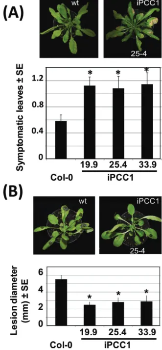
(Sauerbrunn and Schlaich, 2004), iPCC1 plants were sig-nificantly more susceptible than wild-type plants to another oomycete pathogen P. brassicae (Fig. 3A). In turn, iPCC1 plants were surprisingly more resistant than wild-type plants to the necrotrophic fungus B. cinerea, with a reduction of ~50% in the necrotic lesion diameter (Fig. 3B).
Altered lipid content in iPCC1 plants
Alterations to physiological processes as diverse as seed germination and defence responses to pathogens in iPCC1 plants might be indicative of PCC1 affecting either common signalling events or a central metabolic process. Regarding
Fig. 3. Performance of wild-type and iPCC1 plants against
Phytophthora brassicae and Botrytis cinerea. (A and B) The relative symptoms on a scale of 0–4 from the least to the most symptomatic leaves, and the lesion diameter of plants inoculated with P. brassicae and B. cinerea, respectively. Images on top of the graphs corresponding to symptoms for the wild type and iPCC1 25.4 line are also representative for the other iPCC1 lines. Values are the mean ±SE of four replicates and * means that iPCC1 values were all significantly different from wild-type values with a P-value <0.05 by Student’s t-test.
Fig. 2. Effect of ABA on wild-type and iPCC1 stomatal aperture
and root growth. (A) The stomatal aperture, expressed as the ratio of length to width as indicated in the image in the left panel, was calculated for the wild type and two iPCC1 lines either in the absence of exogenously added ABA or in plants treated with 5 μM or 50 μM ABA as indicated. The central panel is a representative image of propidium iodide-stained stomata for every genotype and condition indicated. Values are the mean ±SE of between 35 and 50 stomata for each condition and genotype. (B) Root length of wild-type and iPCC1 seedlings on ABA-supplemented medium. Seedlings were grown in vertical square Petri dishes containing Murashige and Skoog (MS) medium supplemented or not with the indicated concentrations of ABA. Values represent the mean of the main root length (mm) of eight seedlings per genotype and condition ±SD. *Represents significantly different values from wild-type and iPCC1 roots with a P-value <0.05 by Student’s t-test..
this, lipid involvement in seed germination (Theodoulou and Eastmond, 2012) and plant–pathogen interactions (Christensen and Kolomiets, 2011) has gained increasing interest. A lipidomic profile analysis focused on fatty acids and polar lipids for iPCC1 and wild-type leaves was per-formed. Quantification of the total content of polar lipids and their fatty acids by thin-layer chromatography followed by gas chromatography showed that iPCC1 leaves contained ~20% less fatty acids than wild-type leaves (Fig. 4A). This reduction affected the total content of saturated and desatu-rated 16C and 18C fatty acids similarly (Fig. 4A), but when the percentage composition was calculated a significant decrease was only observed in 18:0 (Fig. 4A). On the other hand, quantification of the total content of polar lipids showed no significant difference in iPCC1 leaves when compared with wild-type leaves (Fig. 4B). However, the analyses of the lev-els of different classes of polar lipids showed increases in the abundant chloroplastic monogalactosyl and digaloctosyl dia-cylgycerol (MGDG and DGDG) and phosphatidylglycerol (PG) in iPCC1 plants (Fig. 4B). In turn, the levels of polar lipids widely involved in signalling, such as phosphatidylin-ositol (PI), phosphatidylserine (PS), phosphatidylcholine
(PC), and phosphatidylethanoalmine (PE) were significantly decreased in iPCC1 leaves compared with those of the wild type (Fig. 4B). Of importance, iPCC1 leaves contained only ~30% of the PI content detected in wild-type leaves (Fig. 4B). This dramatic reduction in a lipid class extensively charac-terized as important signalling components in a wide array of plant processes (Munnik and Nielsen, 2011) may help to explain some of the above-mentioned phenotypes as well as some others as yet unexplored. The reduced levels of 18:0 in iPCC1 leaves might suggest that the reduced expression of the PCC1 gene could correlate with enhanced desaturation of fatty acids. Table 1 shows a more detailed quantification of fatty acids in each polar lipid class. The most significant dif-ferences between iPCC1 and wild-type plants were observed in 16:0, 18:0, 18:1, 18:2, and 18:3 fatty acids of PI and PS (Table 1). A 55% decrease in the 18:0 content of iPCC1 PI and the concomitant increase of >2-fold in the unsaturated fatty acids 18:1, 18:2, and 18:3 of PI was observed (Table 1). A similar trend was also observed for the fatty acid compo-sition of PS (Table 1), thus supporting the hypothesis that reduced PCC1 expression correlates well with enhanced desaturation of fatty acids in these classes of polar lipids.
Col0 iPCC1
(A)
(B)
Fa ttya ci ds co mp os itio n( %) Amoun t(Fig. 4. Comparative polar lipid profile of iPCC1 and wild-type plants. The total amounts and percentage composition of each class of
(A) fatty acids and (B) polar lipid are shown from the top to bottom panels as indicated. Phosphatidylinositol (PI), phosphatidylserine (PS), phosphatidylcholine (PC), phosphatidylethanolamine (PE), monogalactosyl and digaloctosyl diacylgycerol (MGDG and DGDG), sulfphoquinovosyl (SQ), and phosphatidylglycerol (PG). Values are the mean of three replicates ±SD. *represents significantly different values from wild-type and iPCC1 leaves with a P-value <0.05 by Student’s t-test.
PCC1-modulated transcriptome
Despite its demonstrated involvement in defence and devel-opment, the molecular function and the mode of action of PCC1 protein in Arabidopsis remain largely unknown. To explore the way PCC1 functions, the effect of reduced expres-sion of the PCC1 gene on the genome-wide transcriptome of Arabidopsis thaliana was explored. Affymetrix microar-ray experiments were performed comparing three different iPCC1 linees, with very low PCC1 expression (Segarra et al., 2010), versus three biological replicates of non-transformed wild-type Col-0 seedlings grown under a long-day photo-period for 12 d. Samples for RNA extraction were harvested at 12 h after dawn when PCC1 expression was highest in wild-type plants (Segarra et al., 2010). A total of 1037 genes were differentially expressed in seedlings with reduced PCC1 expression after applying a cut-off for the P-value corrected for FDR (false discovery rate) values of <0.05 and fold val-ues >1.5 or less than –1.5. Among them, 517 and 520 genes were up- and down-regulated, respectively (Supplementary Table S1 at JXB online). To validate the levels of transcripts observed in the microarray analysis, a randomly selected group of genes were analysed by qRT-PCR, and a good cor-relation between the levels of transcripts detected by both techniques was observed (Supplementary Fig. S3), thus sug-gesting that the results derived from the microarray-based transcriptomic analysis are robust and useful for further bio-logical interpretation.
GO analysis of terms over-represented among differentially expressed genes led to an enrichment (FDR values <0.03) of lipid localization, responses to ABA, glucosinolate bio-synthesis and metabolism, sulphur metabolism, and organic acid transmembrane transporter activity among up-regulated genes (Table 2). The functions significantly over-represented with down-regulated genes were associated with molecular transducer and transmembrane receptor activities as well as defence responses and cell death (Table 2). Several of these functional categories including lipid localization, responses to ABA, and defence responses and cell death are in agree-ment with the above-described phenotypes characterized for iPCC1 plants, suggesting that there is a good correlation between the reduced function of PCC1 and the trancriptomic and phenotypical alterations.
Analysis of promoter sequences of up- and down-regulated genes in iPCC1 plants
An in silico analysis of the 1000 bp promoter sequence upstream the initiation codon of genes that were up- or down-regulated in iPCC1 (Supplementary Table S1 at JXB online) allowed identification of 8-mer motifs that were sig-nificantly over-represented. Six motifs present in at least 15% of the promoter sequences of up-regulated genes were identi-fied (Supplementary Table S2). A GO analysis of those genes points to their involvement in lipid localization and responses to ABA. The motifs A(G)AAAT(G)AAA, TAAATAA(GC) A and AGA(G)TAA(G)A(G)T(G), which were repre-sented in >80, 35, and 20%, respectively, of the promoters
Table 1.
Fatty acid composition of different polar lipid class for wild-type and iPCC1 leaves 16:0
16:1 16:2 16:3 18:0 18:1 18:2 18:3 Col-0 iPCC1 Col-0 iPCC1 Col-0 iPCC1 Col-0 iPCC1 Col-0 iPCC1 Col-0 iPCC1 Col-0 iPCC1 Col-0 iPCC1 PI 51.8 ± 0.2 44.2 ± 3.2* ND ND ND ND ND ND 32.0 ± 2.5 14.3 ± 3.2* 2.0 ± 1.3 4. 9 ± 1.5* 6.6 ± 0.1 18.9 ± 1.7* 7.7 ± 1.0 17.1 ± 1.1* PS 43.5 ± 1.9 32.3 ± 6.8* ND ND ND ND ND ND 38.1 ± 1.3 31.8 ± 4.1* 2.6 ± 0.8 4.2 ± 2.4* 6.9 ± 0.4 16.7 ± 3.6* 9.0 ± 1.1 15.1 ± 3.9* PC 27.2 ± 0.8 24. 8 ± 0.6 0.5 ± 0.1 0.4 ± 0.1 0.1 ± 0.1 ND ND ND 6.1 ± 0.6 5.3 ± 0.46 7.3 ± 0.7 7.1 ± 1.2 29.5 ± 0.4 32.7 ± 0.5* 28.6 ± 1.0 28.9 ± 1.3 PE 32. 6 ± 2.1 31.4 ± 1.4 0.3 ± 0.5 0.2 ± 0.2 ND ND ND ND 6.4 ± 0.7 5.7 ± 0.7 3.2 ± 0.4 3.39 ± 0.1 34.2 ± 0.6 36.3 ± 1.8 22.3 ± 1.3 22.0 ± 0.2 PA 34.1 ± 5.5 34.4 ± 4.0 ND ND ND ND ND ND 22.3 ± 2.0 29.0 ± 7.8 6.3 ± 0.5 4.9 ± 1.3 20.4 ± 4.3 17.8 ± 1.6 16.9 ± 2.5 13.9 ± 0.8 MGDG 2.2 ± 0.2 2.3 ± 0.4 1.6 ± 0.1 1.3 ± 0.1 2.0 ± 0.1 1.6 ± 0.2 26.5 ± 2. 5 28.0 ± 1.9 1.2 ± 0.3 0. 7 ± 0.2 2.0 ± 0.4 1.4 ± 0.1 3.9 ± 0.4 2. 9 ± 0.2 60.7 ± 2.4 61.6 ± 2.5 DGDG 16.9 ± 1.7 17.7 ± 0.9 0.1 ± 0.1 0.2 ± 0.1 0.6 ± 0.1 0.5 ± 0.1 2.0 ± 0.4 2.0 ± 0.4 3.5 ± 0.4 2. 7 ± 0.4 2.0 ± 0.3 1.7 ± 0.1 5.6 ± 0.4 4.9 ± 0.1 69.0 ± 2.5 69.7 ± 1.0 SQ 19.7 ± 3.8 24.0 ± 2.2 1.0 ± 1.4 ND ND ND ND ND 27.0 ± 2.4 20.0 ± 3. 7 8.7 ± 6. 7 5.4 ± 1.4 21.1 ± 4.0 26.3 ± 4.7 22.6 ± 2.7 24.3 ± 2.5 PG 29.5 ± 4.1 28.1 ± 1.1 24.5 ± 1.1 25.4 ± 1.1 ND ND ND ND 7. 6 ± 4.2 3.7 ± 0.4 8.0 ± 0.1 7.1 ± 0. 9 7.6 ± 1.6 8.4 ± 0.7 22.3 ± 5.7 26.7 ± 1.5 Values, expr essed as a per
centage of the fatty acids analysed, ar
e the mean of thr
ee r
eplicates ± SD. Not detected (ND) means values wer
e below the detection limit.
* r
epr
esents significantly dif
fer
ent values fr
om wild-type and iPCC1 leaves with a
P
-value <0.05 by Student’
s
of up-regulated genes in iPCC1 plants, were contained in a consensus 3AF1 binding site tetramer sequence previously characterized as important for the light-responsive promoter of several genes including rbcS-3A from pea (Terzaghi and Cashmore, 1995). Moreover, the first one was also contained in characterized consensus sequences for regulating starch-branching enzyme I gene expression in maize endosperm (Kim and Guiltinan, 1999) and the leghaemoglobin lbc3 gene from soybean (Jensen et al., 1988). The third most repre-sented motif, CTTCT(C)TCT(G), was identified in >25% of the up-regulated genes and it was contained in a previously identified TL1 consensus sequence in the promoter of NPR1-responsive genes (Wang et al., 2005) and also in the TCA-1 (tobacco nuclear protein 1) binding site of SA-inducible genes (Goldsbrough et al., 1993). A CTCTCTCT(CG) motif identi-fied in 15% of the up-regulated genes was present in a CT-rich motif found in the S1 region downstream of the transcrip-tion start site of the Cauliflower mosaic virus 35S promoter, and it may work as an expression enhancer (Pauli et al., 2004). This motif was also present in a consensus sequence described to be involved in phytochrome A-regulated gene expression (Hudson and Quail, 2003). Finally, the motif C(G)A(G)CGTGT(G)C, which was identified in 15% of the up-regulated genes, contains the CACGTG G-box motif extensively present in both induced and repressed phyA-responsive promoters (Hudson and Quail, 2003) as well as the ABRE (ABA-responsive element ACGTGG/TC) motif that has been identified in the promoter region of ABA-inducible genes (Busk and Pagès, 1998; Nakashima et al., 2006) and seems to bind the GBF family of bZIP transcription factors (Menkens et al., 1995; Jakoby et al., 2002).
On the other hand, common motifs in the promoter sequences of down-regulated genes were also found. Nine of them were present in >15% of the promoters of down-regu-lated genes, and the corresponding genes were mostly involved in defence responses and cell death (Supplementary Table S3 at JXB online). The most represented motif, AAATTTT(G) A(G), which was present in 62% of the promoters of down-regulated genes, is contained in a previously character-ized motif (CAAAATTTTGTA) that participates in the
HSR203-responsive element (HSRE), which is involved in the hypersensitive response to pathogens in tobacco (Pontier et al., 2001). The second most represented motif, A(C)CAA(C) A(G)AAT, which was present in 55% of the promoters, is part of the consensus sequence GATACANNAATNTGATG characterized as a methyl jasmonate (JA)-responsive element in tomato lipoxygenases (Beaudoin and Rothstein, 1997). A third motif, GA(G)A(C)GA(G)A(C)GA(CG), was identi-fied in 35% of the down-regulated genes and it was contained in the TL1 consensus sequence CTGAAGAAGAA identi-fied in 13 NPR1-responsive endoplasmic reticulum-resident genes (Wang et al., 2005) and also in the TCA-1 binding site of SA-inducible genes (Goldsbrough et al., 1993).
Discussion
The way in which a small protein such as PCC1 with either no domains typically present in transcription factors or nuclear localization signals regulates several phenotypes in
Arabidopsis is challenging. This work shows that reduced
expression of the PCC1 gene leads to profound changes in both metabolic and signalling events that together or inde-pendently lead to altered developmental and defence-related processes. Previously reported data from bioinformatic analy-sis (Venancio and Aravind, 2010) and from massive proteomic identification of plasma membrane proteins (Marmagne et al., 2004), as well as from unpublished experimental results, suggest that PCC1 is associated with the plasma membrane. Data presented in this work are consistent with PCC1 par-ticipating in lipid-related events in membranes. iPCC1 plants displayed alterations in the lipid content and composition as well as changes in the transcript levels of genes coding for the production, storage, mobilization, and signalling of lipids. First, iPCC1 plants have strongly reduced levels of PI, and to a lesser extent of PS, PE, and PC, as well as an enriched content of desaturated fatty acids of PI, PS, and PC (Fig. 4, Table 1). Secondly, a significant enrichment of the lipid localization GO category was observed among up-regulated genes in iPCC1 plants (Table 2). Those lipid-related genes
Table 2. Enrichment of GO categories in the PCC1-regulated transcriptome
GO term Ontology Description No. input No. BG/Ref. P-value FDR
Up-regulated genes in iPCC1 lines
GO:0010876 P Lipid localization 9 24 1.1e-11 1.1e-08
GO:0019761 P Glucosinolate biosynthesis 6 41 1.4e-05 0.0017
GO:0019760 P Glucosinolate metabolism 7 62 1.5e-05 0.0017
GO:0009737 P Responses to abscisic acid 17 378 1.2e-05 0.0017
GO:0006790 P Sulphur metabolic process 10 220 0.00064 0.027
GO:0005342 F Organic acid transmembrane
transporter activity
7 78 6.9e-05 0.008
Down-regulated genes in iPCC1 lines
GO:0006952 P Defence response 51 766 2.5e-20 2.5e-17
GO:0008219 P Cell death 26 286 2.1e-14 7.1e-12
GO:0050832 P Defence response to fungus 8 108 8.3e-05 0.0055
GO:0004888 F Transmembrane receptor activity 13 171 4.4e-07 0.00011
code for proteins involved in the transport/localization, stor-age, and signalling of lipids (Supplementary Table S4 at JXB online). Because of the plasma membrane-associated loca-tion of PCC1, lipid-related signalling is particularly relevant to explain its widespread effect on plant physiology. iPCC1 plants contained up- and down-regulated genes coding for GDSL-like lipases (Supplementary Table S4), which have been characterized as important components in triggering responses to biotic and abiotic stress factors (Oh et al., 2005; Hong et al., 2008) as well as in hormone-related control of plant growth and development (Cao et al., 2006). Moreover, iPCC1 plants might have an altered PI metabolism and transport as suggested by the reduced levels of PI (Fig. 4B, Table 1) and also by the up-regulation of genes coding for PI-related kinases and phosphatases as well as the down-reg-ulation of genes coding for transporters of the SEC14 family (Supplementary Table S4). On the other hand, iPCC1 plants showed enhanced levels of the chloroplast lipid MGDG. Although this is a mere hypothesis, PCC1 might affect chlo-roplast lipid composition through interaction with signalling events that either end with the modulation of transcription of genes coding for precursor chloroplast proteins or interfere with the lipid trafficking from the endoplasmic reticulum to the chloroplast. Determination of whether PCC1 modulates early or late events and localization or metabolism of lipids in lipid-related signalling would require more work.
A complex pattern of alterations was detected in the defence responses of iPCC1 plants against pathogens with different lifestyles. iPCC1 plants were more susceptible than wild-type plants to the hemibiotrophic oomycete P. brassicae and more resistant to the necrotrophic fungus B. cinerea. Since iPCC1 plants were slightly hypersensitive to ABA (Figs 1, 2), the responses to pathogens of different lifestyles might be related to alterations in ABA signalling. However, it has been recently reported that Arabidopsis mutant plants defective in either the biosynthesis or signalling of ABA were more resistant to the necrotrophic fungus Plectosphaerella cucumerina ( Sánchez-Vallet et al., 2012), thus suggesting that there is not a direct connection between PCC1 and ABA signalling in control-ling defensive responses to different pathogens. In turn, the enhanced susceptibility of iPCC1 plants to Phytophtora (Fig. 3) is consistent with the previously reported enhanced resist-ance of PCC1-overexpressing plants to the downey mildew
P. parasitica (Sauerbrunn and Schlaich, 2004). iPCC1 plants had down-regulated expression of RPP4, RPP5, and RPP13 resistance genes to Hyaloperonospora arabidopsidis (Reignault
et al., 1996; Knoth et al., 2007). RPP4 and RPP5 are targets of
the modulation exerted by Suppressor of npr1-1 Constitutive 1 (SNC1). Microarray data presented in this work indicate that SNC expression is strongly down-regulated (–13.1-fold) in iPCC1 plants (Supplementary Table S1 at JXB online), provid-ing a possible link between the innate immunity and the ubiq-uitin pathway in Arabidopsis (Goritschnig and Zhang, 2007). Interestingly, iPCC1 plants showed a strong down-regulation of the genes coding for UBQ11, UBQ10, and the ubiquitin-conjugating enzyme 37 (UBC37) (Supplementary Table S1) which may be indicative of altered function of the ubiquitin pathway in iPCC1 plants. A PCC1-mediated effect on protein
ubiquitination could also be an efficient way to exert regula-tion on many different targets, thus allowing a wide display of PCC1-regulated phenotypes.
The functional roles of PCC1 in regulating defence responses of Arabidopsis against a wide array of pathogens must be complex enough to explain the enhanced suscep-tibility of iPCC1 plants to P. brassicae and the enhanced resistance to B. cinerea. These two pathogens have different lifestyles and their interactions with the plant are governed by different signalling pathways involving regulatory molecules such as JA, ethylene (ET), and SA. Regarding this, the tran-scriptome of iPCC1 plants showed a large over-representation of down-regulated genes coding for components involved in SA-mediated signalling. Interestingly, PCC1 gene expression is up-regulated by SA (Segarra et al., 2010), and by using
pPCC1::GUS plants it was found that its expression is spread
throughout seedlings (Supplementary Fig. S4 at JXB online). SA-related down-regulated genes include EDS1 that, together with PAD4, has been considered an essential regulatory hub for the establishment of basal resistance to biotrophic and hemi-biotrophic pathogens, by activating SA-mediated resist-ance and by regulating the antagonism with JA- and ET-based defence responses to pathogens (Wiermer et al., 2005). Also the NPR1 gene coding for the ankyrin repeat-containing cen-tral regulator of SA-activated resistance to pathogens (Dong, 2004a) was down-regulated in iPCC1 plants (Supplementary Table S1). In connection with the potential membrane-related site of action for PCC1 discussed above, the ACD6 gene cod-ing for a membrane-associated protein involved in control-ling cell death and SA-mediated defence to pathogens (Dong, 2004b; Lu et al., 2005) was also severely down-regulated in iPCC1 plants (Supplementary Table S1). Enhanced resistance of iPCC1 plants to necrotrophic pathogens such as B. cinerea might be explained by the down-regulation of the SA-related defence signalling and the proposed functional antagonism of that pathway with JA/ET-related defence (Thaler et al., 2012).
The functional interaction between PCC1 and ABA is sup-ported by different sets of data in this work. First, iPCC1 plants are hypersensitive to ABA in several phenotypes, includ-ing seed germination and seedlinclud-ing establishment (Fig. 1), as well as stomatal closure and root growth (Fig. 2). Secondly, the up-regulated transcriptome of iPCC1 plants is enriched in ABA-related genes (Table 1), and many of them contained ABA-responsive elements (Busk and Pagès, 1998; Nakashima
et al., 2006) in their promoters (Supplementary Table S3 at
JXB online). Several reports in the last years have provided
details on the main module of ABA signalling from hormone perception by receptors of the PYR/PYL/RCAR family to downstream components including phosphatases of the PP2C family, kinases of the SnRK2 family, and ABA-modulated transcription factors of different families (Fujii et al., 2009; Ma et al., 2009; Melcher et al., 2009; Park et al., 2009; Cutler
et al., 2010; Raghavendra et al., 2010; Umezawa et al., 2010).
An overall view of the PCC1-modulated transcriptome shows that none of the genes coding for the ABA receptors was sig-nificantly altered in iPCC1 plants (Supplementary Fig. S5). In turn, five out of nine PP2C phosphatase-encoding genes of clade A were significantly up-regulated in iPCC1 plants
(Supplementary Fig. S5). Up-regulation of genes coding for ABA-related PP2Cs may be a response directed to attenu-ate signalling in plants showing hypersensitivity to ABA. Nevertheless, because the transcriptome analysis in this work was performed under non-stressed conditions, the profound changes detected in gene expression correlate well with iPCC1 seeds and plants displaying developmental phenotypes such as reduced seed germination, seedling establishment, and root elongation. These phenotypical alterations are all well explained by the hypersensitivity to ABA of iPCC1 plants even under non-stress conditions.
Taken together, the data presented in this work support an important role for PCC1 as a regulator and as an integrator of responses to the environment and to endogenous develop-mental cues. The likely membrane-associated location points to a potential double role for PCC1 as a modulator of recep-tor systems, including pathogen-derived molecular patterns, as well as a regulator at multiple levels of early downstream signalling. In this way, PCC1 seems to be involved in regulat-ing both ABA-mediated developmental transitions as well as SA- and JA-triggered pathogen-related responses.
Supplementary data
Supplementary data are available at JXB online.
Figure S1. Seedling establishment in osmotic media sup-plemented with mannitol or NaCl.
Figure S2. Water loss in wild-type and iPCC1 seedlings. Figure S3. Comparison of transcript levels detected by microarray and qRT-PCR analysis.
Figure S4. PCC1 expression is induced by SA.
Figure S5. Microarray-derived transcript levels of the core ABA signalling module in Arabidopsis.
Table S1. Comparative transcriptomic analysis of iPCC1 versus wild-type plants.
Table S2. In silico analysis of motifs over-represented in the promoter sequences of genes that were up-regulated in iPCC1 versus Col-0 plants.
Table S3. In silico analysis of motifs over-represented in the promoter sequences of genes that were down-regulated in iPCC1 versus Col-0 plants.
Table S4. Lipid-related genes that were up- and down-regu-lated in iPCC1 compared with wild-type plants.
Acknowledgements
This work was supported by grants BIO2008-00839 and CSD2007-0057 from the Ministerio de Ciencia e Innovación of Spain to JL. A fellowship/contract of the FPU programme of the Ministerio de Educación y Ciencia (Spain) funded the work of RM. MLH was the recipient of a JAE-Doc contract from Consejo Superior de Investigaciones Científicas (CSIC).
References
Adams S, Carré IA. 2011. Downstream of the plant circadian clock:
output pathways for the control of physiology and development. Essays in Biochemistry 49, 53–69.
Beaudoin N, Rothstein SJ. 1997. Developmental regulation of two
tomato lipoxygenase promoters in transgenic tobacco and tomato. Plant Molecular Biology 33, 835–846.
Bligh EG, Dyer WS. 1959. A rapid method of total lipid extraction
and purification. Canadian Journal of Biochemistry and Physiology 37,
911–917.
Busk PK, Pagès M. 1998. Regulation of abscisic acid-induced
transcription. Plant Molecular Biology 37, 425–435.
Cao D, Cheng H, Wu W, Soo HM, Peng J. 2006. Gibberellin
mobilizes distinct DELLA-dependent transcriptomes to regulate seed germination and floral development in Arabidopsis. Plant Physiology
142, 509–525.
Cao FY, Yoshioka K, Desveaux D. 2011. The roles of ABA in plant–
pathogen interactions. Journal of Plant Research 124, 489–499.
Christensen SA, Kolomiets MV. 2011. The lipid language of plant–
fungal interactions. Fungal Genetics and Biology 48, 4–14.
Cutler SR, Rodriguez PL, Finkelstein RR, Abrams SR. 2010.
Abscisic acid: emergence of a core signaling network. Annual Review of Plant Biology 61, 651–679.
Dong X. 2004a. NPR1, all things considered. Current Opinion in Plant Biology 7, 547–552.
Dong X. 2004b. The role of membrane-bound ankyrin-repeat protein
ACD6 in programmed cell death and plant defense. Science’s STKE: signal transduction knowledge environment 2004, pe6.
Fujii H, Chinnusamy V, Rodrigues A, Rubio S, Antoni R, Park SY, Cutler SR, Sheen J, Rodriguez PL, Zhu JK. 2009. In vitro
reconstitution of an abscisic acid signalling pathway. Nature 462,
660–664.
Garcés R, Mancha M. 1993. One-step lipid extraction and fatty
acid methyl esters preparation from fresh plant tissues. Analytical Biochemistry 211, 139–143.
Goldsbrough AP, Albrecht H, Stratford R. 1993. Salicylic
acid-inducible binding of a tobacco nuclear protein to a 10 bp sequence which is highly conserved amongst stress-inducible genes. The Plant Journal 3, 563–571.
Goritschnig S, Zhang Y, Li X. 2007. The ubiquitin pathway is required
for innate immunity in Arabidopsis. The Plant Journal 49, 540–551.
Gubler F, Millar AA, Jacobsen JV. 2005. Dormancy release, ABA and
pre-harvest sprouting. Current Opinion in Plant Biology 8, 183–187.
Hernández ML, Guschina IA, Martínez-Rivas JM, Mancha M, Harwood JL. 2008. The utilization and desaturation of oleate and
linoleate during glycerolipid biosynthesis in olive (Olea europea L.)
callus cultures. Journal of Experimental Botany 59, 2425–2435.
Hong JK, Choi HW, Hwang IS, Kim DS, Kim NH, Choi S, Kim YJ, Hwang BK. 2008. Function of a novel GDSL-type pepper lipase
gene, CaGLIP1, in disease susceptibility and abiotic stress tolerance. Planta 227, 539–558.
Hudson ME, Quail PH. 2003. Identification of promoter motifs
involved in the network of phytochrome A-regulated gene expression by combined analysis of genomic sequence and microarray data. Plant Physiology 133, 1605–1616.
Jackson SD. 2009. Plant responses to photoperiod. New Phytologist 181, 517–531.
Jakoby M, Weisshaar B, Droge-Laser W, Vicente-Carbajosa J, Tiedemann J, Kroj T, Parcy F; bZIP Research Group. 2002. bZIP
transcription factors in Arabidopsis. Trends in Plant Science 7, 106–111.
Jensen EØ, Marcker KA, Schell J, Bruijn FJ. 1988. Interaction of
a nodule specific, trans-acting factor with distinct DNA elements in the soybean leghaemoglobin Ibc(3) 5ʹ upstream region. EMBO Journal 7, 1265–1271.
Kim KN, Guiltinan MJ. 1999. Identification of cis-acting elements
important for expression of the starch-branching enzyme I gene in
maize endosperm. Plant Physiology 121, 225–236.
Knoth C, Ringler J, Dangl JL, Eulgem T. 2007. Arabidopsis
WRKY70 is required for full RPP4-mediated disease resistance and basal defense against Hyaloperonospora parasitica. Molecular Plant-Microbe Interactions 20, 120–128.
Lozano-Juste J, León J. 2011. Nitric oxide regulates DELLA content
and PIF expression to promote photomorphogenesis in Arabidopsis. Plant Physiology 156, 1410–1423.
Lu H, Liu Y, Greenberg JT. 2005. Structure–function analysis of the
plasma membrane-localized Arabidopsis defense component ACD6. The Plant Journal 44, 798–809.
Ma Y, Szostkiewicz I, Korte A, Moes D, Yang Y, Christmann A, Grill E. 2009. Regulators of PP2C phosphatase activity function as
abscisic acid sensors. Science 324, 1064–1068.
Marmagne A, Rouet MA, Ferro M, Rolland N, Alcon C, Joyard J, Garin J, Barbier-Brygoo H, Ephritikhine G. 2004. Identification
of new intrinsic proteins in Arabidopsis plasma membrane proteome. Molecular and Cellular Proteomics 3, 675–691.
Melcher K, Ng LM, Zhou XE, et al. 2009. A gate–latch–lock mechanism
for hormone signalling by abscisic acid receptors. Nature 462, 602–608.
Menkens AE, Schindler U, Cashmore AR. 1995. The G-box: a
ubiquitous regulatory DNA element in plants bound by the GBF family
of bZIP proteins. Trends in Biochemical Sciences 20, 506–510.
Morker KH, Roberts MR. 2011. Light exerts multiple levels of
influence on the Arabidopsis wound response. Plant, Cell and Environment 34, 717–728.
Munnik T, Nielsen E. 2011. Green light for polyphosphoinositide
signals in plants. Current Opinion in Plant Biology 14, 489–497.
Nakashima K, Fujita Y, Katsura K, Maruyama K, Narusaka Y, Seki M, Shinozaki K, Yamaguchi-Shinozaki K. 2006.
Transcriptional regulation of ABI3- and ABA-responsive genes including RD29B and RD29A in seeds, germinating embryos, and
seedlings of Arabidopsis. Plant Molecular Biology 60, 51–68.
Oh IS, Park AR, Bae MS, Kwon SJ, Kim YS, Lee JE, Kang NY, Lee S, Cheong H, Park OK. 2005. Secretome analysis reveals an
Arabidopsis lipase involved in defense against Alternaria brassicicola. The Plant Cell 17, 2832–2847.
Park SY, Fung P, Nishimura N, et al. 2009 Abscisic acid inhibits
type 2C protein phosphatases via the PYR/PYL family of START
proteins. Science 324, 1068–1071.
Pauli S, Rothnie HM, Chen G, He X, Hohn T. 2004. The cauliflower
mosaic virus 35S promoter extends into the transcribed region. Journal of Virology 78, 12120–12128.
Pontier D, Balague C, Bezombes-Marion I, Tronchet M, Deslandes L, Roby D. 2001. Identification of a novel pathogen-responsive element
in the promoter of the tobacco gene HSR203J, a molecular marker of the
hypersensitive response. The Plant Journal 26, 495–507.
Raghavendra AS, Gonugunta VK, Christmann A, Grill E. 2010.
ABA perception and signalling. Trends in Plant Science 15, 395–401.
Reignault P, Frost LN, Richardson H, Daniels MJ, Jones JD, Parker JE. 1996. Four Arabidopsis RPP loci controlling resistance to
the Noco2 isolate of Peronospora parasitica map to regions known to contain other RPP recognition specificities. Molecular Plant-Microbe Interactions 9, 464–473.
Roden LC, Ingle RA. 2009. Lights, rhythms, infection: the role of light
and the circadian clock in determining the outcome of plant–pathogen
interactions. The Plant Cell 21, 2546–2552.
Sánchez A, Shin J, Davis SJ. 2011. Abiotic stress and the plant
circadian clock. Plant Signaling and Behavior 6, 223–231.
Sánchez-Vallet A, López G, Ramos B, et al. 2012. Disruption of
abscisic acid signaling constitutively activates arabidopsis resistance to the necrotrophic fungus Plectosphaerella cucumerina. Plant Physiology 160, 2109–2124.
Sauerbrunn N, Schlaich NL. 2004. PCC1: a merging point for pathogen
defence and circadian signalling in Arabidopsis. Planta 218, 552–561.
Schlaeppi K, Abou-Mansour E, Buchala A, Mauch F. 2010.
Disease resistance of Arabidopsis to Phytophthora brassicae is established by the sequential action of indole glucosinolates and
camalexin. The Plant Journal 62, 840–851.
Segarra S, Mir R, Martínez C, León J. 2010. Genome-wide analyses
of the transcriptomes of salicylic acid-deficient versus wild type plants uncover Pathogen and Circadian Controlled 1 (PCC1) as a regulator of
flowering time in Arabidopsis. Plant, Cell and Environment 33, 11–22.
Terzaghi WB, Cashmore AR. 1995. Light-regulated transcription. Annual Review of Plant Physiology and Plant Molecular Biology 46,
445–474.
Theodoulou FL, Eastmond PJ, 2012. Seed storage oil catabolism: a
story of give and take. Current Opinion in Plant Biology 15, 322–328.
Thaler JS, Humphrey PT, Whiteman NK. 2012. Evolution of jasmonate
and salicylate signal crosstalk. Trends in Plant Science 17, 260–270.
Umezawa T, Nakashima K, Miyakawa T, Kuromori T, Tanokura M, Shinozaki K,Yamaguchi-Shinozaki K. 2010. Molecular basis of
the core regulatory network in ABA responses: sensing, signaling and
transport. Plant and Cell Physiology 51, 1821–1839.
Venancio TM, Aravind L. 2010. CYSTM, a novel cysteine-rich
transmembrane module with a role in stress tolerance across
eukaryotes. Bioinformatics 26, 149–152.
Wang D, Weaver ND, Kesarwani M, Dong X. 2005. Induction of
protein secretory pathway is required for systemic acquired resistance. Science 308, 1036–1040.
Wang GY, Shi JL, Ng G, Battle SL, Zhang C, Lu H. 2011.
Circadian clock-regulated phosphate transporter PHT4;1 plays an
important role in Arabidopsis defense. Molecular Plant 4, 516–526.
Wang W, Barnaby JY, Tada Y, Li H, Tör M, Caldelari D, Lee DU, Fu XD, Dong X. 2011. Timing of plant immune responses by a
central circadian regulator. Nature 470, 110–114.
Wiermer M, Feys BJ, Parker JE. 2005. Plant immunity: the EDS1