Nephrol Dial Transplant (2014) 29: iv113–iv116 doi: 10.1093/ndt/gfu018
The Bench-to-Bedside Transition
Acute metabolic acidosis in a GLUT2-de
ficient patient
with Fanconi
–Bickel syndrome: new pathophysiology insights
Fabrice Mihout
1, Olivier Devuyst
2, Albert Bensman
3, Isabelle Brocheriou
4,5, Christophe Ridel
6, Carsten
A. Wagner
2, Nilufar Mohebbi
2, Jean-Jacques Boffa
1,5,7, Emmanuelle Plaisier
1,5,7and Pierre Ronco
1,5,7 1Department of Nephrology and Dialysis, AP-HP, Tenon Hospital, Paris, France,2Institute of Physiology, University of Zürich, Zürich,Switzerland,3Department of Pediatric Nephrology, AP-HP, Robert Debré Hospital, Paris, France,4Department of Pathology, AP-HP, Tenon Hospital, Paris, France,5Sorbonne Universités, UPMC Univ Paris 06, UMR_S 1155, Paris, France,6Department of Renal Emergency and Kidney Transplantation, AP-HP, Tenon Hospital, Paris, France and7INSERM, UMR_S 1155, Paris, France
Correspondence and offprint requests to: Pierre Ronco; E-mail: pierreronco@yahoo.fr
A B S T R AC T
Fanconi–Bickel syndrome is a rare autosomal-recessive dis-order caused by mutations in the SLC2A2 gene coding for the glucose transporter protein 2 (GLUT2). Major manifestations include hepatomegaly, glucose intolerance, post-prandial hy-poglycaemia and renal disease that usually presents as proximal tubular acidosis associated with proximal tubule dysfunction (renal Fanconi syndrome). We report a patient harbouring a homozygous mutation of SLC2A2 who presented a dramatic exacerbation of metabolic acidosis in the context of a viral infec-tion, owing to both ketosis and major urinary bicarbonate loss. The kidney biopsy revealed nuclear and cytoplasmic accumu-lation of glycogen in proximal tubule cells, a lack of expression of GLUT2, and major defects of key proteins of the proximal tubule such as megalin, cubilin and the B2 subunit of H+– ATPase. These profound alterations of the transport systems most likely contributed to proximal tubule alterations and pro-found bicarbonate loss.
Keywords: Fanconi–Bickel, ketosic acidosis, metabolic acido-sis, proximal tubular nephropathy, SLC12A2
IN TRO DU CT IO N
Fanconi–Bickel syndrome is a rare autosomal-recessive dis-order caused by mutations in the SLC2A2 gene that codes for the glucose transporter protein 2 (GLUT2). Because this transporter is expressed in the liver and pancreas (β-cells),
intestine and kidney, patients typically show hepatomegaly due to glycogen accumulation, glucose intolerance, post-pran-dial hypoglycaemia and proximal tubulopathy. The disease wasfirst described by Fanconi and Bickel in 1949 in a 3-year-old Swiss boy [1]. To date, >200 patients have been reported, and about 34 different mutations of SLC2A2 have been ident-ified [2]. The renal disease usually presents as a moderate proximal tubular acidosis associated with generalized proximal tubule dysfunction (renal Fanconi syndrome). Kidney biopsies are rarely performed, and the mechanisms whereby SLC2A2 mutations induce proximal tubule dysfunction are essentially unknown.
In this brief report, we describe the case of a patient har-bouring a SLC2A2 mutation presenting with acute exacer-bation of acidosis compatible with superimposed acute tubular necrosis. Immunohistochemical studies of the kidney biopsy showed profound alterations in the expression of major pro-teins sustaining reabsorptive functions of the proximal tubule.
CA S E R E PO R T
The patient, of Algerian origin, was diagnosed in 1976 at the age of 19 months with a Toni-Debré-Fanconi syndrome, con-sisting of hyperphosphaturia, glycosuria, aminoaciduria, mod-erate metabolic acidosis and hypercalciuria. The combination of severe growth retardation, hepatomegaly and hypoglycae-mia led to the diagnosis of Fanconi–Bickel syndrome when the patient was 6 years old. Genetic analysis showed a homozy-gous mutation (IVS 3+2t>c/IVS 3+2t>c) in the SLC2A2 gene © The Author 2014. Published by Oxford University Press
on behalf of ERA-EDTA. All rights reserved.
[3]. This splice site mutation produces an irregular amino acid sequence with a premature termination of translation at the 44th codon after the lost splice site [3]. Although the parents claimed no consanguinity, grandparent inbreeding was sus-pected because they were native from the same village. Afirst kidney biopsy, performed at the age of 5, found glycogen de-posits in proximal tubules (Figure1Aa). Therapy was initiated (treatment doses from 2000 onwards) with vitamin D (calci-triol 0.5 μg/day), potassium gluconate (syrup 15% 30 mL/day) and phosphate supplementation (concentrated phosphoric acid, 450 drops/day). Serum creatinine was equilibrated at 70
μmol/L, HCO3 at 25 mmol/L, K at 3.6 mmol/L and PO4 at 0.6 mmol/L (Table1).
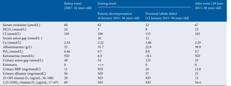
In 2011, at the age of 36, the patient (height 1.44 m, body mass index 18) was admitted to the intensive care unit because of hyperthermia caused by serologically proven myxovirus in-fluenzae A. Investigations (Table1) revealed severe metabolic acidosis (HCO3, 5 mmol/L) with a profound serum anion gap (36 mmol/L) due to severe ketosis (ketonaemia, 6.9 mmol/L) with ketonuria (3+). Glycaemia (6 mmol/L) and serum lactate levels (1.7 mmol/L) were normal. Renal function was stable (serum creatinine 62 μmol/L and eGFRMDRD 93 mL/min/1.73 m²).
F I G U R E 1 :(A) (a) First kidney biopsy at the age of 5 (1980), periodic acid–Schiff staining showing nuclear and cytoplasmic glycogen
inclusions. (b–d) Second kidney biopsy (2011). The trichrome staining shows foci of acute tubular necrosis and proximal tubular lesions (b).
Nuclear and cytoplasmic glycogen inclusions are seen by periodic acid–Schiff stain (c, arrow) and by electron microscopy (d, arrow). (B)
Immu-nohistochemistry for GLUT2, megalin, cubilin and B2 H+–ATPase subunits showing normal localization on proximal tubular cells in control
kidney and the lack of expression in the patient. GLUT2 and B2 H+–ATPase subunits were stained in red whereas actin, megalin/cubilin, was
stained in green and nuclei in blue.
THE B ENCH-T O-BEDSIDE T RANSITION
The patient was rehydrated with sodium bicarbonate 1.4% and placed under respiratory support because of polypnoea and exhaustion. Insulin therapy was started. The serum anion gap returned to the normal range 12 mmol/L, but severe metabolic acidosis of renal origin persisted, with a positive urinary anion gap (125 mmol/L), low ammoniuria level (1 mmol/L), high bi-carbonaturia (55 mmol/L) and urinary pH (7.55). The blood– urine pCO2 gradient was calculated at 40 mmHg, which ex-cluded distal tubular involvement. Molar bicarbonate infusion was started and tubular dysfunction progressively improved. Finally, electrolytes returned to baseline levels (Table1). Bone densitometry revealed a T-score of −3.3.
A second kidney biopsy was performed because of persisting severe renal acidosis. The biopsy revealed severe lesions with acute tubular necrosis with loss of brush border membranes and nuclear and cytoplasmic inclusions compatible with glycogen deposits in proximal tubules (Figure 1Ab–d). Glomerular and interstitial compartments were normal. Electron microscopy confirmed glycogen accumulation (Figure1Ad). Immunoreac-tivity for GLUT2 (AbDSerotec, Kidlington, UK), megalin and cubilin [4] and H+–ATPase pump (subunit B2) [5] was investi-gated by immunofluorescence staining, in comparison with control kidneys. Figure1B shows the basolateral localization of GLUT2 in the human early proximal tubule of control kidneys and the absence of this transporter in the patient. Figure1B also shows dramatically reduced expression of megalin and cubilin and the lack of expression of the B2 H+–ATPase subunit in the patient compared with control kidneys.
DISCUS SION
Fanconi–Bickel syndrome is a well-characterized clinical entity, which combines generalized proximal tubule dys-function, impaired utilization of glucose and galactose, and hepatorenal glycogenosis. The diagnosis of Fanconi–Bickel syndrome was confirmed by the finding of a homozygous mutation (IVS 3+2t>c/IVS 3+2t>c) in SLC2A2, that predicts
the synthesis of a truncated protein. To our knowledge, this mutation is unique to our patient.
The glomerularfiltration rate was normal and stable in the patient, as described by others [6]. In contrast, his presentation included manifestations of severe proximal tubule dysfunction, with a severe, unusual metabolic acidosis.
Severe metabolic acidosis is exceptional in Fanconi–Bickel syndrome. In our patient, severe metabolic acidosis resulted from two mechanisms. First, the high anion gap metabolic acidosis observed initially was due to accumulation of ketone bodies induced by a major defect in glucose and galactose utiliz-ation aggravated by myxovirus influenzae A infection and stress. This defect is primarily caused both by the alteration of the GLUT2 transporter and by insulinopaenia resulting from expression of the mutated GLUT2 in pancreatic beta cells [7]. Notably, ketosis occurred without hyperglycaemia because of urinary glucose loss. The second component of metabolic acido-sis was of renal origin and linked to the proximal tubule defect with hypophosphataemia, glycosuria and low-molecular-weight proteinuria. Acid–base balance was only sustained by large bicarbonate supplementation until recovery of tubular lesions.
Analysis of kidney biopsy provided new insights into the molecular mechanisms of the disease. First, it showed the lack of GLUT2 expression at the basolateral side of proximal tubule cells (compare Figure 1Bd with Figure 1Ba and Bc, arrow), which werefilled with glycogen vacuoles. Secondly, immuno-histochemistry showed highly reduced megalin and cubilin (compare Figure1Bf and Bh with Figure1Be and Bg, respect-ively), both required in tubular absorption of ligands and recy-cling of transporters through the endocytotic compartment [8], accounting for the high urinary loss of retinol binding protein. In addition, the B2 subunit of the H+–ATPase in-volved in proximal tubule reabsorption of bicarbonate and megalin- and cubilin-dependent endocytosis of proteins was also lacking (compare Figure 1Bj with Figure 1Bi, arrow). Similar impaired expression of key molecules of proximal tubular function have been reported in mice and humans with genetic alterations in the hepatic nuclear factor 1 alpha
Table 1. Serum and urinary data before, during the two steps of the exacerbation and after the event
Before event
(2007–32 years old) During event After event (29 June2013–38 years old) Ketosic decompensation
(6 January 2011–36 years old) Proximal tubule defect(12 January 2011–36 years old)
Serum creatinine (μmol/L) 60 62 52 67
HCO3(mmol/L) 24 5 8 23
Cl (mmol/L) 105 106 115 102
Serum anion gap (mmol/L ) – 36 12 –
Ca (mmol/L) 2.54 2.22 1.88 2.29
Albuminaemia (g/L) 52 31.7 22.9 39.9
PO4(mmol/L) 0.46 0.7 0.8 0.7
Ketonaemia (mmol/L) ND 6.9 <0.1 ND
Urinary anion gap (mmol/L) 48 54 125 33
Ketonuria 0 +++ 0 0
Urinary RBP (mg/mmolC) 11 ND 20 12.8
Urinary albumin (mg/mmolC) 56 ND 57 21
25-OH vitamin D3(ng/mL, 30–100) 20 ND ND 12
1,25-(OH)2vitamin D3(pg/mL, 17–67) 69 ND ND 56.4
RBP, retinol binding protein; +++, ketonuria assessed by urinary dipstick on a scale from 0 to +++; ND, not determined.
THE B ENCH-T O-BEDSIDE T RANSITION A c u t e m e t a b o l i c a c i d o s i s r e l a t e d t o F a n c o n i– B i c k e l s y n d r o m e iv115
(HNF1α) and 4 (HNF4) transcription factors leading to a Fanconi syndrome-like phenotype [9,10]. In our patient, these major phenotypic alterations were most likely induced by chronic accumulation of glycogen leading to cell injury. We hypothesize that these alterations account for the severe tubular dysfunction including renal acidosis observed in the Fanconi–Bickel syndrome. It is likely that, in the context of severe metabolic acidosis and superimposed tubular necrosis, these alterations were aggravated. Return of the renal par-ameters, including serum bicarbonate and urinary anion gap to baseline values, suggests that these additional lesions recov-ered, although they may influence the renal outcome later on.
In conclusion, we present a case of severe metabolic acidosis in a patient with Fanconi–Bickel syndrome which led to unravel at least some molecular bases for defective tubular transport.
AC K N OW L E D G E M E N T
We thank Sara Terryn for help in immunohistochemistry. Re-search of the authors is supported by grants from the 7th Fra-mework Programme of the European Community (contract 2012-305608; “European Consortium for High-Throughput Research in Rare Kidney Diseases (EURenOmics)”) and the Klinischer Forschungsschwerpunkt (KFSP) radiz - Rare Disease Initiative Zürich.
CON F LI CT O F IN TE R E S T S TATE M E N T
None declared.
R E F E R E N C E S
1. Santer R, Schneppenheim R, Suter D et al. Fanconi-Bickel syndrome—the original patient and his natural history, historical steps leading to the primary defect, and a review of the litterature. Eur J Pediatr 1998; 157: 783–797
2. Santer R, Calado J. Familial renal glycosuria and SGLT2: from a Mendelian trait to a therapeutic target. Clin J Am Soc Nephrol 2010; 5: 133–141
3. Santer R, Groth S, Kinner M et al. The mutation spectrum of facilitative glucose transporter gene SLC2A2 (GLUT2) in patients with Fanconi-Bickel syndrome. Hum Genet 2002; 110: 21–29.
4. Christensen EI, Devuyst O, Dom G et al. Loss of chloride channel ClC-5 impairs endocytosis by defective trafficking of megalin and cubilin in kidney proximal tubules. Proc Natl Acad Sci USA 2003; 100: 8472–8477 5. Mohebbi N, Mihailova M, Wagner CA. The calcineurin inhibitor FK506
(tacrolimus) is associated with transient metabolic acidosis and altered expression of renal acid–base transport proteins. Am J Physiol Renal Physiol 2009; 297: F499–F509
6. Manz F, Bickel H, Brodehl J et al. Fanconi-Bickel syndrome. Pediatr Nephrol 1987; 1: 509–518
7. Leturque A, Brot-Laroche E, Le Gall M. GLUT2 mutations, translocation, and receptor function in diet sugar managing. Am J Physiol Endocrinol Metab 2009; 296: E985–E992
8. Christensen El, Verroust PJ, Nielsen R. Receptor-mediated endocytosis in renal proximal tubule. Pflugers Arch 2009; 458: 1039–1048
9. Pontoglio M, Barra J, Hadchouel M, Doyen A et al. Hepatocyte nuclear factor 1 inactivation results in hepatic dysfunction, phenylketonuria, and renal Fanconi syndrome. Cell 1996; 84: 575–585
10. Satnescu DE, Hughes N, Kaplan B, Stanley CA et al. Novel presentations of congenital hyperinsulinism due to mutations in the MODY genes: HNF1α and HNF4A. J Clin Endocrinol Metab 2012; 97: E2026–E2030
Received for publication: 14.1.2014; Accepted in revised form: 14.1.2014
THE B ENCH-T O-BEDSIDE T RANSITION