CLINICAL IMAGE
Cystic ameloblastoma in a 5-year-old boy
Veronika Wassermann&Thierry A. G. M. Huisman
Received: 9 November 2007 / Accepted: 26 November 2007 / Published online: 8 January 2008 # Springer-Verlag 2007
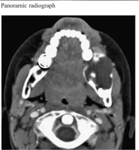
A 5-year-old boy presented with an indolent swelling of the left mandible without displacement or loosening of the teeth. A panoramic radiograph showed a large, well-defined, “bubbly”, multiloculated, lucent expansile lesion within the left posterior mandibular body and angle (Fig.1). A contrast-enhanced CT scan showed a low-attenuation lesion with scalloping and partial resorption of the buccal and lingual mandibular cortex. The adjacent soft tissues were displaced; there was no extraosseous extension (Fig.2). Tumor biopsy confirmed the presumed diagnosis of ameloblastoma. Resec-tion of the left mandibular body/angle with immediate reconstruction was undertaken.
Ameloblastomas are benign epithelial neoplasms that develop from various sources of odontogenic epithelium [1,2]; they represent 10% of all odontogenic tumors. Most ameloblastomas occur in the ramus and posterior body of the mandible (80%). They are usually seen in the third or fourth decade of life, but also occur in children. Their radiographic appearance varies. Differentiation from odon-togenic keratocysts, odonodon-togenic myxomas, aneurysmal bone cysts, and giant cell granulomas is limited [1,2]. CT and MRI are helpful in exactly delineating and determining the extension of the lesion, but imaging is not pathogno-monic [2]. Tumor biopsy remains mandatory.
References
1. Cihangiroglu M, Akfirat M, Yildirim H (2002) CT and MRI findings of ameloblastoma in two cases. Neuroradiology 44:434– 437
2. Asaumi J, Hisatomi M, Yanagi Y et al (2005) Assessment of ameloblastomas using MRI and dynamic contrast-enhanced MRI. Eur J Radiol 56:25–30
Pediatr Radiol (2008) 38:482 DOI 10.1007/s00247-007-0716-6
V. Wassermann
:
T. A. G. M. Huisman (*) Department of Diagnostic Imaging, University Children’s Hospital Zurich, Steinwiesstrasse 75,Zurich CH-8032, Switzerland e-mail: [email protected]
Fig. 1 Panoramic radiograph