Early Miocene herpetofaunas from the Greek localities of Aliveri and Karydia –
bridging a gap in the knowledge of amphibians and reptiles from the early Neogene
of southeastern Europe
Georgios L. Georgalisa,b, Andrea Villab, Martin Ivanovc, Socrates Roussiakisd, Panagiotis Skandalosd and
Massimo Delfinob,e
aDepartment of Geosciences, University of Fribourg, Fribourg, Switzerland; bDipartimento di Scienze della Terra, Università di Torino, Torino, Italy; cFaculty of Science, Department of Geological Sciences, Masaryk University, Brno, Czech Republic; dFaculty of Geology and Geoenvironment, National
& Kapodistrian University of Athens, Athens, Greece; eInstitut Català de Paleontologia Miquel Crusafont, Universitat Autònoma de Barcelona, Edifici
ICTA-ICP, Barcelona, Spain
ABSTRACT
We here describe new remains of amphibians and reptiles from the early Miocene (MN 4) of two different Greek localities, Aliveri and Karydia. The newly described material consists of urodelans, alytids, indeterminate anurans, turtles, crocodylians, lacertids, indeterminate scincomorphs, anguids, colubrids, viperids, and indeterminate snakes. The presence of the frog Latonia cf. gigantea in Greece is documented for the first time. Additionally, the presence of viperids in Aliveri implies a much wider distribution for these snakes during the early Miocene of Europe. Of special interest is the presence of a peculiar colubrid that seems to possess a hitherto unknown vertebral structure, which is herein defined as the ‘paracentral ridge’. Although incomplete, the new material has important taxonomic and biogeographic implications, as it enhances our understanding of southeastern European herpetofaunas from the early Miocene, a time period that was characterised by major dispersal and extinction events and climatic change that affected the whole continent.
Introduction
The early Miocene was an important time interval for European vertebrate faunas, as it witnessed major dispersal events from both Africa and Asia, but also important climatic changes char-acterised by higher temperatures, all resulting in the emergence of new palaeoenvironments, extinction events, and drastic faunal turnovers (Rögl 1999; Böhme 2003). The most important studies about these early Miocene events have so far focused primar-ily on mammals (e.g. Koufos et al. 2005), but knowledge of the respective coeval amphibians and reptiles is far more limited and poorly documented (Ivanov 2001; Delfino et al. 2003; Rage and Roþek 2003; ýerňanský 2012; Rage 2013). The situation is even more puzzling for the southeastern portions of Europe, where the known early Miocene herpetofauna is limited to only few spo-radic occurrences (Đuriü 2016; Georgalis et al. 2013; Georgalis, Villa and Delfino 2016; Vasileiadou et al. 2017).
We try here to fill this gap by describing new fossil amphib-ians and reptiles from two distinct Greek localities, Aliveri and Karydia, both pertaining to the MN 4 zone (Burdigalian, early Miocene). Up to now, only a chamaeleonid lizard (Chamaeleo cf.
andrusovi) had been described from Aliveri (Georgalis, Villa and
Delfino 2016). The turtles from both localities were only prelim-inarily mentioned with no descriptions or figures by Georgalis and Kear (2013) as ‘Emydidae (?) indet.’ (for Aliveri-2) and ‘Testudinata indet.’ (for Karydia-2). We analyze the taxonomic
affinities of the new Aliveri and Karydia specimens and addi-tionally discuss biogeographic implications that enhance our comprehension of the herpetofaunas of southeastern Europe.
Abbreviations: AL1a, Aliveri 1a Site; AL1b, Aliveri 1b Site;
AL1980NQ, Aliveri 1980 New Quarry Site; AL2, Aliveri 2 Site; AMPG, Athens Museum of Palaeontology and Geology, National and Kapodistrian University of Athens, Athens, Greece; HNHM, Hungarian Natural History Museum, Budapest, Hungary; KR2, Karydia-2 Locality; MDHC, Massimo Delfino Herpetological Collection, University of Torino, Torino, Italy; MNCN, Museo Nacional de Ciencias Naturales, Madrid, Spain; MNHN, Muséum national d’Histoire naturelle, Paris, France; NHMW, Naturhistorisches Museum Wien, Vienna, Austria; NMP, Národní Muzeum Praha, Prague, The Czech Republic; UU, University of Utrecht, Utrecht, The Netherlands; ZZSiD, Institute of Systematics and Evolution of Animals, Polish Academy of Sciences, Kraków, Poland.
Materials and methods
The majority of specimens described herein belongs to the collec-tion of the UU, whereas the remaining of the described material belongs to the AMPG. Comparative material includes multiple skeletons of extant frogs, salamanders, turtles, lizards, and snakes held in HNHM, MDHC, MNCN, MNHN, NHMW, NMP, and ZZSiD.
KEYWORDS
Amphibia; Reptilia; Miocene; taxonomy; biogeography; Greece
CONTACT Georgios L. Georgalis dimetrodon82@gmail.com, georgios.georgalis@unifr.ch
http://doc.rero.ch
Published in "Historical Biology 31(8): 1045–1064, 2019"
which should be cited to refer to this work.
represented by the equid Anchitherium, two species of the pal-aeomerycid Lagomeryx, the bovid Eotragus, and the carnivorans
Euboictis aliveriensis and Palaeogale sp. (van den Hoek Ostende
et al. 2015). As for the herpetofauna, up to now, only chamaele-onid lizards have been described from Aliveri (Georgalis, Villa and Delfino 2016), whereas turtles were only briefly mentioned by Georgalis and Kear (2013).
The locality of Karydia is situated in northeastern Greece, in the administrative region of East Macedonia and Thrace (Figure 1). It is located northeast of the town of Komotini, about 800 m south of the Karydia village, and was discovered in 1989 by Hans de Bruijn and Dimitris Foussekis (Doukas 2005). The locality belongs to the Neogene sedimentary sequence of the Thrace Basin. According to Doukas and van den Hoek Ostende (2006), the material was collected from a clay quarry, from three fossiliferous levels around a hill (Karydia-1, -2, and -3 [herein dubbed as KR1–KR3]), and all levels are considered synchro-nous, although the lithology indicates a slightly older age for KR3. Similarly to Aliveri, the Karydia assemblage is attributed to MN 4. However, the rodent fauna implies a younger age for Karydia. Theocharopoulos (2000) argued that Democricetodon
franconicus from Aliveri is more primitive than conspecific
material from Karydia, indicating a slightly younger age for the latter locality. A slightly younger age for Karydia is also sup-ported by the more advanced evolutionary stage of the rodents
Cricetodon and Anomalomys, and the presence in Karydia of Ligerimys instead of Pseudotheridomys (Doukas 2003; Koufos 2006; van den Hoek Ostende et al. 2015). The younger age of Karydia relative to that of Aliveri, is also supported by insec-tivores, as the former locality is characterised by the presence of Galerix kostakii, a species considered as a descendant of G.
symeonidisi, the latter being present in Aliveri (Doukas and van
den Hoek Ostende 2006). Similarly to Aliveri, Karydia shares
Geological settings
The fossiliferous localities of Aliveri and Karydia, together with those of Gavathas and Lapsarna (both on Lesvos Island), Nostimo (Western Macedonia), and possibly of Kalimeriani (Euboea Island), are among the few early Miocene localities in Greece that have yielded fossils of terrestrial vertebrates (Koufos et al. 2003; Koufos 2006; Vasileiadou and Zouros 2012; Georgalis et al. 2013; Koufos 2013; Georgalis, Villa and Delfino 2016; Vasileiadou et al. 2017).
The locality of Aliveri is situated on the island of Euboea (or Evia) in the administrative region of Central Greece (Figure 1) and was discovered in 1977 by a Dutch-Greek team consisting of Albert van der Meulen, Hans de Bruijn and Georgios Katsikatsos (de Bruijn and van der Meulen 1979; Doukas 2003; van den Hoek Ostende et al. 2015). The Aliveri locality represents a lignitic pit in the Neogene Kymi-Aliveri Basin (de Bruijn et al. 1980). The fossil material from the Aliveri locality originates from four dif-ferent sites, Aliveri 1a, Aliveri 1b, Aliveri 2, and Aliveri 1980 New Quarry. All of these sites are considered coeval. Unfortunately, the locality is not accessible anymore due to housing develop-ment in the area. Recently, van den Hoek Ostende et al. (2015) revised the fauna of Aliveri and also provided a history of past discoveries and studies. Though initially correlated to the MN 3 zone (de Bruijn et al. 1980), the fauna of Aliveri is now referred to earliest MN 4, with an estimated age between 18 and 17.5 Ma (Koufos 2006; van den Hoek Ostende et al. 2015). According to van den Hoek Ostende et al. (2015), the Aliveri assemblage is unique in representing the earliest European Neogene local-ity documenting the co-occurrence of eastern immigrants that include the rodents Cricetodon, Eumyarion, Democricetodon,
Megacricetodon, and the insectivore Galerix symeonidisi, in
com-bination with the presence of European taxa. Besides the abun-dant micromammal taxa, large mammals are also known, being
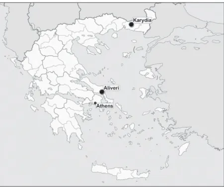
Figure 1. Map of Greece with indication of the localities of Aliveri and Karydia. Source: https://commons.wikimedia.org/wiki/File:Greece_location_map.svg
certain congeneric mammal taxa with early Miocene Anatolian localities (e.g. de Bruijn, 2017). No large mammals are known from Karydia and no herpetofauna has been described to date either. It should be highlighted that Karydia has produced far less fossil material in comparison to Aliveri.
Systematic Palaeontology Amphibia Blainville, 1816 Urodela Duméril, 1806 Urodela indet. (Figure 2)
Material. KR3: a tibia (AMPG KR3 037).
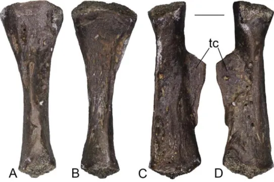
Description. The tibia is medium-sized, with a total length of
6 mm (Figure 2). It is moderately slender and presents a well-de-veloped tibial crest, whose free portion is broken off. A second low ridge is visible on the ventral surface of the bone, running along its entire length.
Remarks. This single, isolated tibia is fully comparable with a
medium-sized urodelan in terms of general morphology. A lack of detailed knowledge about the skeletal anatomy of modern salamanders, however, hinders identifying further taxonomic significant features of the Karydia fossil element.
Anura Fischer von Waldheim, 1813 Alytidae Fitzinger, 1843
Latonia Meyer, 1843
Latonia gigantea (Lartet, 1851)
Latonia cf. gigantea (Figure 3)
Material. KR3: 15 maxillae (AMPG KR3 004, AMPG KR3 005,
AMPG KR3 014–AMPG KR3 016, AMPG KR3 027, AMPG KR3 030, AMPG KR3 048–AMPG KR3 055), a frontoparietal (AMPG KR3 038), two praearticulars (AMPG KR3 028 and AMPG KR3 029), two trunk vertebrae (AMPG KR3 031 and AMPG KR3 033), a sacral vertebra (AMPG KR3 032), and four ilia (AMPG KR3 011, AMPG KR3 026, AMPG KR3 044, and AMPG KR3 045).
Description.
Maxillae. These elements are small and incomplete (Figure 3(A)– (F)). The longest fragments, AMPG KR3 005 and AMPG KR3 015, slightly exceed 5 and 6 mm respectively. The lateral surface is generally smooth. Only AMPG KR3 015 shows a light dermal ornamentation made up by small and indistinct tubercles along the high processus zygomaticomaxillaris, but the presence, or not, of a similar ornamentation in the other specimen cannot be discerned since this part of the bone is missing. Some of the Karydia-3 specimens (e.g. AMPG KR3 005) preserve, at least partially, the anterior end and the processus palatinus. The latter is narrow, anteriorly inclined and gutter-shaped. The portion of the maxilla anterior to the processus palatinus is rather long, being delimited laterally by a high lamina anterior. The lamina horizontalis narrows towards the anterior end. Its dorsal surface displays a rather deep and narrow fossa maxillaris just anterior to the processus palatinus. The posterior end is preserved, at least partially, only in AMPG KR3 015, AMPG KR3 016, and AMPG KR3 027. In these specimens, the lamina horizontalis slightly narrows towards the posterior end and develops a rather long and slender processus pterygoideus. The tooth row extends posteriorly to the end of the lamina horizontalis. A shallow pos-terior depression is recognizable, including a narrow foramen by its contact with the lamina horizontalis. The depression is not marked by ridges anteriorly. The margo orbitalis is strongly concave.
Frontoparietal. The fragmentary frontoparietal is rather small,
with a total preserved length of roughly 4 mm (Figure 3(G)–(H)). The anterior end of the bone is missing, whereas posteriorly the bone misses the entire left corner, most of the right processus paraoccipitalis and the right lateral margin. In the entire pre-served portion of the bone, the facies dorsalis is rather distinctly narrower than the pars contacta, and the tectum supraorbitale
Figure 2. Urodela indet. from Karydia-3: left tibia (AMPG KR3 037) in dorsal (A), ventral (B), lateral (C) and medial (D) views.
Scale bar = 1 mm. Abbreviations: tc, tibial crest.
Praearticulars. These elements are rather small-sized (Figure 3(I)–(J)). They have a slender and horizontal processus para-coronoideus followed posteriorly by a slender and vertically ori-ented processus coronoideus. The latter is completely damaged in AMPG KR3 029, but its base is visible, attesting its original presence. The rather narrow sulcus pro cartilago Meckeli is rather shallow anteriorly, but it deepens strongly by the processus cor-onoideus. The lateral surface displays a large and deep depres-sion, marked ventrally by a sharp crista mandibulae externa. The extremitas spatulata is missing.
Trunk vertebrae. They are small sized and represented only by
the centrum (Figure 3(K)–(L)). The centrum is ophisthocoelous and displays a distinct condylar neck. The shape of the centrum is rather cylindrical, but a slight ventral concavity is visible in lateral view. The concavity is more evident in AMPG KR3 033, however, it is distinctly smaller in AMPG KR3 031. Small por-tions of the rather thin lateral walls of the neural arch are also preserved in both specimens.
Sacral vertebra. This element also preserves only the
verte-bral centrum (Figure 3(M)). It has an anterior condyle and two appears to be developed only as a rather low lamina by each side
of the posterior end of the facies dorsalis. The latter is covered by a moderate dermal ornamentation consisting of low tubercles and grooves. The degree of development of the ornamentation tends to fade both anteriorly and towards the lateral sides. Posteriorly to the facies dorsalis, a rather low longitudinal ridge is present in the middle of the smooth posterior area of the dorsal surface of the frontoparietal. By the end of this ridge, a processus posterior is not developed. A sharper ridge runs also at the middle of the dorsal surface of the preserved portion of the processus paraoc-cipitalis. Medially to the contact between the facies dorsalis and the latter ridge, a small foramen is visible that might represent the opening for the occipital artery. On the lateral sides, the pars contacta is laminar and ventrolaterally extended. On the ventral surface, the incrassatio frontoparietalis is clearly divided into an anterior and a posterior portion, even though the margins of these two portions are poorly marked. Both portions are large. Only the posterior portion of the incrassatio is completely pre-served, showing a circular shape. The surface of the incrassatio frontoparietalis is smooth.
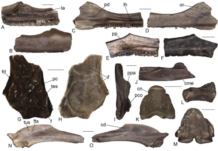
Figure 3. Latonia cf. gigantea from Karydia-3: left maxilla (AMPG KR3 005) in medial (A) and lateral (B) views; left maxilla (AMPG KR3 015) in medial (C) and lateral (D)
views; left maxilla (AMPG KR3 016) in medial (E) and lateral (F) views; frontoparietal (AMPG KR3 038) in dorsal (G) in ventral (H) views; left praearticular in dorsal (I) and lateral (J) views; trunk vertebra (AMPG KR3 033) in ventral (K) and left lateral (L) views; sacral vertebra (AMPG KR3 032) in dorsal view (M); right ilium (AMPG KR3 011) in lateral (N) and medial (O) views.
Scale bars = 1 mm. Abbreviations: cd, crista dorsalis; cme, crista mandibulae externa; cn, condylar neck; f, foramen; fd, facies dorsalis; fts, fossula tuberis superioris; if, incrassatio frontoparietalis; la, lamina anterior; lh, lamina horizontalis; or, ornamentation; pc, pars contacta; pco, processus coronoideus; pd, posterior depression; pp, processus pterygoideus; ppa, processus paracoronoideus; tes, tectum supraorbitale; tus, tuber superior.
of the incrassatio frontoparietalis. The small size of the spec-imen also agrees with a juvenile condition, even though the ornamentation, consisting of low tubercles instead of pits and ridges, might suggest it was not a postmetamorphic individual, but rather a subadult (Roþek 1994, 2013). The identification as a rather young specimen of L. gigantea might be also valid for at least the maxilla AMPG KR3 015, based on the presence of low developed tubercles on the processus zygomaticomaxillaris and the absence of the ridge marking the anterior part of the medial depression in adults (Roþek 1994, 2013). Given the small size and the overall similar morphology shown by the other specimens, it seems possible that all discoglossine fossils from Karydia-3 might belong to young individuals of L. gigantea. Nevertheless, the presence of a foramen for the occipital artery on the fronto-parietal is rather puzzling, and, as such, the identification is here considered only tentative.
cf. Latonia sp. (Figure 4A–G)
Material. AL1a: an atlas (UU AL 3593). AL1980NQ: three
max-illae (UU AL 3552, UU AL 3555, and UU AL 3596), and a right ilium (UU AL 3598).
Description.
Maxillae. All maxillae from Aliveri are represented by small
fragments, the largest one (UU AL 3552) being roughly 4 mm in total length (Figure 4(A)–(B)). They preserve only part of the middle portion of the bone. In medial view, a mediolaterally short and medially convex lamina horizontalis is visible, mark-ing the crista dentalis dorsally. The latter bears pleurodont, close-ly-spaced, and cylindrical teeth, of which none is completely preserved. The number of preserved tooth-positions is at least 12 in UU AL 3552. The dorsal surface of the lamina horizontalis is marked by a narrow and rather deep groove. The preserved portion of the lateral surface is smooth in all specimens. Certain morphological features, such as the shape of the lamina hori-zontalis, the tooth morphology, and the smooth lateral surface resemble the specimens from Karydia-3, described above as
Latonia cf. gigantea.
Atlas. The atlas from Aliveri preserves only the vertebral centrum
(Figure 4(C)–(E)). It is small-sized, strongly dorso-ventrally posterior condyles. Its size is comparable with the largest trunk
vertebra. The condyles are dorsoventrally compressed and the centrum is ventrally flattened.
Ilia. These elements are rather small. AMPG KR3 011 preserves
most of the shaft, but only a small part of the acetabular portion (Figure 3(N)–(O)). A moderately high crista dorsalis is present. Its anterior half is missing, but the preserved portion seems to display a medial bending anteriorly. The posterior end of the crista is characterised by a poorly marked and anteroposteriorly elongated tuber superior. A shallow fossula tuberis superioris (sensu Roþek 1994) is present, housing a small foramen. The supracetabular fossa is deep. Most of the acetabulum is missing, but its ante-rior margin was strongly raised laterally and seems to have been prominent. Both the partes ascendens and descendens are almost completely broken off, but a deep interiliac groove is still visible in medial view. The other specimens are less well-preserved, but their morphology is fully comparable with that of AMPG KR3 011.
Remarks. Maxillae with a long and slender processus
ptery-goideus and a medial depressed area in the posterior portion, together with prearticulars possessing a processus paracoro-noideus associated to the processus coroparacoro-noideus clearly attest the presence of the discoglossine Latonia in Karydia-3 (Roþek 1994, 2013). Other, similar-sized remains showing discogloss-ine features, such as opisthocoelous vertebrae and ilia with a medially-bending crista dorsalis and an interiliac groove, can also be assigned to the same taxon (Roþek 1994, 2013; Bailon 1999). The attribution of the frontoparietal from KR3 to Latonia is supported by its unpaired nature and the split incrassatio fron-toparietalis with a circular posterior portion, but the presence of a foramen for the occipital artery is rather unusual, since this feature is reported to be absent in representatives of the genus (Roþek 1994, 2013; Rage and Hossini 2000). Nevertheless, the general morphology of the frontoparietal is fully comparable with early ontogenetic stages of Latonia gigantea, as described by Roþek (1994) and Rage and Hossini (2000). In particular, the most significant similarities are the narrow facies dorsalis if compared with the pars contacta, the laminar and ventrolater-ally-developed pars contacta, and the poorly-marked margins
Figure 4. cf. Latonia sp. from Aliveri (A–G): left maxilla (UU AL 3552) in medial (A) and lateral (B) views; atlas (UU AL 3593) in dorsal (C), ventral (D) and anterior (E) views; right ilium (UU AL 3598) in lateral (F) and medial (G) views. Anura indet. from Aliveri (H): trunk vertebra (UU AL 3553) in dorsal view (H). Anura indet. from Karydia-2 (I): left humerus (UU KR2 5015) in ventral view (I).
Scale bars = 1 mm.Abbreviations: ec, eminentia capitata; fcv, fossa cubitalis ventralis; fts, fossula tuberis superioris; k, keel; si, spatium interglenoidale.
vertebra was opisthocoelous or procoelous in life. As such, we refrain from formally referring it to cf. Latonia and treat this vertebra as an indeterminate anuran. Furthermore, the recog-nition of the condylar neck is also hindered by a distinct lateral development of the condyle on both sides, possibly indicating a rather high degree of lateral movement of the articulating verte-brae. The respective material from Karydia-2 is relatively better preserved: a medium-sized humerus preserving only the distal epiphysis and a small and very poorly preserved fragment of a radioulna can be assigned only to an indeterminate anuran. The humerus displays a sphaerical eminentia capitata and a mod-erately deep fossa cubitalis ventralis (Figure 4(I)). The epicon-dylus ulnaris is robustly built. The epiconepicon-dylus radialis, on the other hand, is rather small and displays a distinct tubercle on the ventral surface, which is separated from the eminentia capitata by a narrow groove. The bases of both the cristae lateralis and medialis are well developed. On the dorsal surface, the olecranon scar is not recognizable. The fossil remains from Karydia-3 are fragmentary and lack diagnostic characters to the family level, and as such, are here also identified as indeterminate anurans.
Reptilia Laurenti, 1768 Testudines Batsch, 1788 Testudines indet. (Figure 5)
Material. AL2: two shell fragments (UU AL 3504–UU AL 3505).
KR2: three shell fragments (UU KR2 5001–UU KR2 5003).
Description. The two fragments from Aliveri can be joined
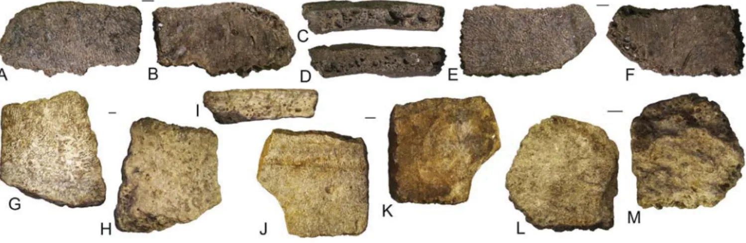
together in a single, larger fragment (about one square cm) that comes from a chelonian shell (Figure 5(A)–(F)). This identifica-tion is supported by the presence of a smooth visceral and a finely vermiculated external surface characterised by a scute sulcus. Moreover, two of the edges of this larger fragment host the typical chelonian sutures. The three small fragments from Karydia are characterised by being thin (2–4 mm) and provided with a rather smooth (visceral) surface and a slightly rough opposite (external) surface (Figure 5(G)–(M)). With the exception of UU KR2 5002, the fragments show a sutural surface on at least one edge. Growth marks are visible on the slightly vermicular external surface of UU KR2 5001. UU KR2 5002 hosts a straight sulcus on the dorsal surface and shows a very modest convexity on its ventral surface, suggesting that this fragment could originate from a costal bone. A rather thin, elongated tubercle, associated with the presence of sutures at three edges, could indicate that UU KR2 5003 is a small fragmentary costal bone as well.
Remarks. The material from Aliveri and Karydia is too
frag-mentary to permit a precise identification of the shell elements. Four clades of non-marine turtles are known from the early Miocene of this region: pan-testudinoids (sensu Joyce et al. 2004), podocnemidoideans, pan-chelydrids, and pan-tri-onychids (Georgalis and Kear 2013; Georgalis et al. 2013; Joyce 2016; Georgalis and Joyce 2017). The absence of a sculpturing pattern clearly denotes that the material described herein does not pertain to pan-trionychids (Georgalis and Joyce 2017). The general morphology is reminiscent of pan-testudinoids but podocnemidoidean and chelydrid affinities cannot be excluded. Therefore, the material from both localities is herein referred to Testudines indet.
Crocodylia Gmelin, 1789 Crocodylia indet. (Figure 6) compressed, and has a subcircular posterior cotyle. Anteriorly,
two kidney-shaped cotyles are present. They are moderately large and dorsoventrally inclined in anterior view. They are not in contact, since a moderately wide spatium interglenoidale sepa-rates them in the middle. A robust keel is visible on the ventral surface of the centrum.
Ilium. The small ilium from Aliveri preserves part of the
ace-tabular portion and the base of the shaft (Figure 4(F)–(G)). On the dorsal margin, part of a rather poorly marked tuber supe-rior is preserved. In life, it was probably confluent with a crista dorsalis, which is now missing. Even though the pars ascendens is missing, the angle between the latter and the tuber superior is rather obtuse in lateral view. Two foramina, a strongly larger posterior one and a rather small anterior one, are visible on the lateral surface of the bone; they are located in a rather shallow fossula tuberis superioris. There is no supracetabular fossa. The acetabulum is strongly eroded, but its anteroventral margin is prominent laterally. The preacetabular area is not expanded. Because the ileoischiadic junction is not preserved, it is not possible to discern whether the interiliac tubercle and groove were present.
Remarks. In spite of their fragmentary nature, a clear similarity
of these specimens is recognizable with Latonia. In particular, the Aliveri specimens resemble Latonia in the shape of the lamina horizontalis of the maxillae, the ventral keel, the dorsoventrally inclined and medially separated anterior cotyles of the atlas, the poorly-marked tuber superior, the obtuse angle between the tuber and the pars ascendens, and the presence of a fossula tub-eris superioris with foramina located into the latter on the ilium (Roþek 1994, 2013; Biton et al. 2016). Among discoglossine alyt-ids, a certain similarity is apparent in at least some bones between
Latonia and Discoglossus (e.g. the ilium; Roþek 1994; Biton et al. 2016), but the Aliveri fossils cannot be assigned to the latter genus due to the fact that the maxillae bear a more slender lamina horizontalis and the ilia have a more prominent tuber superior forming a less obtuse angle with the pars ascendens (Roþek 1994; Bailon 1999; Biton et al. 2016). Due to the poor preservational condition and the scarcity of the fossil material, we here prefer to identify these specimens only tentatively, avoiding also any specific identification, pending the possible discovery of new remains in the future.
Anura indet. (Figure 4H–I)
Material. AL1a: an ilium (UU AL 3595), a trunk vertebra (UU
AL 3553), and three indeterminate elements (UU AL 3554, UU AL 3594). AL1980NQ: an indeterminate element (UU AL 3597). KR2: a left humerus (UU KR2 5015) and a fragment of radioulna (UU KR2 5016). KR3: a premaxilla (AMPG KR3 003), an ilium (AMPG KR3 024), and eight phalanxes (AMPG KR3 006 and AMPG KR3 023).
Description and remarks. These anuran remains from Aliveri
are too fragmentary to allow for rigorous identification. Among them, the sole known, small (roughly 2.7 mm long) trunk verte-bra (UU AL 3553) could probably belong to the above described cf. Latonia sp. on the basis of the cylindrical shape of the centrum. However, the specimen is incomplete and only the cylindrical vertebral centrum is preserved (Figure 4(H)). It displays a cotyle and a condyle, both circular in shape, but since a clear condylar neck is not recognizable, it is not possible to state whether the
Figure 5. Testudines indet. from Aliveri (A–F): shell fragment (UU AL 3504) in external (A), visceral (B), and lateral (C) views; shell fragment (UU AL 3505) in lateral (D), external (E), and visceral (F) views. Testudines indet. from Karydia-2 (G–M): shell fragment (UU KR2 5001) in external (G), visceral (H), and lateral (I); shell fragment, probably a costal (UU KR2 5002) in external (J) and visceral (K) views; shell fragment, probably a costal (UU KR2 5003) in dorsal (L) and ventral (M) views).
Scale bars = 1 mm.
Figure 6. Crocodylia indet. from Aliveri: tooth (UU AL 3536) in labial (A), lingual (B), and mesial (C) views; tooth (UU AL 3537) in labial (D) and lingual (E) views; tooth (UU AL 3538) in labial (F), lingual (G), and mesial (H) views; tooth (UU AL 3539) in labial (I) and lingual (J) views; tooth (UU AL 3540) in labial (K) and lingual (L) views; tooth (UU AL 3541) in labial (M) and lingual (N) views; tooth (UU AL 3576) in labial (O) and lingual (P) views; tooth (UU AL 3577) in labial (Q), lingual (R), and mesial (S) views; tooth (UU AL 3578) in labial (T) and lingual (U) views.
Scale bars = 1 mm.
Chamaeleo cf. andrusovi
Material. AL1a: a skull roofing bone (UU AL 3501).
Remarks. The chamaeleonids from Aliveri were extensively
described by Georgalis, Villa and Delfino (2016) and the reader is referred to that paper for further details. Although they prob-ably all pertain to the same species, only the skull roofing bone possessed enough diagnostic characters for a species determi-nation and was accordingly referred to Chamaeleo cf. andrusovi, whereas the tooth bearing bones were simply considered as inde-terminate chamaeleonids. No further chamaeleonid material has been recovered from Aliveri.
Chamaeleonidae indet.
Material. AL1b: a fragment of tooth-bearing bone (UU AL 3502);
AL1980NQ: a fragment of tooth-bearing bone (UU AL 3503).
Remarks. See Chamaeleo andrusovi above.
Scincomorpha Camp, 1923 (sensu Estes et al. 1988) Lacertidae Oppel, 1811
Lacertidae indet. (Figure 7A–G)
Material. AL1a: a fragment of tooth-bearing bone (UU AL 3520).
AL1b: a dentary (UU AL 3586) and five fragments of tooth-bear-ing bones (UU AL 3557–UU AL 3561). AL1980NQ: a fragment of tooth-bearing bone (UU AL 3542). KR3: two maxillae (AMPG KR3 017–AMPG KR3 018) and an isolated tooth (AMPG KR3 007).
Material. AL1a: six isolated teeth (UU AL 3536–UU AL 3541).
AL1b: ten isolated teeth (UU AL 3576–UU AL 3585). AL1980NQ: an isolated tooth (UU AL 3556).
Description. All teeth from Aliveri share the following characters:
small size (the largest, UU AL 3539, is 2.5 cm long), unserrated mesiodistal carinae, rather acute shape (UU AL 3536 is slightly more massive than the others), lingual surface slightly concave, labial surface slightly convex, smooth lingual and labial surfaces, absence of root, and a concave base (Figure 6). UU AL 3537 is the tip of a slightly larger tooth, but shares with the other specimens all the characters available on the preserved portion of the tooth.
Remarks. Isolated crocodylian teeth are not diagnostic at a lower
taxonomic level (Delfino et al. 2007; ýerňanský et al. 2012; Georgalis, Villa, Vlachos et al. 2016) and therefore the Aliveri material can only be referred to Crocodylia indet. The small size of the teeth and the fact that their root was likely reabsorbed could indicate that they were shed by small individuals (there is no clear evidence of breakage of the root; Frey and Monninger 2010).
Squamata Oppel, 1811 Acrodonta Cope, 1864 Chamaeleonidae Gray, 1825 Chamaeleo Laurenti, 1768
Chamaeleo andrusovi ýerňanský, 2010
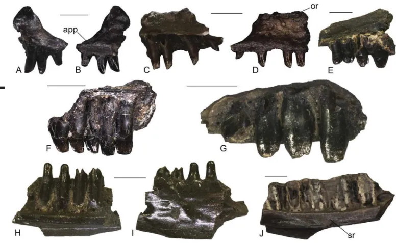
Figure 7. Lacertidae indet. from Karydia-3 (A–D): left maxilla (AMPG KR3 017) in medial (A) and lateral (B) views; left maxilla (AMPG KR3 018) in medial (C) and lateral (D) views. Lacertidae indet. from Aliveri (E–G): fragment of tooth-bearing bone (UU AL 3557) in medial view (E); fragment of tooth-bearing bone (UU AL 3558) in medial view (F); fragment of tooth-bearing bone (UU AL 3559) in medial view (G). Scincomorpha indet. from Aliveri (H–I): left dentary (UU AL 3519) in medial (H) and lateral (I) views; Scincomorpha indet. from Karydia-3 (J): right dentary (AMPG KR3 010) in medial view (J).
Scale bars = 1 mm. Abbreviations: app, anterior premaxillary process; or, ornamentation; sr, subdental ridge.
Material. AL1a: three dentaries (UU AL 3519, UU AL 3521, and
UU AL 3524). AL1980NQ: a dentary (UU AL 3549). KR3: a dentary (AMPG KR3 010).
Description. The specimens from Aliveri are poorly preserved
fragments of dentaries, with a total length of 3.5 mm (UU AL 3519), slightly less than 5 mm (UU AL 3521), roughly 2.5 mm (UU AL 3524), and roughly 3 mm (UU AL 3549) respectively. In medial view, the dentaries display a slender (UU AL 3519) or moderately thick (all other specimens) subdental ridge and a medially open Meckelian fossa. The ventral margin of the bone is missing in all specimens except for UU AL 3549, in which it appears rather straight. At least four (in UU AL 3519), seven (in UU AL 3524) and ten (in UU AL 3521) tooth positions are recog-nizable, but teeth are preserved only in UU AL 3519 (all four of them) and 3524 (a single one). Teeth appear to have been slender, cylindrical and pleurodont. Teeth of UU AL 3519 are strongly eroded at the tip, but appear to have a peculiar, rather abrupt constriction towards the dorsal level of the dentary (Figure 7(H)–(I)), however, it cannot be ascertained whether this was a true feature in life or is simply an artifact of taphonomy and preservation. The preserved tooth of UU AL 3524 is monocuspid. UU AL 3549 preserves the anterior end, displaying a narrow and subhorizontal mandibular symphysis. The lateral surface is smooth, carrying some mental foramina, in all specimens except for UU AL 3519, where it is slightly rugged.
The dentary fragment from Karydia-3 (AMPG KR3 010) displays a moderately thick subdental ridge on its medial side (Figure 7(J)). The Meckel’s groove is open and narrows anteriorly. The ventral margin of the fragment is mostly broken off. The alveolar portion carries 10 tooth positions, but the teeth are not preserved. Nevertheless, they were pleurodont, closely spaced and rather narrow. The lateral surface is smooth, with at least four mental foramina. The total length of the fragment is 6 mm.
Remarks. The presence of a subdental ridge on the medial
side discriminates dentaries of scincomorph lizards from those of anguimorphs, but it is also present in iguanians and gekko-tans (Evans 2008). Nevertheless, the combination of an open Meckel’s groove and the pleurodont dentition allow us to exclude an attribution of these dentaries to the latter two groups, rather favouring a scincomorph (sensu Estes et al. 1988) assignment. The preservational condition of the specimens from Aliveri hin-ders a taxonomic attribution at the family level, but the presence of more than one taxon might be suggested by the difference in the smoothness of the lateral surface of the dentaries. Regarding the Karydia-3 material, if our identification of the other scinco-morphs described above as adults of a small lacertid is correct, then the larger size of this dentary might suggest the presence of a second, though still indeterminate, scincomorph taxon in that locality.
Anguimorpha Fürbringer, 1900 Anguidae Gray, 1825
Anguinae Gray, 1825 Ophisaurus Daudin, 1803 cf. Ophisaurus sp. (Figure 8A–D)
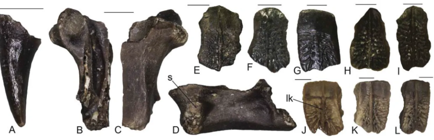
Material. KR3: a tooth (AMPG KR3 035) and a trunk vertebra
(AMPG KR3 034).
Description.
Tooth. The isolated tooth is rather small and pointed, even
though the tip is broken (Figure 8(A)). It has a conical, roughly
Description.
Maxillae. The lacertid maxillae from KR3 are rather small,
fragmentary, and represent only the anterior end of the bone (Figure 7(A)–(D)). The anterior premaxillary process has short anterolateral and anteromedial processes separated by a shallow concavity. Dorsally, the vomeronasal foramen is moderately large and housed in a shallow concave area. It seems that a lappet is absent on the dorsal surface of the anteromedial process. In spite of the strong fragmentary nature of the specimens, a der-mal ornamentation appears to be present on the lateral surface of the facial process, at least in AMPG KR3 018 (Figure 7(D)). Ventrally to the ornamentation, some ventrolateral foramina are present. Teeth are pleurodont, cylindrical, narrow, and closely spaced. AMPG KR3 017 preserves three teeth, whereas AMPG KR3 018 has four preserved teeth plus two empty tooth positions. Only one tooth preserves a monocuspid crown in the former specimen, whereas a bicuspid condition is recognizable in all teeth of the latter except for the anteriormost one in which the crown is not preserved.
Dentary. The dentary UU AL 3586 preserves only part of the
middle portion of the bone. It is 4 mm in length. The Meckelian fossa is wide and opens medially. Teeth are pleurodont but no one is preserved. A moderately slender subdental ridge is present. The ventral margin is distinctly convex in medial view. The labial surface is smooth, though a light roughness seems to be present, however, this could be due to taphonomical reasons. Two mental foramina are present.
Tooth bearing elements. The fragments of indeterminate tooth
bearing bones bear pleurodont, cylindrical, mono-, bi- and tricuspid teeth, which generally show wear (Figure 7(E)–(G)). Teeth of the largest specimens (UU AL 3557 and UU AL 3559) are hypertrophied.
Isolated tooth. AMPG KR3 007 is a single pleurodont and
cylin-drical tooth. It has a bicuspid crown, with a large main cusp and a smaller accessory cusp.
Remarks. The heterodont dentition of these tooth bearing bones
from Aliveri is indicative of lacertid affinities (Bailon 1991), though they cannot be more precisely identified due to their poor preservational condition. In spite of the absence of well-pre-served teeth, the Aliveri dentary is also assigned to the same taxon because of the convex ventral margin, which is also found in lacertids (AV, pers. obs.). It should be noted that the morphol-ogy of the hypertrophied teeth in UU AL 3557 and UU AL 3559 could be reminiscent of certain amblyodont lacertids from the Paleogene and early Neogene of western Europe (Augé 2005), but the Aliveri lizards cannot be attributed to the latter forms, as in amblyodont lizards, the crown is blunt and rounded, and as such, the cusps should be either totally absent or, in some cases, poorly marked.
The overall morphology of the KR3 specimens is consistent with an identification as undetermined lacertids (Bailon 1991; Barahona 1996). Similarly to the Aliveri lacertids, it is difficult to clearly identify the remains at a specific or even generic level, due to their poor preservational status. Nevertheless, their size would suggest the presence of a small-sized taxon in Karydia-3. The presence of the distinct dermal ornamentation on AMPG KR3 018 could testify that, at least this specimen pertains to an adult, and not to a juvenile of a larger species.
Scincomorpha indet. (Figure 7(H)–(J))
Description. Osteoderms from Aliveri are small, but rather thick
and robust (Figure 8(E)–(G)). They show an external surface with a smooth gliding portion, a vermicular ornamentation and a well-developed longitudinal keel on the external surface. The most well preserved ones are subrectangular in shape.
Osteoderms from Karydia-2 are similar to those from Aliveri. They are small, but robustly-built (Figure 8(H)–(I)). The external surface shows a smooth gliding portion, a vermicular ornamen-tation on the rest of the surface, and a well-evident longitudinal keel. Osteoderms from Karydia-3 are small and subrectangular in shape (Figure 8(J)–(L)). They display a vermicular ornamen-tation and, with the sole exception of AMPG KR3 008, a longi-tudinal keel on the external surface. Similar to the osteoderms from Aliveri and Karydia-2, they also show a smooth gliding portion in their external surface.
Remarks. The presence of anguids in Aliveri and Karydia is
tes-tified by a large number of osteoderms showing the typical ver-micular ornamentation on the external surface. The thickness, the presence of a keel, and the subrectangular shape are found in non-Anguis anguine taxa (i.e. either Pseudopus or Ophisaurus), in contrast to smaller, rounded and unkeeled osteoderms in Anguis (Delfino et al. 2011). Regarding the osteoderms from KR3, it is most probable that they pertain to cf. Ophisaurus sp. that was described above from that locality.
Serpentes Linnaeus, 1758 Alethinophidia Nopcsa, 1923 Colubridae Oppel, 1811 Colubridae indet. (Figure 9)
Material. AL1980NQ: an anterior trunk vertebra (UU AL 3590).
KR2: a posterior trunk vertebra (UU KR2 5018), a posterior trunk vertebra (UU KR2 5019), a caudal vertebra (UU KR2 5025), and a fragmentary vertebra (UU KR2 5020). KR3: a frag-ment of a trunk vertebra (AMPG KR3 020).
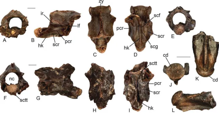
Description. The trunk vertebrae from both Aliveri and Karydia
are incomplete, lacking zygapophyses and synapophyses. The best-preserved specimen, a posterior trunk vertebra (UU KR2 canine shape. The tooth base is not swollen and the apex is not
curved. No striae are present. There are small and sharp carinae both anteriorly and posteriorly.
Trunk vertebra. The vertebra is procoelous and rather
fragmen-tary as only the left side is preserved (Figure 8(B)–(D)). The neural arch is completely missing, as well as the posterior condyle. The preserved portion of the centrum is roughly 4 mm long. The cen-trum is dorsoventrally compressed and has a flat ventral surface. Despite the absence of the condyle, a precondylar constriction does not seem to be present. The left lateral margin of the centrum is oblique and slightly concave in ventral view. The left synapoph-ysisis is eroded, but it is distinctly dorsoventrally elongated. The left prezygapophysis is subelliptical and slightly dorsally tilted.
Remarks. The morphology of both AMPG KR3 034 and
AMPG KR3 035 is reminiscent of that of Ophisaurus. As far as the vertebra is concerned, its attribution to Ophisaurus is sup-ported by the compressed centrum with no precondylar constric-tion, an oblique and concave lateral margin, and a flat ventral surface (Klembara 1981; Estes 1983). The isolated tooth, on the other hand, resembles Ophisaurus in its conical shape and the absence of a strong curvature (Klembara et al. 2014). Despite the fact that extant species of Ophisaurus are reported to have striated tooth crowns, fossil remains with unstriated Ophisaurus-like dentition are also known (e.g. Anguine morphotype I from Merkur-Nord; Klembara 2015). An attribution of these two fos-sils from KR3 to Ophisaurus seems therefore possible, but due to scarcity of material and its poor preservational status we here prefer to treat this identification with caution.
non-Anguis Anguinae indet. (Figure 8E–L)
Material. AL1a: 60 osteoderms (UU AL 3506–UU AL 3518).
AL1b: 14 osteoderms (UU AL 3562–UU AL 3575). AL1980NQ: 22 osteoderms (UU AL 3543–UU AL 3548). KR2: seven oste-oderms (UU KR2 5006–UU KR2 5013). KR3: 11 osteoste-oderms (AMPG KR3 001, AMPG KR3 002, AMPG KR3 008, AMPG KR3 019, AMPG KR3 039–AMPG KR3 043, AMPG KR3 046, AMPG KR3 047, AMPG KR3 056, and AMPG KR3 057).
Figure 8. cf. Ophisaurus sp. from Karydia-3 (A–D): isolated tooth (AMPG KR3 035) in medial view (A); trunk vertebra (AMPG KR3 034) in dorsal (B), ventral (C), and left lateral (D) views. non-Anguis Anguinae indet. from Aliveri (E–G): osteoderm (UU AL 3506) in external view (E); osteoderm (UU AL 3516) in external view (F); osteoderm (UU AL 3545) in external view (G); non-Anguis Anguinae indet. from Karydia-2 (H–I): osteoderm (UU KR2 5007) in external view (H); osteoderm (UU KR2 5008) in external view (I); non-Anguis Anguinae indet. from Karydia-3 (J–L): osteoderm (AMPG KR3 002) in external view (J); osteoderm (AMPG KR3 039) in external view (K); osteoderm (AMPG KR3 040) in external view (L).
Scale bars = 1 mm, except for A, in which it is 0.5 mm. Abbreviations: lk, longitudinal keel; s, synapophysis.
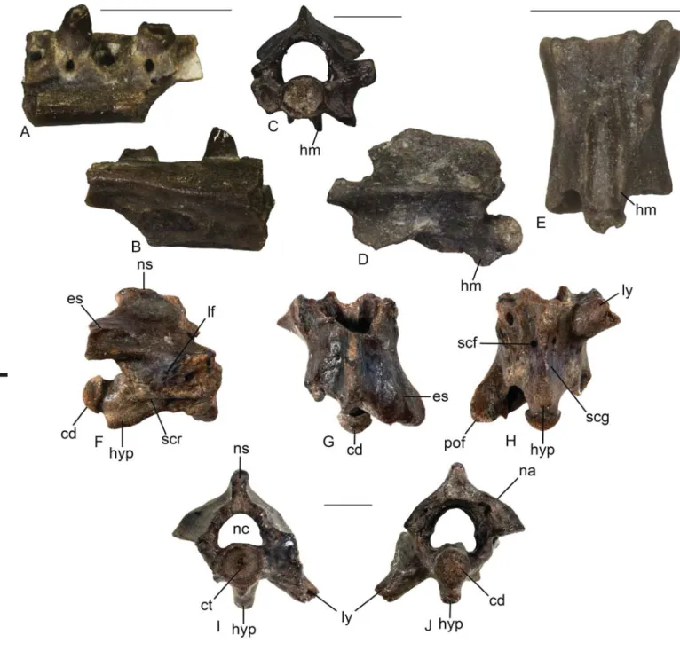
triangular, with distinct subcotylar tubercles, though only the left one is preserved (Figure 9(F)–(I)). In anterior view, the zygos-phenal lip is vaulted dorsally. The neural canal is rounded with short but distinct lateral sinuses. The cotyle, partially preserved in one vertebra, seems to have been most probably circular. UU AL 3590 is an anterior trunk vertebra and is rather incomplete (Figure 9(J)–(L)). Similarly to UU KR2 5018, it is also charac-terised by a large depth of the subcentral grooves.
Remarks. The vertebrae can be attributed to Colubridae on the
basis of their gracile structure, their longer than wide centrum, the narrow haemal keel (or hypapophysis), the distinct subcen-tral ridges and subcensubcen-tral grooves, and the gracile zygosphene (Rage 1984; Szyndlar 1984, 1991a, 1991b; LaDuke 1991; Holman 2000). This taxonomic attribution is further supported by the fact that, although the anterior margin of all vertebrae is damaged, the distinct foramen in one specimen (UU KR2 5018) situated on the right side in anterior view indicates that paracotylar foramina were probably present. Due to the damaged ventral portions of all vertebrae, it is not possible to determine whether the structure on the ventral surface of the centrum represents a haemal keel or a hypapophysis, although the presence of the former structure (haemal keel) might be more probable. As such, we refrain from assigning the Aliveri and Karydia colubrids to either ‘colubrines’ or ‘natricines’ (sensu Szyndlar 1984, 1991a, 1991b), although we must further acknowledge here that the presence or absence of a hypapophysis throughout the vertebral column is a widespread and variable feature and it should be dealt with high caution, when dealing with taxonomic designations and attributions (Pyron et al. 2013; Head et al. 2016).
5018), possesses a relatively small zygosphenal facet of oval to sigmoid outline in lateral view (Figure 9(A)–(E)). A large lateral foramen is situated below the rather sharp interzygapophyseal ridge. Its orifice occurs in the vicinity of the dorsal margin of the rather deep and anteroposteriorly slightly enlarged wide depression. The ventral margin of the depression is bordered by a peculiar, distinct sharp crest, which extends from the caudal margin of the unpreserved synapophysis as far as the two thirds of the length of the subcentral ridge. This highly distinct ridge that lies between and almost parallel to the interzygapophyseal and the subcentral ridges is herein termed as ‘paracentral ridge’. The subcentral ridges are rather prominent. They are straight and extend as far as the posterior border of the pedicle. Due to the incomplete nature of the specimen, it cannot be certain whether the vertebra possessed a haemal keel or a hypapophy-sis, although the former structure (haemal keel) seems to have been more probable. The rather narrow haemal keel (or short hypapophysis) is broken off close to its base. In dorsal view, the zygosphene has distinct lateral lobes and a wide medial lobe. The base of the neural spine rises rather anteriorly, at about half of the length of the zygosphenal facet. In ventral view, the subcentral grooves are rather deep. The subcentral foramina are rather large and are situated at the base of the significantly thin haemal keel/ hypapophysis. Their orifices are directed anteriorly. The posterior section of the haemal keel/hypapophysis is situated on a trian-gular-shaped elevation, this feature providing relative support for the presence of a haemal keel instead of a hypapophysis. The anterior portion of the haemal keel/hypapophysis of the sec-ond posterior trunk vertebra from Karydia (UU KR2 5019) is
Figure 9. Colubridae indet. from Karydia-2 (A–I): posterior trunk vertebra (UU KR2 5018) in anterior (A), right lateral (B), dorsal (C), ventral (D), and posterior (E) views; posterior trunk vertebra (UU KR2 5019) in anterior (F), left lateral (G), dorsal (H), and ventral (I) views; Colubridae indet. from Aliveri (J–L): anterior trunk vertebra (UU AL 3590) in posterior (J), ventral (K), and right lateral (L) views.
Scale bars = 1 mm. Abbreviations: cd, condyle; hk, haemal keel; ir, interzygapophyseal ridge; lf, lateral foramen; nc, neural canal; pcr, paracentral ridge; scf, subcentral foramen; scg, subcentral groove; scr, subcentral ridge; sctt, subcotylar tubercle; zy, zygosphene.
as it clearly does not form a triangle and is not united with the subcentral ridge, and is also well projecting across most of the vertebra’s lateral surface. Furthermore, we do not consider that this, apparently apomorphic, feature is the product of a pathol-ogy, since it is present symmetrically in both lateral views of the vertebrae and is present in both specimens from Karydia (not preserved in the Aliveri specimen). We were only able to identify a similar, but not identical, feature in the trunk vertebrae of the extant Asian snake Boiga sp. (specimen MDHC 137), although it is differently shaped and not as prominent as in the Greek fossils. Curiously, this feature on the vertebrae of Boiga was not men-tioned at all by Ikeda (2007), and it cannot also be ascertained from the photographs provided in that publication. Of course, we are not implying a close relationship among Boiga and the Greek colubrid solely on the basis of this feature. Besides, their vertebrae are rather different in terms of general shape, size, and shape of vertebral structures. In any case, the presence of this unique feature, the paracentral ridge, and the combination of the other characters described above probably denote that the colubrid from Karydia and Aliveri is a new taxon. However, on the basis of the scarceness of the material and the rather incom-plete and fragmentary nature of all specimens, we refrain from naming it as a new species.
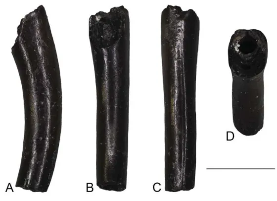
Viperidae Oppel, 1811 Viperidae indet. (Figure 10)
Material. AL1a: an incomplete fang (UU AL 3592).
Description. The fang is incomplete and has its base unpreserved.
The apical termination is slightly curved, with a wide pulpal cavity and venom canal situated anteriorly in central position (Figure 10). In dorsal view, the base of the entrance orifice, which Whereas the caudal vertebra UU KR2 5025 and the two
vertebral fragments (UU KR2 5020 and AMPG KR3 020) are not informative, the other three colubrid specimens (UU AL 3590, UU KR2 5018, and UU KR2 5019) are characterised by a combination of peculiar features and therefore, enable us to provisionally identify them as belonging to the same taxon. More particularly, the most striking features of this taxon are: (1) in lateral view, a highly distinct ridge, herein defined with the newly introduced term ‘paracentral ridge’, which is situated above the subcentral ridge and extends from the posterior mar-gin of the diapophysis up to about half of the centrum length; (2) in ventral view, an almost triangular and rather highly elevated surface surrounding the posterior part of the haemal keel (or hypapophysis); (3) a prominent, deep and narrow haemal keel (or hypapophysis) with the ventral margin being sharp along its entire length; (4) prominent subcentral grooves; (5) rather sharp subcentral ridges which are strongly built; (6) rather large sub-central foramina with their orifices directed anteriorly; (7) large lateral foramina, situated in deep depressions; and (8) in dor-sal view, a trilobate zygosphene, with distinct lateral lobes. The herein newly defined ‘paracentral ridge’ appears to be a unique feature and in fact has never been previously described in fos-sil or extant snakes (e.g. Szyndlar 1984, 1991a, 1991b; Holman 2000; Szyndlar 2005; LaDuke 1991). It is worth noting that in certain colubrids, the anterior portion of the subcentral ridge forms sometimes an elongate triangle whose tip is directed pos-teriorly, as if the ridge was forked anteriorly in two branches, a ventro-medial and a dorso-lateral one (J-C. Rage, pers. comm., December 2017). The paracentral ridge that is observed in our Greek fossil material, however, does not correspond to that case,
Figure 10. Viperidae indet. from Aliveri: isolated fang (UU AL 3592) in lateral (A), posterior (B), anterior (C), and dorsal (D) views.
Scale bars = 1 mm.
originated from the dentine folding throughout ontogeny. This mode of venom canal development is typical for viperids (Jackson 2002; Zahradnicek et al. 2008). The fangs of elapid snakes differ by the presence of a distinct anterior groove con-necting entrance and discharge orifices (Kuch et al. 2006). The distinct lateral grooves which stretch along the entire length of the fragment are rather unusual in viperids although short lateral grooves frequently occur in both crotalines and viperines at the vicinity of the fang base (see Figure 2 in Ivanov 1999; MI, pers. obs.). The preserved specimen from Aliveri is too fragmentary for a more precise determination at the subfamily level. However, is situated in the anteriormost proximal part of the fragment, is
indicated by the distinction of the dentine folds which form the anterior closure of the venom canal (Figure 10(D)). In anterior view, there is a distinct suture close to the distal termination of the fragment. This suture turns proximally into a narrow groove, which diminishes in front of the entrance orifice base where the fang surface is completely smooth. The discharge orifice is not preserved. A wide groove occurs on either lateral side of the fang along its entire length.
Remarks. The single, isolated fang from Aliveri can be assigned
to viperids on the basis of the presence of a venom canal which
Figure 11. Serpentes indet. from Aliveri (A–B): fragment of the anterior part of a left pterygoid (UU AL 3529) in medial (A) and labial (B) views. Serpentes indet. from Karydia-3 (C–E): posterior caudal vertebra (AMPG KR3 021) in anterior (C) and left lateral (D) views; posterior caudal vertebra (AMPG KR3 022) in ventral (E) view. Serpentes indet. from Karydia-2 (F–J): cloacal or anterior caudal vertebra (UU KR2 5022) in right lateral (F), dorsal (G), ventral (H), anterior (I), and posterior (J) views.
Scale bars = 1 mm. Abbreviations: cd, condyle; ct, cotyle; es, epizygapophyseal spine; hm, haemapophysis; hyp, hypapophysis; lf, lateral foramen; ly, lymphapophysis; nc, neural canal; ns, neural spine; pof, postzygapophyseal facet; scf, subcentral foramen; scg, subcentral groove; scr, subcentral ridge.
Material. AL1a: four fragments of tooth-bearing bones (UU AL
3522, UU AL 3523, UU AL 3525, and UU AL 3526), a caudal ver-tebra (UU AL 3527), and fragments of a verver-tebra and a maxilla (UU AL 3528). AL1b: a pterygoid (UU AL 3588) and a humerus (UU AL 3589). KR3: a caudal vertebra (AMPG KR3 025) and an osteoderm (AMPG KR3 009).
Remarks. The above mentioned specimens from Aliveri are
either too poorly preserved or they lack significant diagnostic features for a more precise taxonomic attribution. As such, they are attributed only to indeterminate squamates. Similarly, the two specimens from Karydia-3 represent skeletal elements of lizards that are considered not to bear significant diagnostic features. Nevertheless, the caudal vertebra is rather small-sized (centrum length is less than 2 mm) and could therefore belong to the pre-viously mentioned small-sized lacertid. The osteoderm, on the other hand, is different from anguid ones in shape, general mor-phology and ornamentation, being more similar to supraocular osteoderms of, e.g. lacertids. Given that, it could also belong to that clade.
Discussion
Biogeographic implications of the Aliveri and Karydia herpetofaunas
Both Aliveri and Karydia share common herpetofaunal elements, such as alytids, lacertids, and colubrids (Table 1). Notably, how-ever, crocodylians, chamaeleonids, and viperids are known from Aliveri but are absent from Karydia, whereas the opposite case is known for salamanders. Of course, with the limited material currently available, it is not possible to state whether such faunis-tic differences among the two localities are indeed genuine and could imply ecological differences, or they are simply biased by taphonomical or incomplete collection factors. Definitely, how-ever, the fact that Karydia has yielded significantly lower amount of fossil specimens in comparison with Aliveri might partially explain such faunistic differences among the two localities. a possible taxonomic attribution to Viperinae could be indirectly
supported by a biogeographic rationale, as Crotalinae are only known in Europe with certainty from the late Miocene (MN 9) of Ukraine (Ivanov 1999).
Serpentes indet. (Figure 11)
Material. AL1a: a fragment of the anterior part of a left pterygoid
(UU AL 3529). AL1980NQ: a vertebra (UU AL 3591). KR2: a cloacal or anterior caudal vertebra (UU KR2 5022), two fragmen-tary vertebrae (UU KR2 5023 and UU KR2 5024), and a cloacal or anterior caudal vertebra (UU KR2 5021). KR3: a fragment of a pterygoid (AMPG KR3 013), and two posterior caudal vertebrae (AMPG KR3 021 and AMPG KR3 022).
Remarks. These cranial and postcranial remains from Aliveri
and Karydia are too fragmentary and incomplete to permit a more precise identification within snakes. It is worth noting, however, that one of these specimens, UU KR2 5022, seems to demonstrate a mixed character set between colubroids and boo-ids (Figure 11(F)–(J)). This vertebra most probably originates from the cloacal region, as it can be judged by the presence of a strongly built, short hypapophysis and the preserved base of the ventral ramus of the left lymphapophysis, although an alternative origin from the anterior caudal region cannot be excluded. The relatively massive structure of the vertebra UU KR2 5022, with a strongly built neural spine and short hypapophysis, as well as the absence of paracotylar foramina, is reminiscent of certain Booidea (e.g. Rage 1984; Szyndlar and Rage 2003), but the ratio of a centrum length/neural arch width >1 and a condyle situ-ated on a rather long neck are not typical for cloacal vertebrae of Booidea and are mostly characterising colubroids (e.g. Rage 1984). To make things even more complicated, several non-py-thonid booids, both extinct (e.g. Bavarioboa) and extant (e.g.
Boa), are also known to possess paracotylar foramina, in at least
some of their vertebrae (Szyndlar and Rage 2003), rendering obscure the taxonomic reliability of this character. As such, we herein refrain from identifying UU KR2 5022 as a booid and prefer to refer it as Serpentes indet.
Squamata indet.
Table 1. Known occurrences of amphibians and reptiles in the early Miocene of Greece.
Data from: Aliveri-Georgalis, Villa, and Delfino (2016b) and this paper; Karydia-this paper; Kymi-Römer (1870); Nostimo-Georgalis et al. (2013); Lapsarna Vasileiadou et al. 2017.
Aliveri (MN 4a) Karydia (MN 4a) Kymi (MN 3/4)
Nostimo (Burdigalian) Lapsarna (?MN 3) ?Mioproteus sp. + Urodela indet. + Latonia cf. gigantea + cf. Latonia sp. + Anura indet. + + + Nostimochelone lampra + Testudines indet. + + Crocodylia indet. + + +
Chamaeleo cf. andrusovi and Chamaeleonidae indet. +
Lacertidae indet. + + + Scincomorpha indet. + + cf. Ophisaurus sp. + Anguinae indet. + + Python euboicus + ?Natricinae indet. + Colubridae indet. + + Viperidae indet. + Serpentes indet. + + +
Squamata indet. (non-snake squamates) + + +
The isolated snake fang from Aliveri denotes the presence of viperid snakes in Greece already by the early Miocene, being the oldest representative from that clade in the region, a presence that culminated with the magnificent Laophis crotaloides Owen 1857, one of the largest known viperids, from the Pliocene of Thessaloniki area (Georgalis, Szyndlar, et al. 2016).
Frogs of the genus Latonia in the early Neogene of southeastern Europe
Latonia is a genus of discoglossine alytid frogs that thrived in
Europe from the late Oligocene up to the early Pleistocene (Roþek 1994, 2013; Delfino 2002), becoming the most common alytid in the continent during the Miocene. The genus may have also been present during the early Oligocene, considering a mention of Latonia aff. vertaizoni from Quercy (Rage 2006), however, this material is undescribed and still awaits a formal documentation. Starting from the Pliocene, the European range of Latonia under-went a southward directed contraction, which eventually resulted in its local extirpation during the Pleistocene. It has been sug-gested that this extinction event has been linked to Pleistocene climate change (Roþek 1994). Nevertheless, Latonia has been documented in the early Pleistocene of Anatolia (Vasilyan et al. 2014) and it is also now known that the genus has survived in the Middle East, where the only extant representative, Latonia
nigriventer (Mendelssohn and Steinitz 1943) still exists, being its sole living representative (Biton et al. 2013, 2016). Although several taxa have been assigned to this genus, it is now generally accepted that only four valid extinct species are known from Europe (Roþek 1994): Latonia seyfriedi Meyer, 1843 (type spe-cies), Latonia gigantea (Lartet, 1851), Latonia ragei Hossini, 1993, and Latonia vertaizoni (Friant, 1944).
The remains of Latonia from Karydia-3 share a similar mor-phology and size with juveniles of Latonia gigantea, notwith-standing the possible presence of the foramen for the occipital artery. Assuming that our attribution of these fossils to Latonia cf.
gigantea is correct, Karydia-3 would be one of the southernmost
localities from which this species (or at least a morphologically rather similar form) is reported. As a matter of fact, the new Greek occurrence adds to the tentatively attributed remains from the Gargano palaeoisland in Southern Italy (Delfino 2002) and to the recently described remains from Catalonia in the Iberian Peninsula (Villa et al. 2017). This supports the hypothesis that the seemingly poor representation of L. gigantea in the Mediterranean area, compared with the fossil record of that species in the rest of Europe (Roþek 1994, 2013), might be an artifact of either misidentification or overlooking of fossil remains, rather than a real absence from the area. Further studies on Latonia remains that are currently unassigned to the species level and originate from other localities in the southern European peninsulas might shed more light on this issue. Indeed, recently described remains (an ilium and an urostyle) from the early Miocene of Sibnica, Serbia, have been attributed to Latonia cf. gigantea (Đuriü 2016), though they were not figured and, as such, we cannot confirm their identity. It has to be noted, however, that neither ilia nor urostyles are usually considered diagnostic for Latonia species, and therefore the identification of the Serbian material must be treated with caution. Moreover, remains from Karydia-3 could also represent the oldest occurrence of the species, given that this The single known salamander element from Karydia is not
informative for a precise taxonomic identification, but still rep-resents, along with a recently described probable proteid from Lapsarna (Lesvos) (Vasileiadou et al. 2017), the oldest urodelan remains from Greece, both being also the sole Neogene occur-rences from the country. The frog remains from Karydia (and potentially also Aliveri) document the presence of Latonia for the first time in Greece and are fully concordant with the wide-spread European range of that genus during the early Neogene (Roþek 1994; see also below). The fragmentary nature of the Aliveri and Karydia turtle specimens does not permit any fur-ther biogeographic assumption, but nevertheless, these constitute the oldest such remains from Greece, along with the holotype of Nostimochelone lampra from the Burdigalian of Nostimo (administrative region of Western Macedonia) (Georgalis et al. 2013). Crocodylians are rather rare in the Greek fossil record, and as such, they had only recently been described for the first time from the late Miocene of Plakias (Crete) (Georgalis, Villa, Vlachos et al. 2016) and soon after from the early Miocene of Lapsarna (Lesvos) (Vasileiadou et al. 2017). In any case, the new Aliveri remains demonstrate that crocodylians were more wide-spread in the early Miocene of the region, a situation consistent with similar finds from the Oligo-Miocene of Turkey (Schleich 1994; Sen et al. 2011). As for the lizards, the presence of a cha-maeleonid in Aliveri was recently shown to support a probable Greek pathway for this African clade that could have used the ‘Gomphotherium Landbridge’ for its dispersal (Georgalis, Villa and Delfino 2016). Newly described chamaeleonid remains from the early Miocene (MN 4) of Sibnica, Serbia (Đuriü 2016) suggest that chameleons were more widespread faunal elements in the southern Balkan localities of that time, than what was previously thought. This clade is still absent from the as of yet poor fossil record of squamates from Anatolia. The herein described lacer-tids from Aliveri and Karydia, along with the recently described Lapsarna material (Vasileiadou et al. 2017), demonstrate that these lizards were already widespread in Greece already by the early Miocene and would since then continue to be a common element of the Greek herpetofaunas. Indeed, lacertids were also described from late Miocene localities of the region (Richter 1995; Georgalis et al. 2017a) and they are currently the dominant (in terms of diversity) reptile group on the European continent (Arnold et al. 2007; Sindaco and Jeremþenko 2008). The Aliveri and Karydia anguids are the oldest representatives of this clade from Greece, though they have been described from the early Miocene of Sibnica, Serbia (Đuriü 2016) and various coeval local-ities from Turkey (ýerňanský et al. 2017), confirming their wide distribution in the area already by the early Neogene. The appar-ently bizarre colubrid snake from Karydia and its potentially conspecific form from Aliveri seem to possess unique autapomo-phies that are otherwise unknown in extinct and extant European snakes, most significantly the presence of a new vertebral feature that is herein termed as ‘paracentral ridge’. The skeletal anatomy of extant African and Asian snakes is poorly documented so it is currently impossible to identify the presence of a paracentral ridge in modern taxa and to assess its potential diagnostic impor-tance. Considering, however, that this feature is totally absent in all European extinct and extant snakes, it seems plausible that the herein newly described early Miocene Greek colubrids could represent a shortly lived radiation with African or Asian origin.