1
Thin Film Structural Colouration from Simple Fused Scales in Moths
Cédric Kilchoer 1, Ullrich Steiner 1 and Bodo D. Wilts 1*1 Adolphe Merkle Institute, University of Fribourg, Chemin des Verdiers 4, 1700 Fribourg,
Switzerland
*Author for correspondence: bodo.wilts@unifr.ch
Published in Interface Focus (2018) 20180044: http://dx.doi.org/10.1098/rsfs.2018.0044
Abstract
The metallic colouration of insects often originates from diverse nanostructures ranging from simple thin films to complex three-dimensional photonic crystals. In Lepidoptera, structural colouration is widely present and seems to be abundant in extant species. However, even some basal moths exhibit metallic colouration. Here, we have investigated the origin of the vivid metallic colours of the wing scales of the basal moth M. aureatella by spectrophotometry and scanning electron microscopy. The metallic gold-, bronze- and purple-coloured scales share a similar anatomy formed of a fused lower and upper lamina resulting in a single thin film. The optical response of this thin film scale can be attributed to thin-film interference of incident light, resulting in the colour variations that correlate with film thickness. Subtle variations in the wing scale thickness result in large visible colour changes that gives Micropterix moths their colourful wing patterns. This simple colouration mechanism could provide a hint to understand the evolution of structural colouration in Lepidoptera.
Keywords: Micropterix – iridescence – thin film – insect colours – colour tuning
Background
Colour production mechanisms in animals result from pigmentary or structural colouration, or a combination of both. Pigmentary colouration arises from organic dyes distributed throughout the tissue that selectively absorb part of the incident light and scatter non-absorbed light. For example, melanin pigments have a rather broadband absorption [1] and are responsible for the black and brown colouration of many organisms. In contrast to absorption, structural colouration is produced by structuration on the length scale of the light wavelength (i.e. ~ 100
2
nm), interacting with the incident light. This often interference-based interaction can lead to vivid, metallic and iridescent colours as seen in many insects, fish and birds [2–4]. Colouration in insects can serve a wide range of different functional adaptations, ranging from camouflage to signalling [5,6]. In insects, a particularly diverse range of chitin nanostructures can be found ranging from thin films to photonic crystals [7–12].
Insect structural colours have only recently been linked to phylogenetic studies [13–15] and studied through fossil records [16–18]. Unfortunately, these investigations are often limited to a specific family [13,15] and/or by the number of species investigated. Understanding the links between different production mechanisms of structural colours would give hints concerning their biological functions [19]. In Lepidoptera, a huge diversity of ultrastructures that produce structural colours is encountered. The basal origin of lepidopteran structural colours (or its most ancestral state) is however not yet established, although first hints have been published [20]. The genus Micropterix Hübner 1825 is widely present in Eurasia [21] with more than 70 species that display vivid metallic colouration. The genus is part of the Micropterigidae family that is classified as basally branching within the Lepidoptera by Kristensen [22] and more recent phylogenetic studies [23]. Adult moths of this genus are diurnal, inhabit shrub vegetation and woodland and can be locally abundant on flowering vegetation.
Zhang et al. [20] have very recently reported the evidence of structural colouration in fossil Lepidoptera based on the presence of fused scales. The precise origin of the colouration of extant Micropterix is however still unexplored. Here we investigate the ultrastructural and pigmentary basis of these colourful wing scales by measuring the optical properties of different scales [24]. Based on optical measurements and scanning electron microscopy, we demonstrate that the colouration is highly dependent on the thickness of the scale that is acting as a thin film. A model based on thin film interference was developed and the resulting simulated reflectance confirms experimental measurements.
Material and Methods
Specimen
Micropterix aureatella specimens were captured in Saicourt (Switzerland) in June 2013 and
obtained from R. Bryner, Biel, Switzerland. Photographs of pinned specimens were obtained using a Canon EOS 5D camera (Canon Inc., Tokyo, Japan)
3
Optical Characterization
Optical characterization was performed using a ZEISS (Zeiss AG, Oberkochen, Germany) Axio Scope.A1 polarized light microscope and a xenon light source (Thorlabs SLS401; Thorlabs GmbH, Dachau, Germany). For spectroscopic measurements, an optical fibre (QP230-2-XSR, 230 µm core) collected the light reflected from the scales on the wing with a measurement spot size of approx. 20 µm. Several scales (more than 4 of each type) were measured for three different specimens. The reflected spectra were recorded by a spectrometer (Ocean Optics Maya2000 Pro; Ocean Optics, Dunedin, FL, USA) and a standard white diffuser was used as a white reference sample. Light microscopy images were acquired with a Point Grey (FLIR Integrated Imaging Solutions Inc., Richmond, Canada) GS3-U3-28S5C-C CCD camera. The far-field angular distribution of reflected light was accessed by introducing a Bertrand lens (Zeiss 453671) into the detection pathway of the microscope.
Absorbance measurements were performed using the same microscope in transmission mode on individual scales detached from the wing and placed on a glass slide. Scales were immersed using a refractive index fluid with a refractive index of n = 1.55 (Series A; Cargille Labs, Cedar Grove, NJ, USA) and a coverslip is used to ensure a flat top interface. The reference is taken within a region close to the scale.
Scanning electron microscopy (SEM)
The scale ultrastructure was imaged using a TESCAN (TESCAN, a.s., Brno, Czech Republic) MIRA3 field emission scanning electron microscope. Scales were cut using surgical scissors. To prevent charging, samples were placed on carbon tape and sputtered with a 4 nm thick layer of palladium using a Cressington (Cressington Scientific Instruments, Watford, England) 208 HR sputter coater. The average thickness of an individual scale was determined by measuring and averaging the local thickness at different positions (more than 20) between two parallel ridges.
Optical Simulations
Computational simulations of the light-matter interaction of incident light with the wing scales were performed using a custom-written MATLAB (Mathworks, Natick, MA, USA) code employing a matrix formalism [25,26]. The wing structure was approximated by a thin film. We varied the thickness of the thin film between 100 and 200 nm. The refractive index of the butterfly chitin [27] is described by the Cauchy equation n(λ) = A+B/λ2, with A = 1.517 and B = 8.80·103 nm2. In order to take in account the absorption caused by pigments present within the scale, the absorption coefficient of the melanin [28] was extracted from a refractive
4
matching experiment and was fitted by the exponential function ε = ε0 exp(–λ / λm) with ε0 = 2.74 and λm = 231 nm.
Results
Optical appearance
M. aureatella dorsal forewings have an overall length of around 4 mm and display vivid colours
in a pattern of alternating golden and purple stripes along the length of the wings (Figure 1a). The hindwings and the ventral side of the moth wings are less colourful.
We therefore focus on the colourful forewing. Here, colouration resides in the cover scales where locally a lattice of metallic golden-, bronze- and purple-coloured wing scales can be observed (figure 1b). Ground scales are very sparse, brownish in colour and distinguishable by a smaller shape when removed from the wing (not shown). Purplish scales constitute the major colour of the wing pattern characterised by two golden fasciae positioned at ¼ and ½ of the wing length, measured from the thorax, and an irregular oval shape at ¾ [21]. Bronze-coloured scales are exclusively found at the boundaries of between golden- and purple-coloured areas on the wing. At the borders of the wing, hair-like bristles also exhibit metallic colouration.
Scale reflectance
To characterise the different colours of the dorsal wing scales, we performed spectroscopic measurements using a custom-built microspectrophotometer. Figure 2a,d,g shows reflectance spectra of three differently coloured wing scales. The measurements spots had a diameter of about 20 µm, entirely within single wing scales. Gold-coloured wing scales have a broadband reflection in the visible spectrum. A reflectance minimum is observed at ~370 nm. For a bronze- and a purple-coloured wing scale, the reflectance spectra are bathochromically shifted, i.e. the position of the minimum intensity of reflectance moves to longer wavelengths, to around 430 nm and 550 nm for the bronze and purple wing scales, respectively. Furthermore, the reflectance spectrum of the purple-coloured wing scale exhibits a distinct peak at ~420 nm.
k-space imaging of light reflected from a single wing scale visualises the angular distribution
of reflected and scattered light. Under narrow-aperture light, light is scattered directionally (figure S1) while under an open aperture the iridescence, i.e. its angle dependency, of these wing scales becomes prominent. In figure 2c,f,i, k-space imaging under wide-aperture illumination of a single scale shows a pattern with a radial uniformity. For all scales, a change
5
of colour is observed in reflection between normally incident light (centre of the pattern) and light reflected after light incident at a large angle (exterior of the pattern), indicating a strong iridescence.
Scale pigmentation
To determine the presence of pigments in the wing scales, a refractive index-matching experiment was performed [29]. We recorded the transmission spectrum through single wing scales immersed in a liquid of a refractive index n = 1.55, close to that of cuticular chitin of butterflies [27]. If the scale was pigment-less, a refractive-index-matching experiment would render the immersed scale transparent with a transmittance close to 100%. In the absences of light scattered from the chitin-air interfaces, the measured absorption spectrum can be attributed to the pigments (or scattering) within the scales. The immersed scales appear brown in transmission (figure 3a). The absorbance A = -log10(T), determined from transmittance T, is in good agreement with the absorption spectra of melanin pigments (figure 3b). In reflection, the immersed scale appears grey, indicating that reflected light is primarily caused by scattering from the melanin-containing structures within the scale (figure S2). To confirm that the pigment is melanin-related, we bleached wing scales in warm H2O2. After bleaching, the absorption is significantly reduced, leading to an enhancement of the reflectance and transmittance (see figure S4 and supplementary text).
Scale anatomy
In order to understand the origin of the colouration, we investigated wing scales of different colours by scanning electron microscopy (figure 4). The general scale anatomy is similar for different coloured wing scales; the abwing side consists of around ten parallel rows of ridges spaced by 2.5-3 µm (figure 4a). A high density of crossribs entirely fills the area between longitudinal ridges and we note the absence of open window. The adwing side (figure 4b, ad) appears rather flat and does not show any particular morphology. To inspect the ultrastructure in the third dimension, cross-sectional images reveal an uncommon scale anatomy. No lumen is observed and the scale appears to be comprised of a single layer (figure 4b,c). Moreover, cross-section images exhibit a very small thickness of the scale, with an overall thickness below 200 nm.
Scales of different colours have similar ultrastructures, but display different scale thicknesses. The measured thickness varies with the reflection colour. Gold-coloured scales are thinnest with a scale thickness of 112 ± 8 nm, followed by the bronze-coloured scales with 134 ± 7 nm. Purple
6
scales are thickest with an extracted value of 164 ± 8 nm (number of investigated scales: 5/6/5; error: standard deviation)
Optical Modelling
To understand the interplay of scale thickness, pigmentation and the resulting colour appearance, we calculated reflection spectra of planar thin films as a function of layer thickness in the presence of different amounts of melanin. Figure 5a shows the well known thin film interference, that is, the strong influence of the layer thickness on the reflected spectrum at normal incidence. The thickness of the chitin film determines the wavelength of the constructive and destructive interferences. For instance, an increase in the thickness results in a redshift of the local extrema. Melanin absorption strongly attenuates the reflectance, particularly at short wavelengths (in the UV and blue, slightly red-shifting the wavelengths of the maxima (figure 5b). Assuming an equal melanin distribution throughout the chitin film, indicates from a similar absorption of all scales when normalised by the scale thickness (figure S3), thicker films will be more impacted by the melanin absorption.
Figure 6compares the experimental reflectance spectra with the thin film interference modelling of figure 5. As noticed by the SEM micrographs (figure 4), the main anatomic change between scales of different colouration is their thickness. By using respective scale thicknesses extracted by SEM the simulated spectra are in good agreement with the measurements.
Discussion
Thin film interference causes scale colouration
The wing colours of Micropterix are structurual and are caused by thin film interference from heavily pigmented thin films. Structural colours in Lepidoptera are not unusual and can originate from a wide diversity of composite nanostructures. According to Ghiradella, the basic Bauplan of a butterfly wing scale is the combination of a flat lower lamina and a complex structured upper lamina, consisting of ridges and cross-ribs [30]. The lower lamina can act as a thin film reflector, which can be accessed by measuring and comparing the adwing and adwing reflectance spectra. Stavenga and co-workers [25,31,32] demonstrated that thin film interference determines not only the colouration of the bare wings of many insects but also the colouration of the wing scales of many clades of butterflies. For example in many nymphalid butterflies [31], a lower lamina thickness of 200 nm will result in blue coloured scales. Work
7
by the same group showed that the lower lamina thickness is seemingly tuned according to the spectral absorbance of the pigment present in the wing scales as orange, blue and black scales of the Papilionid P. xuthus [32]. For orange and blue scales, the lower lamina is tuned to have a reflectance peak, respectively, at orange and blue wavelengths, coinciding with the wavelength range where the pigment does not absorb. The adwing reflectance of black scales also has a peak in the blue, which is however strongly suppressed by absorption of melanin. In Micropterix, we show that the scale architecture is a single layer (figure 4) and the resulting optical response can be well understood from a single thin film interference model (figures 5,6). This type of scale has been previously termed “fused scales” (because of the fused upper and lower laminae) by Kristensen [33]. However, this type of scale architecture is rare in Lepidoptera. So far, the wing scales in the glassy, transparent patches of wings of the Common Bluebottle butterfly, Graphium sarpedon, is the only investigated example of colouration originating from fused scales [34]. For these so-called “glass” scales, the average thickness of the fused scale is 400 nm, exhibiting a reflectance peak at 500 nm and which appear green under normal incidence.
Fused scale thicknesses of Micropterix can vary from 100 nm to 170 nm, resulting in a different colouration of each single scale. In an interesting way, a small change of the scale laminar thickness of tens of nanometres results in a large colour shift (figure 5a). Thus, Micropterix takes advantage of its fused scale architecture to colour its wing pattern by simply changing the thickness of a scale. The absorption caused by melanin pigments affects more strongly short-wavelengths. A variation of the absorption coefficient results in a small effective shift of the wavelength maxima and minima. Optically this explains why the thickest scales appear more purple-coloured than blue.
One might argue that the ridges, forming a grating structure, will act like a polarisation dependent reflector [35]. Our results show however no polarisation effects (not shown). Moreover, the spacing between the ridges, greater than 2 µm (figure 4c), are expected to have an influence only at infrared wavelengths. In all scale micrographs, the ridges are always visible, which is not the case for structures that have been reported to act as diffraction gratings [9,13]. The cross-ribs, mostly present at the surface of the upper lamina, have a height varying between 20 and 30 nm that effectively modifies (and smoothes) the local thickness of the film. This local variation increases the effective fused scale thickness. As demonstrated previously [34], a standard deviation smaller than 20 nm does not strongly affect the optical response of the thin film.
8
Functional and biological significance of fused scales in basal moths
Basal moths present an ideal case to study the evolution of taxonomic characters through the entire order of Lepidoptera. Already in 1970, Kristensen noted that dorsal scales of Micropterigidae are different from the other Lepidoptera with a particular fused upper and lower laminae [33]. This type of scale is largely found in other basal moths (e.g. Ecriocraniidae and Heterobathmidae, see [36]). Simonsen classified three different types of scales in basal moths [37]. Type-1, present in Micropterigidae, has fused cover and ground scales, while hollow cover and ground scales are characteristic for type-2 scales. In contrast, Type-3 scales carry a bilayer of hollow cover scales and fused ground scales. Morphological characteristics allowed Simonsen to distinguish between type-1 scales of non-Coelolepidan Lepidoptera (including Micropterigidae) and type-3 scales found in Neolepidoptera. This strongly indicates an independent evolution of fused scales. The analogous scale structure found in Graphium
sarpedon, a non-basal butterfly, also suggests a convergent evolution to achieve an optical
function. Fused type scales are prevailing in basal moths; however, this type is not structurally primitive. The development of common wing scales with a solid thin film in the lower lamina has been described in detail and it starts in the unfused condition [30,38]. During scale elongation, cuticle, produced by the cell extracellularly, is laid down and form the upper and lower laminae. At the eclosion of the butterfly from the chrysalis, the cuticle dries and lead in most cases to a window or lumen between both laminae, where the lower lamina is an unstructured thin film and the upper lamina a network of crossribs and ridges.
Our results potentially suggest that the presence of fused scales in basal moths is linked to the evolutionary advantage of producing a multicolour wing pattern. Figure 7shows colourful forewing patterns of different Micropterix species across Europe. The complexity of the wing pattern varies across species, but it is always a combination of the same metallic colours described for M. aureatella [21]. Considerable pattern variations can also be observed within the same species, possibly influenced by their local habitat [39].
By fusing upper and lower laminae, Micropterix controls the wing pattern colouration with only one dominant parameter, the scale thickness. Although it would also be achievable in hollow scales by controlling the thickness of the lower or upper lamina [40], fused scales offer other advantages. Firstly, a single layer would minimise the amount of light diffusing through its structure. Light diffusion related losses are also impacted by the local roughness and the relative flatness of the scales. As seen by SEM, fused scales are rather flat, which is not necessarily the case in hollow scales, where the distance between both laminae can be as large as several
9
micrometres [38]. Furthermore, fused scales will optimise the weight of the scale, which is beneficial for their flying ability [41,42]. At this early stage in butterfly evolution, fused scales are an efficient way of producing structural colours. Due to its simple thin film morphology, the thickness of a single scale will determine its colouration. Thus, Micropterix produces a colourful wing pattern by simply varying the scale thickness. The biological significance of the pattern still has yet to be investigated [43,44].
Through fossil evidence, the earliest presence of Lepidoptera is attributed to the latest Triassic period [45], ca. 200 million year ago (Ma). Inspection of fossilised scales by SEM highlights the presence of both fused and hollow scales. Fused scales indicate that colouration in the earliest Lepidoptera can bestructural, as already hypothesized by Zhang and co-workers [20].The authors have recently investigated wing scales of fossilised specimens [20], though an in-depth optical characterisation as presented here was not presented. Jurassic lepidopteran fossils and a specimen of the early Cretaceous period (~99 Ma) caught in amber [46] show well preserved scales. Zhang and co-workers claim that the scale ultrastructure, composed of parallel ridges, crossribs and herringbones, diffracts light, dominating the optical properties of the scale. The importance of the scale thickness that dominates the scale structural colouration is however underestimated, since this information is not accessible for most fossils. Nevertheless, amber fossils make it easier to access the scale thickness. If the preservation conditions are ideal, it is possible to retrieve the original colouration by measuring the scale thickness in order to compare these results with the results presented here. Investigation of further fossilised specimens [47–49] could reveal the presence of different scale colouration mechanisms, allow the reconstruction of the original wing pattern of these insects, and eventually provide insights into the evolution of the diversity of wing patterns observed in colourful moths and butterflies nowadays.
Conclusion
In conclusion, the colouration mechanism of Micropterigidae is a combination of thin film interference and optical absorption by melanin pigments. Fused lower and upper laminae have a well-defined thickness that strongly influences its optical response. The colouration of the wing pattern is locally controlled by the thickness of the scales, resulting in metallic gold-, bronze- and purple-coloured scales. Structural colouration based on one of the most basic structural optics phenomena – thin film interference – points towards and early process behind
10
the evolution of structural colours in Lepidoptera. Expanding the investigation into Lepidoptera, including the examination of fossil records, is essential to understand the evolution of the colour formation in Lepidoptera.
Additional statements.
Authors’ contributions. C. K. performed measurements, analysed measurements, performed
modelling, wrote the manuscript, and gave final approval for publication. U. S. wrote the manuscript. B.D. W. performed measurements, analysed measurements, wrote the manuscript and supervised the project. All authors gave final approval for publication.
Competing interests. The authors declare no competing or financial interests. Data accessibility. Data used for analysis are deposited in the Dryad database
(doi:10.5061/dryad.rd3p70d). All other data is included within this manuscript and the supplementary information.
Acknowledgments. We gratefully thank R. Bryner for providing photographs and specimen. Funding. This study was supported by the Adolphe Merkle Foundation, the Swiss National
Science Foundation grant 163220 (US) and the Ambizione programme grant 168223 (BDW), and the National Center of Competence in Research Bio-Inspired Materials.
References
1. Solís-Herrera A, María del Carmen Arias Esparza C, Solís-Arias RI, Solís-Arias PE, Solís-Arias MP. 2010 The unexpected capacity of melanin to dissociate the water molecule fills the gap between the life before and after ATP. Biomed. Res. 21, 224– 226. (doi:10.1111/j.1600-0749.2006.00345.x)
2. Ingram A., Parker A. 2008 A review of the diversity and evolution of photonic structures in butterflies, incorporating the work of John Huxley (The Natural History Museum, London from 1961 to 1990). Philos. Trans. R. Soc. B Biol. Sci. 363, 2465– 2480. (doi:10.1098/rstb.2007.2258)
3. Vukusic P, Sambles JR. 2003 Erratum: Photonic structures in biology. Nature 424,
11 852–855. (doi:10.1038/nature01941)
4. Srinivasarao M. 1999 Nano-Optics in the Biological World: Beetles, Butterflies, Birds, and Moths. Chem. Rev. 99, 1935–1962. (doi:10.1021/cr970080y)
5. Doucet SM, Meadows MG. 2009 Iridescence: a functional perspective. J. R. Soc.
Interface 6, S115–S132. (doi:10.1098/rsif.2008.0395.focus)
6. Hubbs CL. 1942 Adaptive Coloration of Animals. Am. Nat. 76, 500. (doi:10.1086/281046)
7. Stavenga DG, Wilts BD, Leertouwer HL, Hariyama T. 2011 Polarized iridescence of the multilayered elytra of the Japanese jewel beetle, Chrysochroa fulgidissima. Philos.
Trans. R. Soc. B Biol. Sci. 366, 709–723. (doi:10.1098/rstb.2010.0197)
8. Sharma V, Crne M, Park JO, Srinivasarao M. 2009 Structural origin of circularly polarized iridescence in jeweled beetles. Science (80-. ). 325, 449–451.
(doi:10.1126/science.1172051)
9. Vukusic P, Sambles JR, Lawrence CR, Wootton RJ. 1999 Quantified interference and diffraction in single Morpho butterfly scales. Proc. R. Soc. London. Ser. B: Biol. Sci.
266, 1403–1411. (doi:10.1098/rspb.1999.0794)
10. Bartl MH, Galusha JW, Richey LR, Gardner JS, Cha JN. 2008 Discovery of a diamond-based photonic crystal structure in beetle scales. Phys. Rev. E - Stat.
Nonlinear, Soft Matter Phys. 77, 050904. (doi:10.1103/PhysRevE.77.050904)
11. Michielsen K, Stavenga D. 2008 Gyroid cuticular structures in butterfly wing scales: biological photonic crystals. J. R. Soc. Interface 5, 85–94. (doi:10.1098/rsif.2007.1065) 12. Wilts BD, Michielsen K, De Raedt H, Stavenga DG. 2012 Hemispherical Brillouin
zone imaging of a diamond-type biological photonic crystal. J. R. Soc. Interface 9, 1609–1614. (doi:10.1098/rsif.2011.0730)
13. Wilts BD, Matsushita A, Arikawa K, Stavenga DG. 2015 Spectrally tuned structural and pigmentary coloration of birdwing butterfly wing scales. J. R. Soc. Interface 12, 20150717. (doi:10.1098/rsif.2015.0717)
14. Seago AE, Brady P, Vigneron J-P, Schultz TD. 2009 Gold bugs and beyond: a review of iridescence and structural colour mechanisms in beetles (Coleoptera). J. R. Soc.
12
Interface 6, S165–S184. (doi:10.1098/rsif.2008.0354.focus)
15. Giraldo MA, Yoshioka S, Liu C, Stavenga DG. 2016 Coloration mechanisms and phylogeny of Morpho butterflies. J. Exp. Biol. 219, 3936–3944.
(doi:10.1242/jeb.148726)
16. Mcnamara ME. 2013 The taphonomy of colour in fossil insects and feathers.
Palaeontology 56, 557–575. (doi:10.1111/pala.12044)
17. Whalley P. 1986 A review of the current fossil evidence of Lepidoptera. Biol. J. Linn.
Soc. 28, 253–271.
18. Whalley PES. 1985 The systematics and palaeogeography of the Lower Jurassic insects of Dorset, England. Bull. Br. Museum (Natural Hist. Geol. 39, 107–189.
19. Wickham S, Large MCJ, Poladian L, Jermiin LS. 2006 Correction for Wickham et al., Exaggeration and suppression of iridescence: the evolution of two-dimensional
butterfly structural colours. J. R. Soc. Interface 3, 851–851. (doi:10.1098/rsif.2006.2000)
20. Zhang Q et al. 2018 Fossil scales illuminate the early evolution of lepidopterans and structural colors. Sci. Adv. 4, e1700988. (doi:10.1126/sciadv.1700988)
21. Zeller-Lukashort HC, Kurz ME, Lees DC, Kurz MA. 2007 A review of Micropterix Hübner, 1825 from northern and central Europe (Micropterigidae). Nota Lepidopterol.
30, 235–298.
22. Rkristensen N, Scoble MJ, Karsholt O. 2007 Lepidoptera phylogeny and systematics: The state of inventorying moth and butterfly diversity. Zootaxa 747, 699–747.
(doi:http://www.mapress.com/zootaxa/2007f/z01668p747f.pdf)
23. Regier JC et al. 2015 A molecular phylogeny for the oldest (nonditrysian) lineages of extant Lepidoptera, with implications for classification, comparative morphology and life-history evolution. Syst. Entomol. 40, 671–704. (doi:10.1111/syen.12129)
24. Vukusic P, Stavenga D. 2009 Physical methods for investigating structural colours in biological systems. J. R. Soc. Interface 6, S133–S148.
(doi:10.1098/rsif.2008.0386.focus)
25. Stavenga DG. 2014 Thin film and multilayer optics cause structural colors of many
13
insects and birds. Mater. Today Proc. 1, 109–121. (doi:10.1016/j.matpr.2014.09.007) 26. Yeh P, Hendry M. 1990 Optical Waves in Layered Media. Wiley.
(doi:10.1063/1.2810419)
27. Leertouwer HL, Wilts BD, Stavenga DG. 2011 Refractive index and dispersion of butterfly chitin and bird keratin measured by polarizing interference microscopy. Opt.
Express 19, 24061. (doi:10.1364/OE.19.024061)
28. Stavenga DG, Leertouwer HL, Hariyama T, De Raedt HA, Wilts BD. 2012 Sexual Dichromatism of the Damselfly Calopteryx japonica Caused by a Melanin-Chitin Multilayer in the Male Wing Veins. PLoS One 7, 1–7.
(doi:10.1371/journal.pone.0049743)
29. Stavenga DG, Leertouwer HL, Wilts BD. 2013 Quantifying the refractive index dispersion of a pigmented biological tissue using Jamin-Lebedeff interference microscopy. Light Sci. Appl. 2, e100–e100. (doi:10.1038/lsa.2013.56)
30. Ghiradella H. 2010 Insect Cuticular Surface Modifications: Scales and Other Structural Formations. Adv. In Insect Phys. 38, 135–180. (doi:10.1016/S0065-2806(10)38006-4) 31. Stavenga DG, Leertouwer HL, Wilts BD. 2014 Coloration principles of nymphaline
butterflies - thin films, melanin, ommochromes and wing scale stacking. J. Exp. Biol.
217, 2171–2180. (doi:10.1242/jeb.098673)
32. Stavenga DG, Matsushita A, Arikawa K. 2015 Combined pigmentary and structural effects tune wing scale coloration to color vision in the swallowtail butterfly Papilio xuthus. Zool. Lett. 1, 14. (doi:10.1186/s40851-015-0015-2)
33. Kristensen NP. 1970 Morphological observations on the wing scales in some primitive lepidoptera (insecta). J. Ultrasructure Res. 30, 402–410.
(doi:10.1016/S0022-5320(70)80071-5)
34. Stavenga DG, Matsushita A, Arikawa K, Leertouwer HL, Wilts BD. 2012 Glass scales on the wing of the swordtail butterfly Graphium sarpedon act as thin film polarizing reflectors. J. Exp. Biol. 215, 657–662. (doi:10.1242/jeb.066902)
35. Zhang K, Zhou S, Tang Y, Wang G, Zhou H, Fan T, Zhang D. 2014 Polarization-sensitive color in iridescent scales of butterfly Ornithoptera. RSC Adv. 4, 51865–51871. (doi:10.1039/C4RA07988D)
14
36. Kristensen NP, Simonsen TJ. 2003 ‘Hairs’ and scales. In Lepidoptera, Moth and
Butterflies Vol 2: Morphology, Physiology, and Development, pp. 9–22.
37. Simonsen TJ. 2001 The wing vestiture of the non-ditrysian Lepidoptera (Insecta). Comparative morphology and phylogenetic implications. Acta Zool. 82, 275–298. (doi:10.1046/j.1463-6395.2001.00089.x)
38. Dinwiddie A, Null R, Pizzano M, Chuong L, Leigh Krup A, Ee Tan H, Patel NH. 2014 Dynamics of F-actin prefigure the structure of butterfly wing scales. Dev. Biol. 392, 404–418. (doi:10.1016/j.ydbio.2014.06.005)
39. Kozlov M. 1996 Patterns of forest insect distribution within a large city: microlepidoptera in St Peterburg, Russia. J. Biogeogr. 23, 95–103. (doi:10.1046/j.1365-2699.1996.d01-219.x)
40. Trzeciak TM, Wilts BD, Stavenga DG, Vukusic P. 2012 Variable multilayer reflection together with long-pass filtering pigment determines the wing coloration of papilionid butterflies of the nireus group. Opt. Express 20, 8877–8890.
(doi:10.1098/rsfs.2011.0082)
41. Davis AK, Holden MT, Davis AK, Holden MT. 2015 Measuring Intraspecific
Variation in Flight-Related Morphology of Monarch Butterflies ( Danaus plexippus ): Which Sex Has the Best Flying Gear? J. Insects 2015, 1–6. (doi:10.1155/2015/591705) 42. Srygley RB, Kingsolver JG. 2000 Effects of weight loading on flight performance and
survival of palatable Neotropical Anartia fatima butterflies. Biol. J. Linn. Soc. 70, 707– 725. (doi:10.1111/j.1095-8312.2000.tb00225.x)
43. Schachat SR, Brown RL. 2015 Color pattern on the forewing of Micropterix
(Lepidoptera: Micropterigidae): Insights into the evolution of wing pattern and wing venation in moths. PLoS One 10, 1–16. (doi:10.1371/journal.pone.0139972)
44. Schachat SR, Brown RL. 2016 Forewing color pattern in Micropterigidae (Insecta: Lepidoptera): homologies between contrast boundaries, and a revised hypothesis for the origin of symmetry systems. BMC Evol. Biol. 16, 1–25. (doi:10.1186/s12862-016-0687-z)
45. Van Eldijk TJB, Wappler T, Strother PK, Van Der Weijst CMH, Rajaei H, Visscher H, Van De Schootbrugge B. 2018 A triassic-Jurassic window into the evolution of
15
lepidoptera. Sci. Adv. 4, e1701568. (doi:10.1126/sciadv.1701568)
46. Mey W, Wichard W, Müller P, Wang B. 2017 The blueprint of the Amphiesmenoptera - Tarachoptera, a new order of insects from Burmese amber (Insecta,
Amphiesmenoptera). Foss. Rec. 20, 129–145. (doi:10.5194/fr-20-129-2017) 47. Zhang W, Wang J, Shih C, Ren D. 2017 Cretaceous moths (Lepidoptera:
Micropterigidae) with preserved scales from Myanmar amber. Cretac. Res. 78, 166– 173. (doi:10.1016/j.cretres.2017.06.016)
48. Mey W. 2011 On the systematic position of baltimartyria skalski, 1995 and description of a new species from baltic amber (Lepidoptera, Micropterigidae). Zookeys 130, 331– 342. (doi:10.3897/zookeys.130.1480)
49. Delclòs X, Arillo A, Peñalver E, Barrón E, Soriano C, Valle RL Del, Bernárdez E, Corral C, Ortuño VM. 2007 Fossiliferous amber deposits from the Cretaceous (Albian) of Spain. Comptes Rendus - Palevol 6, 135–149. (doi:10.1016/j.crpv.2006.09.003)
16
Figure Legends
Figure 1. (a) The moth Micropterix aureatella. (b) Close-up of the dorsal forewing wing
showing golden-, bronze- and purple-coloured wing scales. Scale bars: (a) 1 mm, (b) 200 µm.
Figure 2. Reflectance spectrum and k-space imaging of single gold- (a,b,c), bronze- (d,e,f)
and purple-coloured (g,h,i) scales. The numerical aperture of the objective was 0.95, resulting in a maximal observed scattering angle of ~71°. Scale bars: 20 µm.
Figure 3. (a) Transmission light micrograph of a single gold-coloured scale immersed in a
refractive-index-matching fluid with n = 1.55. Scale bar: 20 µm. (b) Transmittance and absorbance of gold-, bronze- and purple-coloured scales immersed in liquid with n = 1.55.
Figure 4. Scanning electron microscopy images of M. aureatella wing scales. (a) Top-view of
a single scale. (b) Curved, broken wing scale. ad: adwing side. (c) Cross-section of a golden wing scale. Scale bars, (a) 10 µm, (b, c) 1 µm.
Figure 5. (a) Simulated reflectance of a single chitin layer surrounded by air. (b) Reflectance
of 112 nm, 134 nm and 164 nm thick chitin layers. The dashed lines represent a non-absorbing layer. The solid lines include melanin absorption.
Figure 6. Comparison between measurements and simulations. The dashed lines show the
measured reflectances of single scales. The solid lines are the simulated reflectance spectra for a melanin-pigmented thin films with thicknesses of 112, 134 and 164 nm (see text)
17
Figure 7. The colouration of a range of Micropterix species is very similar. (a) Micropterix
allionella. (b) M. aureatella. (c) M. aureoviridela. (d) M. mansuetella. (e) M. osthelderi. (f) M. paykuellella. (g) M. rothenbachii. (h) M. schaefferi. (i) M. tunbergella. Scale bars: 2 mm.
(b) (a)
(b)
(a)
(d)
(g)
(c)
(e)
(f
)
(h)
(i
)
http://doc.rero.ch
400 500 600 700 800 0 20 40 60 80 100
transmittance absorbance colour gold bronze purple transmittance (%) wavelength (nm) 0.0 0.2 0.4 0.6 0.8 1.0
absorbance (a. u.)
(a)
(b)
(a) (b)
(c)
ad
(a) (b)
(a) (b) (c)
(d) (e) (f)
(g) (h) (i)
1
Supplementary Information
for
Thin Film Structural Colouration from Simple Fused Scales in
Moths
by
Cédric Kilchoer, Ullrich Steiner and Bodo D. Wilts
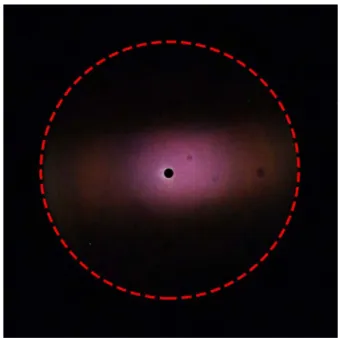
Figure S1. k-space imaging of a metallic purple-coloured scale with narrow aperture illumination in reflection. The presence of only one purple-coloured zone indicates specular reflection and the absence of diffractive effects.
2
Figure S2. (a) Single gold-, bronze- and purple-coloured scales in reflection in air and immersed in a liquid with n = 1.55. (b) Transmitted spectra in air and immersed in an index-matching liquid for the three scales.
Presence of melanin-related pigment
To correctly identify the nature of the pigment present within the scales, for each type of scale, the absorbance depicted in Figure 3b was first normalised by the extracted thickness (112 nm for gold-, 134 nm for bronze- and 164 nm for purple-coloured scale). Figure S3 shows that after this thickness normalisation, the absorbance curves are similar, indicating a uniform melanin-related pigment concentration for all scales. Also the shape is very reminiscent to previously published melanin absorbance curves ([1,2]).
Figure S3. Normalized absorbance of gold-, bronze- and purple-coloured scales.
3
To demonstrate the presence of a melanin-related pigment, a bleaching experiment similar to ref. [1] was carried out. To decrease the absorption due to the presence of melanin pigments, their effect can be reduced after a chemical bleach. In our experiment, scales were immersed for 20h in a 5% solution of H2O2 at 40°C. Figure S4, shows the reflectance and transmittance of a gold-coloured scale without and after bleaching. The effect of bleaching on the photonic response of the scale was simulated by arbitrary setting the absorbance to 0.6 times the initial absorbance. The bleached scale shows a consistent increase in reflection (figure S4.a), where the difference is stronger at shorter wavelengths. In transmittance (figure S4.b), the absorption is also reduced after the bleaching. This result confirms the presence of a melanin-related pigment.
Figure S4. Optical measurement and simulation of a bleaching experiment (a) in reflection and (b) in transmission for a gold-coloured scale without (black curve) and after bleaching (curve). Experimental spectra are shown in dashed lines while continuous lines are used for simulated spectra.
Supplementary References
1. Stavenga DG, Leertouwer HL, Hariyama T, De Raedt HA, Wilts BD. 2012 Sexual Dichromatism of the Damselfly Calopteryx japonica Caused by a Melanin-Chitin Multilayer in the Male Wing Veins. PLoS One 7, e49743.
(doi:10.1371/journal.pone.0049743)
2. Stavenga DG, Leertouwer HL, Wilts BD. 2014 The colouration toolkit of the Pipevine Swallowtail butterfly, Battus philenor: thin films, papiliochromes, and melanin. J.
Comp. Physiol. A 200, 547–561. (doi:10.1007/s00359-014-0901-7)