1239 doi:10.1093/ecco-jcc/jjz088
Advance Access publication May 6, 2019 Original Article
Copyright © 2019 European Crohn’s and Colitis Organisation (ECCO). Published by Oxford University Press. All rights reserved. For permissions, please email: journals.permissions@oup.com
Original Article
Vedolizumab Therapy is Ineffective for
Primary Sclerosing Cholangitis in Patients
With Inflammatory Bowel Disease: A GETAID
Multicentre Cohort Study
Benedicte Caron,
aLaurent Peyrin-Biroulet,
bBenjamin Pariente,
cYoram Bouhnik,
dPhilippe Seksik,
eGuillaume Bouguen,
fLudovic Caillo,
gDavid Laharie,
hFranck Carbonnel,
iRomain Altwegg,
jCatherine Reenaers,
kMelanie Serrero,
lCaroline Trang-Poisson,
mStephane Nancey,
nJerome Filippi,
oVered Abitbol,
pGuillaume Savoye,
qLucine Vuitton,
rStephanie Viennot,
sMathurin Fumery,
tMaud Reymond,
uJean-Pierre Bronowicki,
bJean-Marie Reimund,
aAurelien Amiot
v;
on
be-half of GETAID
aDepartment of Gastroenterology, Hôpitaux Universitaires de Strasbourg [Hôpital de Hautepierre] and INSERM U1113 IRFAC, Université de Strasbourg [Faculté de Médecine], Strasbourg, France bINSERM U954 and Department of Gastroenterology, Université de Lorraine, Nancy, France cDepartment of Gastroenterology, Huriez Hospital, Université of Lille, Lille, France dDepartment of Gastroenterology, IBD and Nutrition Support, Beaujon Hospital, University Paris 7 Denis Diderot, Clichy, France eDepartment of Gastroenterology, AP-HP, Hôpital Saint-Antoine, F-75012, ERL 1057 INSERM/UMRS 7203, UPMC Université Paris 6, Paris, France fDepartment of Gastroenterology, Pontchaillou Hospital and Rennes University, Rennes, France gDepartment of Gastroenterology, Nimes University Hospital, Nimes, France hDepartment of Hepato-Gastroenterology, University Hospital of Bordeaux, Hôpital Haut-Lévêque, Bordeaux, France iDepartment of Gastroenterology, Bicetre University Hospital, APHP, Université Paris Sud, le Kremlin Bicêtre, Paris, France jDepartment of Gastroenterology, Hôpital Saint-Eloi, University Hospital of Montpellier, Montpellier, France kDepartment of Gastroenterology, Liege University Hospital, Liege, Belgium lHôpital Nord, Centre d’investigation clinique Marseille Nord, Université Méditerranée, Marseille, France mDepartment of Gastroenterology, Institut des Maladies de l’appareil Digestif [IMAD], University Hospital of Nantes, Nantes University, Nantes, France nDepartment of Gastroenterology, Hospices Civils de Lyon and University Claude Bernard Lyon 1, Pierre-Benite, France oDepartment of Gastroenterology and Clinical Nutrition, Nice University Hospital, University of Nice Sophia-Antipolis, Nice, France pDepartment of Gastroenterology, Cochin Hospital, University Paris 5 Descartes, Paris, France qDepartment of Gastroenterology, Rouen University and Hospital, Rouen, France rDepartment of Gastroenterology, Besançon University Hospital, Besançon, France sDepartment of Gastroenterology, Caen University Hospital, Caen, France tDepartment of Gastroenterology, Amiens University Hospital, Amiens, France uDepartment of Hepato-Gastroenterology, University Hospital Estaing of Clermont-Ferrand, Université d’Auvergne, Clermont-Ferrand, France vDepartment of Gastroenterology, Henri Mondor Hospital, APHP, EC2M3-EA7375, Paris Est-Créteil Val de Marne University, Creteil, France
Corresponding author: Docteur Aurelien Amiot, 51, Avenue du Marechal de Lattre de Tassigny, Creteil F-94010, France. Tel.: +33-1 49 81 23 62; Fax: +33-1 49 81 23 52; Email: aurelien.amiot@hmn.aphp.fr
Abstract
Background: Whether vedolizumab may be effective as a treatment for primary sclerosing
cholangitis [PSC] in patients with inflammatory bowel disease [IBD] remains controversial.
Methods: We performed a retrospective observational study of consecutive patients with IBD
and PSC, treated with vedolizumab for at least 30 weeks in 22 centres of GETAID from January 2015 to June 2016. The outcomes included a decrease in the serum alkaline phosphatase [ALP] concentration of at least 50% from baseline to Week 30 or 54, a change in any serum liver enzymes concentrations, and an assessment of the efficacy and safety of vedolizumab in IBD.
Results: Among 75 patients with active IBD and PSC treated with vedolizumab, 21 patients
discontinued vedolizumab before Week 30 [due to lack of efficacy in 19 and malignancy in two patients]. In the remaining 54 patients, a decrease in the serum ALP concentration of at least 50% from baseline to Weeks 30 and 54 was observed in four [7%] and four [11%] patients, respectively. No significant change was observed in serum liver enzyme concentrations at week 30 or 54. After a median follow-up period of 19.4 [14.0–29.9] months, nine cases of digestive neoplasia [colorectal neoplasia in seven and cholangiocarcinoma in two] were reported.
Conclusions: In patients with IBD and PSC, vedolizumab did not improve serum liver enzyme
concentrations at week 30 or 54. Nine cases of digestive cancer occurred during the follow-up period, confirming the need for a tight surveillance programme in this population.
Key Words: Crohn’s disease; ulcerative colitis; inflammatory bowel disease; vedolizumab; primary sclerosing cholangitis
1. Introduction
Primary sclerosing cholangitis [PSC] is an idiopathic and progressive cholestatic liver disease characterised by persistent biliary inflamma-tion and fibrosis that occur concurrently with IBD in up to 80% of cases.1,2 The natural history of PSC is dominated by the risk of
bil-iary stenosis, which can lead to acute cholangitis and/or liver failure secondary to biliary obstruction, and an increased risk of developing digestive neoplasia, such as cholangiocarcinoma, with a 400-fold in-creased risk of cholangiocarcinoma and a 10-fold inin-creased risk of colorectal cancer.2,3
There is no effective medical therapy for patients with PSC. Although ursodeoxycholic acid is commonly used in patients with PSC, resulting in reduced and even normal serum liver enzymes, ursodeoxycholic acid failed to demonstrate any improvement in the survival of patients with PSC or any decrease in the risk of asso-ciated complications.4,5 It has been hypothesised that activated T
cell homing from the intestine to the liver might be responsible for PSC-associated biliary inflammation through the upregulation of the adhesion molecule MAdCAM-1 and the chemostatic protein CCL, which may contribute to the recruitment of α4β7 integrin-positive T cells to the biliary endothelium.6–11
Vedolizumab is a fully humanised monoclonal antibody that spe-cifically blocks α4β7 integrin-positive leukocyte migration into the inflamed intestinal tissue, thus making it an attractive treatment for IBD patients with concomitant PSC.12 The efficacy of vedolizumab
for PSC in patients with IBD has only been reported in two retro-spective studies that included 34 and 27 patients with IBD and PSC, respectively.13,14 Due to the relatively small sample sizes and
short-follow-ups [few data beyond Week 30], no definite conclusions can be made from these studies.
We therefore decided to evaluate the efficacy of vedolizumab on serum liver enzyme concentrations, as well as its long-term safety, at Weeks 30 and 54 of treatment in consecutive patients with IBD and PSC who were treated in the centres of the Groupe d’Etude
Therapeutique des Affections Inflammatoires du tube Digestif [GETAID].
2. Methods
2.1. Study population
This was a retrospective observational multicentre study con-ducted in 22 French and Belgian academic centres affiliated with GETAID. From January 2015 to June 2016, all consecutive adult patients with both IBD and PSC, who received at least one injec-tion of vedolizumab, were included. Diagnosis of PSC was con-firmed by two of the following criteria after exclusion of other cholestatic disorders: [1] chronic cholestatic liver disease of at least 6 months duration; [2] intrahepatic and/or extrahepatic biliary duct changes, such as beading or narrowing consistent with PSC as dem-onstrated by magnetic resonance cholangiography; and [3] liver biopsy compatible with the diagnosis of PSC.15 Exclusion criteria
were: age <18 years; PSC without IBD; inactive Crohn’s disease [CD] (Harvey_Bradshaw Index [HBI] <4) or ulcerative colitis [UC] [par-tial Mayo score <3]; and pregnancy or lactation..16,17 Vedolizumab
was administered intravenously at a dose of 300 mg at Weeks 0, 2, and 6, and then at a dose of 300 mg every 8 weeks up to Week 54. The optimisation of vedolizumab therapy at a dose of 300 mg every 4 weeks was allowed for insufficient response according to the investigator’s decision. The concomitant use of steroids and/or immunomodulators for IBD and ursodeoxycholic acid for PSC was allowed according to the investigator’s decision and was recorded at every visit.
The protocol was approved by both the Comité Consultatif sur le Traitement de l’Information en matière de Recherche dans le domaine de la Santé [CCTIRS N°15.888] and the Commission Nationale de l’Informatique et des Libertés [CNIL N°916021]. All authors had access to the study data and reviewed and approved the final manuscript.
2.2. Data collection
Investigators from each participating centre were asked to complete a standardised questionnaire. An on-site visit was then conducted to collect missing data from patient case records. The recorded data in-cluded patient demographics, a detailed account of the IBD and PSC diagnoses and history, smoking status, IBD phenotype according to the Montreal classification, medical and surgical treatment history, and any serious infection history.
All included patients were followed according to routine prac-tice. At each vedolizumab infusion, patients were assessed by physical examination, and the HBI or partial Mayo score was calculated for patients with CD and UC, respectively. In add-ition, routine laboratory tests were conducted, which included the following serum liver enzymes: alkaline phosphatase (ALP, international units [UI]), alanine aminotransferase [ALT, UI], as-partate aminotransferase [AST, UI], γ-glutamyltransferase [γ-GT, UI] and total bilirubin [µmol/L]; C-reactive protein [CRP, mg/L] and haemoglobin [g/dL] levels, as well as leukocyte [/109/L] and platelet [/109/L] counts and serum albumin [g/L], were determined and evaluated for adverse events..16,17 Evaluations of clinical
dis-ease activity and liver tests were collected at Weeks 0, 6, 14, 22, 30, and 54. All adverse events occurring during the follow-up period were collected. Severe adverse events were defined as the occurrence of treatment interruption, hospitalisation, disability, persistent damage, colectomy, or death.
2.3. Outcome measures
The primary efficacy endpoint was the effectiveness of vedolizumab in PSC, as defined by a decrease in the serum ALP concentration of at least 50% from baseline to Week 30 or 54. The percentage of patients with a serum ALP concentration lower than 1.5 times the upper limit of normal [ULN] was also calculated. Changes in the total bilirubin, ALT, AST, γ-GT, CRP, and serum albumin concen-trations were evaluated at Weeks 30 and 54 compared with Week 0. All adverse events occurring during the follow-up period were also collected. Clinical remission of IBD was defined as an HBI score of 4 or less for CD patients, and a partial Mayo Clinic score of less than 3 with a combined stool frequency and rectal bleeding subscore of 1 or less for UC.
2.4. Statistical analysis
The data were expressed as a number [%] for qualitative data and as the mean ± standard deviation [SD] or median [interquartile range] for quantitative data. Liver enzymes were expressed according to the upper limit of normal [ULN]; p-values less than 0.05 were con-sidered significant. Efficacy analyses were performed in patients who received vedolizumab for at least 30 weeks, and a safety analysis was performed in all patients who received at least one infusion of vedolizumab. Pre-treatment and post-treatment laboratory tests and clinical indices were compared between Week 0 and Weeks 30 and 54. The proportion of patients with a decrease in ALP concentration of at least 50%, as well as the proportion of patients with serum ALP concentration >1.5 ULN, was compared between Week 0 and Weeks 30 and 54. Nominal and ordinal data were compared using the chi square test or Fisher’s exact test, as appropriate, whereas parametric data were compared using the Mann-Whitney test and the Wilcoxon matched-pair signed-rank test as appropriate. All analyses were two-tailed, and p-values less than 0.05 were considered significant. All statistical evaluations were performed using SPSS statistical software [SPSS Inc., v17, Chicago, IL, USA].
3. Results
3.1. Study population
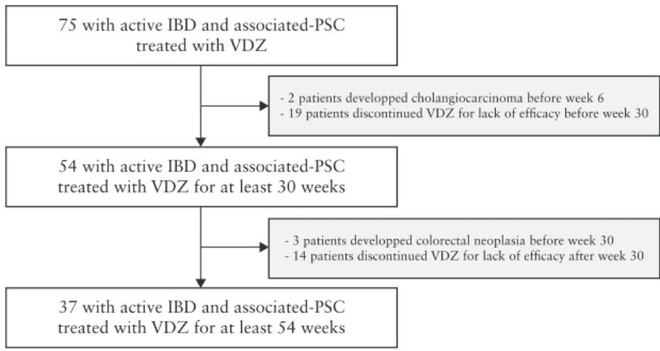
A total of 75 patients with IBD [49 with UC and 26 with CD] and PSC received at least one injection of vedolizumab and were in-cluded in the present study [Table 1]. Nineteen patients were treated for less than 30 weeks due to lack of efficacy in IBD, and two dis-continued vedolizumab therapy between Week 2 and Week 6 after cholangiocarcinoma was diagnosed. These 21 patients were excluded from the analysis of the efficacy of vedolizumab in PSC [Figure 1]. The remaining 54 patients were treated with vedolizumab for at least 30 weeks and were included in the efficacy analysis [Supplementary
Table 1, available as Supplementary data at ECCO-JCC online]. All
75 patients were included in the safety analysis [Table 1].
The demographics, baseline disease characteristics, and medi-cation history of the 75 patients are listed in Table 1. In the overall population [n = 75], the median duration of IBD and PSC was 6.2 [3.7–13.5] and 2.9 [0.4–6.6] years, respectively. All patients had active disease at the time of the enrolment, according to the HBI for CD patients [8.0 ± 4.4] and the total Mayo score for UC patients [7.0 ± 2.7]. Most of the patients had been previously treated for IBD with immunosuppressant [89%] and anti-tumour necrosis factor [TNF] [91%] therapy for IBD. Most of the patients had uncomplicated PSC, with the exclusion of three patients with previous liver transplantation and subsequent PSC recurrence, one patient with a history of gallbladder carcinoma, and one patient with a right colon carcinoma. The two patients with a history of digestive neoplasia had been in remission for at least 5 years
[Table 1].
At baseline, patients were treated with vedolizumab alone in 29 [39%] cases [Table 1]. Vedolizumab was given with concomitant steroid therapy in 38 [51%] cases and concomitant immunosuppres-sant therapy in 23 [31%] cases; 65 patients [87%] were also treated with long-term ursodeoxycholic acid at Week 0 at a dose that did not change throughout the study period. Among patients who were not on ursodeoxycholic acid at baseline, none were treated with ursodeoxycholic acid throughout the study period. The effectiveness of vedolizumab on clinical disease activity of IBD is reported in Table 2 and in Supplementary Tables 2 and 3, available as Supplementary
data at ECCO-JCC online.
3.2. Effect of vedolizumab on serum liver enzymes The effectiveness of vedolizumab on serum liver enzymes was as-sessed in the 54 patients who were treated for at least 30 weeks. Serum liver enzyme concentrations before and after vedolizumab therapy across these 54 patients are shown in Table 2 and Figure 2. There was no significant change in the serum ALP concentration be-tween Week 0 and Weeks 30 or 54 [2.2 ± 2.3 ULN vs 2.3 ± 2.4 ULN and 2.3 ± 2.2 ULN, p = 0.66 and 0.98, respectively], as well as no significant change in other serum liver enzymes concentrations be-tween Week 0 and Weeks 30 or 54. The proportion of patients with an elevated serum ALP concentration [>1.5 ULN] was 37%, 50%, and 46% at Weeks 0, 30, and 54 [p = 0.24 and 0.52], respectively. The proportion of patients with a decrease in the baseline serum ALP concentration of at least 50% was 7% and 11% at weeks 30 and 54, respectively [Table 2 and Figure 2].
We next analysed the effectiveness of vedolizumab on the serum liver enzyme concentrations in patients with an elevated serum ALP concentration at Week 0 and in the subgroup of patients with normal serum ALP concentration at Week 0 [Supplementary Tables 2 and 3,
available as Supplementary data at ECCO-JCC online].
Of the 20 patients with elevated serum ALP concentration at baseline, two patients had a decrease of at least 50% of the baseline serum ALP concentration at Weeks 30 and 54 [10% and 13.3%, respectively] [Supplementary Table 2]. Overall, there was no signifi-cant change in any liver enzyme concentration between Week 0 and Weeks 30 or 54 in patients with an elevated serum ALP concentra-tion at Week 0.
Of the 34 patients with a normal serum ALP concentration at baseline, two patients had a decrease of at least 50% of the baseline
serum ALP concentration at Weeks 30 and 54 [6% and 9%, re-spectively] [Supplementary Table 3]. Overall, there was no signifi-cant change in any liver enzymes between Week 0 and Weeks 30 or 54 in patients with normal serum ALP concentration at Week 0, with the exception of one patient with a significant increase in the serum ALP concentration at Week 30 but not at Week 54. The pro-portion of patients with an elevated serum ALP concentration [>1.5 ULN] was 27%, and 23% at weeks 30 and 54 [p = 0.002 and 0.02], respectively.
Table 1. Demographics, disease characteristics, and medication histories of 75 patients of patients with primary sclerosing cholangitis and
inflammatory bowel disease treated with vedolizumab.
Characteristic Crohn’s disease [n = 26] Ulcerative colitis [n = 49]
Median age at IBD diagnosis, years 20.9 [16.6–34.2]
Median age at PSC diagnosis, years 26.0 [18.3–38.4]
Male gender, n [%] 51 [68.0%]
Body mass index, kg/m2 22.0 ± 3.7
Median duration of IBD, years 6.2 [3.7–13.5]
Median duration of PSC, years 2.9 [0.4–6.6]
Current smoker, n [%] 7 [9%]
Clinical disease activity scoring Harvey-_radshaw Index Mayo Clinic score
8.0 ± 4.4 7.0 ± 2.7
Partial Mayo Clinic score 4.9 ± 2.3
Disease location—Montreal classification L1: 5 [19%] E1: 0
L2: 6 [23%] E2: 3 [8%]
L3: 15 [58%] E3: 45 [92%]
L4: 0
Disease phenotype—Montreal classification B1: 19 [73.1%]
-B2: 3 [11.5%]
-B3: 4 [15.4%]
-p: 6 [8.0%]
-History of intestinal resection, n [%] 12 [46.2%] 0
PSC-related symptoms
Pruritus 3 [6%]
Right upper quadrant abdominal pain 2 [3%]
Jaundice 2 [3%]
Ascites 1 [1%]
Splenomegaly 3 [4%]
Hepatomegaly 6 [8%]
Asymptomatic 65 [87%]
History of IBD treatment, n [%]
Aminosalicylates 62 [83%]
Steroids 73 [97%]
Immunosuppressant 67 [89%]
Anti-TNF therapy 68 [91%]
Biological variables
Alkaline phosphatase [ULN] [normal <120] 2.0 ± 2.6
ALP > 1.5 ULN, n [%] 27 [36%]
Aspartate aminotransferase [ULN] [normal <40] 1.4 ± 1.1
Alanine aminotransferase [ULN] [normal <40] 1.7 ± 1.8
γ-glutamyltransferase [ULN] [normal <40] 5.3 ± 8.3
Total bilirubin [ULN] [normal <17] 1.1 ± 1.7
Serum albumin [g/L] [normal >35] 36.7 ± 5.2
Platelet count, 109/L 342 ± 162 hsCRP level, mg/L 15.8 ± 21.9 Concomitant medications Glucocorticoids 38 [51%] Immunosuppressants 23 [31] Ursodeoxycholic acid 65 [87%]
ALP, alkaline phosphatase; hsCRP, high sensitivity C-reactive protein; IBD, inflammatory bowel disease; PSC, primary sclerosing cholangitis; ULN, upper limit normal; TNF, tumour necrosis factor.
Variables are presented as n [%], mean ± standard deviation, or median [interquartile range].
3.3. Effect of vedolizumab on activity of the inflammatory bowel disease
All 54 patients were assessed for clinical activity at Week 30 and 37 at Week 54. Seventeen patients discontinued vedolizumab between Weeks 30 and 54 for lack of response in 14 cases and after colorectal neoplasia were diagnosed in three cases.
Among the 33 patients with UC, steroid-free clinical remission was achieved in 42%, 48%, and 50% at Weeks 14, 30, and 54, re-spectively. Among the patients with UC, 27 [82%] were assessed for mucosal healing between Week 30 and Week 54. According to the Mayo Clinic endoscopic subscore [0 or 1], mucosal healing was achieved in 11 [42.3%].
Among the 21 patients with CD, steroid-free clinical remission was achieved in 43%, 48%, and 46% at Weeks 14, 30, and 54, respectively. Among the patients with CD, seven [33.3%] were as-sessed for mucosal healing between Weeks 30 and 54, of whom three [42.9%] achieved mucosal healing [absence of ulcer].
No difference was found in the impact of vedolizumab on clinical activity of IBD according to the level of serum ALP concentration at Week 0 [Supplementary Table 4 available as Supplementary data at
ECCO-JCC online]. Patients with elevated serum ALP concentra-tion at Week 0 had higher CRP level at Weeks 0 and 54 and lower albumin level at Week 54.
3.4. Safety and malignancy risk
The analysis of adverse events was performed in the entire cohort [n = 75] of patients who received at least one dose of vedolizumab.
After a median follow-up period of 1.6 [1.2–2.5] years, 19 adverse events occurred in 19 [25%] patients, including 11 [15%] severe ad-verse events. These adad-verse events consisted of infection in 10 cases and digestive neoplasia in nine cases.
The characteristics of the nine patients who developed digestive neoplasia are reported in Table 3. Two cholangiocarcinomas were diagnosed during the vedolizumab induction phase, with symptoms of acute jaundice and cholestasis. One patient subsequently died after not responding to chemotherapy, and the other patient was still alive at the end of the follow-up period but had a progressive disease despite chemotherapy and only received the best supportive care. Three colorectal neoplasias were diagnosed between Weeks 30 and 54. Four additional cases of colorectal neoplasia were diagnosed between 1 and 3 years after the introduction of vedolizumab. Six out of seven cases of colorectal neoplasia were diagnosed during a col-onoscopy screening and consisted of high-grade dysplasia or stage 1 adenocarcinoma, whereas one patient presented with an aggressive form of rectal carcinoma and subsequently died.
4. Discussion
The management of IBD-associated PSC remains challenging. Given the limitations of the available studies, we sought to further investigate whether vedolizumab, by inhibiting the recruitment of α4β7 integrin-positive cells within the liver, might be an effective and safe treatment of PSC in patients with IBD.12 In the 75 patients
with PSC and active IBD included in this study, we first evaluated 75 with active IBD and associated-PSC
treated with VDZ
54 with active IBD and associated-PSC treated with VDZ for at least 30 weeks
37 with active IBD and associated-PSC treated with VDZ for at least 54 weeks
- 2 patients developped cholangiocarcinoma before week 6 - 19 patients discontinued VDZ for lack of efficacy before week 30
- 3 patients developped colorectal neoplasia before week 30 - 14 patients discontinued VDZ for lack of efficacy after week 30
Figure 1. Flow chart of patient with inflammatory bowel disease with primary sclerosing cholangitis, treated with vedolizumab from Week 0 to Week 54. Table 2. Changes in laboratory tests and clinical scoring at Weeks 30 and 54 in patients with primary sclerosing cholangitis and
inflamma-tory bowel disease, treated with vedolizumab.
Changes between Week 30 and Week 0 [n = 54]
p Changes between Weeks 54
and 0 [n = 37]
p
Alkaline phosphatase, ULN 0.1 ± 1.7 0.66 -0.01 ± 2.4 0.98
Aspartate aminotransferase, ULN 0.1 ± 1.2 0.58 -0.2 ± 0.9 0.33
Alanine aminotransferase, ULN -0.01 ± 1.9 0.96 -0.4 ± 1.5 0.22
γ-glutamyltransferase, ULN -0.3 ± 1.6 0.17 -0.4 ± 0.36 0.31
Total bilirubin, ULN 0.5 ± 5.0 0.51 0.1 ± 5.4 0.84
Serum albumin, g/L 2.3 ± 7.7 0.06 -0.5 ± 10.0 0.01
Platelet count, 109/L -27 ± 106 0.02 -43 ± 163 0.03
hsCRP level, mg/L -5.6 ± 20.2 <0.001 -0.5 ± 10.0 0.16
Harvey-Bradshaw index -3.1 ± 6.0 0.03 -4.2 ± 6.5 0.04
Partial Mayo Clinic score -2.0 ± 2.6 <0.001 -2.9 ± 3.0 <0.001
CRP, C-reactive protein; ULN, upper limit of normal.
the effectiveness and safety of vedolizumab therapy in 54 patients treated with vedolizumab for at least 30 weeks. Overall, there was no improvement in the liver tests. Only four patients [7.4%] had a decrease in the serum ALP concentration of at least 50% from base-line to Week 30 and Week 54. Even though the safety profile con-cerning infectious risk was consistent with previous real-world data with vedolizumab, we reported a high rate of digestive neoplasia, especially during the first year of treatment with vedolizumab.18,19
Two retrospective studies have been recently published assessing the effectiveness of vedolizumab on PSC in patients with concomi-tant IBD and PSC.13,14 Christensen et al. included 34 patients [16
with CD and 18 with UC] with a serum liver enzyme assessment
at Week 14 and Week 30, and Tse et al. included 27 patients [10 with CD and 16 with UC] with a serum liver enzyme assessment at 6–8 months in 23 cases and at 12–14 months in 19 cases. There was no improvement in the ALP concentration or in any serum liver enzyme concentration, despite the clear evidence of clinical effect-iveness on IBD activity in the first study. Our study confirmed that vedolizumab was not effective for treating PSC but was clearly ef-fective for treating active IBD. Indeed, there was no change in the ALP concentration or in other serum liver enzyme concentrations. No change in effectiveness in PSC was found by stratifying patients according to an elevated concentration of ALP at baseline. There was no evidence to suggest that the lack of efficacy of vedolizumab in 8 7 Week 0 n = 54 Week 30 n = 54 Week 54 n = 37 Week 0 n = 49 Week 30 n = 48 Week 54 n = 33 6 p = 0.66
A
B
p = 0.98 p = 0.96 0.22 p = 0.51Aspartate aminotransferase (ULN)
p = 0.84 Alkaline phosphatase (ULN)
Alanine aminotransferase (ULN)
p = 0.17 p < 0.001 p = 0.06 p = 0.31 γ-glutamyltransferase (ULN) C-reactive protein (mg/L) p = 0.58 p = 0.33 Total bilirubin (ULN)
5
Serum level Serum level
4 3 2 1 0 5 4 Week 0
n = 49 Week 30n = 50 Week 54n = 36 Week 0n = 49 Week 30n = 50 Week 54n = 34 4
C
D
3 3 Serum level 2 2 1 1 0 4 3 3 Serum level 2 2 1 1 0 18 16 Week 0n = 47 Week 30n = 47 Week 54n = 33 Week 0n = 51 Week 30n = 51 Week 54n = 35 14
E
F
12 10 Serum level 8 6 4 2 0 45 40 35 Serum level 30 25 20 15 10 5 0 3 2 2 1 1 0Figure 2. Serum liver enzymes and C-reactive protein concentrations of 54 patients with inflammatory bowel disease, treated with vedolizumab for at least
30 weeks: [A] alkaline phosphatase; [B] total bilirubin; [C] alanine aminotransferase; [D] aspartate aminotransferase; [E] γ-glutamyltransferase; [F] C-reactive protein. In the panels, the horizontal line in each bow represents the median, the top, and the bottom of the boxes; the interquartile range; and the bars 1.5 times the interquartile range.
Table 3. Characteristics of the nine incident cases of digestive neoplasia in patients with primary sclerosing cholangitis and inflammatory
bowel disease, treated with vedolizumab. Gender,
age [years]
Delay between digestive neoplasia and Type of neoplasia Vedolizumab
therapy at the time of diagnosis of digestive neoplasia
Type, findings. and delay of the last screening test before vedolizumab introduction [months] Staging Outcome diagnosis of IBD [years] diagnosis of PSC [years] intro-duction of VDZ [months] During the induction phase
M, 32.9 11.3 5.5 1.2 Cholangiocarcinoma Yes MRI showing stable
multiple intrahepatic stricture without evidence of cancer. 12.2
T2N1M1 Died
M, 51.1 6.7 0.1 1.3 Cholangiocarcinoma Yes MRI showing
strictures of the left hepatic duct with no evidence of cancer. 2.8
T2N1Mx Alive, palliative
chemotherapy ongoing
During the first year of vedolizumab therapy
M, 29.2 6,0 3.6 5,9 Colorectal neoplasia Yes Colonoscopy
showing active pancolitis [MCES 3; UCEIS 5]. No evi-dence of dysplasia on random biopsies, 6.6 pT2N0M0 Proctocolectomy
M, 44.7 11,0 0.4 6,1 Colorectal neoplasia Yes Colonoscopy
showing active pancolitis [MCES 3; UCEIS 6]. No evi-dence of dysplasia on random biopsies, 6.1 pT2N0Mx pT4N1M1 Died
M, 48.2 13,6 12.8 9,7 Colorectal neoplasia Yes Colonoscopy
showing active pancolitis [MCES 3; UCEIS 4]. No evi-dence of dysplasia on random biopsies, 5.3 HGD Proctocolectomy
After the first year of vedolizumab therapy
M, 36.4 17,5 6.8 14,9 Colorectal neoplasia Yes Colonoscopy
showing pancolitis [MCES 2; UCEIS 3], 18.4
pT2N0Mx Proctocolectomy
F, 34.0 20,4 17.4 35,8 Colorectal neoplasia Yes Colonoscopy
showing pancolitis [MCES 2; UCEIS 3], 16.4
pT1N0Mx Proctocolectomy
F, 27.5 5,2 3.3 22,5 Colorectal neoplasia Yes Colonoscopy
showing active proctitis [MCES 3; UCEIS 6] and inactive col-itis [MCEIS 1; UCEIS 2], 22.5 HGD Proctocolectomy
M, 23.8 8,9 0.2 33,6 Colorectal neoplasia Vedolizumab
discontined after 16.1 months Colonoscopy showing active pancolitis [MCES 3; UCEIS 6]. No evi-dence of dysplasia on random biopsies, 21.4 HGD Proctocolectomy
All screening colonoscopies were complete and performed with a 4-litre polyethyleneglycol preparation and indigo carmine chromoendoscopy.
F, female; HGD, high-grade dysplasia; IBD, inflammatory bowel disease; M, male; MCES, Mayo Clinic Endoscopic Subscore; PSC, primary sclerosing cholan-gitis; UCEIS, Ulcerative Colitis Endoscopic Index of Severity; VDZ, vedolizumab.
PSC was related to a lack of efficacy of vedolizumab in IBD. Indeed, the effectiveness of vedolizumab in IBD was in line with previous reports in patients with IBD and without PSC.19
In the present study, infection was the most frequent adverse event, and there were only two serious adverse events of infection. Importantly, nine cases of digestive neoplasia were reported fol-lowing a median follow-up of 19.4 months. Previous reports in this patient population did not identify such a risk. However, the dur-ation of follow-up and the sample size were smaller in previous re-ports than in this nationwide GETAID study. In particular, five cases of digestive neoplasia were reported in patients with IBD and PSC treated with vedolizumab during the first year of treatment, com-prising two cases of cholangiocarcinoma and three cases of colo-rectal cancer. An additional safety survey focusing on the occurrence of digestive neoplasia was therefore set up, which identified four additional cases with a median follow-up period of 1.6 [1.2–2.5] years. Whether this malignancy risk is related to the impairment of the immunosurveillance of the gastrointestinal [GI] tract induced by anti-integrin antibodies, or to the well-known increased risk of malignancy in patients with both IBD and PSC, will require further investigation.
All the patients included in this study had no evidence of colorectal neoplasia on colonoscopy before enrolment. All the patients with in-cidental colorectal neoplasia had undergone a complete screening colonoscopy, with indigo carmine chromoendoscopy, with a delay of 5.3 to 22.5 months before the introduction of vedolizumab. The majority of patients had endoscopically active disease, which may have biased screening performance. Similarly, the two patients who developed cholangiocarcinoma had magnetic resonance imaging [MRI] assessment before enrolment that showed no evidence of cholangiocarcinoma. Indeed, the risk of cholangiocarcinoma among patients with PSC is 400 times higher than the risk in the general population.2,20 Similarly, the risk of colorectal cancer among patients
with PSC and IBD is four times higher than the risk among patients with IBD alone, and 10 times higher than the risk in the general population.2,21 Although the two patients who were diagnosed with
cholangiocarcinoma presented with a rapidly progressive disease, the occurrence of cancer was noted during the induction phase, sug-gesting a potential underlying pre-neoplastic lesion. The other cases of colorectal neoplasia occurred in patients who had entered a man-datory tight screening programme with various onset delays after the introduction of vedolizumab. No similar safety signal was reported in the study of Christensen et al., whereas no safety data are avail-able in the study of Tse et al.13,14
A meta-analysis estimated the likelihood of experiencing ma-lignancy when patients with IBD without PSC were administered anti-integrin antibodies or a placebo.22 Among the 1146 patients
randomised to receive gut-specific anti-integrin antibodies, there were three [0.3%] malignancies compared with two [0.3%] malig-nancies among the 664 receiving placebo (risk ratio [[RR] = 0.78; 95% confidence interval [CI] 0.15–4.02), with no heterogeneity between studies. In another review, malignancy was reported in 18/2830 IBD patients with vedolizumab exposure compared with 1/504 in placebo-exposed patients with IBD.23 Six out of 18 of the
vedolizumab-exposed patients had gastrointestinal malignancies [three colorectal cancers, one hepatic cancer, one appendiceal car-cinoid metastasis, and one peritoneal metastasis], which were diag-nosed after a median [range 2–41] vedolizumab infusions.
We acknowledge several limitations to the present study due to its retrospective design. However, this study also has several strengths. First, we provide efficacy data up to Week 54, a significantly longer
period than that of previous studies; therefore, this study is probably more appropriate to demonstrate any significant improvements in the liver tests. Second, the sample size for such a patient population is considered clinically relevant. Third, this was a nationwide survey of consecutive patients. Finally, a long-term follow-up allowed us to explore the safety profile of vedolizumab in this specific population.
In conclusion, vedolizumab was not effective to change serum liver enzyme concentrations in patients with IBD and PSC, despite clear evidence of its effectiveness in active intestinal inflammation. Moreover, we reported an unexpected number of digestive neoplasia cases that might be attributable to impaired immunosurveillance induced by vedolizumab therapy but may also be related to the well-known increased risk of malignancy in this patient popula-tion. Overall our findings, together with previous reports, are not in favour of conducting a controlled trial on vedolizumab in PSC. Close monitoring of patients with both IBD and PSC, who have been treated with vedolizumab, is recommended in routine practice, using yearly colonoscopy screening and MRI.
Funding
None.Conflict of Interest
LP-B has received consulting fees from Merck, Abbvie, Janssen, Genentech, Ferring, Norgine, Tillots, Vifor, Shire, Therakos, Pharmacosmos, Pilège, BMS, UCB-Pharma, Hospira, Celltrion, Takeda, Biogaran, Boerhinger-Ingelheim, Lilly, Pfizer, and HAC-Pharma. This author has also received lecture fees from Merck, Abbvie, Takeda, Janssen Cilag, Ferring, Norgine, Tillots, Vifor, Therakos, HAC-Pharma, and Mitsubishi. BP has received consulting fees from Abbvie, MSD, and Pfizer. This author has also received speaking fees from Abbvie, MSD, Ferring, and Takeda. YB has received lecture and consulting fees from Abbvie, Biogaran, Boehringer-Ingelheim, CTMA, Ferring, Gilead, Hospira, ICON, Inception IBD, Janssen, Lilly, Mayoli Spindler, Merck, MSD, Norgine, Pfizer, Robarts Clinical Trials, Roche, Sanofi, Shire, Takeda, UCB, and Vifor Pharma. This author has also stock ownership of Inception IBD, San Diego, CA, USA. PS has received consulting fees from Takeda, Abbvie, Merck-MSD, Astellas, Ferring, and Biocodex, grants from Biocodex, sponsored travel from Merck-MSD and Takeda. GB has received consulting fees from MSD, Janssen, and Abbvie. This author has also received lecture fees from MSD, Abbvie, Pfizer, Takeda, and Ferring. DL has received consulting and lecture fees from AbbVie, Ferring, Janssen Cilag, MSD, Pfizer, and Takeda. RA has received board or lectures fees from Abbvie, Janssen, Pfizer, Takeda. CR has re-ceived consulting fees, lecture fees, and travel accommodations from AbbVie, Takeda, MSD, Mundipharma, Hospira, and Ferring. CT-P has received lec-ture fees from MSD, Abbvie, Vifor Pharma, and Norgine. This author has also received travel accommodations from Biogaran, Takeda, Hospira, MSD, and Mayoly Spindler. SN has received consulting fees from Merck, Abbvie, Takeda, Ferring, Norgine, Vifor Pharma, Novartis, Janssen Cilag, Hospira, Takeda, and HAC‐Pharma. JF has received lecture and consulting fees from Abbvie, Astellas pharma, Covidien, Ferring, Jansen, MSD, Pfizer, and Takeda. VA has received lecture fees from Ferring, MSD, Vifor Pharma, and Abbvie. GS has received lecture fees from Vifor Pharma, Takeda, Pfizer, HAC Pharma, Abbvie, MSD, and Ferring France. This author has also received travel accommodations from Ferring, Abbvie, and MSD France as well as a research grant from Ferring. LV has received lecture fees from Abbvie, MSD, Takeda, Ferring, Janssen, and Pfizer, and research grants from MSD, Takeda, and Pfizer. SV has received con-sulting fees from Abbvie, MSD, Takeda, Vifor Pharma, and Ferring. Mathurin Fumery has received lecture fees from Abbvie, MSD, and Ferring. J-MR has received consulting fees from Hospira. This author has also received lectures fees from Abbvie, Ferring, Janssen, and Takeda. This author has also received travel accommodations from Ferring, Abbvie, MSD, Janssen, Pfizer, Hospira, and Takeda. AA has received consulting fees from Abbvie, Hospira, Takeda, Gilead, and Biocodex, as well as lecture fees and travel accommodations from
Abbvie, Janssen, Biocodex, Hospira, Ferring, Takeda, and MSD. This author has also received advisory board fees from Gilead, Takeda, and Abbvie. No conflict of interest is claimed by the remaining authors.
Author Contributions
Concept and design of the study: AA, LPB, BC, JPB, JMR. Generation, collec-tion, assembly, analysis and/or interpretation of data: BC, LPB, BP, YB, PS, GB, LC, DL, FC, RA, CR, MS, CTP, SN, JF, VA, GS, LV, SV, MF, MR, JPB, JMR, AA. Drafting or revision of the manuscript: BC, LPB, BP, YB, PS, GB, LC, DL, FC, RA, CR, MS, CTP, SN, JF, VA, GS, LV, SV, MF, MR, JPB, JMR, AA. Approval of the final version of the manuscript: BC, LPB, BP, YB, PS, GB, LC, DL, FC, RA, CR, MS, CTP, SN, JF, VA, GS, LV, SV, MF, MR, JPB, JMR, AA.
Supplementary Data
Supplementary data are available at ECCO-JCC online.
References
1. Hirschfield GM, Karlsen TH, Lindor KD, Adams DH. Primary sclerosing cholangitis. Lancet 2013;382:1587–99.
2. Lazaridis KN, LaRusso NF. Primary sclerosing cholangitis. N Engl J Med 2016;375:1161–70.
3. Torres J, Pineton de Chambrun G, Itzkowitz S, Sachar DB, Colombel JF. Review article: colorectal neoplasia in patients with primary sclerosing cholangitis and inflammatory bowel disease. Aliment Pharmacol Ther 2011;34:497–508.
4. Lindor KD. Ursodiol for primary sclerosing cholangitis. Mayo Primary Sclerosing Cholangitis-Ursodeoxycholic Acid Study group. N Engl J Med 1997;336:691–5.
5. Eaton JE, Silveira MG, Pardi DS, et al. High-dose ursodeoxycholic acid is associated with the development of colorectal neoplasia in patients with ulcerative colitis and primary sclerosing cholangitis. Am J Gastroenterol 2011;106:1638–45.
6. Burak K, Angulo P, Pasha TM, Egan K, Petz J, Lindor KD. Incidence and risk factors for cholangiocarcinoma in primary sclerosing cholangitis. Am
J Gastroenterol 2004;99:523–6.
7. Hillan KJ, Hagler KE, MacSween RN, et al. Expression of the mucosal vascular addressin, MAdCAM-1, in inflammatory liver disease. Liver 1999;19:509–18.
8. Grant AJ, Lalor PF, Salmi M, Jalkanen S, Adams DH. Homing of mucosal lymphocytes to the liver in the pathogenesis of hepatic complications of inflammatory bowel disease. Lancet 2002;359:150–7.
9. Grant AJ, Lalor PF, Hübscher SG, Briskin M, Adams DH. MAdCAM-1 expressed in chronic inflammatory liver disease supports mucosal lympho-cyte adhesion to hepatic endothelium [MAdCAM-1 in chronic inflamma-tory liver disease]. Hepatology 2001;33:1065–72.
10. Eksteen B, Grant AJ, Miles A, et al. Hepatic endothelial CCL25 mediates the recruitment of CCR9+ gut-homing lymphocytes to the liver in primary sclerosing cholangitis. J Exp Med 2004;200:1511–7.
11. Eksteen B, Mora JR, Haughton EL, et al. Gut homing receptors on CD8 T cells are retinoic acid dependent and not maintained by liver dendritic or stellate cells. Gastroenterology 2009;137:320–9.
12. Feagan BG, Greenberg GR, Wild G, et al. Treatment of ulcerative colitis with a humanized antibody to the alpha4beta7 integrin. N Engl J Med 2005;352:2499–507.
13. Christensen B, Micic D, Gibson PR, et al. Vedolizumab in patients with concurrent primary sclerosing cholangitis and inflammatory bowel disease does not improve liver biochemistry but is safe and effective for the bowel disease. Aliment Pharmacol Ther 2018;47:753–62.
14. Tse CS, Loftus EV Jr, Raffals LE, Gossard AA, Lightner AL. Effects of vedolizumab, adalimumab and infliximab on biliary inflammation in indi-viduals with primary sclerosing cholangitis and inflammatory bowel dis-ease. Aliment Pharmacol Ther 2018;48:190–5.
15. Karlsen TH, Folseraas T, Thorburn D, Vesterhus M. Primary sclerosing cholangitis - a comprehensive review. J Hepatol 2017;67:1298–323. 16. Rutgeerts P, Sandborn WJ, Feagan BG, et al. Infliximab for
induc-tion and maintenance therapy for ulcerative colitis. N Engl J Med 2005;353:2462–76.
17. Harvey RF, Bradshaw JM. A simple index of Crohn’s-disease activity.
Lancet 1980;1:514.
18. Bye WA, Jairath V, Travis SPL. Systematic review: the safety of vedolizumab for the treatment of inflammatory bowel disease. Aliment Pharmacol Ther 2017;46:3–15.
19. Schreiber S, Dignass A, Peyrin-Biroulet L, et al. Systematic review with meta-analysis: real-world effectiveness and safety of vedolizumab in pa-tients with inflammatory bowel disease. J Gastroenterol 2018;53:1048–64. 20. Boonstra K, Weersma RK, van Erpecum KJ, et al.; EpiPSCPBC Study
Group. Population-based epidemiology, malignancy risk, and outcome of primary sclerosing cholangitis. Hepatology 2013;58:2045–55.
21. Khaderi SA, Sussman NL. Screening for malignancy in primary sclerosing cholangitis [PSC]. Curr Gastroenterol Rep 2015;17:17.
22. Luthra P, Peyrin-Biroulet L, Ford AC. Systematic review and meta-analysis: opportunistic infections and malignancies during treatment with anti-integrin antibodies in inflammatory bowel disease. Aliment Pharmacol
Ther 2015;41:1227–36.
23. Colombel JF, Sands BE, Rutgeerts P, et al. The safety of vedolizumab for ulcerative colitis and Crohn’s disease. Gut 2017;66:839–51.

![Figure 2. Serum liver enzymes and C-reactive protein concentrations of 54 patients with inflammatory bowel disease, treated with vedolizumab for at least 30 weeks: [A] alkaline phosphatase; [B] total bilirubin; [C] alanine aminotransferase; [D] aspartate](https://thumb-eu.123doks.com/thumbv2/123doknet/14052305.460327/6.918.144.776.86.811/reactive-concentrations-inflammatory-vedolizumab-phosphatase-bilirubin-aminotransferase-aspartate.webp)

