Publisher’s version / Version de l'éditeur:
Analytical Chemistry, 89, 8, pp. 4716-4720, 2017-03-27
READ THESE TERMS AND CONDITIONS CAREFULLY BEFORE USING THIS WEBSITE.
https://nrc-publications.canada.ca/eng/copyright
Vous avez des questions? Nous pouvons vous aider. Pour communiquer directement avec un auteur, consultez la
première page de la revue dans laquelle son article a été publié afin de trouver ses coordonnées. Si vous n’arrivez pas à les repérer, communiquez avec nous à [email protected].
Questions? Contact the NRC Publications Archive team at
[email protected]. If you wish to email the authors directly, please see the first page of the publication for their contact information.
NRC Publications Archive
Archives des publications du CNRC
This publication could be one of several versions: author’s original, accepted manuscript or the publisher’s version. / La version de cette publication peut être l’une des suivantes : la version prépublication de l’auteur, la version acceptée du manuscrit ou la version de l’éditeur.
For the publisher’s version, please access the DOI link below./ Pour consulter la version de l’éditeur, utilisez le lien DOI ci-dessous.
https://doi.org/10.1021/acs.analchem.7b00580
Access and use of this website and the material on it are subject to the Terms and Conditions set forth at
Ultrafast separation and analysis of monoclonal antibody aggregates
using membrane chromatography
Madadkar, Pedram; Umatheva, Umatheny; Hale, Geoff; Durocher, Yves;
Ghosh, Raja
https://publications-cnrc.canada.ca/fra/droits
L’accès à ce site Web et l’utilisation de son contenu sont assujettis aux conditions présentées dans le site LISEZ CES CONDITIONS ATTENTIVEMENT AVANT D’UTILISER CE SITE WEB.
NRC Publications Record / Notice d'Archives des publications de CNRC:
https://nrc-publications.canada.ca/eng/view/object/?id=e03330f2-5fe7-4ebe-8beb-a810dc9a9544 https://publications-cnrc.canada.ca/fra/voir/objet/?id=e03330f2-5fe7-4ebe-8beb-a810dc9a9544Ultrafast Separation and Analysis of Monoclonal Antibody
Aggregates Using Membrane Chromatography
Pedram Madadkar,
†Umatheny Umatheva,
†Geoff Hale,
‡Yves Durocher,
§and Raja Ghosh
*
,††
Department of Chemical Engineering, McMaster University, 1280 Main Street West, Hamilton, Ontario L8S 4L7, Canada
‡
Freelance Scientist, Oxford OX3 0SJ, United Kingdom
§
National Research Council of Canada, Montreal, Quebec H4P 2R2, Canada
ABSTRACT: We discuss a method for rapid and cost-effective analysis of monoclonal antibody (mAb) aggregates. Hydrophobic interaction membrane chromatography, which was previously shown to be highly suitable for such separation and analysis, was used in a recently developed format referred to as laterally fed membrane chromatography (or LFMC). A stack of rectangular polyvinylidene fluoride (or PVDF) membranes having 0.22 μm pores housed within a modified analytical-scale LFMC device was used for analyzing aggregate types and content in different monoclonal antibody samples. High-resolution separations could be achieved in less than 1.5 min, this being faster than other currently available techniques such as size exclusion ultraperformance liquid chromatography (SE-UPLC). Moreover, the operating pressure was less than 200 kPa,
which eliminated the need for an expensive high-pressure pump and chromatography system. The resolution obtained using the LFMC was comparable to that obtained using SE-UPLC. The effect of design variations such as change in dead volume and pillar size within the lateral channels within the LFMC device was also examined.
A
ggregation is a major challenge with monoclonal antibod-ies (or mAbs) as aggregates are potentially immunogenic or even toxic.1The presence of soluble aggregates is therefore monitored at the different stages of a mAb manufacturing process. Size exclusion high-performance liquid chromatog-raphy (or SE-HPLC) has been the standard technique for monitoring the aggregate levels.2 Despite advantages such asprocess simplicity due to the isocratic nature of separation, conventional SE-HPLC is very slow, typically averaging about two samples per hour.3,4 The productivity of SE-HPLC could be improved without sacrificing the resolution by using small resin particles within shorter columns, the resultant techniques being referred to as size exclusion ultraperformance liquid chromatography (or SE-UPLC). The so-called SE-UPLC columns contain packing resins with sizes in the range of 1− 3 μm, which reduces the solute diffusion path lengths and thereby dispersion effects. The height equivalent of a theoretical plate (or HETP), obtained with SE-UPLC columns, is considerably lower than that for conventional SE-HPLC columns.5 The typical mAb aggregate analysis times for SE-UPLC are below 10 min which contributes to a significant increase in throughput.3,6,7 Using “zero dead-volume” fittings, minute autosampling volumes, sub-2 μm packing material, and small UV cells, it is now possible to analyze recombinant mAb samples in 3−4 min.8−11 Furthermore, parallelization and
interlacing of sample injections could potentially further bring down the separation time.3
The improved productivity and resolution of SE-UPLC comes at the cost of high back-pressure, its primary shortcoming.12While the typical pressure limit for conventional HPLC is below 400 bar, the corresponding value is often as high as 1000 bar for commonly used SE-UPLC columns (e.g., Waters, AQUITY UPLC).8 This requires costly pumps and seals as well as intricate packing techniques.13 Furthermore, friction heating at such high pressure could potentially affect temperature sensitive proteins to the point of denaturation, leading to the possibility of on-column aggregation.14Sub-3 μm core−shell packing material offers the potential for a lower back pressure.15These particles are superficially porous with a dense central core, on account of which they have higher mass transfer rates, higher packing density, and consequently smaller HETPs. Nonetheless, the back pressure with such core−shell particles is still very high.15 It is expected that size of chromatographic media will go down to 500 nm in the next decade, and challenges such as analyte heat-up at ultrahigh pressures and skewed results will need to be addressed.16 However, some recent studies based on nanofluidics seem to suggest that the pressure required with such fine particles might be lower than that theoretically predicted due to “slip-flow” conditions.5,17
Received: February 15, 2017
Accepted: March 27, 2017
Published: March 27, 2017
pubs.acs.org/ac
Downloaded via NATL RESEARCH COUNCIL CANADA on June 28, 2018 at 12:19:57 (UTC).
Although SE-UPLC is the most versatile method for rapid analysis of closely related species such as mAb aggregates, there are other less commonly used techniques that are also worth mentioning. Techniques such as capillary electrophoresis (CE), light scattering (LS), field flow fractionation, fluorescence spectrophotometry, and analytical ultracentrifugation are also suitable for determining aggregate levels in mAb samples. However, neither of these techniques can deliver the required speed and reliability expected in a standardized analytical method in the biopharmaceutical industry.2,18−21Hydrophobic
interaction membrane chromatography (or HIMC) which has been proposed as a “non-size-based” method for analytical separation of mAb aggregates22,23 is based on the reversible binding of the mAb and the aggregates on microporous polyvinylidene fluoride (or PVDF) membranes. In HIMC, binding occurs in the presence of high concentrations of an antichaotropic salt while elution is achieved using a negative salt gradient. The aggregates are separated from the nonaggregated mAb due to their greater hydrophobicity. In addition to excellent resolution, advantages of HIMC include the high speed and insensitivity of resolution to sample size. In contrast, with SEC, resolution drops significantly with sample size.22,24 The HIMC-based mAb aggregates separation studies reported in the literature are based on the use of stacked disc devices22,23,25which are not very scalable. Both scalability and resolution of membrane chromatography can be dramatically improved using a newly developed format called laterally fed membrane chromatography (or LFMC).26−28
In this paper, we discuss the application of the hydrophobic interaction membrane chromatography in the LFMC format for mAb aggregate analysis. LFMC devices26,28have very low solute dispersion effects and on account of this are suitable for carrying out high-resolution chromatographic separations. In addition, LFMC devices with higher membrane surface area could easily be fabricated and operated at very high flow rates without loss of resolution. The LFMC device used in the current study was an improved version of that reported in the literature26,28 designed specifically for analytical scale chroma-tography. Three layers of rectangular PVDF membrane sheets were stacked within this device, resulting in 0.4 mL membrane bed volume. Different monoclonal antibody samples such as Chinese hamster ovary (or CHO) cell line derived Alemtuzumab (or Campath-1H) and HIgG1-CD4 (or Campath-9) and Human embryonic kidney 293 (HEK) cell line derived IgG1 (trastuzumab), containing varying levels of aggregates, were tested in the current study. Conditions required for achieving high efficiency of aggregate separation were determined. The effects of device dead volume and the design of lateral channels on efficiency of separation were examined.
■
EXPERIMENTAL SECTIONMaterials.Campath-1H (humanized monoclonal antibody monomer-rich and dimer-rich samples) and HIgG1-CD4 (Campath-9)29 were kindly donated by the Therapeutic Antibody Centre, University of Oxford, UK. Human embryonic kidney 293 (HEK) cell derived IgG1 (trastuzumab) was produced at the National Research Council of Canada, Montreal, QC, Canada. Ammonium sulfate (A4418), sodium phosphate monobasic (S0751), sodium phosphate dibasic (S0876), sodium chloride (S7653), hydrochloric acid (258148), 30% acrylamide solution (A3699), sodium dodecyl sulfate (L3771), glycine (G8898), glycerol (G2025),
ammo-nium persulfate (A3678), N,N,N′,N′-tetramethyl ethylenedi-amine (T9281), bromophenol blue (B0126), Brilliant blue R concentrate (B8647), Trizma-hydrochloride (T3253), Trizma base (T1503), and DL-dithiothreitol (43817) were purchased
from Sigma-Aldrich, St. Louis, MO, USA. Acetic acid (1000-1) and methanol (6700-1) were purchased from Caledon laboratories Ltd. (Georgetown, ON, Canada). Hydrophilized poly(vinylidene fluoride) (PVDF) membranes (GVWP14250; 0.22 μm pore size) and Amicon centrifugal filters (30 kDa MWCO) were purchased from EMD Millipore Co. (Billerica, MA, USA). The buffers and solutions used for this work were all prepared using deionized water obtained with a SIMPLICITY 185 water purification unit Millipore (Molsheim, France).
Size Exclusion Chromatography (SEC). Campath-1H monomer-rich and dimer-rich samples were analyzed using SE-HPLC. Stock solutions were diluted to 0.25 mg/mL using 20 mM sodium phosphate buffer (pH 7) containing 250 mM NaCl. An SEC column, TSK-Gel G3000SWXL (7.8 mm × 300 mm Tosoh Bioscience, Montgomeryville, PA), was used with an Alliance HPLC system (Waters Corporations, Milford, MA, USA).
The column was initially equilibrated with the same buffer until stable baseline was achieved. The SE-HPLC experiments were run at 0.3 mL/min flow rate with 10 μL sample volumes. UV absorbance at 280 nm wavelength was monitored and logged to generate chromatograms.
Native Polyacrylamide Gel Electrophoresis. Campath-1H samples were analyzed using acidic native PAGE30by which the protein molecules are separated on the basis of their intrinsic size and charge. The mAb samples were first desalted and concentrated using 30 kDa MWCO centrifugal ultrafilters (3750 rpm, 30 min). They were then mixed with sample buffer containing acetic acid, glycerol, Tris-base, and pyronin and were loaded on 7% polyacrylamide gel. The stacking gel (pH 6.0) was prepared using 0.5 M Tris-HCl, whereas the separating gel (pH 4.8) was made with 1.5 M Tris-HCl buffer. Reverse polarity electrophoresis was carried out at 200 V for 2 h using a Hoefer miniVE system (GE healthcare biosciences, Baie-d’Urfe, QC, Canada). Coomassie Brilliant Blue (diluted to 1 L) dye solution was used to stain the gel for 20 min. The gel was then destained using a solution containing methanol (15%), acetic acid (10%), and water (75%) and was documented using a Bioimaging MiniBis pro system (24-25-PR) (DNR-Imaging Systems, Jerusalem, Israel).
Device Fabrication.The analytical LFMC device consisted of three layers: the top and bottom acrylic plates and the middle spacer made of plastic shims (Figure 1). The outer dimension of all three rectangular components was 150 mm × 40 mm. The plates had an identical design, and each contained a rectangular channel (100 mm × 10 mm) curved at the endings with the radius of 5 mm. The channels contained 140 cylindrical pillars with hexagonal arrays of 3 and 4 over the width of the channel. The pillar height was the same as the depth of the channel (0.2 mm). Two ports were embedded on either side of the channel (109 mm center-to-center distance) which were compatible to luer fittings and were tapered down to 1 mm over the thickness of the plates. The middle spacer contained a rectangular slot which was 2.5 mm wider than the rectangular channels on each side (i.e., 115 mm × 15 mm). Three layers of the PVDF membrane sheet having the same dimension with the rectangular slot were cut, placed within the spacer, and sandwiched between the plates. The spacer
Analytical Chemistry Article
DOI:10.1021/acs.analchem.7b00580 Anal. Chem. 2017, 89, 4716−4720
thickness (317.5 μm) was maintained slightly lower than the thickness of the membrane stack (375 μm) for the purpose of sealing. Nine screws on each side of the device over its length, 15 mm far from one another, were used to hold the three layers together.
The resulting device had the membrane bed volume (MBV) of 0.4 mL. The inlet and outlet ports were on the opposite ends of each plate. The other two additional ports were used for removing the bubbles and priming the device prior to the chromatography experiments and were kept closed at all other times.
HETP Determination.The HETP of the LFMC device was determined using sodium chloride as the tracer agent. The device was integrated with an AKTA prime liquid chromatog-raphy system (GE Healthcare Biosciences, QC, Canada) via the luer fittings and appropriate PEEK tubing. The mobile phase was 0.4 M NaCl, and 5 μL of 0.8 M NaCl was injected as tracer, i.e., about 1% of MBV. The experiment was carried out at 16 mL/min flow rate (40 MBV/min). The HETP values were calculated from the conductivity profiles as described in the literature.31
Hydrophobic Interaction LFMC. The samples were injected using a 250 μL loop. The eluting buffer used in these experiments was 20 mM sodium phosphate buffer (pH 7.0) while the binding buffer consisted of the same containing in addition 1.5 M ammonium sulfate. Linear gradient elution was commenced at the same time as sample injection. All the experiments were carried out using a feed concentration of 0.2 mg/mL and a flow rate of 16 mL/min (40 MBV/min).
■
RESULTS AND DISCUSSIONCHO cell line derived Campath-1H monomer-rich and dimer-rich samples were analyzed by SE-HPLC (Figure 2A) and native PAGE (Figure 2B). The aggregates present in the monomer-rich sample appeared as a prepeak which was further pushed up by the main Campath-1H monomer peak. This phenomenon, i.e., boosting of the minority species peak by the majority species peak, is often seen with SEC chromato-grams.30,32 The dimer-rich Campath-1H sample contained a greater amount of aggregates, and consequently, the monomer peak was pushed up by the aggregate peak. These SE-HPLC results were in general agreement with the Native PAGE results
(Figure 2B); i.e., the monomer-rich sample had more monomer and the dimer-rich sample had more aggregates, the apparent discrepancies in relative amounts being due to the above phenomenon.
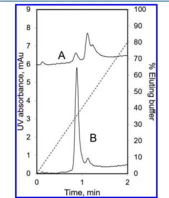
Figure 3shows the LFMC results obtained with the samples discussed above. With the monomer-rich sample (Figure 3B), the separation was achieved in less than 1.5 min with very high monomer/dimer resolution, the pressure drop being about 165 kPa. The peaks were fully resolved. The assay time compares very favorably even with the fastest SE-UPLC techniques
Figure 1.Blowout diagram of the analytical laterally fed membrane chromatography (LFMC) device: (A) acrylic top plate; (B) membrane stack (three layers of hydrophilized PVDF membranes; 0.22 μm pore size; GVWP14250); (C) plastic shim spacer; (D) acrylic bottom plate.
Figure 2.Analysis of the CHO cell line derived Campath-1H samples. (A) SE-HPLC (thick line: monomer-rich sample; thin line: dimer rich sample; column: TSK-Gel G3000SWXL 7.8 mm × 300 mm; feed concentration: 0.25 mg/mL; sample volume: 10 μL; running buffer: 20 mM sodium phosphate pH 7.0 + 250 mM NaCl; flow rate: 0.3 mL/ min); (B) native PAGE, 7% (1: monomer-rich sample; 2: dimer-rich sample).
Figure 3.Analysis of CHO cell line derived Campath-1H aggregates using LFMC. (A) dimer-rich sample; (B) monomer-rich sample; membrane: hydrophilized PVDF, 0.22 μm pore size, GVWP14250; membrane bed volume: 0.4 mL; feed concentration: 0.2 mg/mL; sample volume: 250 μL; eluting buffer: 20 mM sodium phosphate buffer, pH 7.0; binding buffer: eluting buffer + 1.5 M ammonium sulfate; linear gradient: 40 mL; flow rate: 16 mL/min; dashed line: % eluting buffer.
reported in the literature.3,33 More specifically, separation of mAb aggregates with Acquity UPLC BEH200 (4.6 mm × 150 mm column, 1.7 μm resin) was achieved in 3.5 min, the pressure drop being more than 274 bar (27 400 kPa).34
The LFMC technique was also capable of separating higher aggregates, as evident from the back shoulder of mAb dimer peak obtained with the dimer-rich sample (Figure 3A). These results were consistent with SE-HPLC and native PAGE results shown inFigure 1A. As evident by the results in the previous paragraph, very efficient (i.e., both fast and high-resolution separation) mAb aggregate separation and analysis could be achieved using LFMC, at a very low-pressure drop (2 orders of magnitude lower than SE-UPLC). Also, this could be achieved with a bed height of only 375 μm. The PVDF membrane used in the LFMC device had submicrometer pore size, which resulted in low dispersion effects within the chromatographic media. This, combined with the improved fluidic-design attributes of the LFMC device, kept peak broadening to a minimum. This is evident from the very small HETP value obtained for the device used in this study. This was found to be 8.2 μm (equivalent to 122 000 theoretical plates/m) at the operating flow rate. It is worth pointing out that HETP was determined at a much higher superficial velocity than that typically used for resin columns.
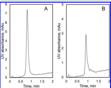
The LFMC device used to attain the above results had top and bottom channels with 1 mm diameter pillars. In order to examine the effects of device dead volume and pillar diameter, a new device with larger pillars was fabricated. The pillar diameter in the channels of this device were 1.75 and 2 mm in the rows of 3 and 4, respectively (seeFigure 1), leading to a decrease in dead volume in each channel from 194 to 139 μL.
Figure 4shows results obtained with LFMC devices having low (A) and high (B) dead volume, operated at identical conditions. The resolution of monomer and dimer peaks was significantly higher with the low dead volume device. Also, the peaks were much sharper and of greater height, clearly indicating lower device dispersion effects. The baseline shift observed in all the
HI-LFMC chromatograms could be attributed to the difference in the ammonium sulfate concentration in the binding and eluting buffers. This is widely reported with hydrophobic interaction chromatography.22,35
The wider applicability of the above LFMC technique was demonstrated by analyzing aggregate content of two other monoclonal antibodies: CHO derived Human IgG1-CD4 and HEK derived IgG1 (trastuzumab). On the basis of the chromatogram shown in Figure 5, it could be inferred that
HIgG1-CD4 contained very low amounts of aggregates while the Herceptin biosimilar did contain some aggregates (mainly dimer). This was consistent with results obtained using SE-HPLC (results not shown).
■
CONCLUSIONSUltrafast, high-resolution separation of monoclonal antibody aggregates was achieved using laterally fed membrane chromatography (LFMC). Previous studies had shown that hydrophobic interaction membrane chromatography was suitable for separating mAb aggregates. In the current study, the separation metrics were significantly improved using the LFMC format. The LFMC-based technique developed as part of this study was able to separate mAb aggregates in less than 1.5 min at an operating pressure of about 165 kPa. The separation time with the LFMC technique was less than half of that reported in the literature for SE-UPLC, while the operating pressure was lower by more than 2 orders of magnitude. The bed height within the LFMC device was only 375 μm, and even with such a small bed height, high-resolution separation could be achieved. The HETP values measured at a much higher superficial velocity than that typically used for resin columns was 8.2 μm (equivalent to 122 000 theoretical plates/m). By decreasing the device dead volume, the separation performance was further enhanced. The rapid, efficient, and cost-effective mAb aggregate separation technique discussed in this paper has
Figure 4. Effect of dead volume on analysis of monomer-rich Campath-1H aggregates using LFMC. (A) 40 mL gradient, lower dead volume; (B) 40 mL gradient, higher dead volume; membrane: hydrophilized PVDF, 0.22 μm pore size, GVWP14250; membrane bed volume: 0.4 mL; feed concentration: 0.2 mg/mL; sample volume: 250 μL; eluting buffer: 20 mM sodium phosphate buffer, pH 7.0; binding buffer: eluting buffer + 1.5 M ammonium sulfate; flow rate: 16 mL/ min.
Figure 5. Analysis of aggregates in different mAb samples using LFMC. (A) HIgG1-CD4; (B) trastuzumab; membrane: hydrophilized PVDF, 0.22 μm pore size, GVWP14250; membrane bed volume: 0.4 mL; feed concentration: 0.2 mg/mL; sample volume: 250 μL; eluting buffer: 20 mM sodium phosphate buffer, pH 7.0; binding buffer: eluting buffer + 1.5 M ammonium sulfate; linear gradient: 40 mL; flow rate: 16 mL/min; device dead volume: 139 μL (per channel).
Analytical Chemistry Article
DOI:10.1021/acs.analchem.7b00580 Anal. Chem. 2017, 89, 4716−4720
significant potential for application at the various stages of process development and analysis of monoclonal antibodies.
■
AUTHOR INFORMATION Corresponding Author *E-mail: [email protected]. ORCID Raja Ghosh:0000-0001-5965-0081 Author ContributionsP.M. conducted the experiments and drafted the manuscript. U.U. helped with conducting the experiments. G.H. and Y.D. kindly donated the model monoclonal antibodies and edited the manuscript. R.G. supervised the project, contributed to the fundamental device and experimental design, and edited the manuscript.
Notes
The authors declare no competing financial interest.
■
ACKNOWLEDGMENTSWe thank the Ontario Research Fund-Research Excellence (ORF-RE) program and the Natural Science and Engineering Research Council (NSERC) of Canada for funding this project. We thank Elna Luckham and the Biointerfaces Institute, McMaster University, for access to their HPLC system. We thank Paul Gatt for fabricating the devices based on the design provided by R.G. and P.M.
■
REFERENCES(1) Fekete, S.; Gassner, A. L.; Rudaz, S.; Schappler, J.; Guillarme, D. TrAC, Trends Anal. Chem. 2013, 42, 74−83.
(2) Fekete, S.; Beck, A.; Veuthey, J.; Guillarme, D. J. Pharm. Biomed. Anal. 2014, 101, 161−73.
(3) Diederich, P.; Hansen, S. K.; Oelmeier, S. A.; Stolzenberger, B.; Hubbuch, J. J. Chromatogr. A 2011, 1218, 9010−9018.
(4) Fekete, S.; Ganzler, K.; Guillarme, D. J. Pharm. Biomed. Anal. 2013, 78−79, 141−149.
(5) Rogers, B. A.; Wu, Z.; Wei, B.; Zhang, X.; Cao, X.; Alabi, O.; Wirth, M. J. Anal. Chem. 2015, 87, 2520−2526.
(6) Wong, C.; Strachan-Mills, C.; Burman, S. J. Chromatogr. A 2012, 1270, 153−161.
(7) Grotefend, S.; Kaminski, L.; Wroblewitz, S.; El Deeb, S.; Kühn, N.; Reichl, S.; Limberger, M.; Watt, S.; Wätzig, H. J. Pharm. Biomed. Anal. 2012, 71, 127−138.
(8) Fekete, S.; Schappler, J.; Veuthey, J.-L.; Guillarme, D. TrAC, Trends Anal. Chem. 2014, 63, 2−13.
(9) Fekete, S.; Guillarme, D. TrAC, Trends Anal. Chem. 2014, 63, 76−84.
(10) Hong, P.; Fountain, K. J. Method Development for Size-Exclusion Chromatography of Monoclonal Antibodies and Higher Order Aggregates; Waters Corporation: Milford, MA, 2011; pp 1−8.
(11) Koza, S.; Lauber, M.; Fountain, K. J. The Analysis of Multimeric Monoclonal Antibody Aggregates by Size-Exclusion UPLC; Waters Corporation: Milford, MA, 2013; pp 1−8.
(12) Fekete, S.; Veuthey, J. L.; McCalley, D. V.; Guillarme, D. J. Chromatogr. A 2012, 1270, 127−138.
(13) Fekete, S.; Kohler, I.; Rudaz, S.; Guillarme, D. J. Pharm. Biomed. Anal. 2014, 87, 105−119.
(14) Nováková, L.; Veuthey, J. L.; Guillarme, D. J. Chromatogr. A 2011, 1218, 7971−7981.
(15) Bobály, B.; Guillarme, D.; Fekete, S. J. Sep. Sci. 2014, 37, 189− 197.
(16) Arnauld, C. H. Chem. Eng. News 2016, 29−33.
(17) Wei, B.; Rogers, B. J.; Wirth, M. J. J. Am. Chem. Soc. 2012, 134, 10780−10782.
(18) Rea, J. C.; Moreno, G. T.; Vampola, L.; Lou, Y.; Van Haan, B.; Tremintin, G.; Simmons, L.; Nava, A.; Wang, Y. J.; Farnan, D. J. Chromatogr. A 2012, 1219, 140−146.
(19) Gourmel, C.; Grand-Guillaume Perrenoud, A.; Waller, L.; Reginato, E.; Verne, J.; Dulery, B.; Veuthey, J. L.; Rudaz, S.; Schappler, J.; Guillarme, D. J. Chromatogr. A 2013, 1282, 172−177.
(20) Hawe, A.; Friess, W.; Sutter, M.; Jiskoot, W. Anal. Biochem. 2008, 378 (2), 115−122.
(21) Liu, J.; Andya, J. D.; Shire, S. J. AAPS J. 2006, 8 (4), E580− E589.
(22) Wang, L.; Hale, G.; Ghosh, R. Anal. Chem. 2006, 78, 6863−7. (23) Wang, L.; Ghosh, R. J. Membr. Sci. 2008, 318, 311−316. (24) Yu, D.; Ghosh, R. Langmuir 2010, 26, 924−929. (25) Ghosh, R.; Wong, T. J. Membr. Sci. 2006, 281, 532−540. (26) Ghosh, R.; Madadkar, P.; Wu, Q. J. Membr. Sci. 2016, 516, 26− 32.
(27) Ghosh, R., Madadkar, P. U.S. Provisional Patent 62304379, 2016.
(28) Madadkar, P.; Wu, Q.; Ghosh, R. J. Membr. Sci. 2015, 487, 173− 179.
(29) Gorman, S. D.; Clark, M. R.; Routledge, E. G.; Cobbold, S. P.; Waldmann, H. Proc. Natl. Acad. Sci. U. S. A. 1991, 88, 4181−4185.
(30) Phillips, J.; Drumm, A.; Harrison, P.; Bird, P.; Bhamra, K.; Berrie, E.; Hale, G. Cytotherapy 2001, 3, 233−242.
(31) Rathore, A. S.; Kennedy, R. M.; O’Donnell, J. K.; Bemberis, I.; Kaltenbrunner, O. BioPharm. Int. 2003, 16, 30−40.
(32) Litzen, A.; Walter, J. K.; Krischollek, H.; Wahlund, K.-G. Anal. Biochem. 1993, 212, 469−480.
(33) Fekete, S.; Guillarme, D. J. Chromatogr. A 2013, 1308, 104−113. (34) Yang, R.; Tang, Y.; Zhang, B.; Lu, X.; Liu, A.; Zhang, Y. T. J. Pharm. Biomed. Anal. 2015, 109, 52−61.
(35) Mayolo-Deloisa, K.; Lienqueo, M. E.; Andrews, B.; Rito-Palomares, M.; Asenjo, J. A. J. Chromatogr. A 2012, 1242, 11−16.