HAL Id: inserm-00732395
https://www.hal.inserm.fr/inserm-00732395
Submitted on 5 Dec 2012
HAL is a multi-disciplinary open access
archive for the deposit and dissemination of sci-entific research documents, whether they are pub-lished or not. The documents may come from teaching and research institutions in France or abroad, or from public or private research centers.
L’archive ouverte pluridisciplinaire HAL, est destinée au dépôt et à la diffusion de documents scientifiques de niveau recherche, publiés ou non, émanant des établissements d’enseignement et de recherche français ou étrangers, des laboratoires publics ou privés.
Antithrombotic properties of water-soluble carbon
monoxide-releasing molecules.
Karol Kramkowski, Agnieszka Leszczynska, Andrzej Mogielnicki, Stefan
Chlopicki, Andrzej Fedorowicz, Elzbieta Grochal, Brian Mann, Tomasz
Brzoska, Tetsumei Urano, Roberto Motterlini, et al.
To cite this version:
Karol Kramkowski, Agnieszka Leszczynska, Andrzej Mogielnicki, Stefan Chlopicki, Andrzej Fedorow-icz, et al.. Antithrombotic properties of water-soluble carbon monoxide-releasing molecules.: Anti-thrombotic properties of CO-RMs. Arteriosclerosis, Thrombosis, and Vascular Biology, American Heart Association, 2012, 32 (9), pp.2149-57. �10.1161/ATVBAHA.112.253989�. �inserm-00732395�
1
Original paper [resub] may 2012 Title:
ANTI-THROMBOTIC PROPERTIES
OF WATER-SOLUBLE
CARBON MONOXIDE-RELEASING MOLECULES (CO-RMs)
Short title: Kramkowski K et al. Anti-thrombotic properties of CO-RMsAuthors
Karol Kramkowski1, Agnieszka Leszczynska1, Andrzej Mogielnicki1, Stefan Chlopicki2,3, Andrzej Fedorowicz2, Elzbieta Grochal2,3, Brian Mann4, Tomasz Brzoska5,Tetsumei Urano5, Roberto Motterlini4and Wlodzimierz Buczko1.
The affiliations 1
Department of Pharmacodynamics, Medical University of Bialystok, Poland
2
Jagiellonian Centre for Experimental Therapeutics (JCET), Jagiellonian University, Krakow, Poland
3
Department of Experimental Pharmacology, Jagiellonian University Medical College, Krakow, Poland
4
INSERM U955, Equipe 3, Université Paris-Est, Faculté de Médecine, 94010 Creteil, France
5
Department of Medical Physiology, Hamamatsu University Medical School of Medicine, 1-20-1, Handa-yama, Hamamatsu, 431-3192, Japan.
Corresponding author:
Prof. Wlodzimierz Buczko, MD, PhD Department of Pharmacodynamics Medical University of Bialystok
Mickiewicza Str. 2C, 15-222 Bialystok, POLAND, EU
2 Abstract word count: 259
A body text word count: 5 077; 6844 (including table, figure legends and references) Total number of figures and tables: 5
3
Abstract
Objective—We compared the antithrombotic effects in vivo of 2 chemically different carbon
monoxide–releasing molecules (CORM-A1 and CORM-3) on arterial and venous thrombus formation and on hemostatic parameters such as platelet activation, coagulation, and fibrinolysis. The hypotensive response to CORMs and their effects on whole blood gas analysis and blood cell count were also examined.
Methods and Results—CORM-A1 (10–30 μmol/kg, i.v.), in a dose-dependent fashion,
significantly decreased weight of electrically induced thrombus in rats, whereas CORM-3 inhibited thrombosis only at the highest dose used (30 μmol/kg). CORM-A1 showed a direct and stronger inhibition of platelet aggregation than CORM-3 in healthy rats, both in vitro and in vivo. The antiaggregatory effect of CORM-A1, but not CORM-3, correlated positively with weight of the thrombus. Concentration of active plasminogen activator inhibitor-1 in plasma also decreased in response to CORM-A1, but not to CORM-3. Neither CORM-A1 nor CORM-3 had an effect on plasma concentration of active tissue plasminogen activator. CORM-3, but not CORM-A1, decreased the concentration of fibrinogen, fibrin generation, and prolonged prothrombin time. Similarly, laser-induced venous thrombosis observed intravitally via confocal system in green fluorescent protein mice was significantly decreased by CORMs. Although both CORM-A1 and CORM-3 (30 μmol/kg) decreased platelets accumulation in thrombus, only CORM-A1 (3–30 μmol/kg) inhibited platelet activation to phosphatidylserine on their surface.
Conclusion—CORM-3 and CORM-A1 inhibited thrombosis in vivo, however CORM-A1,
which slowly releases carbon monoxide, and displayed a relatively weak hypotensive effect had a more pronounced antithrombotic effect associated with a stronger inhibition of platelet aggregation associated with a decrease in active plasminogen activator inhibitor-1 concentration. In contrast, the fast CO releaser CORM-3 that displayed a more pronounced hypotensive effect inhibited thrombosis primarily through a decrease in fibrin generation, but had no direct influence on platelet aggregation and fibrynolysis.
Keywords: carbon monoxide (CO), CO-releasing molecules (CO-RMs), thrombosis, platelet
4 Carbon monoxide (CO) is physiologically present in the human body and its level is regulated through the enzymatic degradation of heme by heme oxygenase enzymes (HO-1 and HO-2)1. Despite being renown as ‘the silent killer’, experimental data have revealed some unexpected benefits of small doses of CO gas in the cardiovascular system, primarily associated with its important effects on vascular tone2, blood pressure3, inflammation4,5, cell proliferation6,7, apoptosis8,9 and cytoprotection against tissue injury10. More recent data indicate that CO may also affect thrombosis, a major complication caused by multiple vascular pathologies. Indeed, CO paradoxically increases the formation of fibrin11 and inhibits fibrinolysis12 in in vitro studies, whereas it exerts anti-thrombotic effect in vivo, which involves activation of fibrinolysis13. Notably, a direct role of CO gas in mitigating the process of thrombosis was assessed by True et al14. These authors found that Hmox-1 knock-out mice display a pro-thrombotic phenotype characterized by damage of endothelial cells and apoptosis, platelet activation, elevation in both tissue factor (TF) and von Willebrand factor (vWF) and reactive oxygen species. In addition, transplantation of HO-1-deficient bone marrow (BM)-derived progenitor cells into wild type HO-1 animals resulted in rapid arterial thrombosis. Interestingly, all these pathological phenotypes were rescued by administration of CO gas. In another study by Chen et al. it was shown that in carotid arteries injury model of Apo-E-deficient mice a significant anti-thrombotic effect can be achieved by exposition for 2 h to CO inhalation (250 ppm) or by treatment with an adenovirus bearing the HO-1 gene (Adv-HO-1)15.
Since Motterlini et al.16 described carbon monoxide-releasing molecules (CO-RMs) as a novel approach to deliver precise amounts of CO within biological systems, several reports confirming the pharmacological efficacy of CO have appeared in the literature17,18. The first class of CO-RMs studied were composed of transition metal carbonyls, complexes of metals surrounded by CO groups as coordinated ligands19. CORM-1 (manganese decacarbonyl t1/2 < 1 min) and CORM-2 (tricarbonyldichlororuthenium(II) dimer, t1/2 ~ 1
5 minare ) , soluble in ethanol and DMSO and release CO either by photodissociation or by ligand substitution20. These carbonyl complexes have been shown to exert important pharmacological effects both in vitro and in vivo (see also supplementary material I). In the context of thrombosis, Chen et al.21 demonstrated that CORM-2, but not an inactive counterpart that does not release CO, rescues Hmox-1 knock-out mice against the development of arterial thrombosis following allogenic aortic transplantation. Interestingly, this anti-thrombotic action of CORM-2 was associated with a markedly reduced platelet aggregation within the graft. On the other hand, it has been shown that CO liberated from CORM-2 in vitro significantly enhances fibrinogen-dependent coagulation kinetics in a concentration-dependent manner in plasma12. Advances in the development of novel CO-RMs led to the synthesis and characterization of water-soluble compounds, the best representation of which is CORM-3 (tricarbonylchloro(glycinato) ruthenium(II), t1/2 ~ 1
min)20. , In vitro and in vivo experiments demonstrated that CO liberated from CORM-3 improves endothelial function22, causes vasodilatation16,23, mediates anti-inflammatory responses24, protects against ischemia-reperfusion injury25, and prevents organ rejection following transplantation18,21,22. Anti-aggregatory activities of CORM-3 have also been demonstrated in human platelets in vitro26. Interestingly, another class of water-soluble
CO-releasing molecules that do not contain metals (boranocarbonates) has been described as pharmacologically active27. CORM-A1, the best characterized of this class, releases CO in
vivo at a much slower rate than CORM-3 and the kinetic of CO release is strictly dependent
on pH and temperature (t1/2 ~ 21 min at pH=7.4 and 37ºC)28. Intuitively, improved
pharmacokinetic profiles would make this particular spontaneous CO releaser an ideal tool to study the physiological properties of CO gas in living organism without the interference of a transition metal carrier28.
The aim of the present work was therefore to compare the anti-thrombotic effects of CORM-A1 and CORM-3 by specifically assessing the effects of these two CO-releasing
6 molecules on haemostatic parameters such as platelet aggregation, coagulation and fibrinolysis, as well as on blood cell count and blood gas analysis.
Materials and methods
Animals
Male Wistar rats (120-150 g) were purchased from and housed in the Centre of Experimental Medicine of Medical University of Bialystok (Poland) and Green fluorescent protein (GFP)-expressing transgenic mice, based on the C57BL/6J strain (GFP mice), were supplied by Dr. Okabe (Osaka University, Osaka, Japan)29 and maintained in Animal Facility of Hamamatsu University Medical School of Medicine (Japan) according to Good Laboratory Practise rules. Animals were housed in a room with a 12 hours light/dark cycle, grouped in cages as appropriate and allowed to have access to tap water and a standard rat/mice chow. All the procedures involving animals were approved by a bioethic committee and conducted in accordance with the institutional guidelines that are in compliance with national and international laws and Guidelines for the Care and the Use of Animals in Biomedical Research30.
Chemicals and drugs
CORM-3 and CORM-A1 were synthesized as previously described24,28. Inactive CORM-3 (iCORM-3) was prepared from CORM-3 by leaving the solution of CORM-3 at room temperature in phosphate buffer solution for 48 h followed by bubbling with N2.
Inactive CORM-A1 (iCORM-A1) was instead prepared by slight acidification of the solution with HCl, then re-buffering and bubbling with N2, as described previously24,28. Pentobarbital
(Vetbutal, Biovet, Poland or Nembutal, Dainippon Pharma, Japan), PBS (Biomed, Poland), Tris buffer (Sigma-Aldrich, Germany), collagen (Chrono-log Corp. USA), Alexa Fluor 568 dye (Invitrogen, Molecular probes, USA), Annexin 5A (KOWA Pharmaceuticals, Tokyo, Japan) and ready-to-use kits for blood cell count and gas blood analysis were used in the
7 study. Routine laboratory reagents to determine prothrombin time (PT), activated partial thromboplastin time (APTT) and fibrinogen (Fg) levels in vitro in rat plasma were purchased from HemosIL, Instrumentation Laboratory (USA). Rat plasminogen activator inhibitor-1 (PAI-1) and tissue plasminogen activator (t-PA) ELISA kits were purchased from Hyphen, BioMed, France.
Administration of CO-RMs: experimental protocol in rats
CORM-3 or CORM-A1 were administered into the right femoral vein at doses of 3 (n=5), 10 (n=17), 30 (n=12) µmol/kg or 3 (n=8), 10 (n=11), 30 (n=11 rats) µmol/kg, respectively. CO-RMs were injected 15 min before the induction of carotid artery injury. Doses of CO-RMs were chosen based on previously published data16,28. Inactive CO-RMs (iCORM-3 or i-CORM-A1, 30 µmol/kg, n=6) were used as negative controls. Vehicle (VEH, 0.9% NaCl, n=32) served as a control to CO-RMs-treated rats and the phosphate buffered saline (PBS, n=5) served as a control to iCO-RMs-treated animals.
Administration of CO-RMs: experimental protocol in mice
CORM-3 or CORM-A1 were administered into the right femoral vein at doses of 3 (n=4), 30 (n=7) µmol/kg or 3 (n=5), 30 (n=9) µmol/kg, respectively. CO-RMs were injected 15 min before the mesenteric vein injury. Doses of CO-RMs were chosen based on previously published data16,28. Inactive CO-RMs (iCORM-3 or i-CORM-A1, 30 µmol/kg, n=3) were used as negative controls. Vehicle (VEH, 0.9% NaCl, n= 9) served as a control to CO-RMs-treated rats and the phosphate buffered saline (PBS, n= 3) served as a control to iCO-RMs-treated animals.
8 Collagen-stimulated platelet aggregation in vitro and ex vivo in a citrated whole blood was evaluated with the impedance method as described previously31 and measured in a Whole Blood Lumi-Aggregometer (Chrono-log Corp., USA).
For in vitro experiments, blood samples were drawn from normal rats into 3.13% trisodium citrate in a volume ratio 10:1. After 13 min of incubation at 37°C with 0.9% NaCl (volume ratio 1:1), samples were mixed with CORM-A1 (final concentrations: 1 mM, 0.5 mM and 0.1 mM) or CORM-3 (final concentrations: 5 mM, 1 mM and 0.5 mM) or VEH (0.9% NaCl). Collagen (5 µg/ml) was added 2 min after.
For ex vivo experiments, blood samples were collected from rats with electrically stimulated thrombosis 75 min after administration of VEH (0.9% NaCl) or CO-RMs in doses of 3, 10, 30 µmol/kg or i-CO-RMs in doses of 30 µmol/kg into 3.13% trisodium citrate in a volume ratio of 10:1 (thrombotic experiment). After 15 min of incubation at 37ºC with 0.9% NaCl (volume ratio 1:1), collagen (5 µg/ml) was added.
Blood samples were collected also from normal rats 15 min after CORM-A1 or CORM-3 administered in doses of 10 µmol/kg and 30 µmol/kg and 60 min after CORM-A1 or CORM-3 administered in doses of 10 µmol/kg. iCO-RMs were administered in doses of 30 µmol/kg 15 min or 60 min before blood collection. After 15 min of incubation at 37ºC with 0.9% NaCl (volume ratio 1:1), collagen (5 µg/ml) was added.
In all in vitro and ex vivo experiments changes in resistance were registered during 6 min. The maximal extension of the aggregation curve at the 6th min was expressed as a percentage of control response.
Induction of arterial thrombosis in rats
Rats were anesthetized with pentobarbital (40 mg/kg, i.p.) and placed in a supine position on a heated (37°C) operating table. The left femoral vein was cannulated to administer the drug. Arterial thrombosis was induced by electrical stimulation of the right common carotid artery as previously described32,33. Briefly, the anode, a stainless steel
L-9 shaped wire, was inserted under the artery and connected with a constant current generator. The cathode was attached subcutaneously to the hindlimb. The artery stimulation (1 mA) took 10 min. 1 h after the beginning of the stimulation (ie. 75 min after drugs administration), the segment of the common carotid artery with the formed thrombus was dissected, then opened lengthwise and the thrombus was completely removed, air-dried at 37 ºC, and weighted after 24 h.
Induction of venous thrombus in mice - in vivo imaging experiments (supplementary
material II)
Intravital fluorescence confocal microscopy was arranged as described previously34.
Laser-induced vessel wall injury34. Mesenteric venules were identified and endothelial
injury was induced by a 514-nm argon-ion laser, (543-GS-A03; Melles Griot Laser Group, CA, USA). The laser beam was aimed at the endothelium through the microscope objective lens and the intensity of laser illumination and duration was kept constant.
GFP mouse preparation for in vivo imaging34. GFP mice were anesthetized with
pentobarbital (50 mg/kg, i.p.) in an atmosphere of diethyl ether. A midline laparotomy incision was made, and then the mesentery of the ileum was pulled out of the abdomen and draped over a plastic mound. The mesentery was continuously perfused with 37°C-warmed saline to prevent the vessels from drying out. For visualizing the surface-exposed platelet phospholipids (PS) in a thrombus, Alexa Fluor 568-labeled annexin V (ANX) (2-μg/g mouse body weight) was administered into the right femoral vein 14 min before laser injury. Previously it was shown, that the fluorescence intensity of ANX directly corresponds to the platelet activation, since the PS are exposed only in activated platelets whose intra-cellular calcium concentration is sustainably elevated. Changes in fluorescence intensity were measured as described previously34.
Image analysis34. A z-stack of 24 optical sections at up to 30 frames per second from the vessel wall to the luminal surface of a thrombus were captured every 5 s (1-μm optical slice
10 thickness, 24 z-sections collected at 1-μm intervals) and analyzed using a Yokogawa Real-Time 3D Workstation and IPLab software (BD Biosciences Bioimaging, MD). A freehand-defined region of interest was traced along the outline of fluorescent areas. The fluorescence intensity of GFP was normalized to the initial value in each experiment, whereas that of ANX was normalized to the last value.
Blood Pressure measurements
Rats were anesthetized with pentobarbital (40 mg/kg, i.p.) and placed in a supine position on a heated (37°C) operating table. The mean blood pressure (MBP) was measured in anaesthetized rats directly through a cannula filled with heparin solution (150 IU/ml), placed in the left common carotid artery and connected to a pressure transducer (Plugsys, Transonics System, USA), as described previously35.
Coagulation parameters and fibrin generation
PT, APTT and Fg levels (Clauss method) were determined according to kit manufacturer’s
instructions using Coag-Chrom 3003 apparatus (Bio-ksel, Poland). A more sensitive assay for fibrin generation was used based on a method described previously36,37 and modified by us38. Fibrin generation curves were created by recalcination of rats’ plasma samples directly in microplate wells with CaCl2 (36 mM) dissolved in Tris buffer (66 mM Tris, 130 mM
NaCl; pH=7.4) in 37°C. Optical density increases in the wells (as a result of fibrin generation) were measured via the microplate reader (Dynex Tech., USA) in 1 min intervals for 14 min and expressed as an area under the curve (AUC). Time-points analysis was calculated using the values of percentage of basal optical density.
11 Concentrations of total plasminogen activator inhibitor-1 (PAI-1) antigen, active PAI-1 and tissue plasminogen activator (t-PA) were analyzed using ELISA techniques using a microplate reader (Dynex Tech., USA) according to kits manufacturer’s instructions.
Blood cell count
Blood cell count was assessed by an Animal Blood Counter (ABC Vet, Horiba, Germany) collected from heart of the rat after thrombus removal.
Blood gas analysis
In anesthetized Wistar rats (BW = 160-170 g) trachea was cannulated and lungs were artificially ventilated (7025 Rodent Ventilator, Ugo Basile) with mixture of oxygen and isoflurane (1.1-1.2 %) with positive pressures (rate of 80 breaths/min, tidal volume =2.4-2.6 ml). Carotid artery was cannulated. CORMs (CORM-A1, CORM-3, iCORM-A1 and iCORM-3) were dissolved in saline, 15 seconds before giving to the dorsal penile vein (dose 30 µmol/kg) and arterial blood samples were collected from the carotid artery before 10 and 65 minutes after CORMs injection. Saline treated rats served as a control group. Blood gasometry parameters were measured by pHox analyzer (Nova Biomedical) immediately after collection of probes.
Statistical analysis
The data are shown as mean±SEM and analyzed using either a Mann-Whitney test (when normality test failed) or an unpaired t-test (when normality test passed). Correlations were analyzed using a Pearson test. P < 0.05 was considered significant.
12
Results
Effect of CO-RMs and its inactive counterparts on platelet aggregation in in normal rats
CORM-3 (0.5-5 mM) and CORM-A1 (0.1-1 mM) exerted a concentration-dependent anti-aggregatory effect on rat’s platelets in vitro in the whole blood (Fig. 1). However, CORM-A1 was much more potent than CORM-3 and at a concentration of 1 mM completely blocked collagen-induced platelet aggregation. In contrast, CORM-3 reached its maximal effect also at 1 mM, but it inhibited platelets aggregation only by 50%. iCO-RMs used at the same concentrations as CO-RMs did not influence platelet aggregation in the same experimental set-up (Fig 1).
In ex vivo experiments, the effect of CORM-A1 (30 µmol/kg) on platelet aggregation was more pronounced as compared to CORM-3 when administered into healthy animals 15 min prior to blood collection (Fig. 2A). Similarly, when CO-RMs (10 µmol/kg) were administered 60 min prior to blood collection, the anti-platelet effect of CORM-A1 measured at 60 min vs 15 min after administration was significantly more pronounced as compared to CORM-3 (Fig. 2B). iCO-RMs did not influence platelet aggregation ex vivo in healthy rats both at 15 and 60 min after administration (Figure 2B).
Effect of CO-RMs and its inactive counterparts on blood gas analysis in normal rats
In the group of rats treated with CORM-A1 (30 µmol/kg), COHb concentration increased significantly 10 min after administration from 2.5±0.66% (baseline) to 8.9±2.1% (p<0.01) but dropped to baseline values 65 min after A1 administration (2.9±0.5% for CORM-A1 vs 2.8±0.46% for control group; ns). No changes in COHb concentration were observed in animals treated with CORM-3 or both iCO-RMs, as compared to control group. Other blood parameters did not change after CORMs administration (supplementary material III).
13
Effect of CO- RMs and its inactive counterparts on platelet aggregation in thrombotic rats
CORM-A1 administered at doses of 10 and 30 µmol/kg inhibited platelet aggregation in whole blood collected immediately after thrombus removal (74.7±20 % and 57.1±10 % of vehicle, respectively). In contrast, the effect of CORM-3 was less pronounced showing only a trend of inhibition on platelet aggregation at the highest dose used (30 µmol/kg, Fig. 3A). iCORM-3 and iCORM-A1 failed to inhibit platelet aggregation (Fig. 3A).
Effect of CO-RMs and its inactive counterparts on electrically induced arterial thrombus formation
CO-RMs inhibited arterial thrombus formation. In the case of CORM-A1, doses of 10 and 30 µmol/kg decreased thrombus weight from 0.81±0.02 mg to 0.69±0.07 mg (p<0.05) and 0.62±0.03 mg (p<0.001), respectively. CORM-3 was only effective at a dose of 30 µmol/kg decreasing thrombus weight to 0.7±0.04 mg (Fig. 3B). iCORM-3 and iCORM-A1 failed to influence thrombus weight (Fig. 3B). It is interesting to note that the inhibition of platelet aggregation correlated with the decrease in thrombus weight only in the case of CORM-A1 (r=0.60; p<0.01, supplementary material IVA) but not in the case of CORM-3 (supplementary material IVB).
Effect of CO-RMs on laser-induced venous thrombosis observed intravitally in mice
CO-RMs inhibited venous thrombus formation. In the case of CORM-A1, only dose of 30 µmol/kg decreased maximal fluorescence intensity of GFP from 4.70±0.58 units to 1.25±0.36 units (p<0.01), while fluorescence intensity of ANX was decreased by doses of 3 and 30 µmol/kg from 1.97±0.23 units to 0.96±0.15 units and 0.59±0.17 units (p<0.05 and p<0.01, respectively, Fig 4. A,B). CORM-3 was significantly effective only at a dose of 30 µmol/kg decreasing fluorescence intensity of GFP from 4.70±0.58 units to 1.92±0.57 units (p<0.05). Moreover, A1 decreased ANX fluorescence slightly stronger than
CORM-14 3 (p=0.06, Fig. 4B). iCO-RMs were without any effect on GFP or ANX fluorescence (data not shown).
Effect of CO-RMs on Blood Pressure
CORM-3 given at a dose of 30 µmol/kg exerted significant hypotensive effect (p<0.05) starting about 10 min after the injection, which increased (p<0.01) about 30 min after the injection. CORM-A1 in the same dose also exerted hypotensive response (p<0.05), but this response appeared later as compared to the effect of CORM-3 (about 20 minutes after the injection) and was less pronounced. CO-RMs used at the lower doses did not change mean blood pressure as compared to control values. Also iCO-RMs did not exert hypotensive effects (supplementary material V A, B).
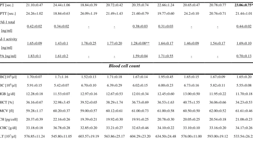
Effect of CO-RMs and iCO-RMs on coagulation parameters in thrombotic rats
CORM-3 significantly reduced the concentration of plasma fibrinogen in vivo at the dose of 30 µmol/kg (1.88±0.06 ng/ml) as compared to vehicle-treated animals (2.21±0.13 ng/ml; p<0.01), while CORM-A1 was without effect (Fig 6.). iCO-RMs failed to affect the concentration of plasma fibrinogen in vivo (2.52±0.1 ng/ml with iCORM-A1 and 2.29±0.10 ng/ml with iCORM-3 vs 2.32±0.11 mg/ml with PBS). Additionally, fibrin generation was significantly inhibited by CORM-3. In fact, fibrin generation decreased from 0.919±0.04 units in the vehicle-treated group to 0.485±0.05 and 0.194±0.14 units in animals treated with 10 and 30 µmol/kg CORM-3, respectively (p< 0.01, Fig. 5). Consequently, PT was prolonged in rats’ plasma treated with 30 µmol/kg CORM-3 (p<0.05, Tab. 1.), whereas APTT did not change in any group. iCORM-A1 (30 µmol/kg) failed to influence coagulation parameters, while 30 µmol/kg iCORM-3 reduced fibrin generation from 0.998±0.069 (PBS-treated group) to 0.722±0.08 units (p<0.05; Fig. 5). iCORM-3 significantly decreased fibrin formation in all examined time points of the clotting curve (supplementary material VI).
15
Effect of CO-RMs and its inactive counterparts on fibrinolysis parameters in thrombotic animals
The concentration of active PAI-1 was significantly decreased in plasma of rats treated with 30 µmol/kg CORM-A1 when compared to the vehicle-treated , p<0.05,), but CORM-3 was without effect at any doses tested (Table 1). The concentrations of total PAI-1 antigen in rats treated with 30 µmol/kg CORM-A1 or CORM-3 did not significantly change when compared to vehicle-treated rats. Similarly, the concentrations of active t-PA did not change in rats treated with 30 µmol/kg CORM-A1 or CORM-3 when compared to vehicle-treated group. iCO-RMs failed to influence fibrinolysis (see Table 1).
Effect of CO-RMs and its inactive counterparts on blood cell count and blood gas analysis in thrombotic animals
No changes were observed on blood cell count in animals treated with CO-RMs as well as with iCO-RMs (Table 1).
Discussion
Previous studies performed on smokers or subjects who inhaled CO gas revealed that CO may influence platelet function, coagulation and fibrinolysis in human39,40. Recent studies using CORM-2 and CORM-3 provided evidence that non-toxic quantities of CO may also exert antithrombotic effects20 and inhibition of platelet aggregation26, 41.
The data presented here also show for the first time that not only 3, but also CORM-A1 inhibits platelet aggregation in vivo. Since we found that the effect of CO-RMs was more pronounced at later time after administration (60 min), we wanted to explore whether CO-RMs may also influence the progression of arterial thrombosis. We were also interested to assess whether the diverse CO releasing profile of CORM-A1 compared to CORM-3 would differentially influence the activity in vivo.
16 Electrically-induced thrombosis is a valuable model for studying the antithrombotic efficacy of novel compounds because it mimics clinical thrombosis31, 32,42. The results of the present study demonstrate that both CORM-3 and CORM-A1 administered intravenously at micromolar doses inhibited arterial thrombus formation in vivo. Another important finding in our study is that the effect of CORM-A1 is strictly dose-dependent and much more potent compared to CORM-3 . Interestingly, a significant decrease in thrombus weight correlated with the inhibition of platelet aggregation only in the group treated with CORM-A1 which is known to releases CO in a constant and gradual manner over time27. The differential effects obtained with these two CO releasers are in line with our recent observations. It is not surprising since the intrinsic chemical nature of these two compounds and especially time course of CO release differs, as presented on the supplementary material I. As we previously showed27, the direct vasorelaxant effect on the vessels and hypotensive effect (confirmed in this study) of both CO-RMs have different nature. Notably, inhibition of NO synthase activity by L-NAME or removal of the endothelium, which significantly inhibited CORM-3-mediated vessel relaxation26, failed to prevent the pharmacological effects of CORM-A127, suggesting that the action of CO exerted through the reactivity of boranocarbonate is independent of the endothelial function. Moreover, the blockade of potassium channels with glibenclamide, which is also known to partially attenuate the vasorelaxation by CORM-313, did not have any effect on the vasoactivity elicited by CORM-A127. We know that CORM-A1, unlike CORM-3, possesses some reducing capacities in addition to its ability to liberate CO27 and thus may differently affect the response of the vessels. It seems the effects of CO-RMs, especially CORM-A1 on the vascular haemostasis may be independent on the hemodynamic activity, since at the doses used in this study it exerted antithrombotic effect in doses that had no influence on blood pressure and it did not exert direct vasorelaxant effect. It is also quite evident that the effects on the thrombosis and platelet of CORM-A1 was more potent when compared to CORM-3. We know that the CORM-A1 releases CO for almost 1 hour, thus for the whole time of the arterial thrombus development and the platelet
17 aggregation in vivo. In contrast, CORM-3 liberates full CO in the first few minutes, thus in the time of platelet activation following vascular injury CO is not present anymore in the circulation. We may assume that high amount of CO released from CORM-3 in short time, but not from CORM-A1, are able to inhibit coagulation factors that are presented in the blood before thrombosis induction. It is interesting to note that severe and prolonged bleeding and a higher incidence of hemorrhagic strokes were reported after intoxication with the inhaled CO43,44.
This hypothesis is also supported by our results on the coagulation parameters.. The fibrin generation inhibited by CORM-3 strongly correlated with the decrease of fibrinogen levels in plasma. The kinetics of fibrin generation process was also significantly inhibited by CORM-3, suggesting a strong anticoagulant effect. In addition, PT was also prolonged at the highest dose of CORM-3. These results indicate that the antithrombotic effects of CORM-3 may be only related to the inhibition of the coagulation cascade. The fact that iCORM-3 reduced fibrin generation suggests that this effect might depend on the presence of the ruthenium metal since no changes on this parameter have been observed after treatment of animals with i-CORM-A1. On the other hand, it cannot be excluded a priori that the effect of iCORM-3 in
vivo is due to the residual CO groups bound to and released from the metal since only 1 CO
per mole is rapidly detected in vitro16.
We also found differences in the effect of CO-RMs on the fibrinolytic parameters. The concentration of active PAI-1 was significantly decreased in plasma of rats treated with CORM-A1 (30 µmol/kg), but not with CORM-3. This discrepancy might also relay on the time course of CO release. During the development of thrombosis, the fibrinolysis process starts after platelets activation and formation of active coagulation plasma factors. Notably, the activated platelets are the main source of active PAI-1 regulating production of plasmin, which limits the growth of thrombus only to the damaged vascular wall. At the time of PAI-1 release from platelets (around 13-20 min after CO-RM iv administration) only CORM-A1 was still liberating CO, which in turn could interfere with the activity of PAI-1. This is also
18 confirmed by previous studies by Soni et al.13 Those authors showed that CORM-3 decreased plasma PAI-1 levels, but in their model CORM-3 was administered during a 10 min infusion at 3 mg/kg (which is equivalent to 100 µmol/kg of CORM-3), thus the liberated CO could have been in contact with the released from platelets PAI-1. On the other hand, Nielsen at al. have shown that CORM-2 inhibits fibrynolysis11,12, 45. Nevertheless their experiments were preformed only in vitro using this lipid-soluble and fast metal-containing CO releaser, which makes it difficult to compare with our results obtained with water-soluble CO-RMs in vivo. Moreover, all our and others previous animal studies provide evidence for the profibrinolytic/thrombotic effects of CO partially due to the suppression of anti-fibrinolytic molecules, such as PAI-115,46, and our study for the first time in vivo shows similar effect of CORM-A1.
To confirm the antithrombotic activity of CO-RMs we have performed intravital laser-induced venous thrombosis in GFP mice, a novel method for real-time evaluation of the progression of thrombosis. Simultaneously relative fluorescence intensities of GFP and ANX are measured. The GFP fluorescence corresponds to platelet accumulation in area of laser-injured endothelium. ANX only binds platelets, that are irreversibly activated and expose PS on their surface. Similarly to reducing thrombus weight in rat, both CO-RMs decreased fluorescence intensity of GFP in laser-induced venous thrombosis in mice. On the other hand, only CORM-A1 inhibited PS exposure on platelet surface (decrease in ANX fluorescence), shoving strong anti-platelet effect. Thus, these data further confirm the anti thrombotic effect of both CO-RMs and the antiplatelet effect of CORM-A1.
In the present study we found that in vitro application of CORM-3 and CORM-A1, respectively a “fast” and “slow” CO releaser, inhibits platelet aggregation in healthy rats in
the same manner as previously reported in human platelets by Chlopicki et al 41. Interestingly, CORM-A1 appears to be a stronger inhibitor of rat platelet aggregation in comparison with CORM-3. Taking into account the pharmacokinetic properties of these two water-soluble CO-RMs20, when the antiaggregatory effect is measured after 6 min, CORM -3
19 has completely released CO, whereas CO from CORM-A1 is still slowly and consistently liberated. This difference in the rate of CO release may partially explain why CORM-A1 is more effective, since CO is still being liberated when platelets are activated, thus inhibiting platelet aggregation more efficiently. These data are in line with our recent observation on human platelets41 showing that in vitro conditions CORM-A1 is indeed more potent in inhibiting platelet aggregation. These new data point out that CORM-A1 display anti-platelet activities that should be exploited further for its therapeutic use as anti-thrombotic drug in vivo40.
In the present study it was important to verify that CO released from CO-RMs did not affect COHb levels in order to ascertain that the doses used did not compromise the oxygen carrying capacity of blood. In blood collected after thrombosis was induced in animals, no changes in blood gas parameters such as HCO3, pCO, pO2 and pH as well as blood COHb
levels were observed after treatment with CORM-3. COHb levels increased slightly after CORM-A1 administration but did not reach toxic values. CO-RMs did not affect blood cell count in any group tested, further excluding a potential toxic effect of CO delivered by these compounds.
In conclusion, we report that CORM-A1 possesses anti-thrombotic activity in vivo in two models of thrombosis. CORM-A1, by releasing CO at a slower rate and at not toxic concentrations, also promotes a strong anti-platelet and fibrynolitic activity, without a significant hypotensive effect. In contrast, CORM-3 that releases CO more rapidly is a weaker inhibitor of thrombosis and acts primarily by inhibition of plasma clotting factors and fibrin generation. For the first time we have compared the effect of CO delivered to living organisms on haemostasis using two carriers that release different amounts of CO with different times and rates. If the presence of CO in the circulation is prolonged this gas may modulate platelets function and fibrinolytic activity. In contrast, sufficiently high concentrations of CO should be reached to significantly inhibit fibrin generation. Importantly, our study indicate that it is possible to achieve a significant anti-thrombotic and
20 anti-aggregatory effect with CO-releasing compounds without major hypotenisve effects with CORMA1-like compounds that release CO at a slow rate. Our findings suggest that CO-RMs provide a novel tool for the design of therapeutic strategies against thrombosis in cardiovascular diseases such as myocardial infarction and stroke, but further studies are required to validate its feasibility in humans.
Acknowledgements
a. We would like to thank Ms Teodora Sienkiewicz for the excellent technical assistance. b. This work was supported by Polish Ministry of Science and Higher Education (grant no. N N405 260437).
c. Supplementary funding was provided by the European Union from the resources of the European Regional Development Fund under the Innovative Economy Programme (grant coordinated by JCET-UJ, No POIG.01.01.02-00-069/09) and Grant-in-Aid for Scientific Research (C:21590230 from the Japan Society for the Promotion of Science (JSPS).
d. Disclosure - Authors disclose any and all relationships that could be perceived as real or apparent conflict(s) of interest.
21 Table 1. Coagulation and blood cell count parameters (plasma) measured in rats treated with 0.9% NaCl (VEH) or CORM-A1 (3, 10, 30 µmol/kg) or CORM-3 (3, 10, 30 µmol/kg) or iCO-RMs (30 µmol/kg).
VEH iCORM-A1 CORM-A1 [µmol/kg]
iCORM-3 CORM-3[µmol/kg]
30 µmol/kg 3 10 30 30 µmol/kg 3 10 30
Coagulation & fibrynolysis
PT [sec.] 21.10±0.47 24.44±1.06 18.84±0.39 20.72±0.42 20.35±0.74 22.66±1.24 20.65±0.47 20.78±0.77 23.06±0.75* APTT [sec.] 24.26±1.02 18.84±0.63 26.09±1.19 21.49±1.43 21.60±0.79 19.77±0.60 24.2±0.10 20.76±0.71 21.44±1.01 PAI-1 total [ng/ml] 0.42±0.02 0.34±0.02 - - 0.38±0.03 0.31±0.03 - - 0.44±0.02 PAI-1 activity [ng/ml] 1.65±0.09 1.43±0.1 1.78±0.25 1.77±0.20 1.28±0.08** 1.64±0.17 1.46±0.09 1.54±0.17 1.69±0.10 t-PA [ng/ml] 1.83±0.1 1.61±0.2 - - 1.59±0.04 1.71±0.55 - - 0.70±0.13
Blood cell count
WBC[106/µl] 1.70±0.07 1.7±1.16 1.52±0.13 1.71±0.18 1.67±0.14 1.95±0.45 1.65±0.15 1.67±0.09 1.65±0.20 RBC [106/µl] 5.91±0.15 5.42±0.07 6.70±0.10 6.39±0.29 6.02±0.15 6.00±0.23 6.73±0.16 5.82±0.11 5.55±0.08 HGB [g/dl] 12.28±0.18 11.53±0.07 12.97±0.16 12.67±0.53 12.01±0.34 12.45±0.60 13.00±0.50 11.95±0.22 11.70±0.18 HCT [%] 36.16±0.67 32.98±3.45 39.52±0.65 38.29±1.74 36.73±0.69 36.53±1.63 40.75±1.55 36.06±0.66 34.23±0.53 MCV [fl] 59.28±1.17 60.20±0.37 59.00±0.57 60.12±0.61 61.00±0.73 61.00±0.58 60.50±0.50 62.00±0.52 61.61±0.46 MCH [pg/cell] 20.37±0.39 22.16±0.26 19.39±0.21 19.92±0.30 19.91±0.25 20.78±0.30 20.05±0.25 20.54±0.18 21.08±0.23 MCHC [g/dl] 33.18±0.18 36.78±0.28 32.85±0.20 33.21±0.27 32.63±0.46 34.10±0.22 33.10±0.10 33.16±0.20 34.17±0.26 PLT [103/µl] 576.85±11.24 545.80±11.05 603.57±19.19 563.86±25.17 604.29±23.20 634.50±24.48 576.00±11.00 593.00±19.12 533.54±26.25
Fg – fibrinogen; PT – prothrombin time; APTT – activated partial thromboplastin time; WBC – white blood cells; RBC – red blood cells; HGB – haemoglobin; HCT – haematocrit; MCV – mean corpuscular volume; MCH – mean corpuscular hemoglobin; MCHC – mean corpuscular hemoglobin concentration; PLT – platelets; PAI-1 – plasminogen activator inhibitor; tPA – tissue plasminogen activator; COHb – blood
carboxyhemoglobin; HCO3 – hydrogen carbonate; pO2 – partial pressure of oxygen; pCO2 – partial pressure of carbon dioxide. * p<0.05 vs VEH. Data are expressed as mean±SEM.
22
Figure legends
Figure 1. Concentration-dependent effects of CO-RMs and iCOMRs on platelet aggregation
in vitro in whole blood of healthy rats (% VEH). *p<0.05, ***p<0.001 vs VEH. Data are
expressed as mean±SEM.
Figure 2. A: Ex vivo anti-aggregatory effect of CO-RMs in whole blood of healthy rats 15 min after drugs administration. B: Time-dependence of ex vivo anti-aggregatory effect of CO-RMs and iCORM-s 15 or 60 min after drugs administration in doses of 30 µmol/kg. *p<0.05, **p<0.01, ***p<0.001 vs VEH; ^p<0.05 vs CORM-3. Data are mean±SEM.
Figure 3A: Collagen-stimulated platelet aggregation in whole blood from thrombotic rats. B: Thrombus weight in rats treated with 0.9% NaCl (VEH for CO-RMs) or CO-RMs (3, 10, 30 µmol/kg) or with PBS (VEH for iCO-RMs) or with iCO-RMs (30 µmol/kg) 10 min before induction of arterial thrombosis. *p<0.05, **p<0.01, ***p<0.001 vs VEH. Data are mean±SEM.
Figure 4: Relative fluorescence intensities of GFP (A) and ANX (B) in mice treated with 0.9% NaCl (VEH) or CO-RMs (30 µmol/kg) 15 minutes before laser injury of vascular endothelial cells on mesenteric vein *p<0.05, **p<0.01 vs. VEH; ^p=0.06 vs. CORM-3.
Figure 5. Fibrinogen levels (ng/ml) (white columns) and fibrin generation (AUC) (black columns) in plasma of rats developing arterial thrombosis treated with CO-RMs 10 min before induction of arterial thrombosis. *p<0.05, **p<0.01, ***p<0.001 vs VEH. Data are expressed as mean±SEM.
23
References
1
Maines MD: Heme oxygenase: function, multiplicity, regulatory mechanism, and clinical applications. FASEB J 1988; 2: 2557-2568.
2
Sammut IA, Foresti R, Clark JE, Exon DJ, Vesely MJ, Sarathchandra P, Green CJ, Motterlini R. Carbon monoxide is a major contributor to the regulation of vascular tone in aortas expressing high levels of haeme oxygenase-1. Br J Pharmacol. 1998; 125: 1437-1444.
3
Motterlini R, Gonzales A, Foresti R, Clark JE, Green CJ, Winslow RM. Heme oxygenase-1-derived carbon monoxide contributes to the suppression of acute hypertensive responses in vivo. Circ Res. 1998; 83: 568-577.
4
Otterbein LE, Bach FH, Alam J, Soares M, Tao Lu H, Wysk M, Davis RJ, Flavell RA, Choi AM. Carbon monoxide has anti-inflammatory effects involving the mitogen-activated protein kinase pathway. Nat Med. 2000; 6: 422-428.
5
Wagener FA, Volk HD, Willis D, Abraham NG, Soares MP, Adema GJ, Figdor CG. Different faces of the heme-heme oxygenase system in inflammation. Pharmacol Rev. 2003; 55: 551-571.
6
Duckers HJ, Boehm M, True AL, Yet SF, San H, Park JL, Clinton Webb R, Lee ME, Nabel GJ, Nabel EG. Heme oxygenase-1 protects against vascular constriction and proliferation. Nat Med. 2001; 7: 693-698.
24
7
Jozkowicz A, Huk I, Nigisch A, Weigel G, Dietrich W, Motterlini R, Dulak J. Heme oxygenase and angiogenic activity of endothelial cells: stimulation by carbon monoxide and inhibition by tin protoporphyrin-IX. Antioxid Redox Signal. 2003; 5: 155-1562.
8
Brouard S, Otterbein LE, Anrather J, Tobiasch E, Bach FH, Choi AM, Soares MP. Carbon monoxide generated by heme oxygenase 1 suppresses endothelial cell apoptosis. J Exp Med. 2000; 192: 1015-1026.
9
Silva G, Cunha A, Grégoire IP, Seldon MP, Soares MP. The antiapoptotic effect of heme oxygenase-1 in endothelial cells involves the degradation of p38 alpha MAPK isoform. J Immunol. 2006; 177: 1894-1903.
10
Morita T. Heme oxygenase and atherosclerosis. Arterioscler Thromb Vasc Biol. 2005; 25: 1786-1795.
11
Nielsen VG, Malayaman SN, Khan ES, Kirklin JK, George JF. Carbon monoxide releasing molecule-2 increases fibrinogen-dependent coagulation kinetics but does not enhance prothrombin activity. Blood Coagul Fibrinolysis. 2010; 21: 349-353.
12
Nielsen VG. The antifibrinolytic effects of carbon monoxide-releasing molecule-2 are fibrin and alpha2-antiplasmin dependent. Blood Coagul Fibrinolysis. 2010; 21:584-587.
13
Soni H, Jain M, Mehta AA. Investigation into the mechanism(s) of antithrombotic effects of carbon monoxide releasing molecule-3 (CORM-3). Thromb Res. 2011; 127: 551-559.
14
True AL, Olive M, Boehm M, San H, Westrick RJ, Raghavachari N, Xu X, Lynn EG, Sack MN, Munson PJ, Gladwin MT, Nabel EG. Heme oxygenase-1 deficiency accelerates
25 formation of arterial thrombosis through oxidative damage to the endothelium, which is rescued by inhaled carbon monoxide. Circ Res. 2007; 101: 893-901.
15
Chen YH, Tsai HL, Chiang MT, Chau LY. Carbon monoxide-induced early thrombolysis contributes to heme oxygenase-1-mediated inhibition of neointimal growth after vascular injury in hypercholesterolemic mice. J Biomed Sci. 2006;13: 721-730.
16
Motterlini R, Clark JE, Foresti R, Sarathchandra P, Mann BE, Green CJ. Carbon monoxide-releasing molecules: characterization of biochemical and vascular activities. Circ Res. 2002; 90: E17-24.
17
Motterlini R, Mann BE, Foresti R. Therapeutic applications of carbon monoxide-releasing molecules. Expert Opin Invest Drugs. 2005; 14: 1305-1318. Review.
18
Motterlini R, Otterbein LE. The therapeutic potential of carbon monoxide. Nat Rev Drug Discov. 2010; 9: 728-743. Review.
19
Herrmann WA. 100 Years of metal carbonyls: a serendipitous chemical discovery of major scientific and industrial impact. J Organomet Chem. 1990; 383: 21-44.
20
Motterlini R. Carbon monoxide-releasing molecules (CORMs): vasodilatory, anti-ischaemic and anti-inflammatory activities. Biochem Soc Trans. 2007; 35: 1142-1146
21
Chen B, Guo L, Fan C, Bolisetty S, Joseph R, Wright MM, Agarwal A, George JF. Carbon monoxide rescues heme oxygenase-1-deficient mice from arterial thrombosis in allogeneic aortic transplantation. Am J Pathol. 2009; 175: 422-429.
26
22
Urquhart P, Rosignoli G, Cooper D, Motterlini R, Perretti M. Carbon monoxide-releasing molecules modulate leukocyte-endothelial interactions dunder flow. J Pharmacol Exp Ther. 2007; 321: 656-662.
23
Clark JE, Naughton P, Shurey S, Green CJ, Johnson TR, Mann BE, Foresti R, Motterlini R. Cardioprotective actions by a water-soluble carbon monoxide-releasing molecule. Circ Res. 2003; 93: E2-8.
24
Sawle P, Foresti R, Mann BE, Johnson TR, Green CJ, Motterlini R. Carbon monoxide-releasing molecules (CORMs) attenuate the inflammatory response elicited by lipopolysaccharide in RAW264.7 murine macrophages. Br J Pharmacol. 2005; 145: 800-810.
25
Guo Y, Stein AB, Wu WJ, Tan W, Zhu X, Li QH, Dawn B, Motterlini R, Bolli R. Administration of a CO-releasing molecule at the time of reperfusion reduces infarct size in vivo. Am J Physiol Heart Circ Physiol. 2004; 286: H1649-1653.
26
Chlopicki S, Olszanecki R, Marcinkiewicz E, Lomnicka M, Motterlini R. Carbon monoxide released by CORM-3 inhibits human platelets by a mechanism independent of soluble guanylate cyclase. Cardiovasc Res. 2006; 71: 393-401.
27
Motterlini R, Sawle P, Hammad J, Bains S, Alberto R, Foresti R, Green CJ. CORM-A1: a new pharmacologically active carbon monoxide-releasing molecule. FASEB J. 2005; 19: 284-486.
28
Foresti R, Bani-Hani MG, Motterlini R. Use of carbon monoxide as a therapeutic agent: promises and challenges. Intensive Care Med. 2008; 34: 649-658.
27
29
Okabe M, Ikawa M, Kominami K, Nakanishi T, Nishimune Y (1997) 'Green mice' as a source of ubiquitous green cells. FEBS Lett 407 (3):313-319)
30
Giles AR. Guidelines for the use of animals in biomedical research. Thromb Haemost 1987; 58: 1078-1084.
31
Kramkowski K, Mogielnicki A, Leszczynska A, Buczko W. Angiotensin-(1-9), the product of angiotensin I conversion in platelets, enhances arterial thrombosis in rats. J Physiol
Pharmacol. 2010; 61: 317-324
32
Schumacher WA, Steinbacher TE, Megill JR, Durham SK. A ferret model of electrical-induction of arterial thrombosis that is sensitive to aspirin. J Pharmacol Toxicol Methods. 1996; 35: 3-10.
33
Mogielnicki A, Kramkowski K, Pietrzak L, Buczko W. N-methylnicotinamide inhibits arterial thrombosis in hypertensive rats. J Physiol Pharmacol. 2007; 58: 515-527.
34
Hayashi T, Mogami H, Murakami Y, Nakamura T, Kanayama N, Konno H, Urano T. Real-time analysis of platelet aggregation and procoagulant activity during thrombus formation in vivo. Pflugers Arch. 2008; 456: 1239-51.
35
Pawlak R, Chabielska E, Golatowski J, Azzadin A, Buczko W. Nitric oxide and
prostacyclin are involved in antithrombotic action of captopril in venous thrombosis in rats. Thromb Haemost. 1998; 79: 1208-12.
28
36
Bjornsson TD, Schneider DE, Berger H Jr. Aspirin acetylates fibrinogen and enhances fibrinolysis. Fibrinolytic effect is independent of changes in plasminogen activator levels. J Pharmacol Exp Ther. 1989; 250: 154-161.
37
He S, Antovic A, Blombäck M. A simple and rapid laboratory method for determination of haemostasis potential in plasma. II. Modifications for use in routine laboratories and research work. Thromb Res. 2001; 103: 355-361.
38
Buczko W, Mogielnicki A, Kramkowski K, Chabielska E. Aspirin and the fibrinolytic response. Thromb Res. 2003; 110: 331-334.
39
Mansouri A, Perry CA. Inhibition of platelet ADP and serotonin release by carbon monoxide and in cigarette smokers. Experientia. 1984; 40: 515-517.
40
Seet RC, Wilder-Smith EP, Lim EC. Hemorrhagic leukoencephalopathy following acute carbon monoxide poisoning. Eur J Neurol. 2008; 15: e49-50.
41
Chlopicki S, Lomnicka M, Grochal E, Fedorowicz A, Kramkowski K, Mogielnicki A, Buczko W, Motterlini R. Inhibition of platelets aggregation by carbon monoxide-releasing molecules (CORMs): comparison with NO-donors. Naunyn-Schmiedeberg Archives of Pharmacology, 2012; 385: 641-650.
42
Wong PC, Luettgen JM, Rendina AR, Kettner CA, Xin B, Knabb RM, Wexler R, Priestley ES. BMS-593214, an active site-directed factor VIIa inhibitor: enzyme kinetics,
29
43
Seet RC, Wilder-Smith EP, Lim EC. Hemorrhagic leukoencephalopathy following acute carbon monoxide poisoning. Eur J Neurol. 2008; 15: e49-50.
44
Weaver LK, Hopkins RO. Hemorrhagic infarction in white matter following acute carbon monoxide poisoning. Neurology. 2005; 64: 1101.
45
Nielsen VG, Kirklin JK, George JF. Carbon monoxide-releasing molecule-2 decreases fibrinolysis in human plasma. Blood Coag Fibr. 2009; 20: 448-455.
46
Matsumoto H, Ishikawa K, Itabe H, Maruyama Y. Carbon monoxide and bilirubin from heme oxygenase-1 suppresses reactive oxygen species generation and plasminogen activator inhibitor-1 induction. Mol Cell Biochem. 2006; 291: 21-28.
30
Supplementary materials
Supplementary material I. Chemical properties of CO-RMs.
CORM-2 CORM-3 CORM-A1
Chemical aspects Ruthenium-CO complex
Ruthenium--CO
complex
boranocarbonate
Half-life with MbCO ~1 min. ~1 min. ~21 min.
Solubility DMSO, ethanol Water Water
Trigger for CO release
ligand substitution ligand substitution pH
Supplementary material II.
Relative changes in the fluorescence intensity of GFP and ANX in the horizontal (X–Y) images.
PS exposure on the platelet surface initiates at a site of laser injury and spatially develops with time during thrombus formation. From focal plane images along the z axis from the vessel wall to the luminal surface of a thrombus taken every 5 s for at least 6 min., the largest horizontal plane was chosen for calculation. The peak intensity of GFP occurred around 50 s after laser injury and then decreased to baseline at 400 s whereas the intensity of ANX gradually increased and reached a plateau at 200 s, which corresponds to the changes of thrombus size and the development of surface-exposed PS, respectively.
31
Supplementary materials III. Effect of CO-RMs and its inactive counterparts on blood gas
analysis in healthy animals 10 and 65 min after administration
Control
n = 5 n = 5 n = 5
0 min 10 min 65 min
pH 7.61 7.69 7.6 pCO2 [kPa] 2.8 2.1 2.4 pO2 [kPa] 50.8 49.5 47.3 tHb [g/l] 15.1 16.0 14.9 sO2 [%] 99.4 99.3 99.1 Hct [%] 33.5 35.4 32.0 FO2Hb [%] 96.5 96.4 96.0 FCOHb [%] 2.7 2.5 2.8 FMetHb [%] 0.2 0.5 0.5 FHHb [%] 0.6 0.7 0.7 TotalF [%] 100 100 100 CORM-A1 iCORM-A1 CORM-A1 iCORM-A1 CORM-A1 iCORM-A1 n = 5 n = 3 n = 5 n = 3 n = 5 n = 3
0 min 10 min 65 min
pH 7.50 7.70 7.59 7.67 7.54 7.51 pCO2 [kPa] 2.6 2.0 2.2 2.0 2.0 2.0 pO2 [kPa] 38.5 42.2 40.1 38.5 49.3 37.8 tHb [g/l] 15.2 12.2 13.7 14.8 15.9 13.1 sO2 [%] 99.7 99.2 99.5 99.4 99.5 99.1 Hct [%] 31.7 15.8 31.8 16.3 33.3 32.3
32 FO2Hb [%] 96.9 95.0 90.1 96.0 96.1 94.9 FCOHb [%] 3.0 4.1 8.9 3.3 2.9 3.7 FMetHb [%] - 0.1 0.3 0.2 0.3 0.4 FHHb [%] 0.1 0.8 0.7 0.5 0.8 0.9 TotalF [%] 100 100 100 100 100 100
CORM-3 iCORM-3 CORM-3 iCORM-3 CORM-3 iCORM-3
n = 5 n = 3 n = 5 n = 3 n = 5 n = 3
0 min 10 min 65 min
pH 7.59 7.67 7.62 7.70 7.59 7.57 pCO2 [kPa] 2.7 2.4 2.1 2.0 1.9 2.0 pO2 [kPa] 53.3 51.8 49.9 61.2 37.1 60.8 tHb [g/l] 14.3 14.8 16.1 17.2 14.7 14.9 sO2 [%] 99.4 99.1 99.2 99.1 99.0 99.4 Hct [%] 38.3 34.5 37.0 37.7 33.0 34.0 FO2Hb [%] 97.2 96.2 96.0 96.0 95.7 96.4 FCOHb [%] 2.3 3.3 2.9 2.8 3.4 2.8 FMetHb [%] 0.2 0.2 0.3 0.2 0.2 0.3 FHHb [%] 0.4 0.4 0.8 0.9 0.8 0.6 TotalF [%] 100 100 100 100 100 100
COHb – blood carboxyhemoglobin; HCO3 – hydrogen carbonate; pO2 – partial pressure of
oxygen; pCO2 – partial pressure of carbon dioxide, tHb – total hemoglobin, Hct -
33
Supplementary material IV. A: Correlation between platelet aggregation and thrombus
weight in rats treated with CORM-A1 (3, 10, 30 µmol/kg). [Thrombus weight(mg)=0.46939+0.00336 * Platelet aggregation (% of VEH), correlation: r=0,60227; p<0.01]. B: Correlation between platelet aggregation and thrombus weight in rats treated with CORM-3 (3, 10, 30 µmol/kg) [Thrombus weight(mg)=0.75061+0.54E-3*Platelet aggregation (% of VEH), correlation: r=0.05673; p=ns.]
34
Supplementary material V. Blood pressure measured directly in carotid artery after iv
35
Supplementary materials VI. Effect of CORM-A1 and CORM-3 and its inactive
counterparts on fibrin generation in plasma of rats developing arterial thrombosis; *p<0.05, **p<0.01, ***p<0.001 vs VEH. Data are expressed as mean±SEM.