HAL Id: hal-02088383
https://hal-amu.archives-ouvertes.fr/hal-02088383
Submitted on 2 Apr 2019
HAL is a multi-disciplinary open access
archive for the deposit and dissemination of
sci-entific research documents, whether they are
pub-lished or not. The documents may come from
teaching and research institutions in France or
abroad, or from public or private research centers.
L’archive ouverte pluridisciplinaire HAL, est
destinée au dépôt et à la diffusion de documents
scientifiques de niveau recherche, publiés ou non,
émanant des établissements d’enseignement et de
recherche français ou étrangers, des laboratoires
publics ou privés.
Theoretical insights into the formation and stability of
radical oxygen species in cryptochrome
Padmabati Mondal, Miquel Huix-Rotllant
To cite this version:
Padmabati Mondal, Miquel Huix-Rotllant. Theoretical insights into the formation and stability of
radical oxygen species in cryptochrome. Physical Chemistry Chemical Physics, Royal Society of
Chemistry, 2019, 21, pp.8874-8882. �10.1039/C9CP00782B�. �hal-02088383�
Jour
nal
Name
Theoretical insights into the formation and stability of
radical oxygen species in cryptochrome
Padmabati Mondal,∗aand Miquel Huix-Rotllant∗a
Abstract
Cryptochrome is a blue-light absorbing flavoprotein containing a flavin adenine dinucleotide (FAD) cofactor. FAD can accept up to two electrons and two protons, which can be subsequently transferred to substrates present in the binding pocket. It is well known that reactive oxygen species are generated when triplet molecular oxygen is present in the cavity. Here, we investigate the formation and stability of radical oxygen species in Drosophila melanogaster cryptochrome using molecular dynamics simulations and electronic structure calculations. We find that superoxide and hydroxyl radicals in doublet spin states are stabilized in the pocket due to attractive electrostatic interactions and hydrogen bonding with partially reduced FAD. These finding validate from a molecular dynamics perspective that [FAD•− -HO•
2] or [FADH•-O •−
2 ] can be alternative radical pairs at the origin of magnetoreception.
1
Introduction
Cryptochromes are a type of flavoproteins that absorb blue-UV/A light.1 They are found throughout the biological kingdom (mi-crobes, plants, animals, etc.)2,3, involved in several biologi-cal processes like plant growth or entrainment of the circadian clock in animals.1,4–7 Cryptochromes contain vitamin B2 acting
as photoactive oxidizing agent, found either in the form of flavin mononucleotide (FMN) or flavin adenine dinucleotide (FAD). The vitamin B2 cofactor, also known as riboflavin, is common to all
flavoproteins, and it is responsible for their functionality. It con-sists of a lumiflavin moiety acting as chromophore and a pentane-2,3,4-triol side chain. Lumiflavin is a good oxidizing agent and a strong base, accepting up to two electrons and two protons. The oxidizing potential is increased upon light absorption.
Initially, the cofactor is usually found in its fully oxidized form. The oxidized FAD (FADox) can then be reduced to the anionic
radical semi-quinone form (FAD•−) via electron transfer from
a residue near the flavin chromophore. In cryptochromes, the most widely accepted mechanism is that this electron is trans-ferred from a nearby tryptophan that is concomitantly oxidized to TrpH•+.8The anionic radical FAD•−is a strong base that can accept a proton to generate the neutral semi-quinone FADH•.
The origin of this proton depends on the type of cryptochrome. For arabidopsis thaliana cryptochrome, an aspartic acid donates a proton to FAD and TrpH•+ subsequently transfer another proton
aAix-Marseille Univ, CNRS, ICR, Marseille, France. E-mail: padma-bati.mondal@gmail.com; miquel.huixrotllant@univ-amu.fr
† Electronic Supplementary Information (ESI) available: [details of any supplemen-tary information available should be included here]. See DOI: 10.1039/b000000x/
to the aspartate, ending up in a neutral tryptophanyl radical Trp• and a neutral semi-quinone FADH•.8For Drosophila melanogaster
cryptochrome, the participating residues in this proton transfer process have not yet been clearly identified. The FADH•can still act as a photooxidizer and a photobase, absorbing in the visible region, leading to the anionic reduced form FADH−when accept-ing one electron and the neutral reduced form FADH2 when
se-quentially accepting a proton.9–11
Cryptochromes have been proposed as plausible candidates for explaining the light-dependent magnetic field sensors, a capacity of certain animals to guide by perceiving the geomagnetic field (so-called magnetoreception).9–11 Spin-correlated radical pairs between flavin and tryptophan (FADH•–Trp•) generated by pho-toreduction in plant cryptochrome have been reported to be at the origin of magnetoreception.8–10,12This is known as the rad-ical pair mechanism of magnetoreception (hereafter referred as “conventional” radical pair). A recent study showed that tryp-tophan triad might not be necessary for magnetoreception in
Drosophila melanogaster,13opening up the route for alternative radical pairs in certain cryptochromes. It has been proposed FADH•can form a radical pair with oxygen superoxide (O•−
2 ) in
lieu of tryptophan.14,15 Recent theoretical studies indicate that the FADH•–O•−2 radical pair could resist better to decoherence phenomena, thus being a better candidate for magnetoreception than the tryptophan-flavin pair.16,17
Superoxide can be formed in cryptochromes during the re-oxidation reaction of the fully reduced anionic form of FAD in the dark environment (FADH−→FADH• →FAD
ox). There are
Fig. 1 (a) Structure of dmCry–O2system. The zoomed active site of
dmCry with FADox–O2is shown as an inset in the right hand side (b)
Structure of flavin adenine dinucleotide.
be present in the active site of cryptochrome.18–21 Combined molecular dynamics simulation and kinetic studies confirm that indeed oxygen molecule can access the flavoprotein binding site via multiple pathways facilitating catalytic oxidation proce-dure.22While several studies focused on the photo-reduction
re-action,8,11,12,23–25not much attention has been given to the study of the re-oxidation reaction.18,26
Here, we perform a thorough molecular dynamics study of dif-ferent oxygen species including the ROS within the Drosophila
melanogaster cryptochrome depending on the redox and
proto-nation states of flavin chromophore. In order to explain the ox-idation state of oxygen complexed with different redox forms of FAD, multi-configurations electronic structure calculations were performed. We find that the ROS stay close to the flavin chro-mophore via strong attractive electrostatic interactions or hydro-gen bonding, while the triplet molecular oxyhydro-gen diffuses out of the flavin pocket when FAD is reduced. These results support the existence of [FAD•−-HO•2] or [FADH•-O•−2 ] as alternative radical pair involving superoxide as the basis for the magnetoreception in Drosophila melanogaster cryptochrome.
2
Computational Details
The initial protein structure (with FAD co-factor, see Figure 1) is taken from the chain B of the crystal structure of Drosophila
melangoster cryptochrome (dmCry), PDBID:4K03. In order to
obtain the initial structure of dmCry–O2 complex, an oxygen
molecule has been placed in the active site according to the gas-phase optimized structure of FAD–O2 complex obtained at
the wB97x-D/6-31G* level.27,28Molecular dynamics simulations based on several random initial conditions of the FAD-O2complex
have been performed to determine the stability of the results (see Supporting Information). The initial dmCry–O2 structure with
FADox–O2(A) is neutralized with 13 Na+ions and solvated in a
105 × 105 × 105Å3 water box rendering the total system size of
114,012 atoms. The other protonation states of the dmCry–O2
complex have been generated from the initial dmCry–O2
struc-ture of the fully oxidized form. All molecular dynamics (MD) simulations were performed using Gromacs software.29The
sol-vated and ionated dmCry–O2structures were then minimized and
equilibrated using NVT (for 200 ps) and NPT (1 ns) ensembles at T=300 K. A production run of 10 ns was performed for each sys-tem. The electrostatic interactions between FAD and oxygen are
represented by the set of charges calculated considering oxygen as superoxide, hydroperoxyl radical or triplet as well as corre-sponding redox and spin state of FAD.
Charmm27 force fields30,31 have been employed for the pro-tein scaffold. The bonded, angular, dihedral and van der Waals parameters of FAD species are taken from CGENFF database and are considered to be the same for all possible redox and protonated states.32The bonded parameters for oxygen species are taken from density functional theory employing the range-separated exchange-correlation functional with dispersion correc-tion (wB97x-D) and 6-31G* atomic basis set. These quantum chemical calculations have been performed using GAUSSIAN16.33
The electrostatic part of the force fields for the FAD and O2species
has been reparameterized based on Mulliken atomic charges ob-tained from density functional theory employing wB97x-D func-tional and 6-31G* atomic basis set.27,28Since Charmm force field relies on Mulliken atomic charges as the initial guess for the pro-tein electrostatic interactions,34,35 we have employed them for the FAD and oxygen species to be consistent. To assess the validity of the force field, we have compared molecular dynamics simula-tions using our Mulliken atomic charges and Merz-Kollman elec-trostatic potential fitting atomic charges, leading to similar evolu-tion for the C4a-OO1 distance, indicating a similar description of
the electrostatic interactions between oxygen and FAD (see Sup-porting Information). Mulliken charges for the optimized struc-ture for the different redox states of FAD, triplet oxygen3O
2and
doublet superoxide2O•−
2 and hydroxyl2HO•2, according to their
total charge and spin multiplicity have been used as the electro-static force fields parameters for the MD simulations. Hereafter, the superscripts on the left indicate the spin multiplicity of the fragment.
The electronic structure calculations for the radical and an-ionic FAD–O2systems were performed using state-averaged
Com-plete Active Space Self Consistent Fields (SA-CASSCF) method with second-order perturbation theory correction (CASPT2). The state-averaged for five electronic states using an active space of (11,12) for radicals and (12,12) for the rest. For multi-configuration calculations, the ANO-RCC-VDZP basis set has been employed.36 For these calculations, OpenMolcas package has been used.37
3
Results and discussion
In Figure 2a, we summarize the redox forms of FAD starting from the blue-light absorption of FADox, supposing that the electron is
donated by a residue near the cavity.9,10 When triplet oxygen is
present inside the cavity, the FAD–O2 complex can exist in
sev-eral spin states (see Figure 2b). Initially, the complex is in a total triplet state3[FAD
ox–O2] composed of singlet1FADox (fully
ox-idized FAD) and triplet molecular oxygen3O
2. Upon reduction
of FAD by tryptophan, the complex can exist either in total dou-blet 2[FAD•−–O
2] or quartet state 4[FAD•−–O2], depending on
the relative orientation of oxygen spin with respect to the FAD electron. Subsequently, a proton can be transferred to neutralize this charge generating again a doublet2[FADH•–O
2] or a
quar-tet4[FADH•–O
2] with the partially reduced neutral semi-quinone 2FADH•. When a second electron is transferred, the complex can
a
b
A
A*
B
C
D
E
F
G
H
I
J
K
Fig. 2 (a) Redox and acid-base reactions path of flavin adenine dinucleotide. The reduction can be induced photochemically by absorption of blue
light by FAD. (b) Redox and electron exchange reactions path involving FAD and oxygen. For convenience, each system is labelled with a one letter tag (A-K) in blue.
be in singlet1[FADH−–O
2] or triplet 3[FADH−–O2]. Here, we
have only considered FAD as the electron/proton acceptor and oxygen as a spectator. However, oxygen can re-oxidize reduced FAD (FADH• or FADH−) and accept protons. This would imply an intramolecular reaction that leaves the total charge and spin of the complex unchanged. The resulting structures produce the ROS,2O•−
2 or hydroxyl 2HO•
2radicals.
The main reaction paths involving FAD and O2are summarized
in Figure 2b. The initial state of cryptochrome is3[FAD ox–O2]
(A). When photoexcited (A∗), it can be reduced by a nearby
tryp-tophan. When the semireduced FAD–O2 complex is in quartet
(B), molecular oxygen is a spectator, and the next proton ( B→ C)
and the subsequent electron transfer (C→F) can only be accepted
by FAD. Instead, when reduced anionic FAD is forming a doublet with O2 (D), the next proton and electron can be accepted by
FAD•−(D→E→F). There is though the possibility that superoxide
is formed (G) and thus an equivalent superoxide channel exists
(G→H→I). Finally, when a proton is transferred to oxygen, two
hydroxyl radicals can be formed with oxidized FAD (J) and
par-tially reduced FAD (K). The systems D and E (total doublet with
triplet oxygen and radical FAD) produce the same partial charges asB and C, respectively. They are only distinguished by the
ori-entation of the triplet spin, either parallel to the radical spin on FAD (quartet) or anti-parallel (doublet). Therefore we performed the simulations for the simpler quartet systemB and C,
assum-ing them equivalent to the systemD and E, respectively, in the
molecular dynamics simulation.
3.1 Presence of oxygen species in the binding pocket
A fundamental issue to investigate the role of O2 in
cryp-tochromes is to know which of these species are stabilized in the active site, near the flavin moiety. For this purpose, we have in-vestigated several flavin-oxygen complexes for different spin and protonation states. For this study, we have defined the active site as the cavity around FAD by a sphere with radius of 5 Å.22Along
the trajectories, we collect the distances between the OO1 atom of the oxygen and the C4a atom (the FAD atom known to interact with oxygen species)26during the 10 ns MD simulations.
The time-evolution of distances is shown in Figure 3. The C4a-OO1 distance for the systems with triplet oxygen (Figure 3a)
A
B
C
F
G
H
I
J
K
Fig. 3 The distance between C4a atom of the chromophore and OO1
atom of the oxygen species for 10 ns of MD simulation of each system. The horizontal dashed black line refers to the radius of the C4a cavity. The distances for the dynamics of neutral and radical oxygen species are separated in the upper (a) and lower panel (b), respectively. The inset of panel b shows the C4a-OO1 distances for radical oxygen species in a optimized y-scale.
A B C
G H F
I J K
Fig. 4 Specific position of the oxygen species (in red CPK
representation) in dmCry–O2for all systems (A to K) as clusters from
200 snapshots taken at 50 ps intervals during 10 ns of simulation. The black arrows indicate the diffusing pathway of oxygen species.
show in general a rather repulsive interaction with FAD. The dis-tance between oxygen and FAD depends strongly on the redox state of FAD. In the initial FADoxform, molecular oxygen is
stabi-lized around 3 Å away from the binding pocket. This is a steady value with small fluctuations on the average distance, in accor-dance with the fact that oxygen is present inside the protein.22,26
For the radical forms (FAD•−and FADH•), oxygen diffuses away from the binding pocket. Large fluctuations are observed for these C4a-OO1 distances, indicating a strong repulsive interaction be-tween reduced FAD and3O
2, the distance of which can be as large
as 20 Å. Finally, for the anionic reduced form (FADH−), oxygen is
stabilized on an average at around 9 Å far from the pocket bound-ary. This suggest a slow reoxidation reaction for FADH−→FADH•
and an even slower FADH•→FADox, in accordance with kinetic
experiment.18Upon electron transfer from reduced flavin to oxy-gen, flavin is reoxidized and superoxide is formed. Subsequently, FAD can be again reduced absorbing a second photon thus form-ing several radical pairs with either superoxide or hydroxyl rad-icals. In Figure 3b, we observe that these radical oxygen species are all stabilized inside the binding pocket, very close to FAD. On average, these distances are around 3-4 Å and undergo very low fluctuation. This indicates a strong attractive electrostatic interaction of oxygen superoxide and hydroxyl radicals with the residues in the binding pocket. This would suggest that absorbing one photon is required to generate superoxide and another pho-ton can generate a radical pair and stabilizes superoxide inside the pocket.
In order to clarify the interaction between the oxygen species and the cavity, we analyze the specific binding location or specific diffusion pathway of the oxygen species. For this purpose, we performed a cluster plot of oxygen species from 200 snapshots
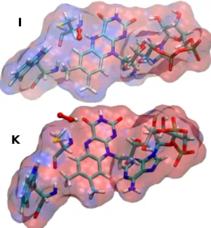
I
K
Fig. 5 The electrostatic potential of FADH•, Trp420 and Cys416 felt by
the superoxide and hydroperoxyl radical for3[FADH•–O•− 2 ] (I) and 3[FAD•−–HO•
2] (K) system. The electrostatic potential (ESP) is
calculated using Adaptive Poisson-Boltzmann Solver (APBS)38in gas
phase. The ESP is shown in a RGB (Red-Green-Blue) color scale in a range of -0.05 to 0.05 kbT/e.
taken at 50 ps intervals during 10 ns trajectories of dmCry–O2
complex. The results are shown in Figure 4. One can observe that the position of the oxygen species is different depending on whether it is a 3O
2 or 2O•−2 or a HO•2. When oxygen is in
su-peroxide form, it stays near the flavin stabilized by the attrac-tive electrostatic interaction dominated by the hydrogen in the N5 position i.e. only in case of2[FADH+–O•−
2 ] (H) and3[FADH•–
O•−2 ] (I) (see Figure 5). The HO•2 species resides always close to the FAD due to electrostatic interaction between the H of HO• 2
and N5 of FAD (in case of2[FAD
ox–HO•2] (J) and3[FAD•−–HO•2]
(K). On the contrary, triplet molecular oxygen diffuses out of the
flavin pocket and that is independent of the redox and protonated states of the FAD. We observe only two distinct paths (shown with black arrows in Figure 4) via which the oxygen species moves out-side of the lumiflavin pocket. After diffusing out from the flavin pocket, triplet oxygen is either stabilized or it hops between very few cryptochrome pockets except in the case of4[FAD•−–O
2] (B)
where the triplet oxygen is hopping between several pockets. Radical superoxide and hydroxyl radicals are essentially stabi-lized by electrostatic interactions and hydrogen bonding. To show this, we have computed the electrostatic potential due to the FAD species, Trp420 and Cys416 felt by the oxygen species for the fi-nal stable structure of3[FADH•–O•−
2 ] (I) and
3[FAD•−–HO• 2] (K)
which are shown in Figure 5. The electrostatic potential (ESP) is calculated using Adaptive Poisson-Boltzmann Solver (APBS)38 implemented in vmd39. The ESP is shown in a RGB
(Red-Green-Blue) color scale in a range of -0.05 to 0.05 kbT/e. For systemI,
we can see that a positive potential (blue), with contribution from FAD, Trp420 and Cys416 group and dominated by the contribu-tion from N5H group of lumiflavin, is created in the region around
the superoxide (shown in CPK representation) and therefore the superoxide, having negative electrostatic potential due to its’ neg-ative charge, is highly stabilized in that region due to strong elec-trostatic interaction. On the other hand, in systemK, the negative
ESP (red) around the FAD•−stabilizes the HO•2radical (shown in CPK representation in the lower panel of Figure 5) since HO•
2
it-self generate a positive electrostatic potential around the H atom. Finally, the triplet oxygen, having zero partial charge, does not feel any attractive potential and therefore can freely move in and around the binding pocket.
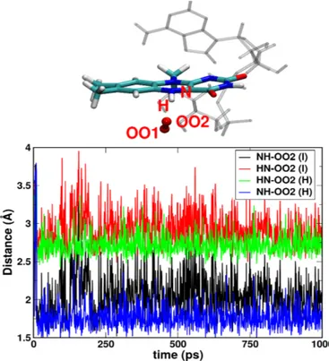
The hydrogen bonding between FAD and radical oxygen species stabilizes further ROS in the binding pocket. We observe that in fact hydrogen bonding induces a stronger binding of oxygen than the pure non-bonded electrostatic interactions. Indeed, the sta-bilization of hydroxyl radical strongly depends on the initial con-ditions, whereas this is not observed for the superoxide or triplet oxygen species. The HO•
2 stays within the cavity only when the
simulation is started from its favorable position (near N5 atom of FAD) which is true only if the O•−2 takes proton from the N5H. This indicates that the hydrogen bonding between the N5H atom of radical or anionic FADH and OO2 atom of hydroxyl radical is the main electrostatic interaction between hydroxyl radical and FAD. If it forms HO•2by taking proton from somewhere else of the protein or from solvent and is placed initially anywhere else than the favored position as shown in Figure 6, the HO•2 flies away from the C4a cavity. On the contrary, for the superoxide or triplet oxygen, the qualitative results that whether the oxygen species stays or leave the C4a cavity remain invariant on its initial posi-tion be it below the lumiflavin ring or in between the lumiflavin ring and adenine (in both case, close to the C4a atom). The bind-ing location of the superoxide in the flavin pocket remain also the same. In order to investigate the possibility of formation of C4a-hydroperoxyflavin within the limit of molecular dynamics simu-lations, the distance between N5 of lumiflavin ring and OO2 of oxygen species is plotted in Figure 6, indicating the formation of a strong hydrogen bonding [N5-H..O2](with average
donor-acceptor distance <3.0 Å) facilitating the formation of FAD•−+ HO•2 as described in Figure 2. The low fluctuation of the ROS due to this hydrogen bonding also indicates towards possibility of formation of C4a-hydroperoxyflavin as suggested by other kinetic experimental studies.18,20,26. In the previous studies, the origin of the H+to form HO•2from O•−2 was never discussed. From our MD simulation results, we propose that the N5 atom of FADH is most likely the proton donor to form the hydroperoxyl radical in-termediate.
3.2 Formation of superoxide and hydroxyl radicals
Recent experiments have been able to detect superoxide and other ROS inside cryptochromes18–21. In this section, we discuss on the possibility of formation of ROS from electronic structure theory, in order to determine the oxidation state of oxygen.
The generation of superoxide implies an electron transfer from radical/anionic flavin to triplet oxygen. From a molecular orbital picture, the transferred electron is initially located on a singly oc-cupied molecular orbital (SOMO) of radical FAD, which is of π∗
Fig. 6 HN5-OO2 and N5H-OO2 distances for a typical trajectory for
2[FADH+–O•−
2 ] (H)3[FADH •–O•−
2 ] system (I) indicating strong tendency
to form HO•2radical. On top one typical structure (indicating the position of O•−2 with respect to the N5 atom of FADH) out of thousands is shown.
character residing between the C4a-N5 atoms. This electron will be transferred to the highest occupied molecular orbital (HOMO) of 3O
2, which consists of πx∗ and πy∗ orbitals each containing
one electron. These orbitals are degenerate when oxygen is in an isotropic environment but, inside the binding pocket of cryp-tochrome, the electron will be transferred to the π∗oxygen orbital
that overlaps with the π∗orbital of FAD.
Upon photo-absorption, an electron is added to the triplet [FADox–O2] complex leading either to a doublet or quartet
to-tal spin, depending on the relative orientation of the independent spins of oxygen and radical FAD, see Figure 7 where the contribut-ing configuration state functions for a radical FAD-O2system with
minimal active space CAS(3,3) are shown. In order to investigate the formation of superoxide, the energies of high and low spin states of different species involving radical oxygen are calculated using SA5-CASSCF+MS-CASPT2 method with larger active space of (11,12) for the radical semi-quinones and (12,12) for the re-duced anion. These results are summarized in Table 1. It should be noted that, after validating that the main interaction with O2
is localized in the lumiflavin ring, we considered only lumiflavin ring (instead of the full FAD cofactor) and oxygen in these calcu-lations. The calculations are done on the structure of lumiflavin– oxygen species extracted from the highest populated dmCry–O2
structure obtained using clustering analysis based on root mean square deviation implemented in Gromacs.
Fig. 7 Representation of the multiconfiguration wavefunction for a
quartet (S=3/2) and doublet (S=1/2). The contributing configuration state functions for the4[FADH–O
2]•(S=3/2) and2[FADH–O2]•(S=1/2) for
the active space of CAS(3,3).
Table 1 SA5-CASSCF+MS-CASPT2 ground and excited state electronic
energies for [FADH–O2]•,[FAD–HO2]•, [FADH–O2]−and [FAD–HO2]−
species. For comparison, the ground state has been set to 0.00 as reference for each species independently. The states (St) are labeled by their spin (S for singlet, D for doublet, T for triplet and Q for quartet). Energies of first three states are shown for each spin. Spin multiplicity on oxygen species (MultO2) is shown based on Mulliken spin density. All
energies are in eV.
[FADH–O2]• [FADox–HO2]•
St Eex MultO2 Eex MultO2 D0 0.09 2.32 0.00 2.00 D1 0.96 1.00 0.63 1.99 D2 0.98 1.00 2.30 – Q0 0.00 2.94 2.27 2.00 Q1 1.70 2.96 2.74 1.99 Q2 3.35 2.03 3.28 2.00
[FADH–O2]− [FADox–HO2]−
St Eex MultO2 Eex MultO2 S0 0.88 – 0.00 – S1 0.97 – 0.53 – S2 1.53 – 2.19 – T0 0.00 2.91 0.04 2.00 T1 1.41 2.05 0.57 1.99 T2 1.65 1.98 2.23 2.00
each spin multiplicities of [FADH–O2]• (doublet or quartet) and
[FADH–O2]− (singlet or triplet) species. In addition, we report
the spin multiplicity of the oxygen fragment, extracted from the Mulliken analysis of the spin densities. First, we discuss the [FADH–O2]• form. The lowest energy state corresponds to the
quartet4[FADH•–O
2], in which oxygen is in a triplet spin state.
However, the lowest energy doublet state is energetically close (only 0.09 eV above Q0), suggesting that the doublet form can also exist (2[FADH+–O•−
2 ]). In that case, the oxygen spin state
could not be precisely determined from the Mulliken analysis, but still its multiplicity is closer to a doublet oxygen form. After the proton transfer between FAD and superoxide, the [FADox–HO2]•
complex is formed. The electronic structure calculations indicate that the doublet is the ground state, in which HO•2has a doublet spin multiplicity. The lowest quartet state for this complex lays 2.26 eV above the doublet. Second, we discuss the [FADH–O2]−
complex. The results for this complex suggest that the ground state is a triplet [FADH–O2]−, in which oxygen is a triplet. The
radical superoxide is only formed in the first excited triplet state (T1) which exists as 3[FADH•–O•−
2 ] and is ≈1.4 eV above the
triplet ground state. On the other hand, in the lowest singlet state (which is 0.88 eV above the triplet ground state) doublet superoxide is also formed. Upon a proton transfer, the complex FAD•−–HO•2 is formed. For this complex, both the lowest singlet and triplets are almost degenerate (∆E =0.038 eV) and both of them contains doublet oxygen species2HO•
2. The HO•2 is formed
in doublet not only in the ground state, but also for all other ex-cited states of singlet and triplet multiplicity.
3.3 The role of Trp420 and Cys416 in Drosophila melanogaster cryptochrome
Trp420 is the closest tryptophan to the FAD. It has been reported that the closest tryptophan or a tryptophan triad containing the closest tryptophan is the electron donor that reduces FAD upon photon absorption,8thus forming TrpH•+and subsequently Trp•. Therefore, Trp420 is likely to be in radical form while consider-ing cryptochrome with reduced FAD. To investigate the effect of Trp• on the position of O•−
2 , we ran further simulations for the 2[FADH+–O•−
2 ] (a typical system where superoxide binds to
lumi-flavin) with the Mulliken charges for Trp•and TrpH•+calculated at the same level of theory as discussed in Section 2. Figure 8a shows the comparison of the position of cluster of superoxide with respect to the FADH•and tryptophan for neutral Trp420 (red) and
TrpH•+ (blue) and Trp• (green). We observe that although the superoxide cluster is closer to the tryptophan for TrpH•+ (blue) than for the neutral Trp420 (red) due to strong attractive electro-static interactions between the negative superoxide and positively charged NH+, it is closer to the FADH•or almost at the same
po-sition for Trp• (green) with respect to the system with Trp420 (red).
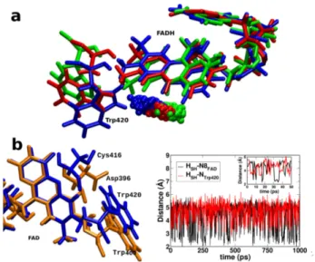
To illuminate on the probable proton donor in dmCry after the photoreduction of FAD, the crystal structure of plant cryp-tochrome and dmCry is superimposed and it is observed that Cys416 in dmCry is in the same position of Asp396 of plant cryp-tochrome (Figure 8b left panel). Since Asp396 is reported to be
the proton donor in plant cryptochrome8, by analogy, we propose
Cys416 may act as a proton donor in dmCry. To further confirm this, an MD simulation run of 1 ns is performed for dmCry and the distance between the -SH of Cys416 and N5 of FAD is recorded and found them to be in close contact over the simulation time. Figure 3 shows the distance between the H of SH (Cys416) and N5 of the ISO(black)/N of the closest tryptophan, Trp420 (red) which indicates SH bond rotates freely- towards N5 of lumiflavin (to donate proton) and then towards NH of Trp420 (to get proton to compensate) which suggests that under favorable condition it is feasible that Cys416 can donate proton to semireduced anionic FAD and can get proton from tryptophan to compensate.
Cysteine has a pKavalue of 8.3 (in solution) which is close to
physiological pH and therefore at the limit of acting as a proton donor. There have been studies in the literature on the possible role of cysteine as a proton donor in dmCry. Some spectroscopic studies suggested that the formation of FADH• in dmCry is hin-dered in physiological conditions, indicating that cysteine has a high pKa value in protein.25,40These would indicate that other
residues in the cavity are responsible for the protonation of an-ionic radical semiquinonic flavin. At variance, Hong et al. used theoretical simulations to show that Cys416 pKa is 9.7 in protein. They conclude that Cys416 could act as the proton donor, de-spite being less efficient than aspartic acid.41Based on their pKa
calculations, the difference (2.1) between the pKa of Asp396 in plant cryptochrome (7.6) and of Cys416 in dmCry (9.7) is smaller than the difference (4.4) of their pKain solution. Therefore, even
though it is evident that FADH• is formed, it remains an open question whether Cys416 act as a possible proton donor or not. Cysteine can possibly act as a slow proton donor only at a higher pH value. A rigorous study based on constant pH molecular dy-namics simulation would be necessary to confirm that.
3.4 FAD–O2as alternative radical pair for magnetoreception
Among the several reactions between FAD and oxygen species, there are four most important reactions involving a radical oxy-gen that is stabilized within the C4a cavity. Some of these reac-tions are also suggested to contribute to radical pair mechanism of magnetoreception by experimental spectral data18). The first two reactions correspond to the electron transfer from FADH•to
O2followed by proton transfer from FADH+to O•−2 , that is,
FADH•+ O2↔ FADH++ O•−2 ↔ FADox+ HO•2. (1)
The third and four reactions correspond to the electron transfer from FADH−to O2followed by proton transfer from FADH•to O•2,
that is,
FADH−+ O2↔ FADH•+ O•−2 ↔ FAD•−+ HO•2. (2)
There exist thus two radical pairs which may act as “alterna-tive” radical pair and they are FADH•+O•−
2 and FAD•−+HO•2.
Still, there are few conditions to be satisfied for the radical pair mechanism for magnetoreception to work best: (i) the radical pair should be closely spaced for spin selective conversion, (ii) the electron nuclear hyperfine interaction should be weak, (iii)
Fig. 8 (a) Comparison of the position of superoxide clusters for different
redox and protonation states of tryptophan e.g. neutral Trp420 (red), Trp•H+(blue) and Trp•(green) . The structures are aligned with respect
to the FADH•. (b) Left: comparison of the core active site (containing FAD, Trp400/Trp420 and Asp396/Cys416 of plant (orange) and Drosophila melanogaster (blue) cryptochrome. Right : the distance between Hydrogen of SH (Cys416) and N8 of lumiflavin ring of FAD (black)/N of Trp420 (red). The inset shows the first 50 ps (zoomed) to detect the frequency of the SH bond rotation.
the spin relaxation of the radical should be slow, and (iv) the ex-change and dipole-dipole spin couplings should be close to zero or mutually cancelling.42With the results we have reported, we can qualitatively say that these conditions are satisfied for both radical pairs involving oxygen. Indeed, from our MD simulations we showed that FADH•–O•−2 and FAD•−–HO•2stays within 2-3 Å. The hyperfine coupling condition is more easily satisfied by oxy-gen species radical pair than by tryptophan radical pair, since its
16O and18O isotopes are not magnetic. Therefore, the
hyper-fine coupling of the radical pair is only due to FADH•. For the spin relaxation, it is known that O•−2 has a fast spin relaxation kinetics18,21, but the presence of Trp•close to the radical pair (as
shown in Figure 8) can act as scavenger to slow down the spin relaxation of oxygen species. In addition, the restricted mobility of radical pair (indicated by low fluctuation of oxygen species in Figure 3) due to strong electrostatic interactions can slow down the spin relaxation. Finally, from electronic structure calculations, we can estimate the exchange coupling strength as the difference between the low and the high spin lowest energies. From this cri-teria, we can conclude that only FAD•−–HO•2has a small exchange coupling (J=400 meV). From these results, we can conclude that FAD•−–HO•
2is a good candidate for explaining magnetorecpetion
via the “alternative” radical pair with oxygen.
4
Conclusion
In this work, we have systematically explored the fate of different oxygen species in the binding pocket of Drosophila melanogaster cryptochrome using atomistic molecular dynamics simulations, taking into account the charge and spin multiplicity of different FAD and oxygen forms that are generated upon blue light
absorp-tion. Additionally, we performed electronic structure simulations of different FAD–O2radical pairs to clarify the oxidation state of
oxygen in each of the complexes.
The molecular dynamics simulations suggest that radical oxy-gen species are strongly stabilized close to FAD due to strong at-tractive electrostatic interactions and hydrogen bonding. On the contrary, triplet oxygen species move out of the cavity when FAD is in radical or charged form. The electrostatic attractive forces between the radical oxygen species and FADH is dominated by the interaction between N5H (of FADH•) and O•2. The results also indicate to a strong tendency of formation of hydroperoxyl radical. The stability of hydroperoxyl radical near C4a atom are in agreement with previous studies suggesting formation of C4a-hydroperoxyflavin intermediate.26
We show that the formation of hydroperoxyl radical is possible by the proton transfer from the proton at N5 position in FADH−.
Furthermore, we have found that the varying electrostatic field of radical or neutral tryptophan has negligible impact on the posi-tion of oxygen. Still, the oxygen species are closer to the caposi-tionic TrpH•+ due to stronger electrostatic interactions. The four im-portant systems that can lead to the formation of radical oxygen species are found to be2[FADH+–O•−
2 ],2[FADox–HO•2],3[FADH•–
O•−2 ],3[FAD•−–HO• 2].
The electronic structure analysis suggests that the radical FADH–O2 system in the ground state is a quartet and exist as 4[FADH•–O
2]. For this particular case, the2[FADH+–O•−2 ] is only
≈0.1 eV above the ground state and therefore it is possible that both spin states are formed in the protein. For the [FADH–O2]−
complex, the triplet radical pair3[FADH•–O•−
2 ] is 1.5 eV above
the ground state3[FADH−–O
2] in which oxygen is in a triplet.
The only radical pair in the ground state is3[FAD•−–HO• 2] which
has a small singlet-triplet energy gap.
In view of “alternative” radical pair, our MD results suggest that two probable radical pairs i.e. FADH•–O•−
2 and FAD•−–HO•2.
Among them, FAD•−–HO•2passes all criteria as discussed in sub-section 3.4. Although, more investigation is necessary on the life-time of the short-lived hydroperoxyl radical. The FADH•–O•−2 rad-ical pair passes all criteria except the small exchange coupling cri-teria. Further investigation is needed on whether the cancellation of dipole and exchange coupling is possible43. Environmental ef-fects could also affect the order of states, but these are expected to be rather small in cryptochromes.44
To summarize, our results from this study focuses not only on the qualitative position of different oxygen species in the flavo-protein active site, but also detect its binding location, diffusion pathway and surrounding important residues depending on vary-ing electrostatic interactions. These results are important not only in view of investigating the "alternative" radical pair mechanism of magnetoreception but also shed light on several other func-tionality of flavoprotein in general e.g. ROS production, anti-oxidant defence. The results from our study (e.g. the specific binding location, diffusion pathway of oxygen species and sur-rounding residues with strong interactions) can help designing mutant flavoprotein to accelerate ROS production as well as to accelerate anti-oxidant defence mechanism.
Acknowledgments
The authors acknowledge financial support by the “Agence Na-tionale pour la Recherche” through the project BIOMAGNET (ANR-16CE29-0008-01). This work was granted access to the HPC resources of Aix-Marseille Université financed by the project Equip@Meso (ANR-10-EQPX-29-01) of the program «In-vestissements d’Avenir» supervised by the Agence Nationale de la Recherche.
References
1 V. Massey, FASEB J., 1995,9, 473–475.
2 I. Chaves, R. Pokorny, M. Byrdin, N. Hoang, T. Ritz, K. Brettel, L. O. Essen, G. T. J. van der Horst, A. Batschauer and M. Ah-mad, Annu. Rev. Plant Bio., 2011,62, 335–364.
3 A. Sancar, Chem. Rev., 2003,103, 2203–2237.
4 P. Sampathkumar, S. Turley, J. E. Ulmer, R. H. G., C. H. Sibley and W. G. Hol, J. Mol. Biol., 2005,352, 1091–1104.
5 H. Myllykallio, G. Lipowski, D. Leduc, J. Filee, P. Forterre and U. Liebl, Science, 2002,297, 105–107.
6 E. Gross, K. D. B., C. A. Kaiser and D. Fass, Cell, 2004,117,
601–610.
7 K. R. Marshall, M. Gong, L. Wodke, J. H. Lamb, D. J. Jones, P. B. Farmer, N. S. Scrutton and A. W. Munro, J. Biol. Chem., 2005,280, 30735–30740.
8 I. A. Solovy’ov, T. Domratcheva, A. R. Moughal Shahi and K. Schulten, J. Am. Chem. Soc., 2012,134, 18046–18052.
9 C. T. Rodgers and P. Hore, Proc. Nat. Acad. Sci., 2009,106,
353–360.
10 T. Ritz, S. Adem and K. Schulten, BioPhys. J., 2000,78, 707–
718.
11 Y.-T. Kao, C. Saxena, T.-F. He, L. Guo, L. Wang, A. sancar and D. Zhong, J. Am. Chem. Soc., 2008,130, 13132–13139.
12 F. Cailliez, P. Müller, M. Gallois and A. de la Lande, J. Am.
Chem. Soc., 2014,136, 12974–12986.
13 R. J. Gegear, L. E. Foley, A. Casselman and S. M. Reppert,
Nature, 2009,463, 804–807.
14 T. Ritz, R. Wiltschko, P. Hore, C. T. Rodgers, K. Stapput, P. Thalau, C. R. Timmel and W. Wiltschko, Biophys. J., 2009,
96, 3451–3457.
15 N. Christine, D. Susanne, S. Katrin, A. Margaret, P. Leo, W. Wolfgang and W. Roswitha, J. Royal Soc. Interface, 2013,
10, 20130638.
16 D. R. Kattnig, J. Phys. Chem. B, 2017,121, 10215–10227.
17 D. R. Kattnig and P. J. Hore, Sci. Rep., 2017,7, 11640–11650.
18 P. Müller and M. Ahmed, J. Biol. Chem., 2011,286, 21033–
21040.
19 L.-D. Arthaut, N. Jourdan, A. Mteyrek, M. Procopio, M. El-Esawi, A. d’Harlingue, P.-E. Bouchet, J. Witczak, T. Ritz, A. Klarsfeld, S. Birman, R. J. Usselman, U. Hoecker, C. F. Mar-tino and M. Ahmad, PLOS ONE, 2017,12, 1–15.
20 L. van Wilderen, G. Silkstone, M. Mason, J. J. van Thor and M. T. Wilson, FEBS open bio, 2015,5, 885–892.
21 H. J. Hogben, O. Efimova, N. Wagner-Rundell, C. R. Timmel and P. J. Hore, Chem. Phys. Lett., 2009,480, 118–122.
22 R. Baron, C. Riley, P. Chenprakhon, K. Thotsaporn, R. T. Win-ter, A. Alfieri, F. Forneris, W. J. H. van Berkel, P. Chaiyen, M. W. Fraaije, A. Mattevi and J. A. McCammon, Proc. Nat.
Acad. Sci., 2009,106, 10603–10608.
23 B. Giovani, M. Byrdin, M. Ahmad and K. Brettel, Nat. Struct.
Biol., 2003,10, 489–490.
24 R. Banerjee, E. Schleicher, S. Kacprzak, J. P. Bouly, M. Picot, W. Wu, A. Berndt, E. Wolf, R. Bittl and M. Ahmad, J. Biol.
Chem., 2007,282, 14916–14922.
25 A. Berndt, T. Kottke, H. Breitkreuz, R. Dvorsky, S. Hennig, . Alexander, M and E. Wolf, J. Biol. Chem., 2007,282, 13011–
13021.
26 V. Massey, J. Biol. Chem., 1994,269, 22459–22462.
27 J.-D. Chai and M. Head-Gordon, Phys. Chem. Chem. Phys., 2008,10, 6615–6620.
28 M. M. Francl, W. J. Pietro, W. J. Hehre, J. S. Binkley, M. S. Gordon, D. J. DeFrees and J. A. Pople, J. Chem. Phys, 1982,
77, 3654–3665.
29 H. J. C. Berendsen, D. van der Spoel and R. van Drunen,
Com-put. Phys. Commun., 1995,91, 43–56.
30 A. D. J. Mackerell, M. Feig and C. L. r. Brooks, J. Comput.
Chem., 2004,25, 1400–1415.
31 P. Bjelkmar, P. Larsson, M. A. Cuendet, B. Hess and E. Lindahl,
J. Chem. Theo. Comput., 2010,6, 459–466.
32 K. Vanommeslaeghe, E. Hatcher, C. Acharya, S. Kundu, S. Zhong, J. Shim, E. Darian, O. Guvench, P. Lopes, I. Vorobyov and A. D. MacKerell (Jr.), J. Comp. Chem., 2010,
31, 671–690.
33 M. J. Frisch, G. W. Trucks, H. B. Schlegel, G. E. Scuseria, M. A. Robb, J. R. Cheeseman, G. Scalmani, V. Barone, G. A. Peters-son, H. Nakatsuji, X. Li, M. Caricato, A. V. Marenich, J. Bloino, B. G. Janesko, R. Gomperts, B. Mennucci, H. P. Hratchian, J. V. Ortiz, A. F. Izmaylov, J. L. Sonnenberg, D. Williams-Young, F. Ding, F. Lipparini, F. Egidi, J. Goings, B. Peng, A. Petrone, T. Henderson, D. Ranasinghe, V. G. Zakrzewski, J. Gao, N. Rega, G. Zheng, W. Liang, M. Hada, M. Ehara, K. Toyota, R. Fukuda, J. Hasegawa, M. Ishida, T. Nakajima, Y. Honda, O. Kitao, H. Nakai, T. Vreven, K. Throssell, J. A. Montgomery, Jr., J. E. Peralta, F. Ogliaro, M. J. Bearpark, J. J. Heyd, E. N. Brothers, K. N. Kudin, V. N. Staroverov, T. A. Keith, R. Kobayashi, J. Normand, K. Raghavachari, A. P.
Ren-dell, J. C. Burant, S. S. Iyengar, J. Tomasi, M. Cossi, J. M. Millam, M. Klene, C. Adamo, R. Cammi, J. W. Ochterski, R. L. Martin, K. Morokuma, O. Farkas, J. B. Foresman and D. J. Fox,
Gaussian 16 Revision A.01, 2016, Gaussian Inc. Wallingford
CT.
34 A. D. MacKerell (Jr.), D. Bashford, M. Bellott, R. L. R. L. Dun-brack (Jr), J. D. Evanseck, M. J. Field, M. Fischer, J. Gao, H. Guo, S. Ha, D. Joseph-McCarthy, L. Kuchnir, K. Kuczera, F. T. K. Lau, C. Mattos, S. Michnick, T. Ngo, D. T. Nguyen, B. Prodhom, W. E. Reiher, B. Roux, M. Schlenkrich, J. C. Smith, R. R. Stote, J. Straub, M. Watanabe, J. Wiøsrkiewicz-Kuczera, D. Yin and M. Karplus, J. Phys. Chem. B, 1998,102,
3586–3616.
35 F. Autenreith, J. Tajkhorshid, E.and Baudry and Z. Luthey-Schulten, J. Comp. Chem., 2004,25, 1613–1622.
36 B. O. Roos, V. Veryazov and P.-O. Widmark, Theor. Chem. Acc., 2004,111, 345–351.
37 F. Aquilante, J. Autschbach, R. K. Carlson, L. F. Chibotaru, M. G. Delcey, L. De Vico, I. Fdez. Galvà ˛an, N. FerrÃl’, L. M. Frutos, L. Gagliardi, M. Garavelli, A. Giussani, C. E. Hoyer, G. Li Manni, H. Lischka, D. Ma, P. Ã. Malmqvist, T. MÃijller, A. Nenov, M. Olivucci, T. B. Pedersen, D. Peng, F. Plasser, B. B. Pritchard, M. Reiher, I. Rivalta, I. Schapiro, J. Segarra-Martí, M. Stenrup, D. G. Truhlar, L. Ungur, A. Valentini, S. Vancoillie, V. Veryazov, V. P. Vysotskiy, O. Weingart, F. Zap-ata and R. Lindh, J. Comput. Chem., 2016,37, 506.
38 N. Baker, D. Sept, S. Joseph, M. J. Holst and M. J. A., Proc.
Nat. Acad. Sci., 2001,98, 10037–10041.
39 W. Humphrey, A. Dalke and K. Schulten, J. Molec. Graphics, 1996,14, 33–38.
40 B. Paulus, C. Bajzath, F. Melin, L. Heidinger, V. Kromm, C. Herkersdorf, U. Benz, L. Mann, P. Stehle, P. Hellwig, S. We-ber and E. Schleicher, FEBS Journal, 2015,282, 3175–3189.
41 G. Hong and R. Pachter, J. Phys. Chem. B, 2015,119, 3883–
3892.
42 P. J. Hore, Annu. Rev. BioPhys., 2016,45, 299–344.
43 O. Efimova and P. J. Hore, BioPhys. J., 2008,94, 1565–1574.
44 E. Cannuccia, O. Pulci, R. D. Sole and M. Cascella, Chem.


![Table 1 SA5-CASSCF+MS-CASPT2 ground and excited state electronic energies for [FADH–O 2 ] • ,[FAD–HO 2 ] • , [FADH–O 2 ] − and [FAD–HO 2 ] − species](https://thumb-eu.123doks.com/thumbv2/123doknet/14333771.498465/7.918.62.417.775.1059/table-casscf-caspt-ground-excited-electronic-energies-species.webp)